is cmv a target in pediatric glioblastoma? expression of ... in children, ... genex) for 2.5 h at 50...
TRANSCRIPT

CLINICAL STUDY
Is CMV a target in pediatric glioblastoma? Expression of CMVproteins, pp65 and IE1-72 and CMV nucleic acids in a cohortof pediatric glioblastoma patients
Amanda Wakefield1,2,3 • Antonella Pignata1,2,3 • Alexia Ghazi1,2,3 • Aidin Ashoori1,2,3 •
Meenakshi Hegde1,2,3 • Daniel Landi1,2,3 • Tara Gray1,2,3 • Michael E. Scheurer2,3 •
Murali Chintagumpala2,3 • Adekunle Adesina2,3,4 • Stephen Gottschalk1,2,3,4 •
John Hicks2,3,4 • Suzanne Z. Powell5 • Nabil Ahmed1,2,3
Received: 28 February 2015 / Accepted: 29 August 2015 / Published online: 4 September 2015
� The Author(s) 2015
Abstract While the 5-year overall survival is better in
pediatric than in adult patients diagnosed with glioblastoma
(GBM), outcomes in children remain very poor. Under-
standing the mechanisms of tumorigenesis and tumor
propagation can identify therapeutic targets to improve
these outcomes. Human cytomegalovirus (CMV) proteins
and nucleic acids are present in the majority of adult GBM.
Indeed, CMV is emerging as a potential glioma-associated
target for anti-CMV agents and cellular therapeutics. Fur-
thermore, CMV appears to contribute to GBM’s malignant
phenotype, although its role in tumorigenesis is less cer-
tain. In this cohort of 25 serially diagnosed pediatric
GBMs, the largest described cohort to date, we used
immunohistochemical staining and in situ hybridization to
show the presence of CMV antigens pp65 and IE1-72 as
well as CMV nucleic acids, respectively. Our cohort indi-
cated either CMV antigen pp65 or IE1-72 was present in
approximately 67 % of pediatric GBM samples. The
majority of samples stained positive for either CMV
antigen showing a cytoplasmic pattern in 25-50 % of cells
within the sample at a moderate intensity, while a few
samples showed nuclear staining and higher grade/inten-
sity. Of 16 samples where in situ hybridization was per-
formed, 13 (81 %) showed specific staining using a CMV
genome specific probe cocktail. ISH positive samples
showed high concordance with being pp65 or IE1-72
positive. These findings, paired with the association of
CMV expression with poor prognosis and overall survival,
indicate the need to further investigate how these antigens
are promoting tumor growth and preventing cell death.
Also, the expression of these antigens in a majority of
tumor tissues should be considered for immunotherapeutic
targets in cases of pediatric GBM.
Keywords Glioblastoma � GBM � Pediatric � CMV �pp65 � IE1-72
Introduction
In children, approximately 65 % of glioblastoma (GBM)
arise in the cerebrum, 20 % in the thalamus and hypotha-
lamus, and 15 % in the posterior fossa, mostly affecting the
cerebellum and brainstem [1]. While GBM in both pedi-
atric and adult patients represents the most anaplastic and
highest grade of gliomas, these tumors appear to differ in
their genetic and molecular underpinnings [1]. Current
treatment includes tumor resection, radiotherapy, and
occasionally in children, adjuvant chemotherapy. This
combination is both toxic and largely ineffective [2, 3].
GBMs exhibit numerous sophisticated defense mecha-
nisms making them resistant to conventional therapies.
They are notorious for microscopically infiltrating healthy
brain, making complete resection difficult. In addition,
& Nabil Ahmed
1 Texas Children’s Hospital, Houston Methodist Hospital,
Center for Cell and Gene Therapy, Baylor College of
Medicine, 1102 Bates Street MC 3-3320, Houston,
TX 77030, USA
2 Texas Children’s Hospital, Texas Children’s Cancer and
Hematology Centers, Baylor College of Medicine, Houston,
TX 77030, USA
3 Departments of Pediatrics, Baylor College of Medicine,
Houston, TX 77030, USA
4 Departments of Pathology and Immunology, Baylor College
of Medicine, Houston, TX 77030, USA
5 Department of Pathology and Genomic Medicine, Houston
Methodist Hospital, Houston, TX 77030, USA
123
J Neurooncol (2015) 125:307–315
DOI 10.1007/s11060-015-1905-z
glioma stem cells (GSC) are quiescent and appear to have
advanced DNA repair mechanisms, anti-apoptosis genes,
and telomerase activity, rendering them resistant to both
chemotherapy and radiation [3].
More effective therapies for GBM are needed, thus
cellular therapies are being developed. Cytotoxic T lym-
phocytes (CTLs) are powerful immune effector cells and
have been successfully used to treat disseminated Epstein–
Barr Virus (EBV) infections and EBV-driven malignancies
[4, 5]. CTLs can be directed to target GBM through ex vivo
expansion of a tumor-specific clone or manipulation of
their T cell receptor (TCR), but effective immunogenic
targets are strongly needed [6–8]. Through identifying
unique targets for cellular therapies and increasing under-
standing of tumor escape mechanisms, we and others are
developing more specific and powerful therapies for GBM
that effectively target tumor cells while sparing the intri-
cate neighboring healthy tissue [6–8].
Recently, the detection of CMV proteins and nucleic acid
in the majority of adult GBM has caused interest in these as a
possible target for immune-based biologics [9–14]. Initial
techniques for detecting CMV proteins and nucleic acids
were varied, but under optimal conditions CMV proteins are
found in the majority of high-grade gliomas [14]. CMV early
or late proteins have also been found in up to 100 % of
neuroblastomas [15] and 40 % of medulloblastomas [16].
CMV has been implicated in promoting GBM pathogenesis;
specifically, CMV appears to enhance telomerase activity
and angiogenesis in adult GBM [17–19]. Increased aware-
ness of the prevalence of CMV expression on GBM tumor
cells and its apparent ability to enhance tumor cell survival
and invasiveness make it an appealing target for
immunotherapy against GBM.
Several reports describe the prevalence of CMV
expression patterns in adult cancers, including adult GBM,
and support the potential for CMV as an immunothera-
peutic target [9–14]. By determining the expression pattern
of CMV antigens in pediatric GBM, we can discern their
potential as a target for cellular and other targeted thera-
pies, as well as pursue a better understanding of their roles
in tumor pathogenesis. Ultimately, this knowledge could
allow us to better determine the utility of CMV for
improving survival in children with GBM.
Materials and methods
Study subjects
GBM tissue samples from 15 pediatric patients were
obtained from Texas Children’s Hospital; one of these
samples was immeasurable due to necrotic tissue. The
remaining 10 samples were sent from collaborating
institutions for analysis. All tissue sections were from
children under the age of 18 at the time of resection, and
serially diagnosed GBM WHO grade IV. All patients were
consented on a human protocol approved by Baylor Col-
lege of Medicine’s internal review board (IRB). Seroposi-
tivity for CMV was unknown. Fifteen of 25 tumors were
primary excisions and 10 were tumor recurrences.
Standard staining
All tissues were received from collaborating institutions as
recently cut 5 lm slides from tumor material fixed in 10 %
formalin and paraffin embedded. H&E stains were also
received completed by collaborating institutions.
Immunohistochemistry (IHC)
To test samples for CMV, IHC staining was performed as
previously described [20]. Briefly, Formalin-fixed, paraffin
embedded sections (6 lm) of primary human GBM were
used. (1) Known CMV-infected lung samples were used as
a positive tissue control for all experiments. All slides were
deparaffinized by heating slides in a xylene bath at 50 �Cfor 1 h and 10 min, followed by a 30-min incubation per-
iod at room temperature. The slides were then washed
additionally in xylene and serially diluted in ethanol baths
(100, 95, 70 and 50 %), post-fixed in neutral buffered
formalin and treated for pepsin digestion (BioGenex, San
Ramon, CA, USA). Freshly prepared 30 % H2O2 was used
to block endogenous peroxidase before performing antigen
retrieval using CitraPlus antigen retrieval solution (Bio-
Genex) for 2.5 h at 50 �C. Avidin, biotin (BioGenex) and
Fc receptor (Innovex Biosciences, Richmond, CA, USA)
blocking reagents were applied to the sections prior to a
4 �C overnight incubation with anti-IE172 (1:100;
Chemicon, Temecula, CA, USA) and anti-pp65 (1:40;
Leica Microsystems Inc., Bannockburn, IL, USA) primary
antibodies. Positive control sections were treated with anti-
actin monoclonal antibody (1:35; BioGenex), while nega-
tive control sections were similarly incubated with no
primary antibody. The sections were then developed using
a biotinylated anti-mouse secondary antibody (1:16.5;
BioGenex), peroxidase-labeled streptavidin (Biogenex)
and 3,30diaminobenzidine (Innovex Biosciences) as a
chromogen. All slides were counterstained in Harris
hematoxylin, dehydrated and coverslipped.
In situ hybridization (ISH)
All paraffin-embedded sections (5 lm) were deparaffinized
and post-fixed in neutral buffered formalin, similar to the
sections for IHC. Pepsin digestion, endogenous peroxidase
block and antigen retrieval were also performed for the ISH
308 J Neurooncol (2015) 125:307–315
123
sections as previously described [20]. For the tests, CMV-
infected lung tissue was used as a positive control. A
human CMV DNA probe cocktail end-labeled with five
fluorescein-linker molecules (Leica) was used to detect
human CMV DNA in the sections. A probe against the
reverse complementary sequence of black beetle virus
RNA2 sequence (Leica) was used as a nonspecific negative
control.
Results
Patient characteristics
A cohort of 25 pediatric patients are reported herein. These
children, aged 9 months to 18 years (Table 1), were seri-
ally-diagnosed with GBM (WHO grade IV), and their
tumors were procured at Texas Children’s Hospital and
other institutions. Pathological diagnosis was confirmed by
3 independent pathologists (two neuropathologist and one
pediatric neuropathologist). All patients were consented on
a human protocol approved by Baylor College of Medi-
cine’s institutional review board (IRB). Seropositivity for
CMV was unknown. Fifteen of 25 tumors were primary
excisions and 10 were tumor recurrences. Sixteen of 25
patients had enough material for both immunohistochem-
istry (IHC) and in situ hybridization (ISH).
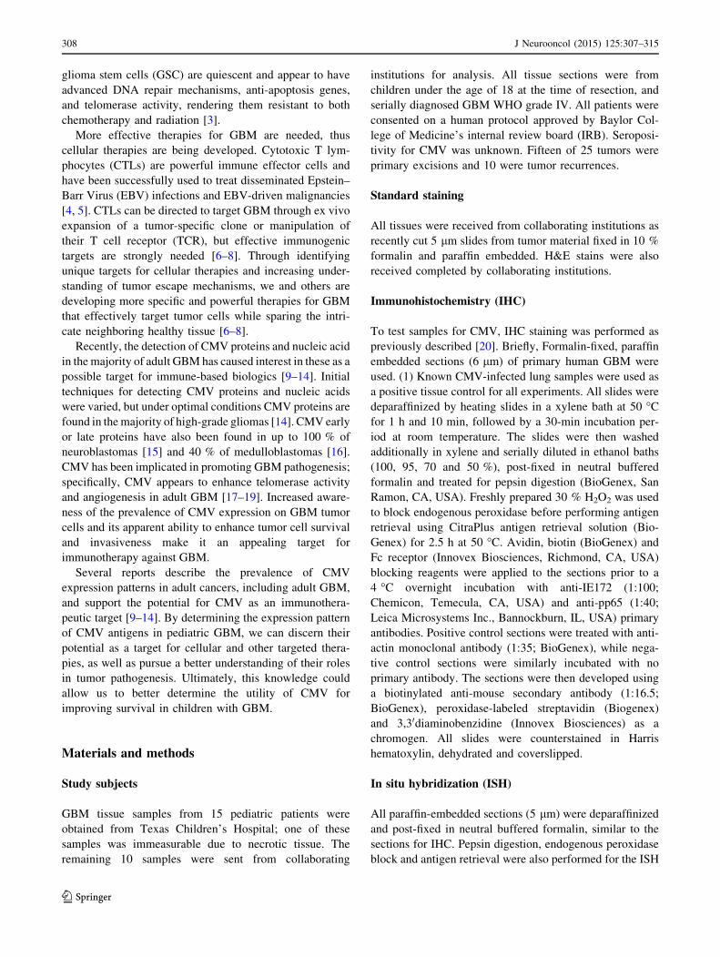
Establishment of a grade and intensity scale for IHC
All IHC stains were given a grade (based on percent pos-
itivity) and an additional score for intensity of the stain
using a pre-determined scheme (Fig. 1). All slides were
reviewed for by 3 independent pathologists and the grading
system was established by all pathologists and verified by
independent sample review. Staining grade ranged from 1
to 4 with grade 1 indicating a positive stain visible in
1–25 % of cells, grade 2 in 26–50 % of cells, grade 3 in
51–75 % of cells, and grade 4 in 76–100 % of cells.
Intensity ranged from 1? to 3? based on the positivity
observed on control slides. Representatives for each grade
and intensity are shown (Fig. 1) including a negative
control (Fig. 1a). Grade distribution is shown increasing
from grade 1 (Fig. 1b), grade 2 (Fig. 1c), grade 3 (Fig. 1d),
and grade 4 (Fig. 1e). Intensity increases from
1? (Fig. 1c), 2? (Fig. 1d), and 3? (Fig. 1e). There was
no correlation between that staining grade or intensity and
any particular pathological features.
Expression of human cytomegalovirus (CMV)
proteins, pp65 and IE1-72
IHC staining was performed on paraffin-embedded sections
obtained from all 25 pediatric patients diagnosed with
GBM (WHO grade IV). One sample was immeasurable
due to extensive necrosis of tumor tissue. Sections were
determined positive for CMV using antibodies specific for
the CMV-encoded late protein (pp65) and CMV-encoded
early protein (IE1-72) and given the grade and intensity
ratings described. Photomicrographs are shown (Fig. 2)
from three representative patients for CMV pp65 (Fig. 2a,
b, c) and CMV IE1-72 (Fig. 2d, e, f). We found pp65
reactivity in 12 of 24 evaluable tumors and IE1-72 reac-
tivity in 14 of 24 evaluable samples. Overall, positivity was
observed for either CMV antigen in 16 of 24 patients. This
data indicates CMV pp65 was observed in 50 % of our
cohort, CMV IE1-72 in 58.3 %, and either CMV pp65 or
CMV IE1-72 in 66.7 %. Grade and intensity of positive
staining for each CMV pp65 and CMV IE1-72 were further
examined to determine the staining pattern of each tested
antigen (Fig. 3). Approximately 60 % of samples positive
for CMV pp65 were observed as grade 1, 15 % observed as
grade 2, 5 % (1 patient) observed as grade 3, and 15 %
observed as grade 4. For CMV IE1-72, approximately
40 % of our samples stained as grade 1, 30 % stained as
grade 2, and 30 % stained as grade 4 (Fig. 3a). Intensity
Table 1 Patient and tumor characteristics
UPN GBM grade Location Age Gender
1 IV Posterior fossa 11 M
2 IV Frontal brain 4 F
3 IV Frontal cortex 11 F
4 IV Frontal brain 18 M
5 IV Intraventricular 1 F
6 IV Frontal brain 9 M
7 IV Frontal cortex 11 M
8 IV Thalamus 9 M
9 IV Frontal brain 6 F
10 IV Side brain 12 M
11 IV Frontal brain 15 M
12 IV Frontal brain 9 m F
13 IV Posterior fossa 10 M
14 IV Posterior fossa 10 M
15 IV Frontal brain 13 F
16 IV Thalamus 17 M
17 IV Left ventricle 13 M
18 IV Frontal brain 4 F
19 IV Front parietal 11 M
20 IV Frontal brain 15 F
21 IV Left anterior 15 M
22 IV Frontal brain 18 M
23 IV Thalamus 17 F
24 IV Frontal brain 14 F
25 IV Temporal 10 F
J Neurooncol (2015) 125:307–315 309
123

distribution for both CMV pp65 and CMV IE1-72 were
similar at about 60–70 % and 20–30 % staining 1? and
2? , respectively, and only 1 patient at 3? (Fig. 3b).
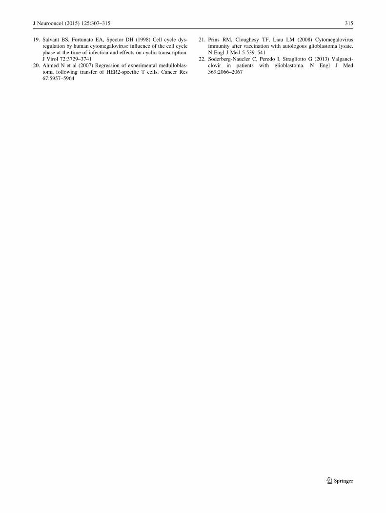
Expression of human CMV nucleic acid using is situ
hybridization (ISH)
To confirm the presence of CMV in GBMs, we performed
ISH analysis using a human CMV DNA probe cocktail.
Sixteen samples of the cohort of 25 were available for
testing. Thirteen out of 16 GBMs were positive for the
CMV genome (Fig. 4), confirming that CMV is detectable
in the majority of primary GBM samples. The staining
pattern was strictly nuclear for positive samples and was
uniformly expressed in the majority of nuclei examined. In
11 out of 13 positive samples concomitant IE1-72 (n = 10)
or pp65 (n = 7) were detected. Six out of 13 samples were
triple positive for CMV nucleic acid using ISH, IE1-72 and
pp65 using IHC and 3 out of 13 were triple negative for all
(Fig. 5). These results confirm results for our group and
those from others demonstrating a high degree of correla-
tion between CMV nucleic acid detection and the presence
of the CMV immunodominant proteins pp65 and IE1-72.
Discussion
In this cohort of 25 pediatric serially diagnosed WHO IV
GBMs, we show that either CMV antigens pp65 or IE1-72
are present in approximately 66.7 % of pediatric GBM
samples. The majority of samples stained positive for either
CMV antigens, pp65 or IE1-72, showing a cytoplasmic
pattern in 25–50 % of cells within the sample at a moderate
intensity, while a few samples showed nuclear staining and
higher grade/intensity. In a sub-cohort of 16 of these
pediatric GBM patients, CMV genome was detectable in
80 % of samples with a high concordance with CMV
protein detection.
Using similar methodology to stain adult GBM for
CMV, we previously reported approximately 45 % posi-
tivity for pp65 and 91 % positivity for IE1-72 in a cohort of
adult GBM [9]. These results are consistent with previous
groups reporting 50-70 % pp65 positivity in adult GBM [9]
indicating a fair similarity of pp65 expression between
adult and pediatric GBM. The majority of previous reports
indicate a higher prevalence of IE1-72 at 90–100 % [9–12]
while Lucas et al. found much lower expression at only
16 % [14]. This variation is possibly due to a difference in
detection methodology and/or interpretation. Interestingly,
Scheurer et al. observed 21 of 21 adult GBM samples
Gra
de 2
: 26
-50%
Inte
nsity
: 1+
Gra
de 3
: 51
-75%
Inte
nsity
: 2+
G
rade
4:
76-1
00%
Inte
nsity
: 3+
Inte
nsity
: 0
Gra
de 1
: 0-
25%
Inte
nsity
: 2+
Neg
ative
control
Pos
itive
control
A
B
C
D
E
Fig. 1 IHC staining showing representative grade and intensity
scoring. a Sample with 0 % positive staining. b Sample staining
positive at Grade: 1 (0–25 %) with Intensity: 2?. c Sample staining
positive at Grade: 2 (26–50 %) with Intensity: 1?. d Sample staining
positive at Grade: 3 (51–75 %) with Intensity: 2?. e Sample staining
positive at Grade: 4 (76–100 %) with Intensity: 3?. Grade and
intensity were measured by three independent pathologist for all
tested patients. Magnification 9100
310 J Neurooncol (2015) 125:307–315
123

Pat
ient
1P
atie
nt 2
Pat
ient
12
CMV IE1-72CMV pp65
A
B
C
D
E
F
Pos
itive
Con
trol
Neg
ativ
e C
ontro
l
Fig. 2 IHC for CMV pp65 and
CMV IE1-72. Results from
three representative patients are
shown for CMV pp65 and CMV
IE1-72For CMV pp65.
(a) Patient 1 stained negative
(b) Patient 2 stained Grade: 2
and Intensity: 2?, and
(c) Patient 12 stained Grade: 1
and Intensity: 2?. For CMV
IE1-72 (d) Patient 1 stained
Grade: 2 and Intensity: 2?
(e) Patient 2 stained Grade: 2
and Intensity: 1?, and
(f) Patient 12 stained Grade: 2
and I: 2?. Positive control is
from CMV infected lung tissue
and negative control has no
primary antibody added.
Magnification 9200. CMV
positive control magnification
9400
J Neurooncol (2015) 125:307–315 311
123

staining positive for CMV, and saw both nuclear and
cytoplasmic staining. This group also reported that
approximately 79 % of cells within a sample staining
positive, indicated by grade 4 in our scheme, which is
much higher than our observation of only 10–20 % of
samples that fell within grade 4. Our staining method was
comparable to what other groups have shown for pp65 and
optimized to eliminate background staining of IE1-72.
Also, consistent with our observations, CMV staining in
these adult cohorts was detectable primarily in the cyto-
plasm of GBM cells [9]. These data suggest the prevalence
of CMV antigens on the majority of GBMs and indicate it
could represent a potential target for novel immunothera-
peutics or antivirals.
While CMV antigens offer utility as a marker whereby
immune-based therapies can target GBM cells, it is pos-
sible that the ubiquitous CMV proteins identified in GBM
contribute to the malignant phenotype of GBM. Therefore,
targeting these molecules could potentially have a direct
therapeutic benefit through disrupting disease pathways.
Indeed, CMV cellular immunity (to CMV pp65), was
demonstrated in a research subject after vaccination with
dendritic cells that are pulsed with an autologous tumor
lysate [21].
The role of CMV in the pathogenesis and propagation of
GBM is the subject of ongoing research. Specifically, CMV
is increasingly implicated in the pathogenesis of GBM and
is being pursued as a target for cellular therapies [9, 21]. It
is well known to be tropic for glial cells and is a cause of
devastating encephalitis and cerebral dysgenesis in human
fetuses and newborns. Interestingly, latent CMV can be
reactivated in astrocytes and astrocyte-derived tumors by
inflammatory stimuli [18]. Cobbs and others have shown
that CMV gene products can corrupt multiple cellular
pathways in GBM including mutagenesis, apoptosis
avoidance, angiogenesis, and microscopic invasion [11,
13].
Other latent viruses, including EBV and human herpes
virus 6 (HHV6), could behave similar to CMV when
expressed on tumor cells and use natural immune system
evasion mechanisms to allow tumor cells to avoid being
targeted by the immune system. These would be interesting
entities to investigate as potential targets in patients whose
tumors do not express CMV.
Some limitations of this study include a relatively small
cohort, although the largest currently reported in pediatric
GBM. Further analysis of pediatric GBM patients would be
warranted to furnish a better understanding of the preva-
lence of these antigens. IHC staining of CMV IE1-72 and
pp65 is variable, however we and others have found
complete concordance with ISH, as previously reported [9–
12]. Indeed, when we performed ISH for CMV genome,
using a cocktail of probes spanning the whole CMV gen-
ome, we found positive nuclear staining in 81 % of a sub-
cohort of 16 samples. These correlated well with being
CMV protein positive. Background staining has been a
consistent problem with IHC for IE1-72, but our protocol
was further optimized to minimize this affect with post-
development incubations. Also, paraffin embedded sections
that are older or not kept at ideal temperatures lose reac-
tivity for CMV over time, potentially resulting in a lower
observed level of CMV expression when compared to
frozen sections or those stained immediately after
resection.
Early efforts targeting both CMV-derived antigens in
patients with GBM have met some success. Interestingly,
retrospective non-randomized data in humans and animal
models have demonstrated improved median overall sur-
vival times in hosts with GBM who receive valganciclovir
[22]. Two ongoing clinical trials are exploring active
StainingGrade
StainingIntensity
A
B
0%
20%
40%
60%
80%
100%
pp65 IE1-72
4: 76-100%
3: 51-75%
2: 26-50%
1: 1-25%
0%
20%
40%
60%
80%
100%
pp65 IE1-72
3+
2+
1+
Perc
ent T
umor
sPe
rcen
t Tum
ors
Fig. 3 Distribution of grade and intensity of CMV pp65 and IE1-72
staining in a cohort of 25 pediatric GBM. Proportion of positive
samples for each Grade (1, 2, 3 and 4) and Intensity (1?, 2? and 3?)
were analyzed for overall staining patterns. Grade and Intensity score
is described in Fig. 1. Distribution of (a) Grade and (b) Intensity for
samples staining positive for each CMV pp65 and CMV IE1-72
(n = 25 represents the 100 % mark)
312 J Neurooncol (2015) 125:307–315
123

immunization strategies targeting CMV-derived epitopes in
patients with GBM (clinicaltrials.gov identifiers:
NCT00639639, NCT00004041). Adoptive cellular therapy
approaches have also been generated to target other CMV
epitopes. While the roles and effector mechanisms of CMV
in adult GBM are becoming better understood and targeted
CM
V-p
robe
Con
trol-p
robe
Patient 12 Control
DB
C EA
F
Patient 2
CM
V-p
robe
Neg
ativ
e-pr
obe
H
G
Fig. 4 CMV genome-specific in situ hybridization (ISH). ISH was
performed on 16 paraffin-embedded primary GBM samples using a
CMV DNA probe. Representative results from one positive patient
and one control are show. a Patient 12 staining positive for CMV
genome using the CMV DNA probe. b Patient 12 staining using a
negative control probe. c, d Magnification of the boxed areas in
(a) and (b). e Staining of a positive control using the CMV DNA
probe. f Staining of a positive control with a negative control probe.
g Patient 2 staining negative for CMV genome using the CMV DNA
probe. h Patient 12 staining using a negative control probe.
Magnification 9100, c, d 9400
J Neurooncol (2015) 125:307–315 313
123
by cellular therapies, these advances must also be explored
and translated for children, based on a sound understanding
of the unique pathogenesis of GBM in these patients.
Historically, treatments in children are extrapolated from
adult pathophysiology or treatment data, which is often a
poor correlate for disease processes and treatment respon-
ses in children. Particularly given the different molecular
and genetic characteristics of pediatric GBM compared to
adult GBM, a better understanding of the prevalence and
function of these tumor-driving antigens in pediatric GBM
is crucial to improve the poor outcomes and low overall
survival rates for these children.
In this study we have shown that CMV proteins and
nucleic acids are expressed on the majority of pediatric
GBM samples at moderate levels. These findings, paired
with the association of CMV expression with poor prog-
nosis and overall survival, indicate the need to further
investigate how these antigens are promoting tumor growth
and preventing cell death. Also, the expression of these
antigens in a majority of tumor tissues should be consid-
ered for immunotherapeutic targets in cases of pediatric
GBM.
Funding This work was funded by the Alliance for Cancer Gene
Therapy (ACGT, Inc), Alex’s Lemonade Stand Pediatric Cancer
Foundation (ALSF) and by a Stand Up To Cancer - St. Baldrick’s
Pediatric Dream Team Translational Research Grant (SU2C-AACR-
DT1113). Stand Up To Cancer is a program of the Entertainment
Industry Foundation administered by the American Association for
Cancer Research.
Compliance with ethical standards
Conflict of interest The Center for Cell and Gene Therapy (CAGT)
has research collaboration with Celgene Inc, to develop genetically-
modified T cells to treat cancer; that is administered by Baylor Col-
lege of Medicine (BCM). NA and SG have patent applications in the
field of T-cell and gene-modified T-cell therapy for cancer.
Open Access This article is distributed under the terms of the
Creative Commons Attribution 4.0 International License (http://crea
tivecommons.org/licenses/by/4.0/), which permits unrestricted use,
distribution, and reproduction in any medium, provided you give
appropriate credit to the original author(s) and the source, provide a
link to the Creative Commons license, and indicate if changes were
made.
References
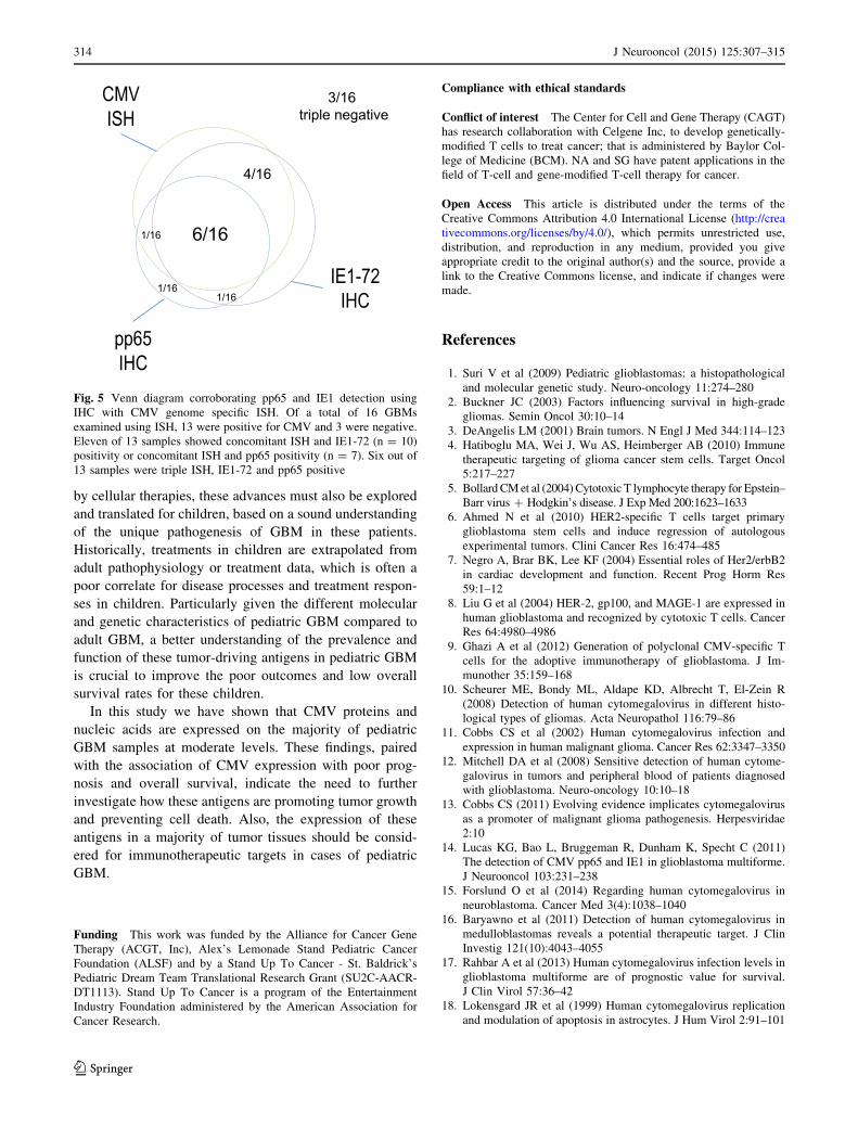
1. Suri V et al (2009) Pediatric glioblastomas: a histopathological
and molecular genetic study. Neuro-oncology 11:274–280
2. Buckner JC (2003) Factors influencing survival in high-grade
gliomas. Semin Oncol 30:10–14
3. DeAngelis LM (2001) Brain tumors. N Engl J Med 344:114–123
4. Hatiboglu MA, Wei J, Wu AS, Heimberger AB (2010) Immune
therapeutic targeting of glioma cancer stem cells. Target Oncol
5:217–227
5. Bollard CM et al (2004) Cytotoxic T lymphocyte therapy for Epstein–
Barr virus ? Hodgkin’s disease. J Exp Med 200:1623–1633
6. Ahmed N et al (2010) HER2-specific T cells target primary
glioblastoma stem cells and induce regression of autologous
experimental tumors. Clini Cancer Res 16:474–485
7. Negro A, Brar BK, Lee KF (2004) Essential roles of Her2/erbB2
in cardiac development and function. Recent Prog Horm Res
59:1–12
8. Liu G et al (2004) HER-2, gp100, and MAGE-1 are expressed in
human glioblastoma and recognized by cytotoxic T cells. Cancer
Res 64:4980–4986
9. Ghazi A et al (2012) Generation of polyclonal CMV-specific T
cells for the adoptive immunotherapy of glioblastoma. J Im-
munother 35:159–168
10. Scheurer ME, Bondy ML, Aldape KD, Albrecht T, El-Zein R
(2008) Detection of human cytomegalovirus in different histo-
logical types of gliomas. Acta Neuropathol 116:79–86
11. Cobbs CS et al (2002) Human cytomegalovirus infection and
expression in human malignant glioma. Cancer Res 62:3347–3350
12. Mitchell DA et al (2008) Sensitive detection of human cytome-
galovirus in tumors and peripheral blood of patients diagnosed
with glioblastoma. Neuro-oncology 10:10–18
13. Cobbs CS (2011) Evolving evidence implicates cytomegalovirus
as a promoter of malignant glioma pathogenesis. Herpesviridae
2:10
14. Lucas KG, Bao L, Bruggeman R, Dunham K, Specht C (2011)
The detection of CMV pp65 and IE1 in glioblastoma multiforme.
J Neurooncol 103:231–238
15. Forslund O et al (2014) Regarding human cytomegalovirus in
neuroblastoma. Cancer Med 3(4):1038–1040
16. Baryawno et al (2011) Detection of human cytomegalovirus in
medulloblastomas reveals a potential therapeutic target. J Clin
Investig 121(10):4043–4055
17. Rahbar A et al (2013) Human cytomegalovirus infection levels in
glioblastoma multiforme are of prognostic value for survival.
J Clin Virol 57:36–42
18. Lokensgard JR et al (1999) Human cytomegalovirus replication
and modulation of apoptosis in astrocytes. J Hum Virol 2:91–101
3/16 triple negative
CMVISH
IE1-72IHC
pp65IHC
1/161/16
6/161/16
4/16
Fig. 5 Venn diagram corroborating pp65 and IE1 detection using
IHC with CMV genome specific ISH. Of a total of 16 GBMs
examined using ISH, 13 were positive for CMV and 3 were negative.
Eleven of 13 samples showed concomitant ISH and IE1-72 (n = 10)
positivity or concomitant ISH and pp65 positivity (n = 7). Six out of
13 samples were triple ISH, IE1-72 and pp65 positive
314 J Neurooncol (2015) 125:307–315
123
19. Salvant BS, Fortunato EA, Spector DH (1998) Cell cycle dys-
regulation by human cytomegalovirus: influence of the cell cycle
phase at the time of infection and effects on cyclin transcription.
J Virol 72:3729–3741
20. Ahmed N et al (2007) Regression of experimental medulloblas-
toma following transfer of HER2-specific T cells. Cancer Res
67:5957–5964
21. Prins RM, Cloughesy TF, Liau LM (2008) Cytomegalovirus
immunity after vaccination with autologous glioblastoma lysate.
N Engl J Med 5:539–541
22. Soderberg-Naucler C, Peredo I, Stragliotto G (2013) Valganci-
clovir in patients with glioblastoma. N Engl J Med
369:2066–2067
J Neurooncol (2015) 125:307–315 315
123