is acrin 6666 annual survey ultrasound place … initials case no. ... philips/atl model ... 14....
TRANSCRIPT
6666 ISb 11-01-05 1 of 27
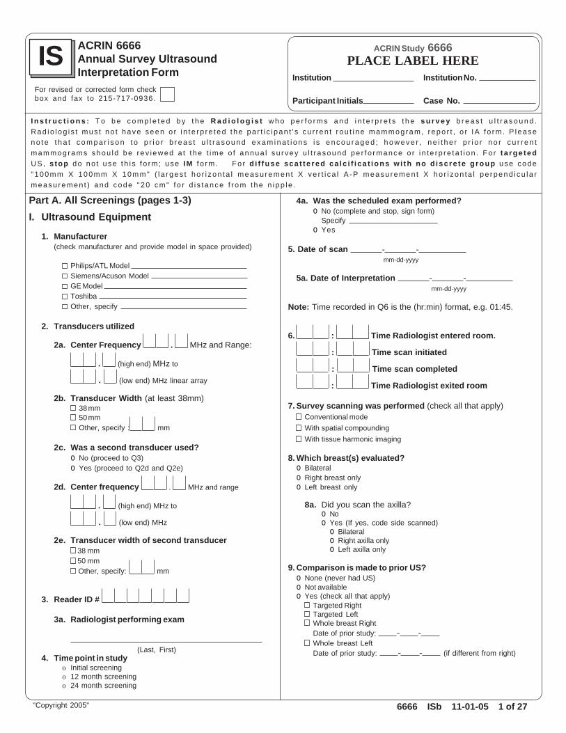
ACRIN 6666Annual Survey UltrasoundInterpretation Form
IS ACRIN Study 6666
Institution No.
Case No.Participant Initials
Institution
PLACE LABEL HERE
I n s t r u c t i o n s : T o b e c o m p l e t e d b y t h e R a d i o l o g i s t w h o p e r f o r m s a n d i n t e r p r e t s t h e s u r v e y b r e a s t u l t r a s o u n d .Rad io log i s t mus t no t have seen o r i n te rp re ted t he pa r t i c i pan t ' s cu r ren t r ou t i ne mammogram, repo r t , o r IA f o rm . P leasen o t e t h a t c o m p a r i s o n t o p r i o r b r e a s t u l t r a s o u n d e x a m i n a t i o n s i s e n c o u r a g e d ; h o w e v e r , n e i t h e r p r i o r n o r c u r r e n tmammograms shou ld be rev iewed a t t he t ime o f annua l su rvey u l t r asound pe r fo rmance o r i n te rp re ta t i on . Fo r t a rge tedUS, stop do no t use th i s f o rm ; use IM f o rm . Fo r d i f fuse sca t te red ca lc i f i ca t ions w i th no d iscre te g roup use code"100mm X 100mm X 10mm" ( l a rges t ho r i zon ta l measu remen t X ve r t i ca l A -P measu remen t X ho r i zon ta l pe rpend i cu la rmeasu remen t ) and code "20 cm" f o r d i s tance f r om the n ipp le .
4a. Was the scheduled exam performed?o No (complete and stop, sign form)
Specify o Yes
5. Date of scan - - mm-dd-yyyy
5a. Date of Interpretation - - mm-dd-yyyy
Note: Time recorded in Q6 is the (hr:min) format, e.g. 01:45.
6. : Time Radiologist entered room.
: Time scan initiated
: Time scan completed
: Time Radiologist exited room
7. Survey scanning was performed (check all that apply)Conventional modeWith spatial compoundingWith tissue harmonic imaging
8. Which breast(s) evaluated?o Bilateralo Right breast onlyo Left breast only
8a. Did you scan the axilla?o Noo Yes (If yes, code side scanned)
o Bilateralo Right axilla onlyo Left axilla only
9. Comparison is made to prior US?o None (never had US)o Not availableo Yes (check all that apply)
Targeted RightTargeted LeftWhole breast RightDate of prior study: - -Whole breast LeftDate of prior study: - - (if different from right)
Part A. All Screenings (pages 1-3)I. Ultrasound Equipment
1. Manufacturer(check manufacturer and provide model in space provided)
Philips/ATL Model Siemens/Acuson Model GE Model Toshiba Other, specify
2. Transducers utilized
2a. Center Frequency . MHz and Range:
. (high end) MHz to
. (low end) MHz linear array
2b. Transducer Width (at least 38mm)38 mm50 mmOther, specify : mm
2c. Was a second transducer used?o No (proceed to Q3)o Yes (proceed to Q2d and Q2e)
2d. Center frequency . MHz and range
. (high end) MHz to
. (low end) MHz
2e. Transducer width of second transducer 38 mm 50 mm Other, specify: mm
3. Reader ID #
3a. Radiologist performing exam
(Last, First)4. Time point in study
o Initial screeningo 12 month screeningo 24 month screening
"Copyright 2005"
For revised or corrected form checkbox and fax to 215-717-0936.

6666 ISb 11-01-05 2 of 27
ACRIN Study 6666
Institution No.
Case No.Participant Initials
Institution
PLACE LABEL HERE IS If this is a revised or correctedform, please check box
"Copyright 2005"
10. Greatest depth (thickness) of Breast by ultrasound
Right Lefto < 2 cm o < 2 cmo 2.0-2.9 cm o 2.0-2.9 cmo 3.0-3.9 cm o 3.0-3.9 cmo 4.0-4.9 cm o 4.0-4.9 cmo 5.0-5.9 cm o 5.0-5.9 cmo 6.0-6.9 cm o 6.0-6.9 cmo >7 cm o >7 cmo not applicable o not applicable
11. Background EchotextureR L
HomogeneousDiffusely HeterogeneousFocally Heterogeneous (If focally heterogenous, code all applicable quadrants)Right Left
UOQ UOQ UIQ UIQ LOQ LOQ LIQ LIQ
12. Were any simple cysts identified?o No (proceed to Q13)o Yes (If yes, proceed to Q12a)
12a. Right o Solitary o 2-3 o numerous (>4) Left o Solitary o 2-3 o numerous (>4)
12b. Detail Largest Cyst
o R o L o' clock cm . cm mm
12c. Are any previously enumerated lesions from any prior sonograms now gone?o No (proceed to Q13)o Yes (If yes, detail below)
______ Number of previously enumerated lesions now gone since last annual screening.
Note: Do not reuse Lesion # once it has been reported as gone.Lesion # ____
Lesion # ____
Lesion # ____
Lesion # ____
Lesion # ____
Lesion # ____
Lesion # ____
Lesion # ____
13. Were any discrete lesions other than simple cysts identified?o No (proceed to Q14)o Yes (complete and proceed to Q20)
o Bilateralo Right breast onlyo Left breast only
Depth fromskin to center
of cyst(to nearest 0.5 cm)
Distance fromthe nipple
ClockfaceBreast Largest Dimension(report on hour and 1/2 houre.g. 7:00 = 0700, 12:30 = 1230)
6666 ISb 11-01-05 3 of 27
ACRIN Study 6666
Institution No.
Case No.Participant Initials
Institution
PLACE LABEL HERE IS If this is a revised or correctedform, please check box
"Copyright 2005"
14. Final Assessment of Right Breast
14a. Not on study (proceed to Q17)
14b. % Likelihood of malignancyfor the right breast (best guess from 0-100)
15. Final assessment for the entire right breast
o 1 Negativeo 2 Benigno 3 Probably Benigno 4A Low Suspicion of Malignancyo 4B Intermediate Suspiciono 4C Moderately High Suspiciono 5 Highly Suggestive of Malignancy
16. Recommendation for right breasto Routine screening in one yearo Diagnostic follow-up in one yearo Short-interval follow-up in 6 months with USo Intervention and/or Additional Imaging
(detail intervention and/or additional imaging)Interventiono Aspiration w/core biopsy if solido US-guided core biopsyo Vacuum-assisted biopsy, guidance by USo Vacuum-assisted biopsy, guidance by Mammoo Excisional biopsyAdditional Imaging (check all that apply)
Comparison to current mammograms is required(lesion seen on US)Comparison to prior mammograms is requiredAdditional mammographic projections
17. Final Assessment of Left Breast
17a. Not on study (sign and date form)
17b. % Likelihood of malignancyfor the left breast (best guess from 0-100)
18. Final assessment for the entire left breast
o 1 Negativeo 2 Benigno 3 Probably Benigno 4A Low Suspicion of Malignancyo 4B Intermediate Suspiciono 4C Moderately High Suspiciono 5 Highly Suggestive of Malignancy
19. Recommendation for left breasto Routine screening in one yearo Diagnostic follow-up in one yearo Short-interval follow-up in 6 months with USo Intervention and/or Additional Imaging
(detail intervention and/or additional imaging)Interventiono Aspiration w/core biopsy if solido US-guided core biopsyo Vacuum-assisted biopsy, guidance by USo Vacuum-assisted biopsy, guidance by Mammoo Excisional biopsyAdditional Imaging (check all that apply)
Comparison to current mammograms is required(lesion seen on US)Comparison to prior mammograms is requiredAdditional mammographic projections
Stop, sign and date form.
Comments:
- -Signature of Radiologist responsible for the data 1 Date Form Completed (mm-dd-yyyy)
Signature of person entering data onto web 2

6666 ISb 11-01-05 4 of 27
ACRIN Study 6666
Institution No.
Case No.Participant Initials
Institution
PLACE LABEL HERE IS If this is a revised or correctedform, please check box
"Copyright 2005"
Part B. Positive Findings (pages 4-27) as needed20. List lesions other than simple cysts (maximum of 4 per breast)
20a. Number of solid findings/lesions other than simple cysts: Right Breast Left Breast (Note: If there are multiple bilateral similar-appearing circumscribed masses, code this as one bilateral lesion).
20b. Lesion # (e.g. UR1, UB1, UL1 etc.)(Retain lesion numbering from initial study survey sonogram. If this is the first examination, begin with R1 for the first lesion in the rightbreast, R2 for the second lesion in the right breast etc. If the finding is new since a prior study sonogram, use next sequential #. Describeany new or suspicious findings first. Location, distance from nipple, depth to lesion center and measurements are completedfor all reportable findings).
Was this "lesion" seen on a previous sonogram including any sonograms performed prior to study enrollment?o Not applicable, no prior breast sonogramso Noo Yes
o Goneo Decreased in size since previous examo Stable in size since previous examo Multiple bilateral circumscribed masses fluctuating
in size since previous examo Increased in size since previous examo Other suspicious changeo Increasing and other suspicious change
Is this "lesion" multiple bilateral circumscribed masses? If yes, describe location and measurement of largest mass.o Noo Yes
o R o L o' clock cm . cm20c. Lesion Size
mm mm mm mm3
20d. Is this lesion at the site of prior biopsy?o Noo Yes (if yes, select prior procedure)
o Core/vacuum biopsy with clip (if procedure performed, select diagnosis)o Core/vacuum biopsy without marker (if procedure performed, select diagnosis)o Surgical biopsy site (if procedure was performed, select diagnosis)
o Benigno Atypical/high-risk lesiono Cancer siteo Unknown
o Biopsy details unknowno FNAB
o Not applicable, multiple bilateral circumscribed masses
20e. Special Case (see choices below)o Noo Yes (if yes, detail below then proceed to Q20n)
(Special Case Features)o Complicated Cyst (Note: Do not use this term for "complex cystic masses".
For complex cystic masses code "No" for Q20e, proceed to Q20f and indicate "complex cystic" at Q20j.)Homogeneous low-level echoesFluid-Debris LevelMobile internal echoesMultiple bilateral complicated cysts in company of simple cysts
o Multiple bilateral solid oval, circumscribed masseso Mass in or on skino Clustered microcystso Intraductal masso Lymph nodeo Calcifications without a masso Foreign bodyo Post-Surgical scaro Other, specify:
U
Note: Volume is pro-grammed to be calcu-lated on line; however,as verification, pleasecalculate volume basedon horizontal, verticaland perpendicular mea-surements as a valida-tion.
Depth from skin tocenter of lesion
(to nearest 0.5 cm)
Distance fromthe nipple
Clockface(report on the hour)Breast
(report on hour and 1/2 houre.g. 7:00 = 0700, 12:30 = 1230)
HorizontalPerpendicular Meas
(mm) D3Vertical A-P
meas (mm) D2
Measured PlaneLargestHorizontal
Meas (mm) D1
SecondMeasured Plane
Volume D1XD2XD3 2..
o Trvo Sago Rado Arado Oblique
o Trvo Sago Rado Arado Perpendicular Oblique
X X
6666 ISb 11-01-05 5 of 27
ACRIN Study 6666
Institution No.
Case No.Participant Initials
Institution
PLACE LABEL HERE IS If this is a revised or correctedform, please check box
"Copyright 2005"
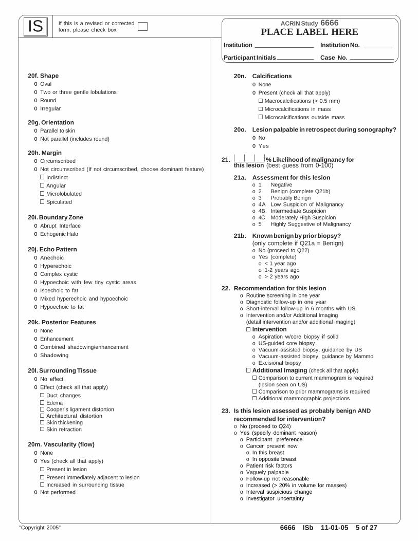
20f. Shapeo Ovalo Two or three gentle lobulationso Roundo Irregular
20g. Orientationo Parallel to skino Not parallel (includes round)
20h. Margino Circumscribedo Not circumscribed (If not circumscribed, choose dominant feature)
IndistinctAngularMicrolobulatedSpiculated
20i. Boundary Zoneo Abrupt Interfaceo Echogenic Halo
20j. Echo Patterno Anechoico Hyperechoico Complex cystico Hypoechoic with few tiny cystic areaso Isoechoic to fato Mixed hyperechoic and hypoechoico Hypoechoic to fat
20k. Posterior Featureso Noneo Enhancemento Combined shadowing/enhancemento Shadowing
20l. Surrounding Tissueo No effecto Effect (check all that apply)
Duct changesEdemaCooper’s ligament distortionArchitectural distortionSkin thickeningSkin retraction
20m. Vascularity (flow)o Noneo Yes (check all that apply)
Present in lesionPresent immediately adjacent to lesionIncreased in surrounding tissue
o Not performed
20n. Calcificationso Noneo Present (check all that apply)
Macrocalcifications (> 0.5 mm)Microcalcifications in massMicrocalcifications outside mass
20o. Lesion palpable in retrospect during sonography?o Noo Yes
21. % Likelihood of malignancy forthis lesion (best guess from 0-100)
21a. Assessment for this lesiono 1 Negativeo 2 Benign (complete Q21b)o 3 Probably Benigno 4A Low Suspicion of Malignancyo 4B Intermediate Suspiciono 4C Moderately High Suspiciono 5 Highly Suggestive of Malignancy
21b. Known benign by prior biopsy?(only complete if Q21a = Benign)o No (proceed to Q22)o Yes (complete)
o < 1 year agoo 1-2 years agoo > 2 years ago
22. Recommendation for this lesiono Routine screening in one yearo Diagnostic follow-up in one yearo Short-interval follow-up in 6 months with USo Intervention and/or Additional Imaging
(detail intervention and/or additional imaging)Interventiono Aspiration w/core biopsy if solido US-guided core biopsyo Vacuum-assisted biopsy, guidance by USo Vacuum-assisted biopsy, guidance by Mammoo Excisional biopsyAdditional Imaging (check all that apply)
Comparison to current mammogram is required(lesion seen on US)Comparison to prior mammograms is requiredAdditional mammographic projections
23. Is this lesion assessed as probably benign ANDrecommended for intervention?o No (proceed to Q24)o Yes (specify dominant reason)
o Participant preferenceo Cancer present now
o In this breasto In opposite breast
o Patient risk factorso Vaguely palpableo Follow-up not reasonableo Increased (> 20% in volume for masses)o Interval suspicious changeo Investigator uncertainty
6666 ISb 11-01-05 6 of 27
ACRIN Study 6666
Institution No.
Case No.Participant Initials
Institution
PLACE LABEL HERE IS If this is a revised or correctedform, please check box
"Copyright 2005"
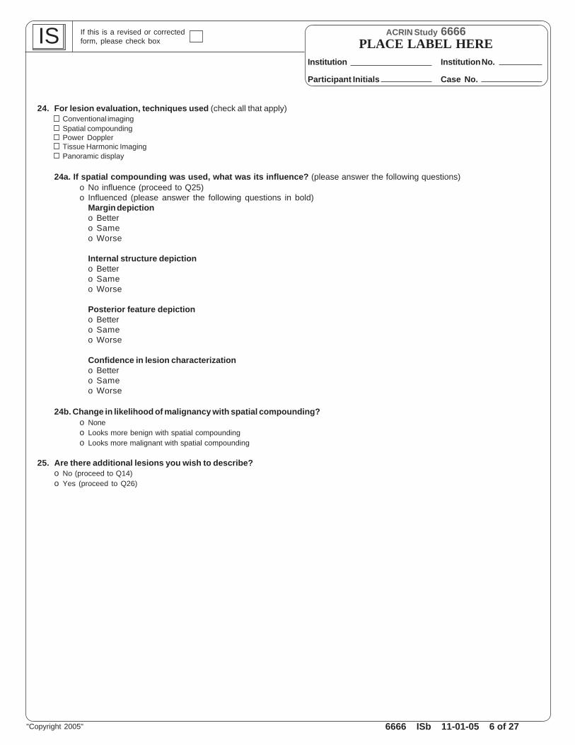
24. For lesion evaluation, techniques used (check all that apply)Conventional imagingSpatial compoundingPower DopplerTissue Harmonic ImagingPanoramic display
24a. If spatial compounding was used, what was its influence? (please answer the following questions)o No influence (proceed to Q25)o Influenced (please answer the following questions in bold)
Margin depictiono Bettero Sameo Worse
Internal structure depictiono Bettero Sameo Worse
Posterior feature depictiono Bettero Sameo Worse
Confidence in lesion characterizationo Bettero Sameo Worse
24b. Change in likelihood of malignancy with spatial compounding?o Noneo Looks more benign with spatial compoundingo Looks more malignant with spatial compounding
25. Are there additional lesions you wish to describe?o No (proceed to Q14)o Yes (proceed to Q26)

6666 ISb 11-01-05 7 of 27
ACRIN Study 6666
Institution No.
Case No.Participant Initials
Institution
PLACE LABEL HERE IS If this is a revised or correctedform, please check box
"Copyright 2005"
26. Additional lesions other than simple cysts (maximum of 4 per breast)
26a. Lesion # (e.g. UR1, UB1, UL1 etc.)(Retain lesion numbering from initial study survey sonogram. If this is the first examination, begin with R1 for the first lesion in the rightbreast, R2 for the second lesion in the right breast etc. If the finding is new since a prior study sonogram, use next sequential #. Describeany new or suspicious findings first. Location, distance from nipple, depth to lesion center and measurements are completedfor all reportable findings).
26b. Was this "lesion" seen on a previous sonogram including any sonograms performed prior to study enrollment?o Not applicable, no prior breast sonogramso Noo Yes
o Goneo Decreased in size since previous examo Stable in size since previous examo Multiple bilateral circumscribed masses fluctuating in size since previous examo Increased in size since previous examo Other suspicious changeo Increasing and other suspicious change
Is this "lesion" multiple bilateral circumscribed masses? If yes, describe location and measurement of largest mass.o Noo Yes
o R o L o' clock cm . cm
26c. Lesion Size
mm mm mm mm3
26d. Is this lesion at the site of prior biopsy?o Noo Yes (if yes, select prior procedure)
o Core/vacuum biopsy with clip (if procedure performed, select diagnosis)o Core/vacuum biopsy without marker (if procedure performed, select diagnosis)o Surgical biopsy site (if procedure was performed, select diagnosis)
o Benigno Atypical/high-risk lesiono Cancer siteo Unknown
o Biopsy details unknowno FNAB
o Not applicable, multiple bilateral circumscribed masses
26e. Special Case (see choices below)o Noo Yes (if yes, detail below then proceed to Q26n)
(Special Case Features)o Complicated Cyst (Note: Do not use this term for "complex cystic masses".
For complex cystic masses code "No" for Q26e, proceed to Q26f and indicate "complex cystic" at Q26j.)Homogeneous low-level echoesFluid-Debris LevelMobile internal echoesMultiple bilateral complicated cysts in company of simple cysts
o Multiple bilateral solid oval, circumscribed masseso Mass in or on skino Clustered microcystso Intraductal masso Lymph nodeo Calcifications without a masso Foreign bodyo Post-Surgical scaro Other, specify:
U
Note: Volume is pro-grammed to be calcu-lated on line; however,as verification, pleasecalculate volume basedon horizontal, verticaland perpendicular mea-surements as a valida-tion.
Depth from skin tocenter of lesion
(to nearest 0.5 cm)
Distance fromthe nipple
Clockface(report on the hour)Breast
(report on hour and 1/2 houre.g. 7:00 = 0700, 12:30 = 1230)
HorizontalPerpendicular Meas
(mm) D3Vertical A-P
meas (mm) D2Measured Plane
LargestHorizontal
Meas (mm) D1
SecondMeasured Plane Volume D1XD2XD3 2
.
.
X Xo Trvo Sago Rado Arado Oblique
o Trvo Sago Rado Arado Perpendicular Oblique
6666 ISb 11-01-05 8 of 27
ACRIN Study 6666
Institution No.
Case No.Participant Initials
Institution
PLACE LABEL HERE IS If this is a revised or correctedform, please check box
"Copyright 2005"
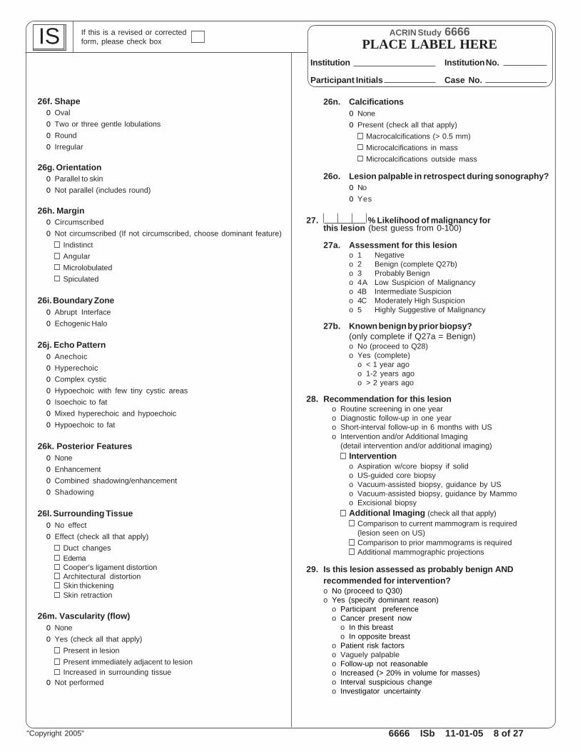
26f. Shapeo Ovalo Two or three gentle lobulationso Roundo Irregular
26g. Orientationo Parallel to skino Not parallel (includes round)
26h. Margino Circumscribedo Not circumscribed (If not circumscribed, choose dominant feature)
IndistinctAngularMicrolobulatedSpiculated
26i. Boundary Zoneo Abrupt Interfaceo Echogenic Halo
26j. Echo Patterno Anechoico Hyperechoico Complex cystico Hypoechoic with few tiny cystic areaso Isoechoic to fato Mixed hyperechoic and hypoechoico Hypoechoic to fat
26k. Posterior Featureso Noneo Enhancemento Combined shadowing/enhancemento Shadowing
26l. Surrounding Tissueo No effecto Effect (check all that apply)
Duct changesEdemaCooper’s ligament distortionArchitectural distortionSkin thickeningSkin retraction
26m. Vascularity (flow)o Noneo Yes (check all that apply)
Present in lesionPresent immediately adjacent to lesionIncreased in surrounding tissue
o Not performed
26n. Calcificationso Noneo Present (check all that apply)
Macrocalcifications (> 0.5 mm)Microcalcifications in massMicrocalcifications outside mass
26o. Lesion palpable in retrospect during sonography?o Noo Yes
27. % Likelihood of malignancy forthis lesion (best guess from 0-100)
27a. Assessment for this lesiono 1 Negativeo 2 Benign (complete Q27b)o 3 Probably Benigno 4A Low Suspicion of Malignancyo 4B Intermediate Suspiciono 4C Moderately High Suspiciono 5 Highly Suggestive of Malignancy
27b. Known benign by prior biopsy?(only complete if Q27a = Benign)o No (proceed to Q28)o Yes (complete)
o < 1 year agoo 1-2 years agoo > 2 years ago
28. Recommendation for this lesiono Routine screening in one yearo Diagnostic follow-up in one yearo Short-interval follow-up in 6 months with USo Intervention and/or Additional Imaging
(detail intervention and/or additional imaging)Interventiono Aspiration w/core biopsy if solido US-guided core biopsyo Vacuum-assisted biopsy, guidance by USo Vacuum-assisted biopsy, guidance by Mammoo Excisional biopsyAdditional Imaging (check all that apply)
Comparison to current mammogram is required(lesion seen on US)Comparison to prior mammograms is requiredAdditional mammographic projections
29. Is this lesion assessed as probably benign ANDrecommended for intervention?o No (proceed to Q30)o Yes (specify dominant reason)
o Participant preferenceo Cancer present now
o In this breasto In opposite breast
o Patient risk factorso Vaguely palpableo Follow-up not reasonableo Increased (> 20% in volume for masses)o Interval suspicious changeo Investigator uncertainty

6666 ISb 11-01-05 9 of 27
ACRIN Study 6666
Institution No.
Case No.Participant Initials
Institution
PLACE LABEL HERE IS If this is a revised or correctedform, please check box
"Copyright 2005"
30. For lesion evaluation, techniques used (check all that apply)Conventional imagingSpatial compoundingPower DopplerTissue Harmonic ImagingPanoramic display
30a. If spatial compounding was used, what was its influence? (please answer the following questions)o No influence (proceed to Q31)o Influenced (please answer the following questions in bold)
Margin depictiono Bettero Sameo Worse
Internal structure depictiono Bettero Sameo Worse
Posterior feature depictiono Bettero Sameo Worse
Confidence in lesion characterizationo Bettero Sameo Worse
30b. Change in likelihood of malignancy with spatial compounding?o Noneo Looks more benign with spatial compoundingo Looks more malignant with spatial compounding
31. Are there additional lesions you wish to describe?o No (proceed to Q14)o Yes (proceed to Q32)

6666 ISb 11-01-05 10 of 27
ACRIN Study 6666
Institution No.
Case No.Participant Initials
Institution
PLACE LABEL HERE IS If this is a revised or correctedform, please check box
"Copyright 2005"
32. Additional lesions other than simple cysts (maximum of 4 per breast)
32a. Lesion # (e.g. UR1, UB1, UL1 etc.)(Retain lesion numbering from initial study survey sonogram. If this is the first examination, begin with R1 for the first lesion in the rightbreast, R2 for the second lesion in the right breast etc. If the finding is new since a prior study sonogram, use next sequential #. Describeany new or suspicious findings first. Location, distance from nipple, depth to lesion center and measurements are completedfor all reportable findings).
32b. Was this "lesion" seen on a previous sonogram including any sonograms performed prior to study enrollment?o Not applicable, no prior breast sonogramso Noo Yes
o Goneo Decreased in size since previous examo Stable in size since previous examo Multiple bilateral circumscribed masses fluctuating in size since previous examo Increased in size since previous examo Other suspicious changeo Increasing and other suspicious change
Is this "lesion" multiple bilateral circumscribed masses? If yes, describe location and measurement of largest mass.o Noo Yes
o R o L o' clock cm . cm
32c. Lesion Size
mm mm mm mm3
32d. Is this lesion at the site of prior biopsy?o Noo Yes (if yes, select prior procedure)
o Core/vacuum biopsy with clip (if procedure performed, select diagnosis)o Core/vacuum biopsy without marker (if procedure performed, select diagnosis)o Surgical biopsy site (if procedure was performed, select diagnosis)
o Benigno Atypical/high-risk lesiono Cancer siteo Unknown
o Biopsy details unknowno FNAB
o Not applicable, multiple bilateral circumscribed masses
32e. Special Case (see choices below)o Noo Yes (if yes, detail below then proceed to Q32n)
(Special Case Features)o Complicated Cyst (Note: Do not use this term for "complex cystic masses".
For complex cystic masses code "No" for Q32e, proceed to Q32f and indicate "complex cystic" at Q32j.)Homogeneous low-level echoesFluid-Debris LevelMobile internal echoesMultiple bilateral complicated cysts in company of simple cysts
o Multiple bilateral solid oval, circumscribed masseso Mass in or on skino Clustered microcystso Intraductal masso Lymph nodeo Calcifications without a masso Foreign bodyo Post-Surgical scaro Other, specify:
U
Note: Volume is pro-grammed to be calcu-lated on line; however,as verification, pleasecalculate volume basedon horizontal, verticaland perpendicular mea-surements as a valida-tion.
Depth from skin tocenter of lesion
(to nearest 0.5 cm)
Distance fromthe nipple
Clockface(report on the hour)Breast
(report on hour and 1/2 houre.g. 7:00 = 0700, 12:30 = 1230)
HorizontalPerpendicular Meas
(mm) D3Vertical A-P
meas (mm) D2Measured Plane
LargestHorizontal
Meas (mm) D1
SecondMeasured Plane Volume D1XD2XD3 2
.
.o Trvo Sago Rado Arado Oblique
o Trvo Sago Rado Arado Perpendicular Oblique
X X

6666 ISb 11-01-05 11 of 27
ACRIN Study 6666
Institution No.
Case No.Participant Initials
Institution
PLACE LABEL HERE IS If this is a revised or correctedform, please check box
"Copyright 2005"
32f. Shapeo Ovalo Two or three gentle lobulationso Roundo Irregular
32g. Orientationo Parallel to skino Not parallel (includes round)
32h. Margino Circumscribedo Not circumscribed (If not circumscribed, choose dominant feature)
IndistinctAngularMicrolobulatedSpiculated
32i. Boundary Zoneo Abrupt Interfaceo Echogenic Halo
32j. Echo Patterno Anechoico Hyperechoico Complex cystico Hypoechoic with few tiny cystic areaso Isoechoic to fato Mixed hyperechoic and hypoechoico Hypoechoic to fat
32k. Posterior Featureso Noneo Enhancemento Combined shadowing/enhancemento Shadowing
32l. Surrounding Tissueo No effecto Effect (check all that apply)
Duct changesEdemaCooper’s ligament distortionArchitectural distortionSkin thickeningSkin retraction
32m. Vascularity (flow)o Noneo Yes (check all that apply)
Present in lesionPresent immediately adjacent to lesionIncreased in surrounding tissue
o Not performed
32n. Calcificationso Noneo Present (check all that apply)
Macrocalcifications (> 0.5 mm)Microcalcifications in massMicrocalcifications outside mass
32o. Lesion palpable in retrospect during sonography?o Noo Yes
33. % Likelihood of malignancy forthis lesion (best guess from 0-100)
33a. Assessment for this lesiono 1 Negativeo 2 Benign (complete Q33b)o 3 Probably Benigno 4A Low Suspicion of Malignancyo 4B Intermediate Suspiciono 4C Moderately High Suspiciono 5 Highly Suggestive of Malignancy
33b. Known benign by prior biopsy?(only complete if Q33a = Benign)o No (proceed to Q34)o Yes (complete)
o < 1 year agoo 1-2 years agoo > 2 years ago
34. Recommendation for this lesiono Routine screening in one yearo Diagnostic follow-up in one yearo Short-interval follow-up in 6 months with USo Intervention and/or Additional Imaging
(detail intervention and/or additional imaging)Interventiono Aspiration w/core biopsy if solido US-guided core biopsyo Vacuum-assisted biopsy, guidance by USo Vacuum-assisted biopsy, guidance by Mammoo Excisional biopsyAdditional Imaging (check all that apply)
Comparison to current mammogram is required(lesion seen on US)Comparison to prior mammograms is requiredAdditional mammographic projections
35. Is this lesion assessed as probably benign ANDrecommended for intervention?o No (proceed to Q36)o Yes (specify dominant reason)
o Participant preferenceo Cancer present now
o In this breasto In opposite breast
o Patient risk factorso Vaguely palpableo Follow-up not reasonableo Increased (> 20% in volume for masses)o Interval suspicious changeo Investigator uncertainty

6666 ISb 11-01-05 12 of 27
ACRIN Study 6666
Institution No.
Case No.Participant Initials
Institution
PLACE LABEL HERE IS If this is a revised or correctedform, please check box
"Copyright 2005"
36. For lesion evaluation, techniques used (check all that apply)Conventional imagingSpatial compoundingPower DopplerTissue Harmonic ImagingPanoramic display
36a. If spatial compounding was used, what was its influence? (please answer the following questions)o No influence (proceed to Q37)o Influenced (please answer the following questions in bold)
Margin depictiono Bettero Sameo Worse
Internal structure depictiono Bettero Sameo Worse
Posterior feature depictiono Bettero Sameo Worse
Confidence in lesion characterizationo Bettero Sameo Worse
36b. Change in likelihood of malignancy with spatial compounding?o Noneo Looks more benign with spatial compoundingo Looks more malignant with spatial compounding
37. Are there additional lesions you wish to describe?o No (proceed to Q14)o Yes (proceed to Q38)
6666 ISb 11-01-05 13 of 27
ACRIN Study 6666
Institution No.
Case No.Participant Initials
Institution
PLACE LABEL HERE IS If this is a revised or correctedform, please check box
"Copyright 2005"
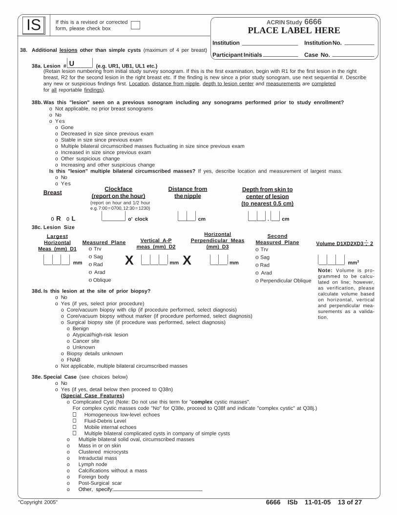
38. Additional lesions other than simple cysts (maximum of 4 per breast)
38a. Lesion # (e.g. UR1, UB1, UL1 etc.)(Retain lesion numbering from initial study survey sonogram. If this is the first examination, begin with R1 for the first lesion in the rightbreast, R2 for the second lesion in the right breast etc. If the finding is new since a prior study sonogram, use next sequential #. Describeany new or suspicious findings first. Location, distance from nipple, depth to lesion center and measurements are completedfor all reportable findings).
38b. Was this "lesion" seen on a previous sonogram including any sonograms performed prior to study enrollment?o Not applicable, no prior breast sonogramso Noo Yes
o Goneo Decreased in size since previous examo Stable in size since previous examo Multiple bilateral circumscribed masses fluctuating in size since previous examo Increased in size since previous examo Other suspicious changeo Increasing and other suspicious change
Is this "lesion" multiple bilateral circumscribed masses? If yes, describe location and measurement of largest mass.o Noo Yes
o R o L o' clock cm . cm38c. Lesion Size
mm mm mm mm3
38d. Is this lesion at the site of prior biopsy?o Noo Yes (if yes, select prior procedure)
o Core/vacuum biopsy with clip (if procedure performed, select diagnosis)o Core/vacuum biopsy without marker (if procedure performed, select diagnosis)o Surgical biopsy site (if procedure was performed, select diagnosis)
o Benigno Atypical/high-risk lesiono Cancer siteo Unknown
o Biopsy details unknowno FNAB
o Not applicable, multiple bilateral circumscribed masses
38e. Special Case (see choices below)o Noo Yes (if yes, detail below then proceed to Q38n)
(Special Case Features)o Complicated Cyst (Note: Do not use this term for "complex cystic masses".
For complex cystic masses code "No" for Q38e, proceed to Q38f and indicate "complex cystic" at Q38j.)Homogeneous low-level echoesFluid-Debris LevelMobile internal echoesMultiple bilateral complicated cysts in company of simple cysts
o Multiple bilateral solid oval, circumscribed masseso Mass in or on skino Clustered microcystso Intraductal masso Lymph nodeo Calcifications without a masso Foreign bodyo Post-Surgical scaro Other, specify:
U
Note: Volume is pro-grammed to be calcu-lated on line; however,as verification, pleasecalculate volume basedon horizontal, verticaland perpendicular mea-surements as a valida-tion.
HorizontalPerpendicular Meas
(mm) D3Vertical A-P
meas (mm) D2Measured Plane
LargestHorizontal
Meas (mm) D1
SecondMeasured Plane Volume D1XD2XD3 2
.
.
Depth from skin tocenter of lesion
(to nearest 0.5 cm)
Distance fromthe nipple
Clockface(report on the hour)Breast
(report on hour and 1/2 houre.g. 7:00 = 0700, 12:30 = 1230)
o Trvo Sago Rado Arado Oblique
o Trvo Sago Rado Arado Perpendicular Oblique
X X

6666 ISb 11-01-05 14 of 27
ACRIN Study 6666
Institution No.
Case No.Participant Initials
Institution
PLACE LABEL HERE IS If this is a revised or correctedform, please check box
"Copyright 2005"
38f. Shapeo Ovalo Two or three gentle lobulationso Roundo Irregular
38g. Orientationo Parallel to skino Not parallel (includes round)
38h. Margino Circumscribedo Not circumscribed (If not circumscribed, choose dominant feature)
IndistinctAngularMicrolobulatedSpiculated
38i. Boundary Zoneo Abrupt Interfaceo Echogenic Halo
38j. Echo Patterno Anechoico Hyperechoico Complex cystico Hypoechoic with few tiny cystic areaso Isoechoic to fato Mixed hyperechoic and hypoechoico Hypoechoic to fat
38k. Posterior Featureso Noneo Enhancemento Combined shadowing/enhancemento Shadowing
38l. Surrounding Tissueo No effecto Effect (check all that apply)
Duct changesEdemaCooper’s ligament distortionArchitectural distortionSkin thickeningSkin retraction
38m. Vascularity (flow)o Noneo Yes (check all that apply)
Present in lesionPresent immediately adjacent to lesionIncreased in surrounding tissue
o Not performed
38n. Calcificationso Noneo Present (check all that apply)
Macrocalcifications (> 0.5 mm)Microcalcifications in massMicrocalcifications outside mass
38o. Lesion palpable in retrospect during sonography?o Noo Yes
39. % Likelihood of malignancy forthis lesion (best guess from 0-100)
39a. Assessment for this lesiono 1 Negativeo 2 Benign (complete Q39b)o 3 Probably Benigno 4A Low Suspicion of Malignancyo 4B Intermediate Suspiciono 4C Moderately High Suspiciono 5 Highly Suggestive of Malignancy
39b. Known benign by prior biopsy?(only complete if Q39a = Benign)o No (proceed to Q40)o Yes (complete)
o < 1 year agoo 1-2 years agoo > 2 years ago
40. Recommendation for this lesiono Routine screening in one yearo Diagnostic follow-up in one yearo Short-interval follow-up in 6 months with USo Intervention and/or Additional Imaging
(detail intervention and/or additional imaging)Interventiono Aspiration w/core biopsy if solido US-guided core biopsyo Vacuum-assisted biopsy, guidance by USo Vacuum-assisted biopsy, guidance by Mammoo Excisional biopsyAdditional Imaging (check all that apply)
Comparison to current mammogram is required(lesion seen on US)Comparison to prior mammograms is requiredAdditional mammographic projections
41. Is this lesion assessed as probably benign ANDrecommended for intervention?o No (proceed to Q42)o Yes (specify dominant reason)
o Participant preferenceo Cancer present now
o In this breasto In opposite breast
o Patient risk factorso Vaguely palpableo Follow-up not reasonableo Increased (> 20% in volume for masses)o Interval suspicious changeo Investigator uncertainty

6666 ISb 11-01-05 15 of 27
ACRIN Study 6666
Institution No.
Case No.Participant Initials
Institution
PLACE LABEL HERE IS If this is a revised or correctedform, please check box
"Copyright 2005"
42. For lesion evaluation, techniques used (check all that apply)Conventional imagingSpatial compoundingPower DopplerTissue Harmonic ImagingPanoramic display
42a. If spatial compounding was used, what was its influence? (please answer the following questions)o No influence (proceed to Q43)o Influenced (please answer the following questions in bold)
Margin depictiono Bettero Sameo Worse
Internal structure depictiono Bettero Sameo Worse
Posterior feature depictiono Bettero Sameo Worse
Confidence in lesion characterizationo Bettero Sameo Worse
42b. Change in likelihood of malignancy with spatial compounding?o Noneo Looks more benign with spatial compoundingo Looks more malignant with spatial compounding
43. Are there additional lesions you wish to describe?o No (proceed to Q14)o Yes (proceed to Q44)

6666 ISb 11-01-05 16 of 27
ACRIN Study 6666
Institution No.
Case No.Participant Initials
Institution
PLACE LABEL HERE IS If this is a revised or correctedform, please check box
"Copyright 2005"
44. Additional lesions other than simple cysts (maximum of 4 per breast)
44a. Lesion # (e.g. UR1, UB1, UL1 etc.)(Retain lesion numbering from initial study survey sonogram. If this is the first examination, begin with R1 for the first lesion in the rightbreast, R2 for the second lesion in the right breast etc. If the finding is new since a prior study sonogram, use next sequential #. Describeany new or suspicious findings first. Location, distance from nipple, depth to lesion center and measurements are completedfor all reportable findings).
44b. Was this "lesion" seen on a previous sonogram including any sonograms performed prior to study enrollment?o Not applicable, no prior breast sonogramso Noo Yes
o Goneo Decreased in size since previous examo Stable in size since previous examo Multiple bilateral circumscribed masses fluctuating in size since previous examo Increased in size since previous examo Other suspicious changeo Increasing and other suspicious change
Is this "lesion" multiple bilateral circumscribed masses? If yes, describe location and measurement of largest mass.o Noo Yes
o R o L o' clock cm . cm
44c. Lesion Size
mm mm mm mm3
44d. Is this lesion at the site of prior biopsy?o Noo Yes (if yes, select prior procedure)
o Core/vacuum biopsy with clip (if procedure performed, select diagnosis)o Core/vacuum biopsy without marker (if procedure performed, select diagnosis)o Surgical biopsy site (if procedure was performed, select diagnosis)
o Benigno Atypical/high-risk lesiono Cancer siteo Unknown
o Biopsy details unknowno FNAB
o Not applicable, multiple bilateral circumscribed masses
44e. Special Case (see choices below)o Noo Yes (if yes, detail below then proceed to Q44n)
(Special Case Features)o Complicated Cyst (Note: Do not use this term for "complex cystic masses".
For complex cystic masses code "No" for Q44e, proceed to Q44f and indicate "complex cystic" at Q44j.)Homogeneous low-level echoesFluid-Debris LevelMobile internal echoesMultiple bilateral complicated cysts in company of simple cysts
o Multiple bilateral solid oval, circumscribed masseso Mass in or on skino Clustered microcystso Intraductal masso Lymph nodeo Calcifications without a masso Foreign bodyo Post-Surgical scaro Other, specify:
U
Note: Volume is pro-grammed to be calcu-lated on line; however,as verification, pleasecalculate volume basedon horizontal, verticaland perpendicular mea-surements as a valida-tion.
Depth from skin tocenter of lesion
(to nearest 0.5 cm)
Distance fromthe nipple
Clockface(report on the hour)Breast
(report on hour and 1/2 houre.g. 7:00 = 0700, 12:30 = 1230)
HorizontalPerpendicular Meas
(mm) D3Vertical A-P
meas (mm) D2Measured Plane
LargestHorizontal
Meas (mm) D1
SecondMeasured Plane Volume D1XD2XD3 2
.
.o Trvo Sago Rado Arado Oblique
o Trvo Sago Rado Arado Perpendicular Oblique
X X
6666 ISb 11-01-05 17 of 27
ACRIN Study 6666
Institution No.
Case No.Participant Initials
Institution
PLACE LABEL HERE IS If this is a revised or correctedform, please check box
"Copyright 2005"
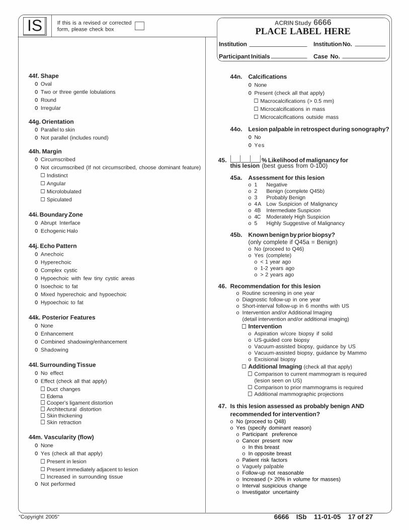
44f. Shapeo Ovalo Two or three gentle lobulationso Roundo Irregular
44g. Orientationo Parallel to skino Not parallel (includes round)
44h. Margino Circumscribedo Not circumscribed (If not circumscribed, choose dominant feature)
IndistinctAngularMicrolobulatedSpiculated
44i. Boundary Zoneo Abrupt Interfaceo Echogenic Halo
44j. Echo Patterno Anechoico Hyperechoico Complex cystico Hypoechoic with few tiny cystic areaso Isoechoic to fato Mixed hyperechoic and hypoechoico Hypoechoic to fat
44k. Posterior Featureso Noneo Enhancemento Combined shadowing/enhancemento Shadowing
44l. Surrounding Tissueo No effecto Effect (check all that apply)
Duct changesEdemaCooper’s ligament distortionArchitectural distortionSkin thickeningSkin retraction
44m. Vascularity (flow)o Noneo Yes (check all that apply)
Present in lesionPresent immediately adjacent to lesionIncreased in surrounding tissue
o Not performed
44n. Calcificationso Noneo Present (check all that apply)
Macrocalcifications (> 0.5 mm)Microcalcifications in massMicrocalcifications outside mass
44o. Lesion palpable in retrospect during sonography?o Noo Yes
45. % Likelihood of malignancy forthis lesion (best guess from 0-100)
45a. Assessment for this lesiono 1 Negativeo 2 Benign (complete Q45b)o 3 Probably Benigno 4A Low Suspicion of Malignancyo 4B Intermediate Suspiciono 4C Moderately High Suspiciono 5 Highly Suggestive of Malignancy
45b. Known benign by prior biopsy?(only complete if Q45a = Benign)o No (proceed to Q46)o Yes (complete)
o < 1 year agoo 1-2 years agoo > 2 years ago
46. Recommendation for this lesiono Routine screening in one yearo Diagnostic follow-up in one yearo Short-interval follow-up in 6 months with USo Intervention and/or Additional Imaging
(detail intervention and/or additional imaging)Interventiono Aspiration w/core biopsy if solido US-guided core biopsyo Vacuum-assisted biopsy, guidance by USo Vacuum-assisted biopsy, guidance by Mammoo Excisional biopsyAdditional Imaging (check all that apply)
Comparison to current mammogram is required(lesion seen on US)Comparison to prior mammograms is requiredAdditional mammographic projections
47. Is this lesion assessed as probably benign ANDrecommended for intervention?o No (proceed to Q48)o Yes (specify dominant reason)
o Participant preferenceo Cancer present now
o In this breasto In opposite breast
o Patient risk factorso Vaguely palpableo Follow-up not reasonableo Increased (> 20% in volume for masses)o Interval suspicious changeo Investigator uncertainty

6666 ISb 11-01-05 18 of 27
ACRIN Study 6666
Institution No.
Case No.Participant Initials
Institution
PLACE LABEL HERE IS If this is a revised or correctedform, please check box
"Copyright 2005"
48. For lesion evaluation, techniques used (check all that apply)Conventional imagingSpatial compoundingPower DopplerTissue Harmonic ImagingPanoramic display
48a. If spatial compounding was used, what was its influence? (please answer the following questions)o No influence (proceed to Q49)o Influenced (please answer the following questions in bold)
Margin depictiono Bettero Sameo Worse
Internal structure depictiono Bettero Sameo Worse
Posterior feature depictiono Bettero Sameo Worse
Confidence in lesion characterizationo Bettero Sameo Worse
48b. Change in likelihood of malignancy with spatial compounding?o Noneo Looks more benign with spatial compoundingo Looks more malignant with spatial compounding
49. Are there additional lesions you wish to describe?o No (proceed to Q14)o Yes (proceed to Q50)

6666 ISb 11-01-05 19 of 27
ACRIN Study 6666
Institution No.
Case No.Participant Initials
Institution
PLACE LABEL HERE IS If this is a revised or correctedform, please check box
"Copyright 2005"
50. Additional lesions other than simple cysts (maximum of 4 per breast)
50a. Lesion # (e.g. UR1, UB1, UL1 etc.)(Retain lesion numbering from initial study survey sonogram. If this is the first examination, begin with R1 for the first lesion in the rightbreast, R2 for the second lesion in the right breast etc. If the finding is new since a prior study sonogram, use next sequential #. Describeany new or suspicious findings first. Location, distance from nipple, depth to lesion center and measurements are completedfor all reportable findings).
50b. Was this "lesion" seen on a previous sonogram including any sonograms performed prior to study enrollment?o Not applicable, no prior breast sonogramso Noo Yes
o Goneo Decreased in size since previous examo Stable in size since previous examo Multiple bilateral circumscribed masses fluctuating in size since previous examo Increased in size since previous examo Other suspicious changeo Increasing and other suspicious change
Is this "lesion" multiple bilateral circumscribed masses? If yes, describe location and measurement of largest mass.o Noo Yes
o R o L o' clock cm . cm
50c. Lesion Size
mm mm mm mm3
50d. Is this lesion at the site of prior biopsy?o Noo Yes (if yes, select prior procedure)
o Core/vacuum biopsy with clip (if procedure performed, select diagnosis)o Core/vacuum biopsy without marker (if procedure performed, select diagnosis)o Surgical biopsy site (if procedure was performed, select diagnosis)
o Benigno Atypical/high-risk lesiono Cancer siteo Unknown
o Biopsy details unknowno FNAB
o Not applicable, multiple bilateral circumscribed masses
50e. Special Case (see choices below)o Noo Yes (if yes, detail below then proceed to Q50n)
(Special Case Features)o Complicated Cyst (Note: Do not use this term for "complex cystic masses".
For complex cystic masses code "No" for Q50e, proceed to Q50f and indicate "complex cystic" at Q50j.)Homogeneous low-level echoesFluid-Debris LevelMobile internal echoesMultiple bilateral complicated cysts in company of simple cysts
o Multiple bilateral solid oval, circumscribed masseso Mass in or on skino Clustered microcystso Intraductal masso Lymph nodeo Calcifications without a masso Foreign bodyo Post-Surgical scaro Other, specify:
U
Note: Volume is pro-grammed to be calcu-lated on line; however,as verification, pleasecalculate volume basedon horizontal, verticaland perpendicular mea-surements as a valida-tion.
Depth from skin tocenter of lesion
(to nearest 0.5 cm)
Distance fromthe nipple
Clockface(report on the hour)Breast
(report on hour and 1/2 houre.g. 7:00 = 0700, 12:30 = 1230)
HorizontalPerpendicular Meas
(mm) D3Vertical A-P
meas (mm) D2Measured Plane
LargestHorizontal
Meas (mm) D1
SecondMeasured Plane Volume D1XD2XD3 2
.
.o Trvo Sago Rado Arado Oblique
o Trvo Sago Rado Arado Perpendicular Oblique
X X

6666 ISb 11-01-05 20 of 27
ACRIN Study 6666
Institution No.
Case No.Participant Initials
Institution
PLACE LABEL HERE IS If this is a revised or correctedform, please check box
"Copyright 2005"
50f. Shapeo Ovalo Two or three gentle lobulationso Roundo Irregular
50g. Orientationo Parallel to skino Not parallel (includes round)
50h. Margino Circumscribedo Not circumscribed (If not circumscribed, choose dominant feature)
IndistinctAngularMicrolobulatedSpiculated
50i. Boundary Zoneo Abrupt Interfaceo Echogenic Halo
50j. Echo Patterno Anechoico Hyperechoico Complex cystico Hypoechoic with few tiny cystic areaso Isoechoic to fato Mixed hyperechoic and hypoechoico Hypoechoic to fat
50k. Posterior Featureso Noneo Enhancemento Combined shadowing/enhancemento Shadowing
50l. Surrounding Tissueo No effecto Effect (check all that apply)
Duct changesEdemaCooper’s ligament distortionArchitectural distortionSkin thickeningSkin retraction
50m. Vascularity (flow)o Noneo Yes (check all that apply)
Present in lesionPresent immediately adjacent to lesionIncreased in surrounding tissue
o Not performed
50n. Calcificationso Noneo Present (check all that apply)
Macrocalcifications (> 0.5 mm)Microcalcifications in massMicrocalcifications outside mass
50o. Lesion palpable in retrospect during sonography?o Noo Yes
51. % Likelihood of malignancy forthis lesion (best guess from 0-100)
51a. Assessment for this lesiono 1 Negativeo 2 Benign (complete Q51b)o 3 Probably Benigno 4A Low Suspicion of Malignancyo 4B Intermediate Suspiciono 4C Moderately High Suspiciono 5 Highly Suggestive of Malignancy
51b. Known benign by prior biopsy?(only complete if Q51a = Benign)o No (proceed to Q52)o Yes (complete)
o < 1 year agoo 1-2 years agoo > 2 years ago
52. Recommendation for this lesiono Routine screening in one yearo Diagnostic follow-up in one yearo Short-interval follow-up in 6 months with USo Intervention and/or Additional Imaging
(detail intervention and/or additional imaging)Interventiono Aspiration w/core biopsy if solido US-guided core biopsyo Vacuum-assisted biopsy, guidance by USo Vacuum-assisted biopsy, guidance by Mammoo Excisional biopsyAdditional Imaging (check all that apply)
Comparison to current mammogram is required(lesion seen on US)Comparison to prior mammograms is requiredAdditional mammographic projections
53. Is this lesion assessed as probably benign ANDrecommended for intervention?o No (proceed to Q54)o Yes (specify dominant reason)
o Participant preferenceo Cancer present now
o In this breasto In opposite breast
o Patient risk factorso Vaguely palpableo Follow-up not reasonableo Increased (> 20% in volume for masses)o Interval suspicious changeo Investigator uncertainty

6666 ISb 11-01-05 21 of 27
ACRIN Study 6666
Institution No.
Case No.Participant Initials
Institution
PLACE LABEL HERE IS If this is a revised or correctedform, please check box
"Copyright 2005"
54. For lesion evaluation, techniques used (check all that apply)Conventional imagingSpatial compoundingPower DopplerTissue Harmonic ImagingPanoramic display
54a. If spatial compounding was used, what was its influence? (please answer the following questions)o No influence (proceed to Q55)o Influenced (please answer the following questions in bold)
Margin depictiono Bettero Sameo Worse
Internal structure depictiono Bettero Sameo Worse
Posterior feature depictiono Bettero Sameo Worse
Confidence in lesion characterizationo Bettero Sameo Worse
54b. Change in likelihood of malignancy with spatial compounding?o Noneo Looks more benign with spatial compoundingo Looks more malignant with spatial compounding
55. Are there additional lesions you wish to describe?o No (proceed to Q14)o Yes (proceed to Q56)

6666 ISb 11-01-05 22 of 27
ACRIN Study 6666
Institution No.
Case No.Participant Initials
Institution
PLACE LABEL HERE IS If this is a revised or correctedform, please check box
"Copyright 2005"
56. Additional lesions other than simple cysts (maximum of 4 per breast)
56a. Lesion # (e.g. UR1, UB1, UL1 etc.)(Retain lesion numbering from initial study survey sonogram. If this is the first examination, begin with R1 for the first lesion in the rightbreast, R2 for the second lesion in the right breast etc. If the finding is new since a prior study sonogram, use next sequential #. Describeany new or suspicious findings first. Location, distance from nipple, depth to lesion center and measurements are completedfor all reportable findings).
56b. Was this "lesion" seen on a previous sonogram including any sonograms performed prior to study enrollment?o Not applicable, no prior breast sonogramso Noo Yes
o Goneo Decreased in size since previous examo Stable in size since previous examo Multiple bilateral circumscribed masses fluctuating in size since previous examo Increased in size since previous examo Other suspicious changeo Increasing and other suspicious change
Is this "lesion" multiple bilateral circumscribed masses? If yes, describe location and measurement of largest mass.o Noo Yes
o R o L o' clock cm . cm
56c. Lesion Size
mm mm mm mm3
56d. Is this lesion at the site of prior biopsy?o Noo Yes (if yes, select prior procedure)
o Core/vacuum biopsy with clip (if procedure performed, select diagnosis)o Core/vacuum biopsy without marker (if procedure performed, select diagnosis)o Surgical biopsy site (if procedure was performed, select diagnosis)
o Benigno Atypical/high-risk lesiono Cancer siteo Unknown
o Biopsy details unknowno FNAB
o Not applicable, multiple bilateral circumscribed masses
56e. Special Case (see choices below)o Noo Yes (if yes, detail below then proceed to Q56n)
(Special Case Features)o Complicated Cyst (Note: Do not use this term for "complex cystic masses".
For complex cystic masses code "No" for Q56e, proceed to Q56f and indicate "complex cystic" at Q56j.)Homogeneous low-level echoesFluid-Debris LevelMobile internal echoesMultiple bilateral complicated cysts in company of simple cysts
o Multiple bilateral solid oval, circumscribed masseso Mass in or on skino Clustered microcystso Intraductal masso Lymph nodeo Calcifications without a masso Foreign bodyo Post-Surgical scaro Other, specify:
U
Note: Volume is pro-grammed to be calcu-lated on line; however,as verification, pleasecalculate volume basedon horizontal, verticaland perpendicular mea-surements as a valida-tion.
HorizontalPerpendicular Meas
(mm) D3Vertical A-P
meas (mm) D2Measured Plane
LargestHorizontal
Meas (mm) D1
SecondMeasured Plane Volume D1XD2XD3 2
.
.
Depth from skin tocenter of lesion
(to nearest 0.5 cm)
Distance fromthe nipple
Clockface(report on the hour)Breast
(report on hour and 1/2 houre.g. 7:00 = 0700, 12:30 = 1230)
o Trvo Sago Rado Arado Oblique
o Trvo Sago Rado Arado Perpendicular Oblique
X X
6666 ISb 11-01-05 23 of 27
ACRIN Study 6666
Institution No.
Case No.Participant Initials
Institution
PLACE LABEL HERE IS If this is a revised or correctedform, please check box
"Copyright 2005"
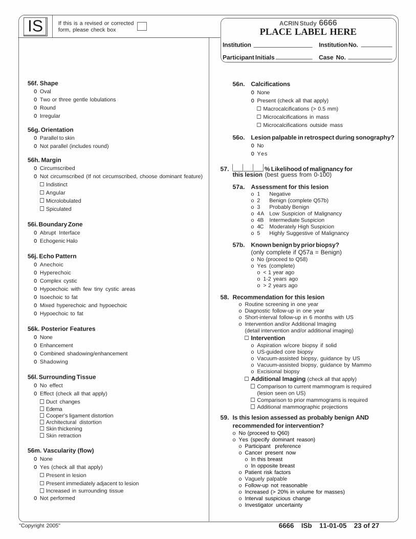
56f. Shapeo Ovalo Two or three gentle lobulationso Roundo Irregular
56g. Orientationo Parallel to skino Not parallel (includes round)
56h. Margino Circumscribedo Not circumscribed (If not circumscribed, choose dominant feature)
IndistinctAngularMicrolobulatedSpiculated
56i. Boundary Zoneo Abrupt Interfaceo Echogenic Halo
56j. Echo Patterno Anechoico Hyperechoico Complex cystico Hypoechoic with few tiny cystic areaso Isoechoic to fato Mixed hyperechoic and hypoechoico Hypoechoic to fat
56k. Posterior Featureso Noneo Enhancemento Combined shadowing/enhancemento Shadowing
56l. Surrounding Tissueo No effecto Effect (check all that apply)
Duct changesEdemaCooper’s ligament distortionArchitectural distortionSkin thickeningSkin retraction
56m. Vascularity (flow)o Noneo Yes (check all that apply)
Present in lesionPresent immediately adjacent to lesionIncreased in surrounding tissue
o Not performed
56n. Calcificationso Noneo Present (check all that apply)
Macrocalcifications (> 0.5 mm)Microcalcifications in massMicrocalcifications outside mass
56o. Lesion palpable in retrospect during sonography?o Noo Yes
57. % Likelihood of malignancy forthis lesion (best guess from 0-100)
57a. Assessment for this lesiono 1 Negativeo 2 Benign (complete Q57b)o 3 Probably Benigno 4A Low Suspicion of Malignancyo 4B Intermediate Suspiciono 4C Moderately High Suspiciono 5 Highly Suggestive of Malignancy
57b. Known benign by prior biopsy?(only complete if Q57a = Benign)o No (proceed to Q58)o Yes (complete)
o < 1 year agoo 1-2 years agoo > 2 years ago
58. Recommendation for this lesiono Routine screening in one yearo Diagnostic follow-up in one yearo Short-interval follow-up in 6 months with USo Intervention and/or Additional Imaging
(detail intervention and/or additional imaging)Interventiono Aspiration w/core biopsy if solido US-guided core biopsyo Vacuum-assisted biopsy, guidance by USo Vacuum-assisted biopsy, guidance by Mammoo Excisional biopsyAdditional Imaging (check all that apply)
Comparison to current mammogram is required(lesion seen on US)Comparison to prior mammograms is requiredAdditional mammographic projections
59. Is this lesion assessed as probably benign ANDrecommended for intervention?o No (proceed to Q60)o Yes (specify dominant reason)
o Participant preferenceo Cancer present now
o In this breasto In opposite breast
o Patient risk factorso Vaguely palpableo Follow-up not reasonableo Increased (> 20% in volume for masses)o Interval suspicious changeo Investigator uncertainty

6666 ISb 11-01-05 24 of 27
ACRIN Study 6666
Institution No.
Case No.Participant Initials
Institution
PLACE LABEL HERE IS If this is a revised or correctedform, please check box
"Copyright 2005"
60. For lesion evaluation, techniques used (check all that apply)Conventional imagingSpatial compoundingPower DopplerTissue Harmonic ImagingPanoramic display
60a. If spatial compounding was used, what was its influence? (please answer the following questions)o No influence (proceed to Q61)o Influenced (please answer the following questions in bold)
Margin depictiono Bettero Sameo Worse
Internal structure depictiono Bettero Sameo Worse
Posterior feature depictiono Bettero Sameo Worse
Confidence in lesion characterizationo Bettero Sameo Worse
60b. Change in likelihood of malignancy with spatial compounding?o Noneo Looks more benign with spatial compoundingo Looks more malignant with spatial compounding
61. Are there additional lesions you wish to describe?o No (proceed to Q14)o Yes (proceed to Q62)

6666 ISb 11-01-05 25 of 27
ACRIN Study 6666
Institution No.
Case No.Participant Initials
Institution
PLACE LABEL HERE IS If this is a revised or correctedform, please check box
"Copyright 2005"
62. Additional lesions other than simple cysts (maximum of 4 per breast)
62a. Lesion # (e.g. UR1, UB1, UL1 etc.)(Retain lesion numbering from initial study survey sonogram. If this is the first examination, begin with R1 for the first lesion in the rightbreast, R2 for the second lesion in the right breast etc. If the finding is new since a prior study sonogram, use next sequential #. Describeany new or suspicious findings first. Location, distance from nipple, depth to lesion center and measurements are completedfor all reportable findings).
62b. Was this "lesion" seen on a previous sonogram including any sonograms performed prior to study enrollment?o Not applicable, no prior breast sonogramso Noo Yes
o Goneo Decreased in size since previous examo Stable in size since previous examo Multiple bilateral circumscribed masses fluctuating in size since previous examo Increased in size since previous examo Other suspicious changeo Increasing and other suspicious change
Is this "lesion" multiple bilateral circumscribed masses? If yes, describe location and measurement of largest mass.o Noo Yes
o R o L o' clock cm . cm
62c. Lesion Size
mm mm mm mm3
62d. Is this lesion at the site of prior biopsy?o Noo Yes (if yes, select prior procedure)
o Core/vacuum biopsy with clip (if procedure performed, select diagnosis)o Core/vacuum biopsy without marker (if procedure performed, select diagnosis)o Surgical biopsy site (if procedure was performed, select diagnosis)
o Benigno Atypical/high-risk lesiono Cancer siteo Unknown
o Biopsy details unknowno FNAB
o Not applicable, multiple bilateral circumscribed masses
62e. Special Case (see choices below)o Noo Yes (if yes, detail below then proceed to Q62n)
(Special Case Features)o Complicated Cyst (Note: Do not use this term for "complex cystic masses".
For complex cystic masses code "No" for Q62e, proceed to Q62f and indicate "complex cystic" at Q62j.)Homogeneous low-level echoesFluid-Debris LevelMobile internal echoesMultiple bilateral complicated cysts in company of simple cysts
o Multiple bilateral solid oval, circumscribed masseso Mass in or on skino Clustered microcystso Intraductal masso Lymph nodeo Calcifications without a masso Foreign bodyo Post-Surgical scaro Other, specify:
U
Note: Volume is pro-grammed to be calcu-lated on line; however,as verification, pleasecalculate volume basedon horizontal, verticaland perpendicular mea-surements as a valida-tion.
HorizontalPerpendicular Meas
(mm) D3Vertical A-P
meas (mm) D2Measured Plane
LargestHorizontal
Meas (mm) D1
SecondMeasured Plane Volume D1XD2XD3 2
.
.
Depth from skin tocenter of lesion
(to nearest 0.5 cm)
Distance fromthe nipple
Clockface(report on the hour)Breast
(report on hour and 1/2 houre.g. 7:00 = 0700, 12:30 = 1230)
o Trvo Sago Rado Arado Oblique
o Trvo Sago Rado Arado Perpendicular Oblique
X X
6666 ISb 11-01-05 26 of 27
ACRIN Study 6666
Institution No.
Case No.Participant Initials
Institution
PLACE LABEL HERE IS If this is a revised or correctedform, please check box
"Copyright 2005"
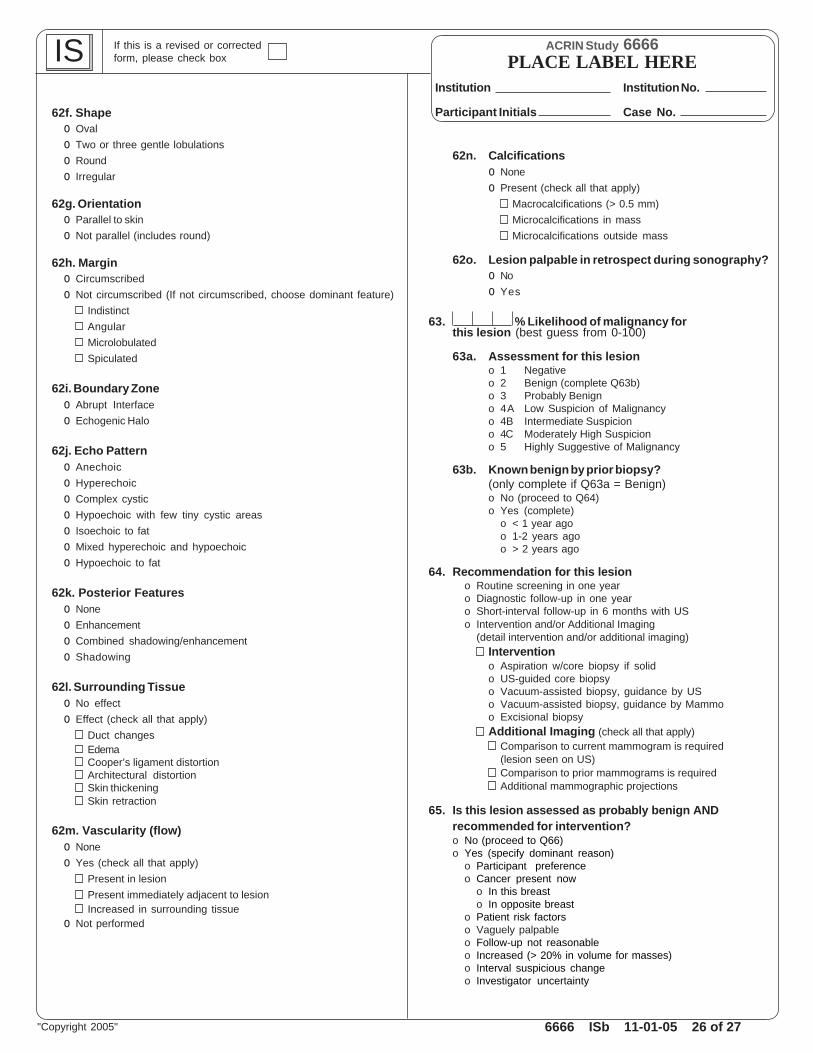
62f. Shapeo Ovalo Two or three gentle lobulationso Roundo Irregular
62g. Orientationo Parallel to skino Not parallel (includes round)
62h. Margino Circumscribedo Not circumscribed (If not circumscribed, choose dominant feature)
IndistinctAngularMicrolobulatedSpiculated
62i. Boundary Zoneo Abrupt Interfaceo Echogenic Halo
62j. Echo Patterno Anechoico Hyperechoico Complex cystico Hypoechoic with few tiny cystic areaso Isoechoic to fato Mixed hyperechoic and hypoechoico Hypoechoic to fat
62k. Posterior Featureso Noneo Enhancemento Combined shadowing/enhancemento Shadowing
62l. Surrounding Tissueo No effecto Effect (check all that apply)
Duct changesEdemaCooper’s ligament distortionArchitectural distortionSkin thickeningSkin retraction
62m. Vascularity (flow)o Noneo Yes (check all that apply)
Present in lesionPresent immediately adjacent to lesionIncreased in surrounding tissue
o Not performed
62n. Calcificationso Noneo Present (check all that apply)
Macrocalcifications (> 0.5 mm)Microcalcifications in massMicrocalcifications outside mass
62o. Lesion palpable in retrospect during sonography?o Noo Yes
63. % Likelihood of malignancy forthis lesion (best guess from 0-100)
63a. Assessment for this lesiono 1 Negativeo 2 Benign (complete Q63b)o 3 Probably Benigno 4A Low Suspicion of Malignancyo 4B Intermediate Suspiciono 4C Moderately High Suspiciono 5 Highly Suggestive of Malignancy
63b. Known benign by prior biopsy?(only complete if Q63a = Benign)o No (proceed to Q64)o Yes (complete)
o < 1 year agoo 1-2 years agoo > 2 years ago
64. Recommendation for this lesiono Routine screening in one yearo Diagnostic follow-up in one yearo Short-interval follow-up in 6 months with USo Intervention and/or Additional Imaging
(detail intervention and/or additional imaging)Interventiono Aspiration w/core biopsy if solido US-guided core biopsyo Vacuum-assisted biopsy, guidance by USo Vacuum-assisted biopsy, guidance by Mammoo Excisional biopsyAdditional Imaging (check all that apply)
Comparison to current mammogram is required(lesion seen on US)Comparison to prior mammograms is requiredAdditional mammographic projections
65. Is this lesion assessed as probably benign ANDrecommended for intervention?o No (proceed to Q66)o Yes (specify dominant reason)
o Participant preferenceo Cancer present now
o In this breasto In opposite breast
o Patient risk factorso Vaguely palpableo Follow-up not reasonableo Increased (> 20% in volume for masses)o Interval suspicious changeo Investigator uncertainty

6666 ISb 11-01-05 27 of 27
ACRIN Study 6666
Institution No.
Case No.Participant Initials
Institution
PLACE LABEL HERE IS If this is a revised or correctedform, please check box
"Copyright 2005"
66. For lesion evaluation, techniques used (check all that apply)Conventional imagingSpatial compoundingPower DopplerTissue Harmonic ImagingPanoramic display
66a. If spatial compounding was used, what was its influence? (please answer the following questions)o No influence (proceed to Q14)o Influenced (please answer the following questions in bold)
Margin depictiono Bettero Sameo Worse
Internal structure depictiono Bettero Sameo Worse
Posterior feature depictiono Bettero Sameo Worse
Confidence in lesion characterizationo Bettero Sameo Worse
66b. Change in likelihood of malignancy with spatial compounding? (complete then proceed to Q14, Final Assessment)o Noneo Looks more benign with spatial compoundingo Looks more malignant with spatial compounding