irf4-rearranged large b-cell lymphoma on waldeyer’s ring
TRANSCRIPT

292
Turk J Hematol 2020;37:286-309LETTERS TO THE EDITOR
cases of CLL with DM were reported, and one patient received methotrexate with cyclophosphamide. The skin rash in DM does not mean malignant skin involvement; therefore, DM treatment should be initiated separately, as in our case. However, DM with associated malignancy responds poorly compared to idiopathic cases [5]. Long-term outcomes of CLL with DM are unknown, and so are data regarding the use of chemoimmunotherapy in such patients, although rituximab has shown efficacy in DM [6]. It is prudent for the overworked clinician to not overlook neurological or musculoskeletal symptoms in CLL patients, and a keen search for hematological malignancy is justified in known cases of inflammatory myopathies.
Keywords: CLL, Cutaneous involvement, Rash
Anahtar Sözcükler: KLL, Deri tutulumu, Döküntü
Informed Consent: Informed consent has been obtained before writing this manuscript.
Authorship Contributions
Concept: S.K., A.G.; Design: S.K., A.G.; Data Collection or Processing: R.G., S.M., A.G.; Writing: S.K., A.G., R.G., S.M.
Ethics Committee Approval: This study has followed ethical standards.
Financial Disclosure: No funding was received for this study.
References1. Hamblin TJ. Non-hemic autoimmunity in CLL. Leuk Res 2009;33:366-367.
2. Stübgen JP. Inflammatory myopathies and lymphoma. J Neurol Sci 2016;369:377-389.
3. Kay NE. Abnormal T-cell subpopulation function in CLL: excessive suppressor (T gamma) and deficient helper (T mu) activity with respect to B-cell proliferation. Blood 1981;57:418-420.
4. Gunawardena H, Betteridge ZE, McHugh NJ. Myositis-specific autoantibodies: their clinical and pathogenic significance in disease expression. Rheumatology (Oxford) 2009;48:607-612.
5. Marie I, Guillevin L, Menard JF, Hatron PY, Cherin P, Amoura Z, Cacoub P, Bachelez H, Buzyn A, Le Roux G, Ziza JM, Brice P, Munck JN, Sarrot-Reynauld F, Piette JC, Larroche C. Hematological malignancy associated with polymyositis and dermatomyositis. Autoimmun Rev 2012;11:615-620.
6. Levine TD. Rituximab in the treatment of dermatomyositis: an open-label pilot study. Arthritis Rheum 2005;52:601-607.
Address for Correspondence/Yazışma Adresi: Ajay Gogia, MD, AIIMS, Department of Medical Oncology, New Delhi, India Phone : 9013000642E-mail : [email protected] ORCID: orcid.org/0000-0001-6500-1061
Received/Geliş tarihi: June 15, 2020Accepted/Kabul tarihi: August 19, 2020
DOI: 10.4274/tjh.galenos.2020.2020.0331
©Copyright 2020 by Turkish Society of HematologyTurkish Journal of Hematology, Published by Galenos Publishing House
IRF4-Rearranged Large B-Cell Lymphoma on Waldeyer’s Ring: A Case ReportWaldeyer Halkasında IRF4 Yeniden Düzenlenmesi Büyük B-hücreli Lenfoma Olgusu
Deram Büyüktaş1, Serdar Örnek2, Fatma Tokat3, Tülay Tecimer4, Burhan Ferhanoğlu1
1Koç University Faculty of Medicine, Department of Hematology, İstanbul, Turkey2American Hospital, Clinic of Hematology, İstanbul, Turkey3Acıbadem University Faculty of Medicine, Department of Pathology, İstanbul, Turkey4Acıbadem Health Group, Pathology Laboratory, İstanbul, Turkey
To the Editor,
Large B-cell lymphoma (LBCL) with IRF4 rearrangement is a
newly recognized and rare entity. LBCL is characterized by co-
expression of MUM1 and BCL6. It has been associated with
young age and a favorable outcome. Most patients present with
predominantly Waldeyer’s ring or neck or head lymph node
involvement [1,2,3]. Here, we present a case of LBCL with IRF4
rearrangement in an older male.
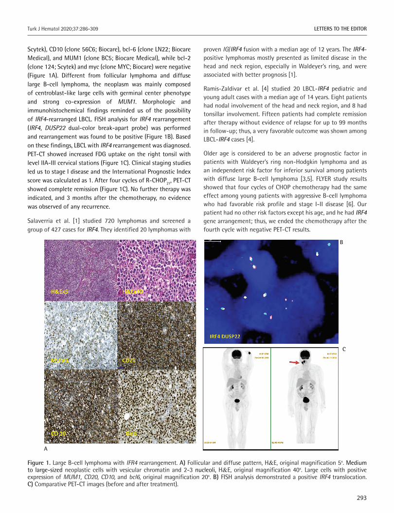
A 67-year-old man was admitted to the hospital with sore throat, dysphagia, and a lump in the right cervical region. Physical examination showed that the right palatine tonsil was enlarged and there was 2 cm of cervical lymphadenopathy unilaterally. After one week of antibiotic therapy, the lymphadenopathy still persisted. The patient underwent an excisional lymph node biopsy, which was consistent with LBCL of germinal center type, with a mainly follicular and focally diffuse pattern. The immunohistochemistry panel was positive for CD20 (clone L26;
293
Turk J Hematol 2020;37:286-309 LETTERS TO THE EDITOR
Scytek), CD10 (clone 56C6; Biocare), bcl-6 (clone LN22; Biocare Medical), and MUM1 (clone BC5; Biocare Medical), while bcl-2 (clone 124; Scytek) and myc (clone MYC; Biocare) were negative (Figure 1A). Different from follicular lymphoma and diffuse large B-cell lymphoma, the neoplasm was mainly composed of centroblast-like large cells with germinal center phenotype and strong co-expression of MUM1. Morphologic and immunohistochemical findings reminded us of the possibility of IRF4-rearranged LBCL. FISH analysis for IRF4 rearrangement (IRF4, DUSP22 dual-color break-apart probe) was performed and rearrangement was found to be positive (Figure 1B). Based on these findings, LBCL with IRF4 rearrangement was diagnosed. PET-CT showed increased FDG uptake on the right tonsil with level IIA-III cervical stations (Figure 1C). Clinical staging studies led us to stage I disease and the International Prognostic Index score was calculated as 1. After four cycles of R-CHOP21, PET-CT showed complete remission (Figure 1C). No further therapy was indicated, and 3 months after the chemotherapy, no evidence was observed of any recurrence.
Salaverria et al. [1] studied 720 lymphomas and screened a group of 427 cases for IRF4. They identified 20 lymphomas with
proven IG/IRF4 fusion with a median age of 12 years. The IRF4-positive lymphomas mostly presented as limited disease in the head and neck region, especially in Waldeyer’s ring, and were associated with better prognosis [1].
Ramis-Zaldivar et al. [4] studied 20 LBCL-IRF4 pediatric and young adult cases with a median age of 14 years. Eight patients had nodal involvement of the head and neck region, and 8 had tonsillar involvement. Fifteen patients had complete remission after therapy without evidence of relapse for up to 99 months in follow-up; thus, a very favorable outcome was shown among LBCL-IRF4 cases [4].
Older age is considered to be an adverse prognostic factor in patients with Waldeyer’s ring non-Hodgkin lymphoma and as an independent risk factor for inferior survival among patients with diffuse large B-cell lymphoma [3,5]. FLYER study results showed that four cycles of CHOP chemotherapy had the same effect among young patients with aggressive B-cell lymphoma who had favorable risk profile and stage I-II disease [6]. Our patient had no other risk factors except his age, and he had IRF4 gene arrangement; thus, we ended the chemotherapy after the fourth cycle with negative PET-CT results.
Figure 1. Large B-cell lymphoma with IFR4 rearrangement. A) Follicular and diffuse pattern, H&E, original magnification 5x. Medium to large-sized neoplastic cells with vesicular chromatin and 2-3 nucleoli, H&E, original magnification 40x. Large cells with positive expression of MUM1, CD20, CD10, and bcl6, original magnification 20x. B) FISH analysis demonstrated a positive IRF4 translocation. C) Comparative PET-CT images (before and after treatment).
A
B
C

294
Turk J Hematol 2020;37:286-309LETTERS TO THE EDITOR
In conclusion, IRF4 gene analysis should be considered in patients of any age with Waldeyer’s ring LBCL with germinal center origin, follicular and/or diffuse pattern, and strong MUM1 expression on pathological examination. The presence of IRF4 rearrangement may affect the prognosis of the disease and the duration of chemotherapy.
Keywords: Large B- cell lymphoma, Waldeyer’s ring, IRF4, MUM1
Anahtar Sözcükler: Büyük B-hücreli lenfoma, Waldeyer halkası, IRF4, MUM1
Authorship Contributions
Surgical and Medical Practices: D.B., S.Ö., B.F.; Concept: B.F.; Design: D.B, B.F.; Data Collection or Processing: D.B., S.Ö., T.T., F.T.; Analysis or Interpretation: D.B., B.F.; T.T., F.T.; Literature Search: D.B.; Writing: D.B., S.Ö., T.T., B.F.
Conflict of Interest: No conflict of interest was declared by the authors.
Financial Disclosure: This research did not receive any specific grant from any funding agency in the public, commercial, or not-for-profit sectors.
References 1. Salaverria I, Philipp C, Oschlies I, Kohler CW, Kreuz M, Szczepanowski M,
Burkhardt B, Trautmann H, Gesk S, Andrusiewicz M, Berger H, Fey M, Harder L, Hasenclever D, Hummel M, Loeffler M, Mahn F, Martin-Guerrero I, Pellissery S, Pott C, Pfreundschuh M, Reiter A, Richter J, Rosolowski M, Schwaenen C, Stein H, Trümper L, Wessendorf S, Spang R, Küppers R, Klapper W, Siebert R; Molecular Mechanisms in Malignant Lymphomas Network Project of
the Deutsche Krebshilfe; German High-Grade Lymphoma Study Group; Berlin-Frankfurt-Münster-NHL trial group. Translocations activating IRF4 identify a subtype of germinal center-derived B-cell lymphoma affecting predominantly children and young adults. Blood 2011;118:139-147.
2. de Leval L, Bonnet C, Copie-Bergman C, Seidel L, Baia M, Brière J, Molina TJ, Fabiani B, Petrella T, Bosq J, Gisselbrecht C, Siebert R, Tilly H, Haioun C, Fillet G, Gaulard P. Diffuse large B-cell lymphoma of Waldeyer’s ring has distinct clinicopathologic features: a GELA study. Ann Oncol 2012;23:3143-3151.
3. Swerdlow SH, Campo E, Harris NL, Jaffe ES, Pileri SA, Stein H, Thiele J, Arber DA, Hasserjian RP, Lea Beau M, Orazi A, Siebert R. WHO Classification of Tumours of Haematopoietic and Lymphoid Tissues. Lyon, IARC Press, 2017.
4. Ramis-Zaldivar JE, Gonzalez-Farré B, Balagué O, Celis V, Nadeu F, Salmerón-Villalobos J, Andrés M, Martin-Guerrero I, Garrido-Pontnou M, Gaafar A, Suñol M, Bárcena C, Garcia-Bragado F, Andión M, Azorín D, Astigarraga I, Sagaseta de Ilurdoz M, Sábado C, Gallego S, Verdú-Amorós J, Fernandez-Delgado R, Perez V, Tapia G, Mozos A, Torrent M, Solano-Páez P, Rivas-Delgado A, Dlouhy I, Clot G, Enjuanes A, López-Guillermo A, Galera P, Oberley MJ, Maguire A, Ramsower C, Rimsza LM, Quintanilla-Martinez L, Jaffe ES, Campo E, Salaverria I. Distinct molecular profile of IRF4-rearranged large B-cell lymphoma. Blood 2020 23;135:274-286.
5. Ezzat AA, Ibrahim EM, El Weshi AN, Khafaga YM, AlJurf M, Martin JM, Ajarim DS, Bazarbashi SN, Stuart RK, Zucca E. Localized non-Hodgkin’s lymphoma of Waldeyer’s ring: clinical features, management, and prognosis of 130 adult patients. Head Neck 2001;23:547-558.
6. Poeschel V, Held G, Ziepert M, Witzens-Harig M, Holte H, Thurner L, Borchmann P, Viardot A, Soekler M, Keller U, Schmidt C, Truemper L, Mahlberg R, Marks R, Hoeffkes HG, Metzner B, Dierlamm J, Frickhofen N, Haenel M, Neubauer A, Kneba M, Merli F, Tucci A, de Nully Brown P, Federico M, Lengfelder E, di Rocco A, Trappe R, Rosenwald A, Berdel C, Maisenhoelder M, Shpilberg O, Amam J, Christofyllakis K, Hartmann F, Murawski N, Stilgenbauer S, Nickelsen M, Wulf G, Glass B, Schmitz N, Altmann B, Loeffler M, Pfreundschuh M; FLYER Trial Investigators; German Lymphoma Alliance. Four versus six cycles of CHOP chemotherapy in combination with six applications of rituximab in patients with aggressive B-cell lymphoma with favourable prognosis (FLYER): a randomised, phase 3, non-inferiority trial. Lancet 2020;394:2271-2281.
Address for Correspondence/Yazışma Adresi: Burhan Ferhanoğlu, MD, Koç University Faculty of Medicine, Department of Hematology, İstanbul, TurkeyE-mail : [email protected] ORCID: orcid.org/0000-0002-4257-549X
Received/Geliş tarihi: February 25, 2020Accepted/Kabul tarihi: April 28, 2020
DOI: 10.4274/tjh.galenos.2020.2020.0086
©Copyright 2020 by Turkish Society of HematologyTurkish Journal of Hematology, Published by Galenos Publishing House
An Unconventional Presentation of Multiple Myeloma: Bazex SyndromeMultipl Myelomun Olağan Dışı Bir Prezentasyonu: Bazex Sendromu
Özlem Kandemir Alibakan1, Naciye Demirel2, Nihan Nizam3, Rafet Eren2
1University of Health Sciences Turkey, Prof. Dr. Cemil Taşçıoğlu Training and Research Hospital, Clinic of Internal Medicine, İstanbul, Turkey2University of Health Sciences Turkey, Prof. Dr. Cemil Taşçıoğlu Training and Research Hospital, Clinic of Hematology, İstanbul, Turkey3İstanbul University İstanbul Faculty of Medicine, Department of Internal Medicine, İstanbul, Turkey
To the Editor,
Multiple myeloma patients may exhibit signs and symptoms of skin involvement secondary to either malignant cell infiltration or disease-specific treatment. There are also anecdotal reports
of paraneoplastic skin diseases including Sweet syndrome,
leukocytoclastic vasculitis, and neutrophilic dermatosis associated
with multiple myeloma [1]. In this report, we share a patient with
a remarkably rare skin presentation of multiple myeloma.