involvement of elongation factor-1α in cytokinesis without
TRANSCRIPT

Involvement of Elongation Factor-1α in Cytokinesis without Actomyosin Contractile Ring in the Primitive Red Alga
Cyanidioschyzon merolae
Yuuta Imoto1,2, Keiji Nishida2, Fumi Yagisawa2, Yamato Yoshida2, Mio Ohnuma2, Masaki Yoshida2, Takayuki Fujiwara2, Haruko Kuroiwa2,
Shigeyuki Kawano1 and Tsuneyoshi Kuroiwa2,* 1 Department of Integrated Bioscience, Graduate School of Frontier Science, University of Tokyo, Kashiwa, Chiba 277–8562, Japan
2 Laboratory of Cell Biology, Department of Life Science, College of Science, Research Information Centre for Extremophile, Rikkyo University, 3–34–1 Nishi-ikebukuro, Toshima, Tokyo 171–8501, Japan
Received August 31, 2011; accepted September 14, 2011
Summary Cytokinesis is a pivotal event in cell division in all living organisms. In mammals, cyto-kinesis progresses via constriction of the contractile ring, a bundle of actin filaments and myosin that is located on the equatorial plane. The eukaryotic elongation factor 1α (eEF-1α) is associated with the contractile ring in sea urchin eggs. Although eEF-1α is a ubiquitous protein translation factor and is highly conserved in eukaryotes, archaea, and prokaryotes, the archaebacterial EF-1α and the pro-karyotic EF-1α ortholog EF-Tu do not function in cytokinesis, suggesting that the association between the contractile ring and EF-1α appeared at about the same time as the establishment of eukaryotic cells. However, the role of EF-1α in cytokinesis in primitive cells is unclear. In this study, we show that the primitive alga Cyanidioschyzon merolae elongation factor-lα (CmEF-1α) is localized in the contractile division plane, but not in the actomyosin contractile ring. The genome of C. merolae contains 2 unexpressed actin genes and lacks a myosin gene. Immunoblotting analyses revealed that the protein level of CmEF-1α remained constant during the G1 to M phase, and then peaked during cytokinesis. Immunofluorescent microscopy revealed 2 patterns of localization of CmEF-1α during the cell cycle: it was dispersed throughout the cytoplasm from G1 to early M phase, and then localized to the contractile region during cytokinesis. From the results of current study on cytokinesis in the primitive red algae C. merolae, our findings led us to hypothesize that when first established in the lower eukaryotes, cytokinesis fundamentally relied on eEF-1α, and the actomyosin system was acquired later during evolution.
Key words eEF-1α, Cyanidioschyzon merolae, Cytokinesis, Microtubule, Actomyosin.
Eukaryotic cells contain many types of membrane-bound organelles: the nucleus, mitochon-drion, plastid, endoplasmic reticulum, Golgi apparatus, lysosome, and microbody, all of which pro-liferate via individual division cycles (Imoto et al. 2011). During cell division, the cell controls the number of these membrane-bound organelles by physically and accurately splitting them between the 2 daughter cells. This process, known as cytokinesis, is the final step in the cell cycle.
In animal cells, the separation into 2 daughter cells that occurs during cytokinesis is controlled by a contractile apparatus known as the contractile ring, which is composed of a bundle of actin fil-aments and myosin, and lies beneath the plasma membrane (Shroeder 1968). Myosin generates the motive force for the contraction of actin filaments, which drives the constriction of the contractile
* Corresponding author, e-mail: [email protected]
© 2011 The Japan Mendel Society Cytologia 76(4): 431–437

432 Y. Imoto et al. Cytologia 76(4)
ring (Mabuchi and Okuno 1986). In plant cells, cytokinesis is controlled by actin microfilaments that are involved in the development of the cell plate (Higaki et al. 2008). Although these actomyo-sin-based systems are found in most eukaryotic cells, the fundamental mechanisms of cytokinesis are unknown. In mammals, contraction of the contractile ring is followed by the completion of cell division, but for a considerable time the daughter cells remain connected by a narrow bridge with a central midbody, which is typically filled with residual spindle microtubules (Byers and Abramson 1968; Mullins and Biesele 1973, 1977, Mullins and McIntosh 1982). In Saccharomyces cerevisiae, the deletion of actin or myosin considerably delayed cell division, but cytokinesis was still com-pleted (Bi et al. 1998). Therefore, it is assumed that there are other proteins besides actin filaments and myosin that have more fundamental functions in cytokinesis.
Recent studies have shown that the eukaryotic transition elongation factor 1α (eEF-1α) local-izes to the cleavage furrow, and plays a role in cytokinesis in higher eukaryotic cells (Numata et al. 2000, Fujimoto and Mabuchi 2010). eEF-1α is a prokaryotic elongation factor thermo-unstable (EF-Tu) homolog. It is a highly conserved member of the GTPase superfamily, which includes pro-tein factors involved in translation processes and a number of other nontranslational cellular pro-cesses, such as interactions with cytoskeletal proteins (Baldauf et al. 1996, Keeling et al. 1998, Negrutskii and El’skaya 1998). The universality of eEF-1α suggests that it is involved in cytokine-sis in primitive eukaryotes, and that it plays a fundamental role. To clarify this point we used the primitive red algae Cyanidioschyzon merolae, which has 2 non-expressed actin genes in the ge-nome (Takahashi et al. 1996, Matsuzaki et al. 2004) and lacks a myosin gene (Suzuki et al. 1995).
In this study, we examined the localization of C. merolae elongation factor-1α (CmEF-1α) throughout the cell cycle and observed that it was localized at the region of the contractile ring. The localization of CmEF-1α at the contractile ring was continuous during cytokinesis and was followed by the completion of cell division. Our results strongly suggest that eEF-1α plays a significant role in primitive cytokinesis. This is the first report of cytokinesis in a primitive eukaryotic cell that uses eEF-1α, but not actomyosin. This presumably reflects ancestral cell division associated with eEF-1α. These results, together with those from other reports, lead to the hypothesis that the cyto-skeletal or transport systems in higher eukaryotes were acquired courtesy of evolutionary pro-cesses, and eukaryotic cell division in early eukaryotes fundamentally relied on eEF-1α.
Materials and methods
Synchronous culture and drug treatmentWe used the Cyanidioschyzon merolae 10D-14 strain (Toda et al. 1998) in these experiments.
The cells were cultured and synchronized as described previously (Suzuki et al. 1994). For syn-chronization, the cell culture was maintained in 2×Allen’s medium at pH 2.5 in a flask with shaking under continuous light (40 W/m2) at 40°C. The cells were subcultured for 3 h in the dark to achieve a concentration of 1×10⁷ cells/ml, and then synchronized under a 12-h light/12-h dark cycle at 40°C while the medium was aerated. Experiments with synchronized cultures were initiated at the com-mencement of the second light period.
AntibodiesTo generate anti-eukaryotic elongation factor 1 alpha antiserum, the coding region for the
amino acid residues of the predicted 450-amino acid sequence of CMH226C eEF-1α protein was amplified from C. merolae genomic DNA by PCR by using the following primers 5′-ATGG-GTAAGGAAAAGACGCA-3′ (forward) and 5′-TGCCTTCTTCGCTGCTGCTT-3′ (reverse). The DNA fragment was cloned into the pQE80 vector by restriction digestion and ligation in Escherichia coli strain XL1Blue. The recombinant protein was produced as a fusion protein with a 6-histidine tag at the N terminus and purified using a nickel affinity column. The purified protein

2011 eEF-1α in Cytokinesis of Cyanidioschyzon merolae 433
was injected into a guinea pig. The anti-α-tubulin antisera used have been described in detail else-where (Fujiwara et al. 2009). For immunblotting analyses, 20 μg total protein extracted from C. merolae was separated by 12.5% SDS-PAGE and then blotted onto a polyvinylidene difluoride membrane. After blocking with 5% skim milk in Triton X-100 Tris-buffered saline (TTBS), the membranes were incubated with primary antisera or antibodies at dilutions of 1/1000 for anti-CmEF-1α or 1/1000 for anti-α-tubulin. The secondary antibodies, namely, alkaline phosphatase-conjugated goat anti-guinea pig and anti-rabbit IgG (Kirkegaard and Perry Laboratories, Gaithersburg, MD), were each used at a dilution of 1/1000 and detected using an AP-conjugate substrate kit (Bio-Rad, Hercules, CA) and quantified via VIMPCS (Hamamatsu Photonics) (Kuroiwa et al. 1986).
Microscopy Cell fixation and immunofluorescence microscopy were performed as described previously
(Nishida et al. 2005). The anti-eEF-1α antiserum was used at a dilution of 1/500. Highly cross-ad-sorbed goat anti-mouse IgG conjugated with Alexa 488 was used as the secondary antibody at a di-lution of 1/1000. The length and relative signal intensity of CmEF-1α at the contractile region were measured with VIMPCS as described previously (Kuroiwa et al. 1986, Imoto et al. 2010)
Results and discussion
C. merolae contains a single CmEF-1α gene (Matsuzaki et al. 2004, Nozaki et al. 2007). To reveal the function of the CmEF-1α in cytokinesis of C. merolae, we generated antibodies against CmEF-1α in a guinea pig. Immunoblotting analysis showed that the raised antiserum specifically detected a 49-kD band from the total cell lysate of C. merolae (Fig. 1a), which is nearly consistent with the predicted size of CmEF-1α in C. merolae. To elucidate the role of CmEF-1α during the
Fig. 1. (a) Immunoblotting analysis with preimmune antisera or anti-CmEF1-α antibodies. Numbers on the left indicate molecular weight (kD). (b) Immunoblotting analysis of proteins associated with micro-tubules and organelle division throughout the cell cycle. Cells were harvested at indicated time points after the start of incubation from the same synchronous culture. Each lane contained 20 μg total protein. Proteins were detected with anti-CmEF1-α antibodies or anti-α-tubulin antibodies. (c) Ratio of protein levels of CmEF1-α and α-tubulin. Protein level was normalized to the intensity of the band at initiation of cell synchronization (0 h). Values are mean±SD; n=3.

434 Y. Imoto et al. Cytologia 76(4)
Fig. 2. Immunofluorescence images of CmEF1-α showing 2 patterns of localization during cell cycle. (A) Immunofluorescence and phase-contrast images showing behavior of CmEF1-α (CmEF1-α: green) and α-tubulin (α-tubu: red) during progression of mitotic cycle. (B) Diameter, width and signal in-tensity of the CmEF1-α at the contractile region versus diameter at the equator plane of cell divi-sion. The length and relative signal intensity were measured with VIMPCS. Scale bar=2 μm. White arrowhead indicates contractile plane. Nu: cell nucleus, Mt: mitochondrion, Pt: Plastid, SP: spindle pole.

2011 eEF-1α in Cytokinesis of Cyanidioschyzon merolae 435
mitotic phase, synchronous cultures were harvested every 2 h for 24 h after initiation of the second light period. The mitotic stages of C. merolae have been identified previously based on microtubule formation and morphology (Nishida et al. 2005, Imoto et al. 2010). The samples were then sub-jected to immunoblotting analyses with antisera against CmEF-1α and α-tubulin (Fig. 1b, c). The protein level of CmEF-1α remained constant during the G1 and the S phase, and increased during the late M phase and cytokinesis until the early G1 phase. The protein level of α-tubulin increased from S phase and reached a peak at early M phase, which coincides with the period of organelle di-vision. This result suggested that EF-1α plays a significant role just after the division of organelles during cytokinesis.
To characterize the localization of CmEF-1α throughout the cell cycle, cells at various phases of the cell cycle were examined by immunofluorescence microscopy after triple staining with the anti-CmEF-1α antibody (CmEF-1α) and the anti-α-tubulin antibody (α-tubu), which shows the lo-cation of CmEF-1α (Fig. 2A). As shown in a previous study, the cleavage furrow begins to appear at the equator of the cell, which consists of a central spindle area that forms immediately after the division of the nucleus, mitochondrion, plastid, and microbody. This occurs during the G2 phase to the M phase (Imoto et al. 2010, Imoto et al. 2011), and then cytokinesis proceeds normally for an-other 2 h (Miyagishima et al. 1999). During the G1 to early M phase (Fig. 2Aa–d), CmEF-1α sig-nals were located throughout the entire cytoplasm. From late M phase to cytokinesis, the CmEF-1α signals were localized along the contractile region of cytokinetic division (Fig. 2Ae–g). The CmEF-1α signals became denser along the contractile region as it thickened, and then steadily de-composed as cytokinesis proceeded (Fig. 2Af, g). At the final phase of cytokinesis, the 2 daughter cells remained connected for a considerable period by a narrow bridge, and no CmEF-1α signals were detected (Fig. 2Ah). These observations suggested that CmEF-1α is involved in the contrac-tion of the cell but not in the final fission of the cell membrane. To clarify the morphological changes of the CmEF-1α, the diameter, width and the total intensityof the CmEF-1α at the contrac-tile region were plotted versus the diameter of cell division plane at the equator in the same manner (Fig. 2B). The diameter of CmEF-1α at the contractile region decreased as cytokinesis proceeded while the width increased (Fig. 2Ba). The total fluorescence intensity of the CmEF-1α at the con-tractile region decreased along with contraction, suggesting that the total number of CmEF-1α mol-ecules that localize the contractile region decreases with contraction (Fig. 2Bb).These changes in the CmEF-1α localization during contraction is similar to that of the inner PD ring or Z ring deter-mined previously (Miyagishima et al. 1999, 2001). Therefore, CmEF-1α seems to operate with a mechanism similar to that of the inner PD ring or Z ring during contraction and involved in cytoki-nesis.
In Saccharomyces pombe, three out of four genes encoding eEF-1α are involved in control of the actomyosin cytoskeleton (Suda et al. 1999). However, C. merolae contains a single CmEF-1α (Matsuzaki et al. 2004) and it was localized throughout the entire cytoplasm during the G1 to early M phase, but in the contractile region during late M phase until cytokinesis. These results suggested that CmEF-1α has a dual function, playing roles in translation and cytokinesis, and that it is regu-lated by the cell cycle.
Cytokinesis of C. merolae shows some unusual features compared with that of other eukary-otes. Firstly, when cells were examined by electron microscopy, no structures reminiscent of the contractile ring were visible beneath the dividing plane (Suzuki et al. 1995). Secondly, cell prolifer-ation was not inhibited by addition of cytochalasin, an inhibitor of polymerization of actin mono-mers (Suzuki et al. 1995). Because C. merolae lacks a myosin gene but contains an unexpressed actin gene (Takahashi et al. 1998, Matsuzaki et al. 2004), the contractile ring based on actomyosin did not appear (Takahashi et al. 1998). However, in this study, it was suggested that C. merolae di-vides using CmEF-1α, which is a simpler mechanism than that of actomyosin. In contrast, red alga Cyanidium caldarium RK-1, a close relative of C. merolae, cells divide using a contractile ring
436 Y. Imoto et al. Cytologia 76(4)
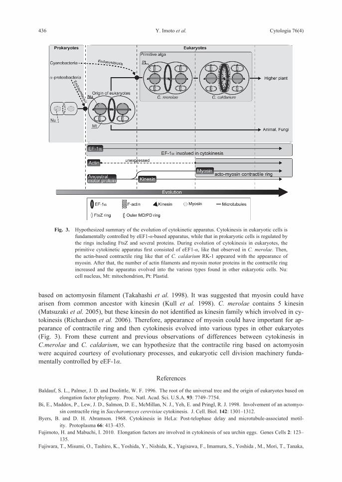
based on actomyosin filament (Takahashi et al. 1998). It was suggested that myosin could have arisen from common ancestor with kinesin (Kull et al. 1998). C. merolae contains 5 kinesin (Matsuzaki et al. 2005), but these kinesin do not identified as kinesin family which involved in cy-tokinesis (Richardson et al. 2006). Therefore, appearance of myosin could have important for ap-pearance of contractile ring and then cytokinesis evolved into various types in other eukaryotes (Fig. 3). From these current and previous observations of differences between cytokinesis in C.merolae and C. caldarium, we can hypothesize that the contractile ring based on actomyosin were acquired courtesy of evolutionary processes, and eukaryotic cell division machinery funda-mentally controlled by eEF-1α.
References
Baldauf, S. L., Palmer, J. D. and Doolittle, W. F. 1996. The root of the universal tree and the origin of eukaryotes based on elongation factor phylogeny. Proc. Natl. Acad. Sci. U.S.A. 93: 7749–7754.
Bi, E., Maddox, P., Lew, J. D., Salmon, D. E., McMillan, N. J., Yeh, E. and Pringl, R. J. 1998. Involvement of an actomyo-sin contractile ring in Saccharomyces cerevisiae cytokinesis. J. Cell. Biol. 142: 1301–1312.
Byers, B. and D. H. Abramson. 1968. Cytokinesis in HeLa: Post-telophase delay and microtubule-associated motil-ity. Protoplasma 66: 413–435.
Fujimoto, H. and Mabuchi, I. 2010. Elongation factors are involved in cytokinesis of sea urchin eggs. Genes Cells 2: 123–135.
Fujiwara, T., Misumi, O., Tashiro, K., Yoshida, Y., Nishida, K., Yagisawa, F., Imamura, S., Yoshida , M., Mori, T., Tanaka,
Fig. 3. Hypothesized summary of the evolution of cytokinetic apparatus. Cytokinesis in eukaryotic cells is fundamentally controlled by eEF1-α-based apparatus, while that in prokaryotic cells is regulated by the rings including FtsZ and several proteins. During evolution of cytokinesis in eukaryotes, the primitive cytokinetic apparatus first consisted of eEF1-α, like that observed in C. merolae. Then, the actin-based contractile ring like that of C. caldarium RK-1 appeared with the appearance of myosin. After that, the number of actin filaments and myosin motor proteins in the contractile ring increased and the apparatus evolved into the various types found in other eukaryotic cells. Nu: cell nucleus, Mt: mitochondrion, Pt: Plastid.
2011 eEF-1α in Cytokinesis of Cyanidioschyzon merolae 437
K. and Kuroiwa, T. 2009. Periodic gene expression patterns during the highly synchronized cell nucleus and or-ganelle division cycles in the unicellular red alga Cyanidioschyzon merolae. DNA Res. 16: 59–72.
Higaki, T., Kustuna, N., Sano, T. and Hasezawa, S. 2008. Quantitative analysis of changes in actin microfilament contribu-tion to cell plate development in plant cytokinesis. BMC Plant Biol. 8: 80.
Imoto, Y., Fujiwara, T., Yohida, Y., Kuroiwa, H., Maruyama, S. and Kuroiwa, T. 2010. Division and segregation of cell nu-cleus, mitochondrion and microbody mediated by centrosomes (mitotic spindle pole bodies) in the primitive red alga Cyanidioschyzon merolae. Protoplasma 241: 63–74.
, Yoshida, Y., Yagisawa, F., Kuroiwa, H. and Kuroiwa, T. 2011. The cell cycle, including the mitotic cycle and organelle division cycles, as revealed by cytological observations J Electro Microsc (Tokyo) 60: 117–136.
Keeling, P. J., Fast, N. M. and McFadden, G. I. 1998. J. Mol. Evol. 47: 649–655.Kull, F. J., Vale, R. D. and Fletterick, R. J. 1998. The case for a common ancestor: kinesin and myosin motor proteins and
G proteins. J. Muscle Res. Cell Motil. 19: 877–886. Kuroiwa, T., Nakamura, S., Kawano, S., Hizume, M., Thoe, A., Miyakawa, I. and Sando, N. 1986. Cytological character-
ization of a synaptonemal complex-less nucleolar-organizing region on a bivalent in the bivalent of Saccharomyces cerevisiae using a video-intensified microscope system. Exp. Cell Res. 165: 199–206.
and Okuno, M. 1986. The effect of myosin antibody on the division of the starfish blasomeres. J. Cell Biol. 74: 251–263.
Matsuzaki, M., Misumi, O., Shin-I, T., Maruyama, S., Takahara, M., Miyagishima, S., Mori, T., Nishida, K.,Yagisawa, F., Nishida, K., Yoshida, Y., Nishimura,Y., Nakao, S., Kobayashi, T., Momoyama,Y., Higashiyama,T., Minoda, A., Sano, M., Nomoto, H., Oishi, K., Hayashi, H., Ohta, F., Nishizaka, S., Haga, S., Miura, S., Morishita, T., Kabeya, Y., Terasawa, K., Suzuki, Y., Ishii, Y., Asakawa, S., Takano, H., Ohta, N., Kuroiwa, H., Tanaka, K., Shimizu, N., Sugano, S., Sato, N., Nozaki, H., Ogasawara, N., Kohara, Y. and Kuroiwa, T. 2004. Genome sequence of the ultra-small unicellular red alga Cyanidioschyzon merolae 10D. Nature 428: 653–657.
Miyagishima, S., Itoh, R, Toda, K., Kuroiwa, H. and Kuroiwa, T. 1999. Realtime analyses of chloroplast and mitochondrial division and differences in the behavior of their dividing rings during contraction. Planta 207: 343–353.
, Takahara, M., Mori, T., Kuroiwa, H., Higashiyama, T. and Kuroiwa, T. 2001. Plastid division is driven by a complex mechanism that involves differential transition of the bacterial and eukaryotic division rings. Plant Cell. 13: 2257–2268.
Mullins, J. M., and Biesele, J. J. 1973. Cytokinetic activities in a human cell line: the midbody and the intercellular bridge. Tissue Cell. 5: 47–61.
and Biesele, J. J. 1977. Terminal phase of cytokinesis in D-985 cells. J. Cell Biol. 73: 672–684. and McIntosh, J. R. 1982. Isolation and initial characterization of the mammalian midbody. J. Cell Biol. 94: 654–661.Nishida, K., Yagisawa, F., Kuroiwa, H., Nagata, T. and Kuroiwa, T. 2005. Cell cycle-regulated, microtubule-independent
organelle division in Cyanidioschyzon merolae. Mol. Biol. Cell. 16: 2493–2502Nozaki, H., Takano, H., Misumi, O., Terasawa, K., Matuzaki, M., Maruyama, S., Nishida, K., Yagisawa, F., Yoshida, Y.,
Fujiwara, T., Takio, S., Tamura, K., Chung, j-Chung., Nakamura, S., Kuroiwa, H., Tanaka, K., Sato, N. and Kuroiwa, T. 2007. The first 100% complete eukaryotic genome sequences from the red alga Cyanidioschyzon merolae 10D. BMC Biol. 5: 28.
Negrutskii, B. S. and El’skaya, A. V. 1998. Prog. Nucleic Acid Res. Mol. Biol. 60: 47–78.Numata, O., Kurosawa, Y., Gonda, K. and Watanabe, Y. 2000. Tetrahymena elongation factor-lα is localized with calmod-
ulin in the division furrow. J. Biochem. 127: 51–56.Suda, M., Fukui, M., Sogabe, Y., Sato, K., Morimatsu, A., Arai, R., Motegi, F., Miyakawa, T., Mabuchi, I. and Hirata, D.
1999. Overproduction of elongation factor 1α, an essential translational component, causes aberrant cell morphol-ogy by affecting the control of growth polarity in fission yeast. Genes Cells 4: 517–527.
Richardson, D. N., Simmons, M. P. and Reddy, A. S. N. 2006. Comprehensive comparative analysis of kinesins in photo-synthetic eukaryotes. BMC Genomics 7: 18.
Shroeder, T. E. 1968. Cytokinesis: Filaments in the cleavage furrow. Exp. Cell. Res. 53: 272–276.Suzuki, K., Ehara, T., Osafune, T., Kuroiwa, H., Kawano, S. and Kuroiwa, T. 1994. Cooperation behavior of mitochondria,
chloroplast and their nuclei during the mitotic cycle in the ultra-microalga Cyanidioschyzone merolae. Eur J Cell Biol 63: 280–288.
Takahashi, H., Takano, H., Kawano, S., Tohe, A. and Kuroiwa, T. 1996. Isolation, characterization and chromosomal map-ping of an actin gene from the primitive red alga Cyanidioschyzon merolae. Curr. Genet. 28: 484–490.
, , Kuroiwa, H., Itoh, H,. Toda, S., Kawano, S. and Kuroiwa, T. 1998. A possible role of actin dots in the formation of the contractile ring in the ultramicro alga Cyanidium caldarium RK-1. Protoplasma 201: 115–119.
Toda, K., Takano, H., Miyagishima, S., Kuroiwa, H. and Kuroiwa, T. 1998. Characterization of a chloroplast isoform of serine acetyltransferase from the thermo-acidiphilic red alga Cyanidioschyzone merolae. Biochem. Biophys. Acta 140: 72–84.