investigations and treatment of tuberculosis · pdf fileiap ug teaching slides 2015‐16...
TRANSCRIPT

IAP UG Teaching slides 2015‐16
INVESTIGATIONS AND TREATMENT OF TUBERCULOSIS
1

IAP UG Teaching slides 2015‐16
INVESTIGATIONS
Direct evidence– Bacteriological examination for isolation of bacilli– Histopathology– Molecular diagnosis
Indirect evidence– Biochemical markers– Immunological techniques– Supportive investigations – Tuberculin test, BCG test– Blood examination– Radiology – Family screening
2
IAP UG Teaching slides 2015‐16
BACTERIOLOGY
• Specimen• Sputum – by expectoration, Bronchoscopic aspiration
• from trachea & bronchi• Gastric fluid – aspirated early in the morning before• the child swallows secretions over night – for 3 • consecutive days• CSF• Serous fluids• Urine • Pus
3
IAP UG Teaching slides 2015‐16
STAINING
• Ziehl‐Neelsen Staining• Auramine – Rhodamine stain & fluoresent microscopy
• Sodium hydroxide (2%)used as liquefying agent and digestant
• Concentration by centrifugation• AFB in conc. > 10000/ ml can be detected by smear examination.
4

IAP UG Teaching slides 2015‐16
ZIEHL‐NEELSEN STAINING• Heat fix cells on glass microscope slide. • Flood the slide with carbol fuchsin stain. • Heat the slide gently until it steams (5 min). • Pour off the carbol fuchsin. • Wash slide thoroughly with water. • Decolorize with acid‐alcohol 20 – 30 sec. • Wash slide thoroughly with water. • Flood slide with methylene blue counterstain for 1 min.
• Wash with water. • Blot excess water and dry in hand over
5

IAP UG Teaching slides 2015‐16
AURAMINE – RHODAMINE STAIN & FLUORESENT MICROSCOPY
• Flame slides to heat fix• Flood the slide with Auramine Rhodamine stain and • allow to stain for 20 minutes.• Rinse the slide with water • Flood the slide with 0.5% Acid Alcohol and allow to • decolorize for 5 minutes.• Rinse off the 0.5% Acid Alcohol with water• Flood each slide with Potassium Permanganate and allow to quench for 1 minute.
• Wash off the Potassium Permanganate
6

IAP UG Teaching slides 2015‐16
CULTURE
• Solid Media – – Lowenstein – Jensen Medium, – Dorsets Medium, – Petroff’s Medium
• Liquid Media – Middle – brooks Medium• Disadvantages
– Difficult to collect sputum, CSF & others, – Takes 2 – 8 weeks for result, – Only 5% results come true positive
7

IAP UG Teaching slides 2015‐16
RECENT CULTURE TECHNIQUES
• Bactec: – Radiometric culture system– duration time 8 – 14 days– Radiolabelled substrate is used– Growth of AFB is detected radiometrically by measuring the metabolite radiolabelled CO2 that is released
• Septicheck : modified middlebrook broth used• Rapid slide culture method• Mycobacterium growth inhibitor tubes
8

IAP UG Teaching slides 2015‐16
HISTOPATHOLOGY
• Histological examination shows evidence of a delayed hypersensitivity reaction
• Classical appearance is of caeseating necrosis • Tuberculosis follicle consists of central caseaous necrosis
• Surrounded by lymphocytes, multi‐nucleate giant cells and epitheloid macrophages
• Organisms may be identified within the macrophages
9

IAP UG Teaching slides 2015‐16
MOLECULAR DIAGNOSIS• PCR
– used for detection of very small amount of mycobacterial DNA.
– The clinical sample in collected & incubated. – DNA is extracted, amplified using DNA polymerase & analyzed by electrophoresis on agarose gel & identified by ethidium bromide stain.
– Sensitivity – 2 – 100%.– Specificity – 60 – 100%.– Advantages – rapid, sensitive, specific, requires very little DNA (< 1 AFB).
– Limitations – contamination
10

IAP UG Teaching slides 2015‐16
CONT…
• Nucleic acid probes• Ligase chain reaction
11

IAP UG Teaching slides 2015‐16
BIOCHEMICAL MARKERS
• Adenosine deaminase (ADA) – – level co–relates with proliferation and differentiation of lymphocytes.
– Normal levels – 13 – 60 units / ml• Bromide partition test – CSF bromide levels < 1.6 indicates tuberculous meningities
• High performance liquid chromatography• Tuberculostearic acid detection by gas chromatography
12

IAP UG Teaching slides 2015‐16
IMMUNODIAGNOSIS
• Antibody detection– Antibodies to crude antigen/ specific antigen (35 KDa, P 64, P 32, 38 KDa etc.)
• Antigen detection– Protein antigens : using polyclonal antibodies / monoclonal antibodies
– ELISA / RIA test used
13

IAP UG Teaching slides 2015‐16
TUBERCULIN SKIN TEST
• Delayed type of hypersensitivity • T cells sensitized by prior infection are recruited to the skin, where they release lymphokines that induce induration through local vasodilatation, edema, fibrin deposition, and recruitment of other inflammatory cells to the area
• A negative mantoux does not rule out Tuberculosis
14

IAP UG Teaching slides 2015‐16
PROCEDURE
1. Locate the injection site 5–10 cm (2–4 inches) below elbow joint. Select an area free of barriers (e.g. scars, sores) 2. Prepare syringe. Check expiry date on vial. Use a single‐dose tuberculin syringe with a short (¼‐ to ½‐inch) 27‐gauge needle with a short bevel. Fill the syringe with 0.1 ml tuberculin.
15

IAP UG Teaching slides 2015‐16
3. Inject tuberculin. Insert the needle slowly, bevel up, at an angle of 5–15 °. Needle bevel should be visible just below skin surface.4. Check injection site. After injection, a flat intradermal wheal of 8–10 mm diameter should appear. If not, repeat the injection at a site at least 5 cm (2 inches) away from the original site.
16

IAP UG Teaching slides 2015‐16
• Record all the information required • Date and time of test administration, Injection site location
5 TU of tuberculin PPD‐S / 2 TU of tuberculin PPD RT23
• The results should be read between 48 and 72 hours after administration.
• A patient who does not return within 72 hours will probably need to be rescheduled for another TST.
17
IAP UG Teaching slides 2015‐16
READING OF TUBERCULIN SKIN TEST
1. Inspect site ‐ Visually inspect injection site under good light, and measure induration (thickening of the skin), not erythema (reddening of the skin).2. Palpate induration ‐‐‐ Use fingertips to find
margins of induration3. Mark induration ‐ Use fingertips as a guide for marking widest edges of induration across the forearm
18

IAP UG Teaching slides 2015‐16
POSITIVE TUBERCULIN SKIN TEST
Induration >5 mm :1) Children in close contact with known/suspected contagious people with tuberculosis disease2) Children suspected to have tuberculosis disease ‐ Findings on chest radiographs consistent with active or previous tuberculosis disease ‐ Clinical evidence of tuberculosis disease
19

IAP UG Teaching slides 2015‐16
3) Children receiving immunosuppressive therapy or with immunosuppressive conditions including HIV infection
Induration >10 mm :1) Children at increased risk of disseminated TB :
‐ Children younger than 4 years of age‐ Children with other medical conditions like Hodgkin’s disease, lymphoma, diabetes, chronic renal failure or malnutrition
20

IAP UG Teaching slides 2015‐16
Cont..
2) Children with increased exposure to TB disease :‐ Children born in high‐prevalence regions of world ‐ Children frequently exposed to adults who are HIV infected, homeless, users of illicit drugs, residents of nursing homes or migrant farm workers‐ Children who travel to high‐prevalence regions of world
21

IAP UG Teaching slides 2015‐16
Induration > 15mm :1) Children 4 years of age or older without any risk factors
22

IAP UG Teaching slides 2015‐16
BLOOD EXAMINATION
• CBC – Lymphocytosis• ESR – High. May be more than 100
23

IAP UG Teaching slides 2015‐16
CSF IN TB MENINGITIS
• Macroscopy – fluid will be opalescent and on standing a cob web coagulum
• Cytology – cell count – 60 – 400 cell / cu.mm,– early – polymorphs, – late – mononuclear cells
• Biochemistry – – protein – > 40 mg / dl, – sugar – low absent or normal, – chlorides ‐ < 600
• Microbiology – tuberculosis bacilli may present
24

IAP UG Teaching slides 2015‐16
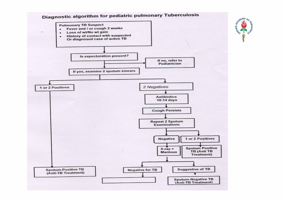
X RAY CHEST FINDINGS
• Enlarged hilar & mediastinal nodes• Pneumonia, bronchopneumonia• Pleural effusion• Mediastinal shift – pleural effusion tension cavity• Cavitation in unusual • Obstructive emphysema – hypertransluence• Bronchiectasis• Miliary mottling
25
IAP UG Teaching slides 2015‐16
CONT..
Abnormalities often seen in apical or posterior segments of upper lobe or superior segments of lower lobe may have unusual appearance in HIV‐positive persons . It cannot confirm diagnosis of TB.
26
IAP UG Teaching slides 2015‐16 27

IAP UG Teaching slides 2015‐16
DEFINITIONS
New case ‐ Patient who had not taken ATT previously or has taken for less than 4 weeks.Relapse ‐ Patient declared cured/completed therapy in past and has evidence of recurrence.Treatment after default ‐ A patient who has taken treatment for at least 4 weeks and comes after interruption of treatment for 2 months and has active disease.
28

IAP UG Teaching slides 2015‐16
DEFINITIONS
Failure to respond A case of pediatric tuberculosis who fails to have bacteriological conversion to negative status or fails to respond clinically and /or deteriorates after12weeks of compliant intensive phase shall be deemed to have failed to respond
29

IAP UG Teaching slides 2015‐16
TREATMENT
CATEGORY TYPE OF PATIENT INTENSIVE PHASE
CONTINUATION PHASE
New cases • New smear positive Pulmonary TB
• New smear negative Pulmonary TB
• New extrapulmonary Tuberculosis
2 (HRZE) 3 4 (HR) 3
Previously treated Cases
Relapse Failure to respond Treatment after default
2(HRZES)3 +1 (HRZE) 3
5 (HRE) 3
30
IAP UG Teaching slides 2015‐16
ROLE OF INTENSIVE PHASE
Four drugs in the intensive phase (IP) :•To achieve rapid killing of actively multiplying bacillary population•To eliminate naturally occurring drug resistant mutants •To prevent the further emergence of drug resistant mutants•An optimal minimum duration of two months in new cases is essential for achieving smear conversion of 90% and above, thereby significantly reducing the infectiousness of the patient
31
IAP UG Teaching slides 2015‐16
ROLE OF CONTINUATION PHASE
Fewer drugs for a longer time for :•Elimination of persisters which are responsible for relapses. •The optimum duration of continuation phase is four months in new cases.
32

IAP UG Teaching slides 2015‐16
BACILLE CALMETTE GUERIN (BCG)
• Albert Calmette & Camille Guerin • In India produced in Chennai (Guindy)• Danish 1331 strain• Freeze dried (3 Months) & Liquid vaccine (1Month)• Stored at 2‐4°C• Each vaccine dose has 0.1‐ 0.4 million viable bacilli• Reconstituted with NS as distilled water is irritant • Reconstituted vaccine to be used within 3 hrs• Dose – 0.1ml (0.1mg)
33

IAP UG Teaching slides 2015‐16
(BCG) CONT…
• Timing – at Birth or at time of earliest contact with the child, preferably before 1 year of age
• 25/26 Guage needle, Intradermal, Left upper arm at insertion of deltoid
• IUGR should not be a reason for delaying vaccination• 50% effective in preventing pulmonary tuberculosis in adults and children
• The protective effect for disseminated and meningeal tuberculosis : 50‐80% of cases
34

IAP UG Teaching slides 2015‐16
CONTRAINDICATIONS
• Impaired immunity in leukemia, lymphoma, other malignancy, HIV and congenital immunodeficiency
• Immunosuppressive therapy – steroids, antimetabolites, irradiation and alkylating agents
• Any viral infection (Measles, Varicella and Hepatitis‐B) give vaccine 4‐6 weeks later
• If received immunoglobulin postpone vaccination for 3 months
35
IAP UG Teaching slides 2015‐16
EVENTS FOLLOWING BCG VACCINATION
• 0.1ml Intradermal raises a wheal of 8mm• Wheal gets absorbed in half‐an‐hour • Induration at injection site at 3 weeks• Lump of 6‐10 mm at 6th week ( Not painful but tender) ‐‐‐ softens with pus formation & discharges
• Healing complete by 12 weeks with a scar of 5‐7mm
36

IAP UG Teaching slides 2015‐16
THANK YOU
37