investigation and validation of intersite fmri studies ... · investigation and validation of...
TRANSCRIPT

Original Research
Investigation and Validation of Intersite fMRIStudies Using the Same Imaging Hardware
Bradley P. Sutton, PhD,1,2* Joshua Goh, MA,2–4 Andrew Hebrank, BA,2,5
Robert C. Welsh, PhD,6 Michael W.L. Chee, MD,4 and Denise C. Park, PhD2,3,5
Purpose: To provide a between-site comparison of func-tional MRI (fMRI) signal reproducibility in two laboratoriesequipped with identical imaging hardware and software.Many studies have looked at within-subject reliability andmore recent efforts have begun to calibrate responsesacross sites, magnetic field strengths, and software. Bycomparing identical imaging hardware and software, weprovide a benchmark for future multisite comparisons.
Materials and Methods: We evaluated system compatibilitybased on noise and stability properties of phantom scans andcontrast estimates from repeated runs of a blocked motor andvisual task on the same four subjects at both sites.
Results: Analysis of variance (ANOVA) and region of inter-est (ROI) analysis confirmed that site did not play a signif-icant role in explaining variance in our large fMRI dataset.Effect size analysis shows that between-subject differencesaccount for nearly 10 times more variance than site effects.
Conclusion: We show that quantitative comparisons of con-trast estimates derived from cognitive experiments can reli-ably be compared across two sites. This allows us to establishan effective platform for comparing group differences betweentwo sites using fMRI when group effects are potentially con-founded with site, as in the study of neurocultural differencesbetween countries or multicenter clinical trials.
Key Words: functional MRI; reproducibility; intersite com-parisons; effect size; cultural neuroscienceJ. Magn. Reson. Imaging 2008;28:21–28.© 2008 Wiley-Liss, Inc.
SINCE THE INTRODUCTION of functional MRI (fMRI),many studies have been conducted to explore humancognition. The use of fMRI technology is both costly andtime-consuming, resulting in relatively small numbersof subjects in treatment conditions—commonly be-tween 10 and 20 subjects per condition. As researchhas become more sophisticated, there is increasing de-mand that larger numbers of subjects or patients beincluded in studies, allowing researchers to investigateboth intergroup differences and interindividual differ-ences within groups. As it may be difficult to recruitadequate numbers of volunteers or patients from anygiven site, and because it is almost impossible to de-scribe population characteristics adequately at a singlesite, multicenter studies are becoming an increasinglyimportant aspect of neuroimaging research.
Site differences are a particularly important issue inthe burgeoning area of cultural neuroscience, the studyof cultural differences in neurocognitive processes (1–5). In order to address the critical questions in thissubdiscipline, it is typically necessary to collect datafrom multiple sites that are often located in two or moredifferent countries, and then compare group differencesin specific neurocognitive processes. In order to directlycompare neural activation patterns across sites (as incultural studies) or to treat aggregate data from sub-jects tested at different sites (as in multisite patientstudies), it is important to demonstrate that the scan-ning site is not a significant source of systematic vari-ance in observed neural activation patterns so that onedoes not falsely conclude that there are differences dueto cultural experience that are really due to site.
Given evidence for good reliability of single site re-sults of fMRI for within-subject (for example, Refs. 6,7)and between-subject (for example, Refs. 8,9) activa-tions, multisite studies require reliability of activationdetection across sites using different magnets. Earlyintersite comparisons have largely focused on produc-ing similar thresholded activation maps at differentsites for the same stimulus paradigm. Casey et al (10)reported results from four different sites, using differentpulse sequences, different fMRI processing software,and 1.5T MR scanners made by two different manufac-turers and found reliable patterns of activation acrosssites. Similarly, Ojemann et al (11) compared fMRI re-sults from two sites with positron emission tomography
1Bioengineering Department, University of Illinois at Urbana-Cham-paign, Urbana, Illinois.2Beckman Institute, University of Illinois at Urbana-Champaign, Ur-bana, Illinois.3Department of Psychology, University of Illinois at Urbana-Cham-paign, Urbana, Illinois.4Cognitive Neuroscience Laboratory, Duke-NUS Graduate MedicalSchool, Singapore.5Center for Brain Health, University of Texas at Dallas, Dallas, Texas.6Radiology, University of Michigan, Ann Arbor, Michigan.Contract grant sponsor: National Institutes of Health (NIH); Contractgrant number: 5R01AG015047-08.*Address reprint requests to: B.P.S., Bioengineering, 3120 DCL, 1304W. Springfield Ave., Urbana, IL 61801. E-mail: [email protected] October 25, 2007; Accepted March 20, 2008.DOI 10.1002/jmri.21419Published online in Wiley InterScience (www.interscience.wiley.com).
JOURNAL OF MAGNETIC RESONANCE IMAGING 28:21–28 (2008)
© 2008 Wiley-Liss, Inc. 21
(PET) results obtained using the same word-stem com-pletion task. Even though the fMRI results originatedfrom two different institutions and two different groupsof subjects using MRI hardware produced by differentmanufacturers with different pulse sequences andanalysis techniques, the investigators found highly re-producible areas of activation that agreed well withother findings from PET data. These qualitative findingssuggest that fMRI results are fairly robust across arange of conditions.
In light of the promising results from Casey et al (10)and Ojemann et al (11), a number of recent studies havebeen conducted to provide a concerted effort to examinereproducibility of fMRI responses from different sites. Aconservative criterion for the demonstration of multi-center compatibility would be to demonstrate a highlyreproducible blood oxygenation level-dependent(BOLD) response rather than simply similar thresh-olded activation maps, as has been used in previousstudies. One of the larger efforts in this direction hasbeen conducted by fBIRN (www.nbirn.net), involving 14different MR laboratories that use magnets of threedifferent field strengths with three different scannermanufacturers and different pulse sequences (12–17).The initial work has highlighted many factors impor-tant to ensure compatibility between different sites. Forexample, the fBIRN group has shown the importance ofsimple but regular standardized quality control mea-sures to maintain comparability of scanners using bothphantoms (14) and human data (18). In Friedman andBirn (12) and Friedman et al (13), differences in thesmoothness and sensitivity of fMRI images were com-pared among 10 sites. Significant smoothness and sen-sitivity differences were found that related to imagingsequence, gradient performance, image reconstructionand filtering methods, and field strength. Due to differ-ent sensitivities to the BOLD response at different sites,these studies have also motivated an effort to calibratefMRI responses across sites and individuals using abreath-hold task (15).
In the present study we evaluate the contributions ofsite, subject, and session to fMRI data variance. Be-cause we are particularly interested in comparing dif-ferences in signal patterns in groups tested at two dif-ferent sites (eg, we are interested in whether EastAsians tested in Asia show subtle differences in neuralcircuitry for processing objects and scenes compared toWesterners tested in the US), it was critically importantto demonstrate that observed differences between cul-tural groups could not be attributed to systematic in-tersite variability, but rather to systematic differencesin neural function between groups. We hypothesizedthat we would be able to assess intersite reliability if weimaged the same individuals repeatedly at the two sites.Specifically, we hypothesized that we would find thatthe variability within subjects imaged at two differentsites was no greater than the differences within sub-jects imaged repeatedly at a single site, and less thanvariability between subjects at a single site. Unlike pre-vious intersite comparisons, we compared sites thathad the same imaging hardware, identical pulse se-quences, and data analysis strategies using the samesubjects at both sites. We will show that within-subject
variance across site was small compared to between-subject variance within a single site. This enablesquantitative group comparisons across sites using con-trasted parameter estimates rather than merely thresh-olded activation maps.
We examined reproducibility in terms of system sta-bility and noise based on both phantom quality controlmeasures and reproducibility of contrast estimatesfrom motor and visual functional tasks. The motor andvisual tasks were performed repeatedly by four subjectsand treated in a three-way analysis of variance (ANOVA)to assess main effects and interactions of subject, site,and task.
MATERIALS AND METHODS
The two sites involved in this study, one located in Asiaand the other in the US, are henceforth referred to as“site 1” and “site 2.” Each site was equipped with aSiemens 3T Allegra MR scanner (Erlangen, Germany)and a USA Instruments (Aurora, OH) headcoil. Proto-cols for both the phantom quality control (QC) and thehuman studies were equalized between sites using elec-tronic transfer of protocols.
Visual stimuli were presented using an LCD projectorbackprojecting onto a screen. The geometry of thescreen was matched between sites and the luminanceof the projected images was matched using a lightmeter. Ambient light conditions were matched by run-ning human experiments with scanner room lights off.Auditory stimuli were delivered using an identicalheadphone system from Resonance Technologies(Northridge, CA).
Phantom Studies
First, we examined system noise using a copper sulfate-doped cylindrical phantom. The phantom is the stan-dard Siemens QA cylindrical phantom. The phantomuses a special holder and markings to ensure properpositioning of the phantom compared to the head coil,so positioning of the phantom between sites was repro-ducible. The phantom was scanned daily at both MRsites as part of a quality control monitoring routine,with 78 datasets acquired at site 1 and 220 acquired atsite 2. The phantom was scanned using an echo planarimaging (EPI) sequence with ramp sampling (36 � 3 mmthick slices, 0.3 mm slice gap, TR 2 sec, TE 25 msec, flipangle 90°, field of view [FOV] 220 � 220 mm, 64 � 64matrix size, bandwidth of 2894 Hz/pixel). A total of 256volumes were acquired for a total imaging time of 516seconds, with 4 seconds of discarded acquisitions.Peak-to-peak (PTP) noise and normalized root-mean-squared (NRMS) noise were used to examine stability inthe resulting time courses on a slice-by-slice basis. ThePTP and NRMS noise were determined from the timecourse of the mean over a circular 110 mm area insidethe center of phantom. The peak-to-peak noise wascalculated by subtracting the minimum value over themean time course from the maximum and then normal-izing by the mean. The NRMS noise was calculated as:
NRMS � �1T�
t�1
T
�s�t� � s� �2� s� , [1]
22 Sutton et al.
where s(t) is the value of the mean time course at vol-ume t, s� is the mean of the mean time course, and T isthe total number of volumes acquired.
The mean, NRMS, and PTP noise were compared be-tween sites to determine if there was a system noisedifference between the two sites. This system noise rep-resents only one component of the noise in a functionalimaging data, with physiological noise potentially dom-inating system noise. The phantom analysis can serveto indicate when system maintenance is required byexamining the daily noise properties versus the historyor by comparing the system noise to residual meanerror after fitting functional imaging data.
Reproducibility of Contrast Estimates as aFunction of Subject and Site
Two tasks were presented to four subjects scanned atboth sites—a motor task and a visual task. The func-tional acquisition was an EPI sequence with ramp sam-pling (32 � 4 mm thick slices, 0.4 mm slice gap, TR 2sec, TE 25 msec, flip angle 80°, FOV 220 � 220 mm,64 � 64 matrix size, bandwidth 2894 Hz/pixel). Foursubjects (males, ages 24, 26, 27, and 28; mean 26.25years) were repeatedly scanned at both sites on the twotasks (same four subjects at both sites), with the taskdesign modeled after McGonigle et al (6). All subjectswere right-handed and visual acuity was corrected withMR-compatible lenses to 20/30 using a Snellen Chart.For each task there were �30 scan sessions per subject(15 at each site) spread over 3 days at each site, with atotal of 230 scan sessions across all tasks and subjects.The first task was a motor task: button pressing pacedby auditory tones (1 Hz). Subjects performed alternat-ing 20-second blocks of button pressing and rest with atotal of six button-pressing blocks and seven restblocks in each session; 3 seconds of visual instructions(indicating whether to press or rest) preceded eachblock. A single finger on each hand was used for press-ing a button on a response box (Rowland Institute USBfMRI response boxes, Cambridge, MA). The second taskwas a visual paradigm with 20-second blocks alternat-ing between a fixation cross and an 8 Hz reversingcheckerboard. Three rest and three checkerboardblocks were presented to the subject per run.
During each scan session the subjects underwent alocalizer scan in addition to performing the motor andvisual tasks. A T2-weighted turbo-spin-echo (TSE)high-resolution anatomical scan with the same sliceprescription as the EPI acquisition was used for imagecoregistration. The subjects were removed from thescanner between each scan session. The 15 sessions ateach site were spread out over 3 days of scanning.During one session a magnetization-prepared rapid ac-quisition of gradient echo (MPRAGE) sequence wasused to acquire a high-resolution 3D structural scanused for normalization. The scan acquired whole brainwith isotropic 0.8 mm resolution and an inversion prep-aration time of 1100 msec.
Functional analysis was carried out using FEAT(FMRI Expert Analysis Tool) v. 5.4, part of FSL (FMRIB’sSoftware Library, www.fmrib.ox.ac.uk/fsl). The follow-ing preprocessing steps were applied at the first-level
analysis: slice-timing correction using Fourier-spacetime-series phase-shifting; motion correction usingMCFLIRT (19); nonbrain removal using BET (20); spa-tial smoothing using a Gaussian kernel of full-width athalf-maximum (FWHM) 8 mm; mean-based intensitynormalization of all volumes by the same factor; high-pass temporal filtering (Gaussian-weighted LSFstraight line fitting, with sigma � 50.0 sec). Time-seriesstatistical analysis was carried out using FILM withlocal autocorrelation correction (21). Registration pro-ceeded in three steps using FLIRT (19,22). First, the EPIimages were registered to the TSE scans, which werethen registered to the MPRAGE anatomical image, andfinally to the standard (MNI) image. Higher-level analy-sis was carried out using FLAME (FMRIB’s Local Anal-ysis of Mixed Effects) (23,24). A fixed effects three-wayANOVA was performed in MatLab (MathWorks, Natick,MA) on all 230 functional runs from the study (4 sub-jects � 2 tasks � 2 sites with runs [13–15 per subjects]as a random variable) to assess the effect of subject,site, and task on the functional results.
Besides qualitatively examining the maps of signifi-cant voxels in the main effects and interactions result-ing from the ANOVA analysis (thresholded at P � 0.001uncorrected), two other analyses of the functional datawere performed. We examined the spatial extent of ac-tivation in single subjects across the two sites and weanalyzed the percent signal change over a visual andtwo motor regions-of-interest (ROIs) for each subject ateach site. Additionally, the effect size from the ANOVAwas examined to determine the proportion of variancedue to site as compared to intersubject variance.
RESULTS
Signal Stability in Phantoms and Volunteers
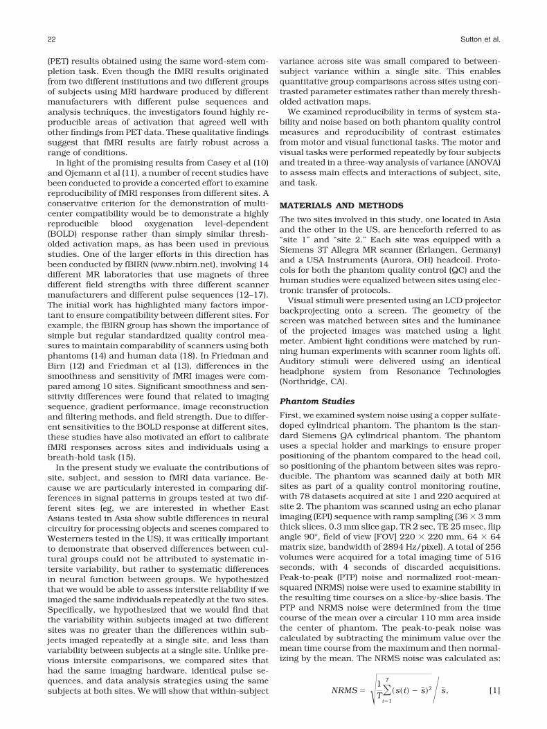
The resulting mean of the time series and NRMS noisevalues from several months of quality control scans ofthe cylindrical phantom at both sites are shown in Fig.1 as strip plots. The PTP noise looked similar to theNRMS noise and is not shown. The phantom resultimages show the mean and NRMS noise for each slicedown the columns and for each day across the rows.Note that at each site there were a few days or clustersof days that had noise values that were significantlyhigher than surrounding days. This suggests the im-portance of daily quality control. On days when thenoise was unacceptably high the MRI service engineerwas notified that maintenance was required on the sys-tem and all scanning sessions were cancelled for thatday.
Although the means of the time series appear equiv-alent across sites, Fig. 1 shows obvious differences be-tween the noise characteristics. In fact, grouping all 36slices into a t-test between the two sites resulted insignificant differences for mean, NRMS noise, and PTPnoise (all at P � 0.01). Figure 2a plots the results ofNRMS noise as a percent of the mean signal for slice 18of 36. Note that more phantom scans were performed atsite 2 than site 1. The differences in noise may resultfrom actual system noise, environmental differencesbetween scanning sites, or a difference in the number of
Validation of Intersite fMRI 23

items passed through the patch panel that may leaksmall amounts of noise.
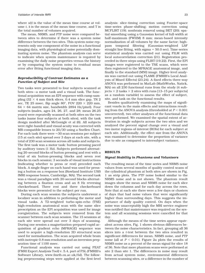
At first glance, these results might seem to suggest aninconsistency between the systems. However, the sys-tem noise must be considered relative to the physiolog-ical noise present in a functional run. The physiologicalnoise may dominate the relevant noise properties of afunctional run, rendering slight differences in systemnoise as relatively inconsequential. In order to examinethe relationship between this system noise and thenoise in the functional runs, we examined fMRI noise attwo levels: 1) looking at the supraventricular white mat-ter in unprocessed functional data, and 2) examiningthe residual error after the first-level functional analy-sis in the 230 runs from our four volunteers.
Signal noise was analyzed by calculating the NRMSnoise from the supraventricular white matter in the rawfunctional images. Data in this region are not affectedby function, but reflect the stability in the baselinesignal. A conservative mask was made of the supraven-tricular white matter consisting of 2192 voxels in thehigh-resolution standard space (2 mm isotropic resolu-tion). After motion-correcting the raw functional timeseries relative to the middle volume, the white mattermask was transformed into the subject’s local space byusing the FSL transformations determined during thefirst level analysis. For each timepoint in the functionalseries the mean over the white matter mask was calcu-lated. The time series of the mean was transformed intoNRMS according to Eq. [1]. The NRMS as a percentageof the mean is plotted in Fig. 2b.
Additionally, for each functional run the residual er-ror (�2) was computed for all voxels that showed signif-icant activation (P � 0.001 uncorrected) to the task in asingle run at the first-level analysis. This residual errormap was formed into an NRMS measure by averagingthe �2 across voxels, taking the square root, and nor-malizing by the mean of the functional imaging timecourse. The plot of NRMS error from the functionalscans is plotted in Fig. 2c.
While it is tempting to quantitatively compare theNRMS measure from the human scans to those in Fig.2a from the phantom scans, note, however, that theslice thickness is different between the phantom andhuman acquisitions. Instead, it is better to compare theNRMS error in Fig. 2b,c between sites. Note that nosignificant differences existed in this error measure be-tween sites (P � 0.38 for both), while differences be-tween subjects were observable. This indicated that thedifferences between system noise were insignificant,
Figure 1. Phantom quality control runs at both sites, showing the mean and noise properties across all 36 slices of the QAacquisitions. Top row is mean and bottom row is root mean square noise of the time series of the averaged ROI.
Figure 2. Normalized root mean square error (or noise) as apercentage of the mean for (a) phantom quality control runs,(b) supraventricular white matter, and (c) residual mean errorin the motor and visual functional tasks. Note that V1 indi-cates subject 1, visual task and M2 indicates subject 2, motortask. Subject/task labels are valid for (b) and (c). [Color figurecan be viewed in the online issue, which is available atwww.interscience.wiley.com.]
24 Sutton et al.

dominated by other sources of noise, and did not affectthe ability to quantitatively compare the functional re-sults across sites.
Statistical Maps
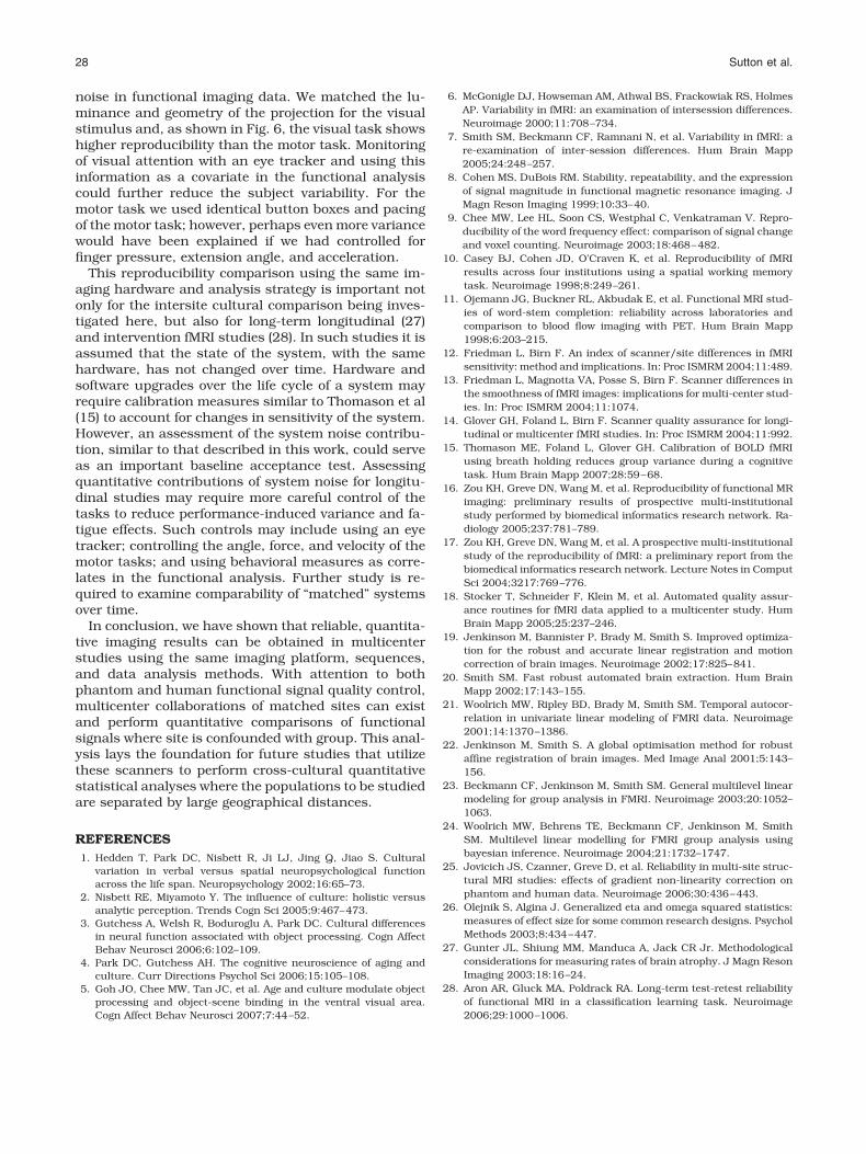
A three-way ANOVA analysis was performed in Matlabon the 230 functional runs with main effects of subject,site, and task and their interactions being investigated.For reference, thresholded (P � 0.001 uncorrected) z-score maps of the mean activations for each task overall subjects and both sites are shown in Fig. 3. Therobust activations resulted from the two reproducibletasks used in this analysis and the 115 functional runsused per map. The thresholded (P � 0.001 uncorrected)ANOVA results for the main effects of subject, site, andtask are shown in Fig. 4. There were extensive regionsshowing significant main effects of subject and task,but there were very few voxels with main effect of sitesurviving P � 0.001 uncorrected. The main effect oftask occurred as expected, due to the significantly dif-ferent brain areas activated by the motor versus visualtasks. These large task differences were included in theANOVA analysis for completeness and to remove theseeffects as a source of variance. The site effects wereminimal—there were several regions that indicated dif-ferences: regions at the most inferior and posteriorparts of the brain. These effects, however, were quitemodest and sparse when compared to the subject dif-ferences, which were much larger, as shown in Table 1,which is described later. Slight differences in placementof the subject’s head could have resulted in these dif-ferences at the lower periphery of the FOV due to shim-ming differences, RF coil sensitivity, or gradient nonlin-earity effects. The scanner’s automatic gradient
shimming routine was employed to shim over the pre-scribed slices; however, differences in head placementcould affect the shimming convergence. Gradient non-linearity effects can be addressed by warping algo-rithms and this has shown promising results for 3Dmorphometry studies (25). Validation of these methods
Figure 3. Thresholded z-scores for all subjects for each task(P � 0.001 uncorrected).
Figure 4. Main effects of the three-way ANOVA, thresholdedwith P � 0.001 (uncorrected). Results are displayed as –log10(p)shown from P � 10e-3 to 10e-40.
Table 1Effect Size Analysis From the ANOVA Results for Site and Subject
Site Subject
Mean �G2 Max �G2 Mean �G2 Max �G2
Visual 0.0112 0.0356 0.0810 0.2342Motor left 0.0120 0.0248 0.0966 0.2574Motor right 0.0085 0.0152 0.0880 0.2501
Validation of Intersite fMRI 25

for functional imaging applications is still lacking;therefore, extra care in placement of the subject’s headwill be exercised in future studies.
The interaction effects are shown in Fig. 5. The moststriking effect is that there was a pronounced Subject �Task interaction, but that interactions involving siteoccurred in relatively few brain areas. Specifically, wefound a significant three-way interaction among site,subject, and task in the medial occipital region (Fig. 5,top left panel). Examining the two-way interactionssuggested that this might stem from the difference inactivation across subjects in these regions being signif-icantly different across sites (Fig. 5, top right). However,note that there was a greater interaction effect betweensubject and task (Fig. 5, lower left), suggesting that thedifferences across site did not affect our ability to detectinterindividual or task differences when data from bothsites were collapsed together.
In order to assess functional sensitivity at both siteswe compared the spatial extent of activation acrosssites. Using cluster in FSL we computed the number ofvoxels in the largest cluster using a threshold of P �0.001, uncorrected, for each subject and each task. Acluster includes all contiguous voxels that are belowthe threshold of 0.001. Then we performed two-samplet-tests between sites for the cluster sizes for each of theapproximately 15 runs. The P-value for the t-test isgiven in Table 2 for each subject and task. Only one ofthe eight pairings yielded statistically significant differ-ences, suggesting that the spatial extent is wellmatched between sites. As further evidence, Fig. 6shows a scatterplot of the pairings of cluster sizes be-
tween site 2 vs. site 1. The scatterplot shows pairings ofrun 1 at site 1 with run 1 at site 2, etc. This scatterplotindicates that there is good agreement between sites inthe number of voxels in the largest cluster.
An ROI analysis was performed on percent signalchange data from three functionally defined areas, onein the visual cortex, one in the right motor area, and onein the left motor area. The ROIs were defined by thresh-olding the group mean activation z-scores in the visualand motor tasks at a level near the peak, then erodingand dilating the mask such that only one contiguousregion of voxels was selected around the peak of inter-est. The number of voxels in the visual ROI was 63, inthe left motor 102 voxels, and in the right motor 54voxels. The ROIs were used to examine percent signalchange at both sites. A pairing of run 1 at site 1 with run1 at site 2, and so on, resulted in the scatterplots in Fig.
Figure 5. Interaction effects from the ANOVA, thresholded with P � 0.001 (uncorrected). Results are displayed as –log10(p)shown from P � 10e-3 to 10e-40.
Table 2Results From Two-Sample t-Test for Comparison of MaximumCluster Size Between Two Sites
Task t-Statistic P-value
Subject 1 Motor 0.398 0.6937Visual 1.556 0.1309
Subject 2 Motor 0.274 0.7863Visual 1.526 0.1381
Subject 3 Motor 0.513 0.6124Visual 2.572 0.0164*
Subject 4 Motor 0.049 0.9612Visual 0.338 0.7384
*Significance at the 0.05 level.
26 Sutton et al.

7. As can be seen in these figures, the percent signalchanges were correlated between sites and intersubjectdifferences were readily observable beyond any site dif-ferences. This was especially true in the visual ROI, asthe activations were much stronger than in the motorareas. Also notice that the left motor cortex activationshows higher consistency than the right motor cortex,likely reflecting the right-handedness of the subjects.The average percent signal change inside the visual ROIfor every visual task run was compared between siteswith a two-sample t-test to assess if the means wereequal. Likewise, the motor ROIs were used to calculateaverage percent signal change from motor runs andassessed for equal means via a two-sample t-test. Theresult (dof � 112 for all ROIs) for the Visual ROI was P �0.89, Left Motor ROI P � 0.16, Right Motor ROI P �0.38. This provides further evidence suggesting that thesite effect is smaller than subject-related variability inROI analyses.
Finally, the ROIs above were used to examine gener-alized effect size of the site and subject componentsfrom the ANOVA. We used the generalized �G
2 fromOlejnik and Algina (26) to provide a measure of effectsize that should be robust to the specific effects (includ-ing task) that were included in the ANOVA analysis. Thegeneralized �G
2 describes the proportion of the varianceexplained by each effect. We treated site as a manipu-lated factor and subject as a measured factor and usedthe formula provided in table 2 of Olejnik and Algina(26). The results in Table 1 show that the site accounted
for around 1% of the variance, whereas subject ac-counted for 8%–10%. This suggests that the effect ofsite is much smaller than the intersubject variance.
DISCUSSION
A methodology is currently being developed to take ad-vantage of the collective resources available acrossmulticenter collaborations. Currently these collabora-tions involve different imaging hardware platforms, dif-ferent sequences, and different data analysis strategies.Quantitative comparison of results from these collabo-rations will rely on either signal processing to makesignals comparable from different sites or on calibra-tion signals obtainable during a normal functional eval-uation (15). In the current study we showed that quan-titative comparisons of functional data can be madewithout calibration steps if two sites are matched onimaging hardware, sequences, analysis methods, scan-ner performance, and quality control.
Our results examine comparability of fMRI data gen-erated at two sites in terms of phantom noise and sta-bility measurements as well as in fMRI experiments.The phantom data show that it is important to monitorsystem noise to ensure that it is low. With careful qual-ity control measures, however, the impact of small dif-ferences in system noise will not have an impact on thenoise level in functional imaging data, which is largelydominated by physiological noise. The three-wayANOVA analysis from repeated scanning of four sub-jects on two tasks indicated a much greater main effectof subject than site. However, there were some signifi-cant voxels showing site differences, especially inferiorregions and cerebellum. This could be due to system-atic head placement differences due to different subjectpositioning pads, an effect that will be controlled infuture studies. The main effect and interaction effectsthat include site show very small numbers of significantvoxels (P � 0.001 uncorrected) with low mean z-scorescompared to other effects. By comparing generalizedeffect sizes, we see that there is a nearly 10 timesgreater effect of subject than site, rendering the plat-form reliable for comparisons using group studies.
Although we matched some parameters of the taskperformance, even more careful control in even thesesimple tasks could provide more information about thecontribution of intra- and intersubject variability to the
Figure 6. Scatterplot of number of voxels in largest cluster atsite 1 vs. site 2. Data labels are defined in Fig. 2.
Figure 7. Scatterplot of percent signal change (% SC) in ROIs for the visual, right motor, and left motor activations. Thescatterplots are mean percent signal change at site 1 vs. site 2. Individual subject scatterplots are evident by color-coding thesubjects as follows: Subject 1 (Black), Subject 2 (Blue), Subject 3 (Green), Subject 4 (Red).
Validation of Intersite fMRI 27
noise in functional imaging data. We matched the lu-minance and geometry of the projection for the visualstimulus and, as shown in Fig. 6, the visual task showshigher reproducibility than the motor task. Monitoringof visual attention with an eye tracker and using thisinformation as a covariate in the functional analysiscould further reduce the subject variability. For themotor task we used identical button boxes and pacingof the motor task; however, perhaps even more variancewould have been explained if we had controlled forfinger pressure, extension angle, and acceleration.
This reproducibility comparison using the same im-aging hardware and analysis strategy is important notonly for the intersite cultural comparison being inves-tigated here, but also for long-term longitudinal (27)and intervention fMRI studies (28). In such studies it isassumed that the state of the system, with the samehardware, has not changed over time. Hardware andsoftware upgrades over the life cycle of a system mayrequire calibration measures similar to Thomason et al(15) to account for changes in sensitivity of the system.However, an assessment of the system noise contribu-tion, similar to that described in this work, could serveas an important baseline acceptance test. Assessingquantitative contributions of system noise for longitu-dinal studies may require more careful control of thetasks to reduce performance-induced variance and fa-tigue effects. Such controls may include using an eyetracker; controlling the angle, force, and velocity of themotor tasks; and using behavioral measures as corre-lates in the functional analysis. Further study is re-quired to examine comparability of “matched” systemsover time.
In conclusion, we have shown that reliable, quantita-tive imaging results can be obtained in multicenterstudies using the same imaging platform, sequences,and data analysis methods. With attention to bothphantom and human functional signal quality control,multicenter collaborations of matched sites can existand perform quantitative comparisons of functionalsignals where site is confounded with group. This anal-ysis lays the foundation for future studies that utilizethese scanners to perform cross-cultural quantitativestatistical analyses where the populations to be studiedare separated by large geographical distances.
REFERENCES1. Hedden T, Park DC, Nisbett R, Ji LJ, Jing Q, Jiao S. Cultural
variation in verbal versus spatial neuropsychological functionacross the life span. Neuropsychology 2002;16:65–73.
2. Nisbett RE, Miyamoto Y. The influence of culture: holistic versusanalytic perception. Trends Cogn Sci 2005;9:467–473.
3. Gutchess A, Welsh R, Boduroglu A, Park DC. Cultural differencesin neural function associated with object processing. Cogn AffectBehav Neurosci 2006;6:102–109.
4. Park DC, Gutchess AH. The cognitive neuroscience of aging andculture. Curr Directions Psychol Sci 2006;15:105–108.
5. Goh JO, Chee MW, Tan JC, et al. Age and culture modulate objectprocessing and object-scene binding in the ventral visual area.Cogn Affect Behav Neurosci 2007;7:44–52.
6. McGonigle DJ, Howseman AM, Athwal BS, Frackowiak RS, HolmesAP. Variability in fMRI: an examination of intersession differences.Neuroimage 2000;11:708–734.
7. Smith SM, Beckmann CF, Ramnani N, et al. Variability in fMRI: are-examination of inter-session differences. Hum Brain Mapp2005;24:248–257.
8. Cohen MS, DuBois RM. Stability, repeatability, and the expressionof signal magnitude in functional magnetic resonance imaging. JMagn Reson Imaging 1999;10:33–40.
9. Chee MW, Lee HL, Soon CS, Westphal C, Venkatraman V. Repro-ducibility of the word frequency effect: comparison of signal changeand voxel counting. Neuroimage 2003;18:468–482.
10. Casey BJ, Cohen JD, O’Craven K, et al. Reproducibility of fMRIresults across four institutions using a spatial working memorytask. Neuroimage 1998;8:249–261.
11. Ojemann JG, Buckner RL, Akbudak E, et al. Functional MRI stud-ies of word-stem completion: reliability across laboratories andcomparison to blood flow imaging with PET. Hum Brain Mapp1998;6:203–215.
12. Friedman L, Birn F. An index of scanner/site differences in fMRIsensitivity: method and implications. In: Proc ISMRM 2004;11:489.
13. Friedman L, Magnotta VA, Posse S, Birn F. Scanner differences inthe smoothness of fMRI images: implications for multi-center stud-ies. In: Proc ISMRM 2004;11:1074.
14. Glover GH, Foland L, Birn F. Scanner quality assurance for longi-tudinal or multicenter fMRI studies. In: Proc ISMRM 2004;11:992.
15. Thomason ME, Foland L, Glover GH. Calibration of BOLD fMRIusing breath holding reduces group variance during a cognitivetask. Hum Brain Mapp 2007;28:59–68.
16. Zou KH, Greve DN, Wang M, et al. Reproducibility of functional MRimaging: preliminary results of prospective multi-institutionalstudy performed by biomedical informatics research network. Ra-diology 2005;237:781–789.
17. Zou KH, Greve DN, Wang M, et al. A prospective multi-institutionalstudy of the reproducibility of fMRI: a preliminary report from thebiomedical informatics research network. Lecture Notes in ComputSci 2004;3217:769–776.
18. Stocker T, Schneider F, Klein M, et al. Automated quality assur-ance routines for fMRI data applied to a multicenter study. HumBrain Mapp 2005;25:237–246.
19. Jenkinson M, Bannister P, Brady M, Smith S. Improved optimiza-tion for the robust and accurate linear registration and motioncorrection of brain images. Neuroimage 2002;17:825–841.
20. Smith SM. Fast robust automated brain extraction. Hum BrainMapp 2002;17:143–155.
21. Woolrich MW, Ripley BD, Brady M, Smith SM. Temporal autocor-relation in univariate linear modeling of FMRI data. Neuroimage2001;14:1370–1386.
22. Jenkinson M, Smith S. A global optimisation method for robustaffine registration of brain images. Med Image Anal 2001;5:143–156.
23. Beckmann CF, Jenkinson M, Smith SM. General multilevel linearmodeling for group analysis in FMRI. Neuroimage 2003;20:1052–1063.
24. Woolrich MW, Behrens TE, Beckmann CF, Jenkinson M, SmithSM. Multilevel linear modelling for FMRI group analysis usingbayesian inference. Neuroimage 2004;21:1732–1747.
25. Jovicich JS, Czanner, Greve D, et al. Reliability in multi-site struc-tural MRI studies: effects of gradient non-linearity correction onphantom and human data. Neuroimage 2006;30:436–443.
26. Olejnik S, Algina J. Generalized eta and omega squared statistics:measures of effect size for some common research designs. PsycholMethods 2003;8:434–447.
27. Gunter JL, Shiung MM, Manduca A, Jack CR Jr. Methodologicalconsiderations for measuring rates of brain atrophy. J Magn ResonImaging 2003;18:16–24.
28. Aron AR, Gluck MA, Poldrack RA. Long-term test-retest reliabilityof functional MRI in a classification learning task. Neuroimage2006;29:1000–1006.
28 Sutton et al.