investigating brain connectivity heritability in a twin
TRANSCRIPT
Investigating brain connectivity heritability in a twin study using diffusion imaging data
K.-K. Shen1, S. Rose1, J. Fripp1, K. L. McMahon2, G. I. de Zubicaray3, N. G. Martin4, P. M. Thompson5, M. J. Wright4, O. Salvado1
1. CSIRO Computational Informatics, Herston QLD 4029, Australia; 2. Centre for Advanced Imaging, University of Queensland, Brisbane, Australia;
3. School of Psychology, University of Queensland, Brisbane, Australia; 4. Queensland Institute of Medical Research, Brisbane, Australia;
5. Imaging Genetics Center, Institute for Neuroimaging & Informatics, University of South California, Los Angeles, CA, USA
Abstract
Heritability of brain anatomical connectivity has been studied with diffusion-weighted
imaging (DWI) mainly by modeling each voxel’s diffusion pattern as a tensor (e.g., to
compute fractional anisotropy), but this method cannot accurately represent the many
crossing connections present in the brain. We hypothesized that different brain networks (i.e.,
their component fibers) might have different heritability and we investigated brain
connectivity using High Angular Resolution Diffusion Imaging (HARDI) in a cohort of twins
comprising 328 subjects that included 70 pairs of monozygotic and 91 pairs of dizygotic
twins. Water diffusion was modeled in each voxel with a Fiber Orientation Distribution
(FOD) function to study heritability for multiple fiber orientations in each voxel. Precision
was estimated in a test-retest experiment on a sub-cohort of 39 subjects. This was taken into
account when computing heritability of FOD peaks using an ACE model on the monozygotic
and dizygotic twins. Our results confirmed the overall heritability of the major white matter
tracts but also identified differences inheritability between connectivity networks. Inter-
hemispheric connections tended to be more heritable than intra-hemispheric and cortico-
spinal connections.
Introduction
White matter (WM) structures develop and change throughout life. These changes are
influenced by both genetics and environment, and can be monitored using diffusion-weighted
imaging (DWI) (Wozniak and Lim, 2006; Sullivan and Pfefferbaum, 2006, Cascio et al.,
2007). Axon myelination is involved in the plasticity of cognitive functions, and may be
altered by changes in myelin genes or mutation in neurological or psychiatric disorders
(Fields, 2008). Based on the quantification of WM integrity using Diffusion Tensor Imaging
(DTI), investigations into the heritability of WM structures usually estimate the genetic
influence based on scalar measures such as fractional anisotropy (FA) or mean diffusivity
(Jahanshad et al., 2013; for a review, see Kanchibhotla, et al., 2013). For example, the Tract-
Based Spatial Statistics method revealed a significant genetic component influencing whole
brain FA and radial diffusivity (Kochunov et al., 2010). Strong genetic influence on WM was
shown at different stages of life (Pfefferbaum et al., 2001, Chiang et al., 2009, Brouwer et al.
2010).
DWI captures the complex microscopic features of axons, but a DTI model assumes a
single dominant direction of water diffusion. This can be inadequate for representing voxels
containing crossing or diverging fibers. New DWI techniques such as High Angular
Resolution Diffusion Imaging (HARDI, Tuch et al., 2002) can resolve fiber microstructure
more accurately. A voxel-wise diffusion orientation density function can be reconstructed by
diffusion spectrum imaging (Lin et al., 2003), q-ball imaging (Tuch, 2004), or hybrid
diffusion imaging (Zhan et al., 2011).
Our approach uses the Fiber Orientation Distribution technique (FOD, Tournier et al.,
2004) to describe the intra-voxel structure of WM fibers. As the FOD is proportional to the
fraction of fibers oriented along the respective direction, it can provide a biologically
meaningful representation of the fiber structure in each voxel.
We hypothesized that there may be a different degree of genetic influence on distinct
brain networks and their components. We investigated this hypothesis on a large cohort of

twins comprising 328 subjects that included 70 pairs of monozygotic (MZ) and 91 pairs of
dizygotic (DZ) twins. We used FOD-based measurements to study crossing fibers
individually and estimate genetic influences on intra-voxel fiber structures. A test-retest
experiment was also conducted with repeated scans to evaluate the reliability of our image
processing and analysis framework. We estimated the heritability of the FOD amplitudes,
translating into heritability estimates for intra-voxel fiber populations, and estimated the
average heritability along each tract by sampling the FOD. We further projected each tract’s
heritability onto the cortical areas it innervates, to study heritability pattern for various
cortical networks. This links the heritability of WM organization with heritability patterns for
cortical structure that have been reported before (Lenroot et al., 2009, Winkler et al., 2010,
Joshi et al., 2011, Eyler et al., 2012). The overall heritability of the major WM tracts was
high and we were also able to map some regional differences in heritability. In particular,
inter-hemispheric connections tended to be more genetically influenced than intra-
hemispheric and cortico-spinal connections.
Materials and Methods
Participants. The cohort consisted of 328 subjects (118M, 210F) with average age 22.7(2.3)
years. Among the subjects, there were 71 pairs (N=142, 48M, 94F) of MZ twins with average
age 22.8(2.2) years, and 90 pairs (N=180, 69M, 111F) of DZ twins with average age 22.6(2.4)
years. A subset of 39 subjects (16M, 23F) with average age of 23.1(2.4) years was analyzed
to estimate test-retest reliability. For this purpose, subjects were scanned twice at 3-month
interval.
Image Acquisition. HARDI data were acquired using a 4T Bruker Medspec MRI scanner.
Each dataset consisted of 11 images without diffusion sensitization (b=0), and diffusion
weighted images (DWI) with 94 gradient directions at b = 1159s/mm2.

Diffusion Magnetic Resonance (MR) Image Processing. The overall image processing
pipeline is shown in Figure 1. The DWI images were pre-processed using point spread
function mapping (Zaitsev et al., 2004). The bias field was corrected using the N4 method
(Tustison et al., 2010). Interleaving artifacts due to subject motion within the same MR
volume were corrected using the inverse interpolation method (Rohlfing et al., 2008), and
inter-volume motion was corrected by rigid registration of brain masks (Raffelt et al., 2012).
Image intensity was normalized across subjects (Raffelt et al., 2012). Spherical harmonic
deconvolution (Tournier et al., 2008) was used to estimate the distribution of the fiber
population in each voxel. In this approach, the observed HARDI signal in each voxel is
modeled as a superposition of signals from the partial volumes of fibers aligned in various
orientations. The fiber partial volume is modeled by a continuous distribution of fiber
orientations. The sampled HARDI signal is estimated as a convolution of the FOD and the
response signal from coherently aligned fibers (Leow et al., 2009), and this was estimated
from a region of interest in the corpus callosum defined on the common atlas.
Spatial normalization of FOD images to a common atlas space. Once corrected, all the
subjects’ datasets were aligned to a common template (Figure 1). We created an atlas from the
data set by iteratively computing a non-rigid registration of each subject to the current
template, followed by averaging all the subjects’ transformed data to estimate a new template.
At the first iteration a randomly chosen subject was used as the template. The non-rigid
registration was performed on the FOD images using a symmetric diffeomorphic registration
(Raffelt et al., 2011). Briefly, during registration the FOD of each voxel represented by
spherical harmonic coefficients was interpolated using B-spline interpolation once the image
was warped to the template. Besides interpolation, the spatial transformation was also used to
modify each FOD to reorient the fiber population within each voxel. Each FOD was
reoriented by an affine transformation that approximated the local deformation field. In
addition, to correct for the local deformation of the transformation field, a modulation step
was required. The FOD amplitude in each spatial orientation was rescaled by a modulation
factor computed from the local Jacobian (Raffelt et al., 2012).
After the final iteration, the template represented the sample average with each voxel
modeled as the average of all the subjects. An FA map was computed using a tensor model
for each subject. The same transformation field for each subject was also used to transform
the FA map of each subject and to create an average FA map. By registering the average FA
map to the FA map from the JHU DTI atlas (Mori et al., 2005, Wakana et al., 2007, Hua et
al., 2008), we realigned the FOD maps for each subject to the standard MNI coordinates with
a dense spatial correspondence between the sampled population and the MNI space.
Detection of the FOD peaks. Once all the subjects’ images had been spatially normalized to
the common template, voxel-wise statistical analysis was performed (Figure 1). As the FOD
describes the distribution of underlying WM fibers within the voxel, the amplitude of the
FOD peaks estimates the proportion of axons aligned in different orientations (Raffelt et al.,
2012). We measured the amplitude of the FOD peaks, and used this to estimate a measure of
WM heritability. The three principal FOD peak amplitudes were estimated in each voxel of
the average FOD template using MRtrix (Tournier et al., 2012). The same estimate for each
subject also yielded three main FOD peaks in each voxel, which were matched to the most
likely peaks from the template, based on angular error. Finally, for each voxel, the three FOD
peak amplitudes were ranked and the two largest used in analyses described below, unless the
second highest FOD peak was lower than 0.1, in which case only one FOD peak was
considered (Jeurissen et al., 2013).
Statistical analysis. Voxel-based analysis was performed on the FOD peak amplitude (i.e.,
two peaks or one depending on the previous step). Test-retest reliability (precision) of the
FOD peaks’ amplitude was estimated for each voxel. We computed the average the reliability
of the largest two FOD peaks, respectively, over the WM regions of interest (ROI) according
to the probabilistic JHU DTI tractography atlas (Hua et al., 2008) in which the ROIs defined
by probability greater than 25% (Jahanshad et al., 2010) were used. Subdivisions of corpus
callosum and internal capsule were also included using the JHU ICBM labels (Wakana et al.,
2007).
To estimate the reliability of the MR diffusion measurements, we compared images of
the same subjects at two different time points. We used the intraclass correlation (ICC, Shrout
and Fleiss, 1979) to evaluate the test-retest reliability of the FOD peak amplitudes. The ICC
was calculated for each of the FOD peak amplitudes in each voxel, and was defined as:
ICC =BMS −WMS
BMS +WMS
where BMS is the between-subject mean square variance, and WMS is the mean square
variance within the subject between test and retest time points. Negative ICC estimates were
clamped to zero (Bartko, 1976), such that the variance remained non-negative, consistent
with ICC interpretation. Measurements were inverse-normalized and corrected for age and
sex.
One can expect the relative influence of genetic and environmental factors to be
different among MZ twins and DZ twins as MZ twins share identical genes whereas DZ
twins share on average only half of their genes. Using an ACE model, FOD peak amplitudes
were assumed to be subject to the influence of three factors: additive genetics A, common
environment C, and residual E due to unique environment and measurement errors which are
independent between individuals. We thus assumed that
FOD ��� = � + � + �.
The measurements of FOD peak amplitude were used to fit the covariance structure of the

ACE model. A non-negative least squares estimation (Lawson and Hanson, 1987, Chen et al.,
2013) was used to estimate the variance of each component and the heritability index
estimates for additive genetic influences, within the total variance defined as:
ℎ� =Var(�)
Var(���� !").
A likelihood ratio test (LRT) comparing the ACE model and CE model of the environment
factors only (i.e. common environment C and unique environment E) was used to assess the
significance of the additive genetic component A for each FOD peak. The p-value of the LRT
was computed by 1000 permutations.
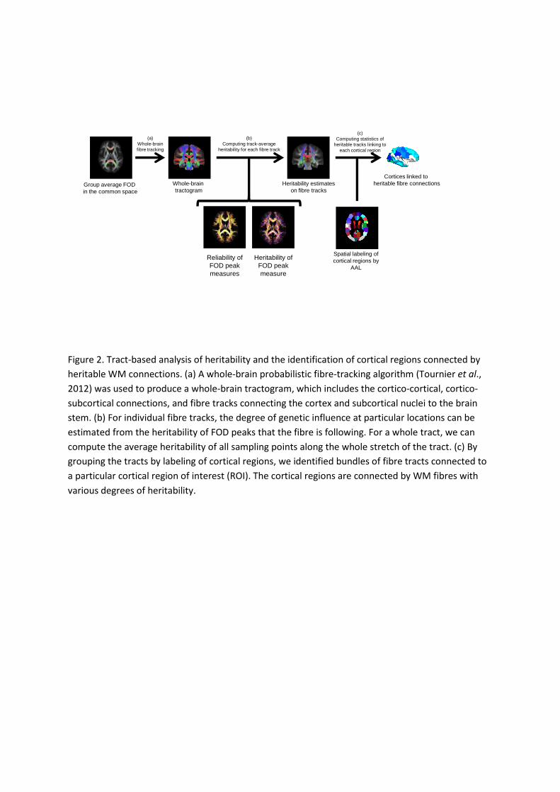
Tract-based analysis. A diagram illustrating our tract-based analysis is shown in Figure 2. We
first performed whole brain probabilistic fiber tracking (Tournier et al., 2012) on the
population average FOD template, creating a tractogram. Only the tracts connecting the
cortical mantle (gray matter, GM), linking cerebral cortex with subcortical nuclei, such as
cortico-striatal and cortico-thalamic tracts, and those travelling through the brain stem were
kept. All other tracts were excluded as they do not represent anatomically plausible pathways.
Voxels with FA > 0.3 in the average template, and voxels most probably containing WM (as
opposed to GM and cerebrospinal fluid) according to the a priori probability of the ICBM
152 atlas (Fonov et al., 2011) were delineated as the WM ROI. The cerebral cortex and
subcortical nuclei were parcellated using the Anatomical Automatic Labeling atlas (AAL,
Tzourio-Mazoyer, 2002), and the brain stem mask was defined as in the Harvard-Oxford atlas
(Markis et al., 2006).
We assumed that FOD peaks characterized the underlying fiber structure in each
voxel, and thus the heritability FOD peaks described genetic influence on the fiber tracts
travelling through the voxel along the direction of the FOD peaks. This allowed us to project
the test-retest reliability ICC and heritability index h2 of FOD peaks onto each of the fiber
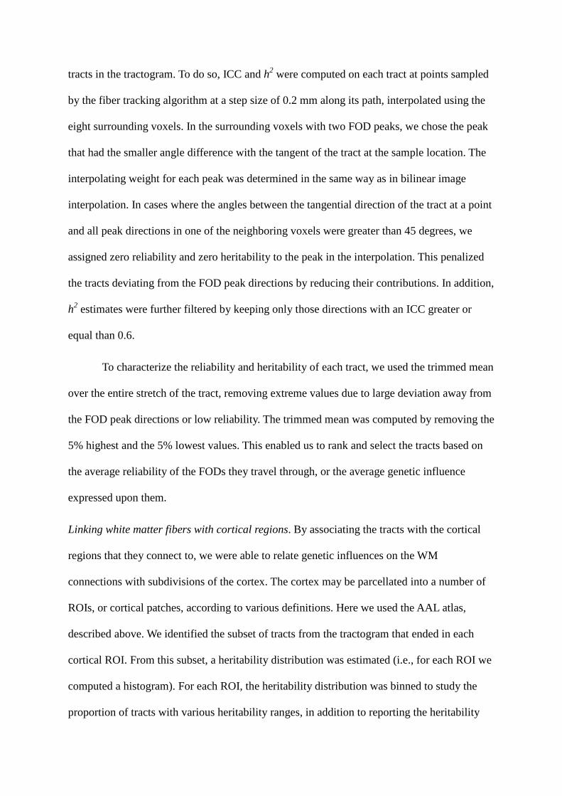
tracts in the tractogram. To do so, ICC and h2 were computed on each tract at points sampled
by the fiber tracking algorithm at a step size of 0.2 mm along its path, interpolated using the
eight surrounding voxels. In the surrounding voxels with two FOD peaks, we chose the peak
that had the smaller angle difference with the tangent of the tract at the sample location. The
interpolating weight for each peak was determined in the same way as in bilinear image
interpolation. In cases where the angles between the tangential direction of the tract at a point
and all peak directions in one of the neighboring voxels were greater than 45 degrees, we
assigned zero reliability and zero heritability to the peak in the interpolation. This penalized
the tracts deviating from the FOD peak directions by reducing their contributions. In addition,
h2 estimates were further filtered by keeping only those directions with an ICC greater or
equal than 0.6.
To characterize the reliability and heritability of each tract, we used the trimmed mean
over the entire stretch of the tract, removing extreme values due to large deviation away from
the FOD peak directions or low reliability. The trimmed mean was computed by removing the
5% highest and the 5% lowest values. This enabled us to rank and select the tracts based on
the average reliability of the FODs they travel through, or the average genetic influence
expressed upon them.
Linking white matter fibers with cortical regions. By associating the tracts with the cortical
regions that they connect to, we were able to relate genetic influences on the WM
connections with subdivisions of the cortex. The cortex may be parcellated into a number of
ROIs, or cortical patches, according to various definitions. Here we used the AAL atlas,
described above. We identified the subset of tracts from the tractogram that ended in each
cortical ROI. From this subset, a heritability distribution was estimated (i.e., for each ROI we
computed a histogram). For each ROI, the heritability distribution was binned to study the
proportion of tracts with various heritability ranges, in addition to reporting the heritability
average that could be color-coded over the brain surface for each patch.
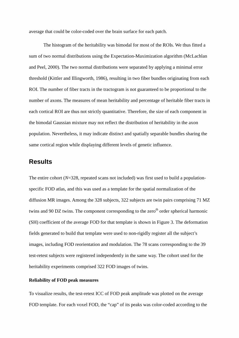
The histogram of the heritability was bimodal for most of the ROIs. We thus fitted a
sum of two normal distributions using the Expectation-Maximization algorithm (McLachlan
and Peel, 2000). The two normal distributions were separated by applying a minimal error
threshold (Kittler and Illingworth, 1986), resulting in two fiber bundles originating from each
ROI. The number of fiber tracts in the tractogram is not guaranteed to be proportional to the
number of axons. The measures of mean heritability and percentage of heritable fiber tracts in
each cortical ROI are thus not strictly quantitative. Therefore, the size of each component in
the bimodal Gaussian mixture may not reflect the distribution of heritability in the axon
population. Nevertheless, it may indicate distinct and spatially separable bundles sharing the
same cortical region while displaying different levels of genetic influence.
Results
The entire cohort (N=328, repeated scans not included) was first used to build a population-
specific FOD atlas, and this was used as a template for the spatial normalization of the
diffusion MR images. Among the 328 subjects, 322 subjects are twin pairs comprising 71 MZ
twins and 90 DZ twins. The component corresponding to the zeroth order spherical harmonic
(SH) coefficient of the average FOD for that template is shown in Figure 3. The deformation
fields generated to build that template were used to non-rigidly register all the subject’s
images, including FOD reorientation and modulation. The 78 scans corresponding to the 39
test-retest subjects were registered independently in the same way. The cohort used for the
heritability experiments comprised 322 FOD images of twins.
Reliability of FOD peak measures
To visualize results, the test-retest ICC of FOD peak amplitude was plotted on the average
FOD template. For each voxel FOD, the “cap” of its peaks was color-coded according to the

ICC value of each peak. A close-up view of the coronal section with crossing fibers of the
corona radiata is shown in Figure 4, where the two main peaks of FODs with fibers crossing
can be seen.
To investigate precision, a test-retest ICC map was created corresponding to the first FOD
peak in the WM region (Figure 5). The average of estimated test-retest ICC in the WM was
0.670. The test-retest ICC for voxels with a second peak (FOD > 0.1) is shown in Figure 6.
The ICC average was 0.547 for the second peak. ROI based analysis was carried out using
the labeling from the JHU DTI atlas. The average test-retest ICC in each ROI is listed in
Figure 7.
Heritability of WM measured by FOD peak
The heritability index h2 for the first FOD peak in WM is shown in Figure 8. After applying
the binary mask to suppress less reliable estimates (ICC < 0.6), the heritability of the first
FOD peak averaged over the entire WM region was 0.194. Significant component of additive
genetic influence was found in major WM tracts, where the average over all the major WM
tracts defined in the JHU atlas was 0.291. The results for the second FOD peak are shown in
Figure 9, and the average within the WM was 0.129.The average heritability of WM in each
ROI of the JHU atlas is listed in Figure 10.
Relatively high heritability was found for the corpus callosum, where the average heritability
of the FOD peak was 0.552 in the body, 0.519 in the genu, and 0.499 in the splenium. The left
and right inferior fronto-occipital fasciculus had a heritability of 0.347 and 0.381,
respectively. The superior longitudinal fasciculus (h2: left 0.307, right 0.381) and the inferior
longitudinal fasciculus (h2: left 0.204, right 0.259) were found to be less heritable. The
heritability of FOD peak measurements in cortico-spinal tracts were estimated to be 0.287
(left) and 0.418 (right) along the tracts.

For areas known to include crossing fibers such as the corona radiata (labeled as cortico-
spinal tract in the ROI definition), moderate heritability of the second FOD peak was found
(left 0.232, right 0.220). There was also a moderately heritable component found in the
second FOD peaks in the superior longitudinal fasciculus (left 0.155, right 0.179) in addition
to more pronounced heritability in the first peak (left 0.307, right 0.381).
Heritable WM fiber tracts
We built a whole brain tractogram on the population average FOD template using a
probabilistic fiber tracking algorithm. In total 250,000 tracts were generated, among which
122,000 were kept because both ends were located in the cortex, connecting to subcortical
nuclei, or passing through the brain stem. We selected among these tracts a reliable subset
with track-average test-retest ICC greater than 0.6, comprising 98,000 tracts, which were then
ranked according to their average heritability index, h2. Projections of the tractogram are
shown in Figure 11, which shows the reliable subset of tracts from the fiber tracking
algorithm, and the tracts with average heritability greater than 0.45. In Figure 12, the
tractogram was binned into 6 classes according to h2, showing the WM fiber tracks with
heritability ranging from below 0.2 to greater than 0.45.The fibers with different heritability
appeared to form different connection networks. For instance, in Figure 12, the less heritable
fiber connections are almost absent in the corpus callosum, whereas the commissural fibers
transiting via the corpus callosum appeared exclusively in groups with higher heritability.
Cortical regions linked to heritable WM tracks
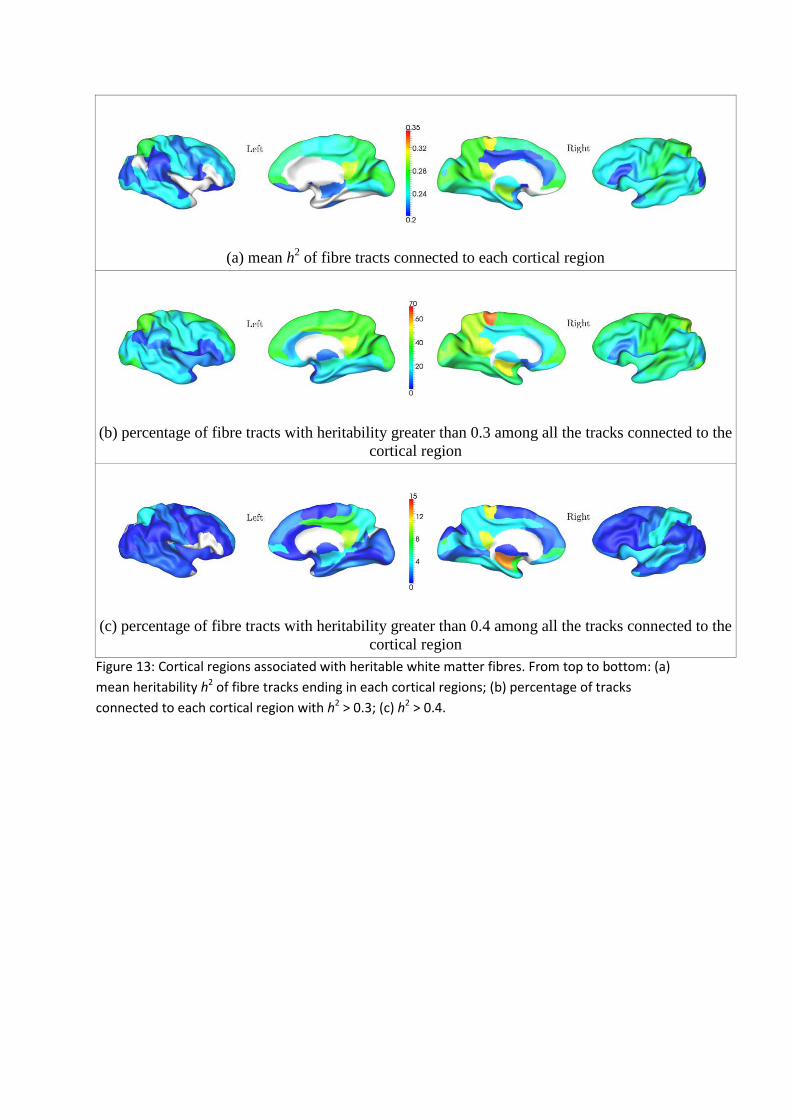
Figure 13 shows the results after mapping the average heritability along each tract on the
cortical surface where it connects. In addition, for each cortical ROI defined by the AAL
atlas, we plotted the mean heritability of all of the tracts originating or ending in that ROI
(Figure 13a). We also plotted the percentage of tracks terminating in each ROI that were
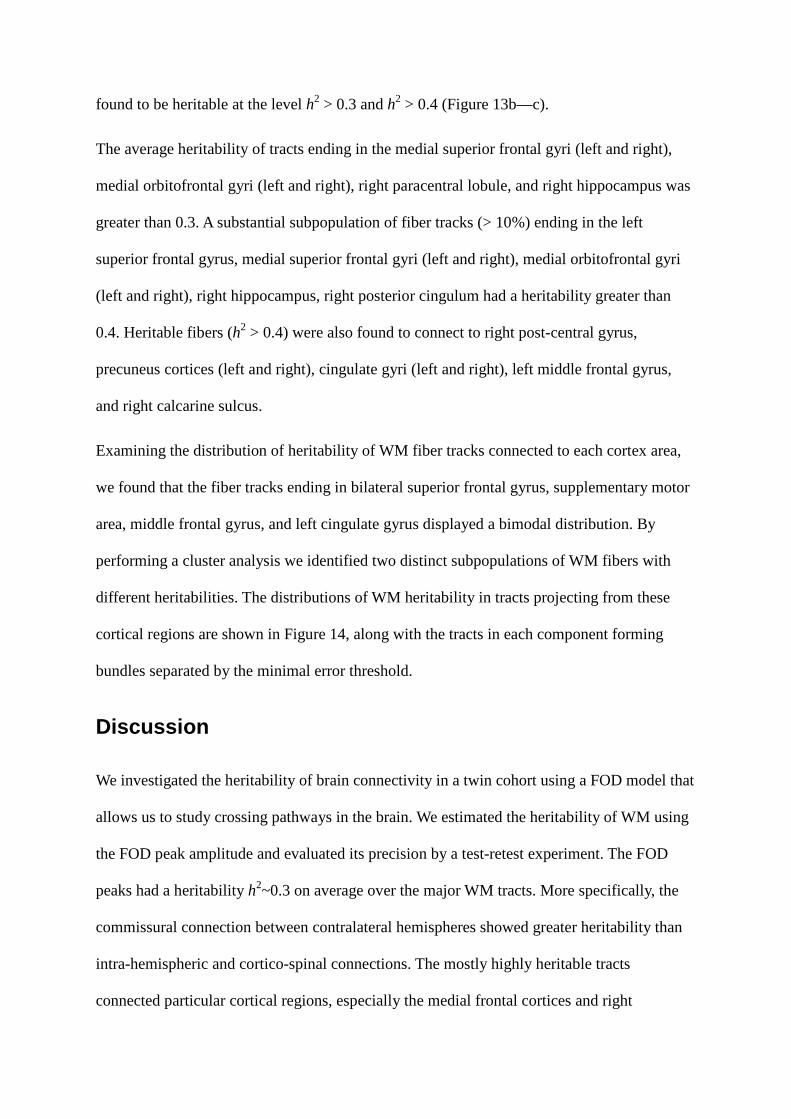
found to be heritable at the level h2 > 0.3 and h2 > 0.4 (Figure 13b—c).
The average heritability of tracts ending in the medial superior frontal gyri (left and right),
medial orbitofrontal gyri (left and right), right paracentral lobule, and right hippocampus was
greater than 0.3. A substantial subpopulation of fiber tracks (> 10%) ending in the left
superior frontal gyrus, medial superior frontal gyri (left and right), medial orbitofrontal gyri
(left and right), right hippocampus, right posterior cingulum had a heritability greater than
0.4. Heritable fibers (h2 > 0.4) were also found to connect to right post-central gyrus,
precuneus cortices (left and right), cingulate gyri (left and right), left middle frontal gyrus,
and right calcarine sulcus.
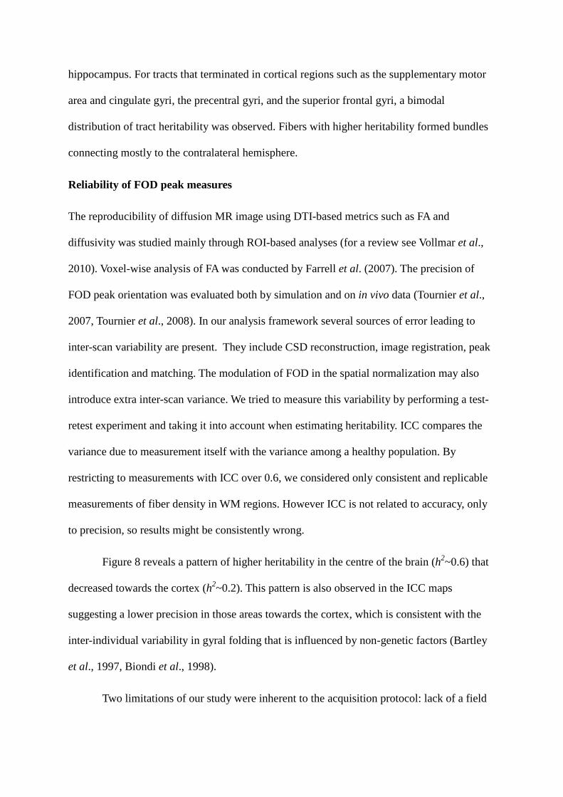
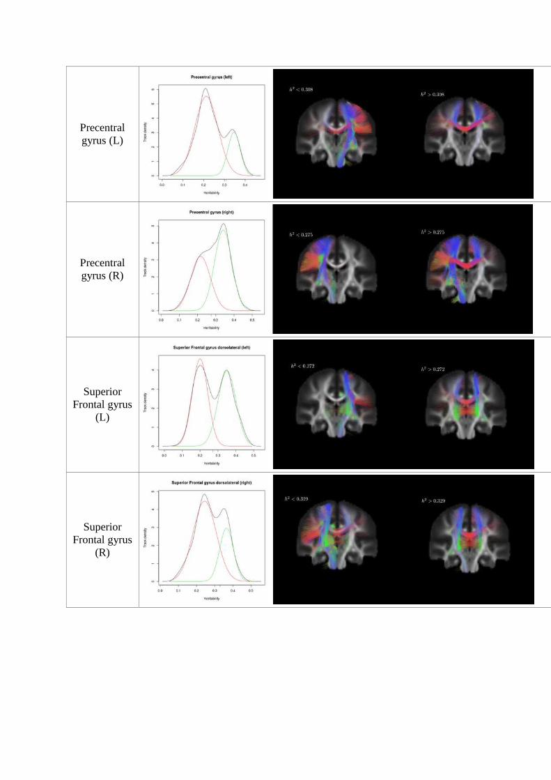
Examining the distribution of heritability of WM fiber tracks connected to each cortex area,
we found that the fiber tracks ending in bilateral superior frontal gyrus, supplementary motor
area, middle frontal gyrus, and left cingulate gyrus displayed a bimodal distribution. By
performing a cluster analysis we identified two distinct subpopulations of WM fibers with
different heritabilities. The distributions of WM heritability in tracts projecting from these
cortical regions are shown in Figure 14, along with the tracts in each component forming
bundles separated by the minimal error threshold.
Discussion
We investigated the heritability of brain connectivity in a twin cohort using a FOD model that
allows us to study crossing pathways in the brain. We estimated the heritability of WM using
the FOD peak amplitude and evaluated its precision by a test-retest experiment. The FOD
peaks had a heritability h2~0.3 on average over the major WM tracts. More specifically, the
commissural connection between contralateral hemispheres showed greater heritability than
intra-hemispheric and cortico-spinal connections. The mostly highly heritable tracts
connected particular cortical regions, especially the medial frontal cortices and right
hippocampus. For tracts that terminated in cortical regions such as the supplementary motor
area and cingulate gyri, the precentral gyri, and the superior frontal gyri, a bimodal
distribution of tract heritability was observed. Fibers with higher heritability formed bundles
connecting mostly to the contralateral hemisphere.
Reliability of FOD peak measures
The reproducibility of diffusion MR image using DTI-based metrics such as FA and
diffusivity was studied mainly through ROI-based analyses (for a review see Vollmar et al.,
2010). Voxel-wise analysis of FA was conducted by Farrell et al. (2007). The precision of
FOD peak orientation was evaluated both by simulation and on in vivo data (Tournier et al.,
2007, Tournier et al., 2008). In our analysis framework several sources of error leading to
inter-scan variability are present. They include CSD reconstruction, image registration, peak
identification and matching. The modulation of FOD in the spatial normalization may also
introduce extra inter-scan variance. We tried to measure this variability by performing a test-
retest experiment and taking it into account when estimating heritability. ICC compares the
variance due to measurement itself with the variance among a healthy population. By
restricting to measurements with ICC over 0.6, we considered only consistent and replicable
measurements of fiber density in WM regions. However ICC is not related to accuracy, only
to precision, so results might be consistently wrong.
Figure 8 reveals a pattern of higher heritability in the centre of the brain (h2~0.6) that
decreased towards the cortex (h2~0.2). This pattern is also observed in the ICC maps
suggesting a lower precision in those areas towards the cortex, which is consistent with the
inter-individual variability in gyral folding that is influenced by non-genetic factors (Bartley
et al., 1997, Biondi et al., 1998).
Two limitations of our study were inherent to the acquisition protocol: lack of a field
map to fully correct for susceptibility-related distortion and a DWI parameter of
b=1159mm/s2 which is not the optimal value for computing the FOD model. Despite those,
we believe that our findings still provide interesting insights into the heritability of brain
connectivity.
WM tract heritability
FOD peak amplitude displays high heritability across major WM tracts. In particular, for the
corpus callosum, cortico-spinal tracts, the inferior fronto-occipital fasciculus, and the superior
longitudinal fasciculus, it shows how FOD-based measures can capture the genetic effects on
WM structures. The detected heritability was rather conservative since the heritability of
FOD peaks with low reliability was not considered. We might have underestimated
heritability in fibers passing through these voxels. The results with lower heritability should
be interpreted cautiously: a low heritability estimate may not necessarily suggest strong
environmental influences, but low precision estimated from the test-retest. However, when all
WM voxels were included (including those with low precision), similar qualitative results
were observed (not shown).
The corpus callosum communicates between both sides of the brain. The high
heritability of WM measures along the corpus callosum suggests a strong influence of genes
upon the inter-hemisphere connection. Corpus callosum growth continues into adulthood
(Pujol et al., 1993, Luders et al., 2010, Paul, 2011), which is guided and regulated by a wide
range of genes and environmental factors (Paul et al., 2007). Our finding of highly heritable
corpus callosum is consistent with DTI-based findings suggesting that WM integrity is under
heavy genetic influence in the corpus callosum (Pfefferbaum et al., 2001, Chiang et al., 2009,
Brouwer et al., 2010). Voxel-based morphometry found strong genetic contributions to WM
density in corpus callosum (Hulshoff Pol et al., 2006), results that we confirmed, observing
the highest heritability in the genu of corpus callosum. This area showed high heritability
regardless of imaging protocol (Jahanshad et al., 2012). In the posterior parts of the corpus
callosum, a pattern of more rapid growth during the development has been reported
(Rajapakse et al., 1996, Giedd et al., 1999, Thompson et al., 2000, Chung et al., 2001); these
regions displayed lower heritability using our method, compared to the anterior parts.
In the superior frontal gyrus, middle frontal gyrus, the middle part of the cingulate
gyrus and supplementary motor area, the fiber cluster with higher average heritability
corresponded to bundles connecting these cortices to the contralateral hemisphere. For
instance, in the left supplementary motor area, the heritability of tracts connected to this part
of the cortex distinctly formed a bimodal distribution. A majority of tracts along the
ipsilateral projection or association was less heritable, but the commissural connection had a
higher heritability consistent with the high heritability in the corpus callosum.
The heritabilities for the inferior fronto-occipital fasciculus and the superior
longitudinal fasciculus were relatively high, whereas the inferior longitudinal fasciculus was
lower. These former structures are significantly influenced by genetic effects in DTI studies
(Chiang et al., 2009, Kochunov et al., 2010, Brouwer et al., 2010). High heritability has been
reported in the right superior longitudinal fasciculus (Brouwer et al., 2010), consistent with
our findings. The genetic influences on the superior longitudinal fasciculus are shared with
performance measures of a spatial delayed response task (Karlsgodt et al., 2010).
Among the tracts ending in the orbitofrontal cortex, a bimodal distribution in
heritability was observed. The cluster with higher heritability (usually h2>0.3) included the
inter-hemisphere connections in the forceps minor and the inferior fronto-occipital fasciculi
connecting the orbital surface.
Genetic specifications are critical to the early development of the cortico-spinal
system, which plays an important role in the control of skilled limb movements. Its
maturation and the long-term function of the motor system also depend on activity and
experience (Martin et al., 2007). Cortico-spinal tracts descending from motor cortex through
the posterior limb of internal capsule to the spinal cord were heritable (h2>0.3), as in previous
studies (Hulshoff Pol et al., 2006). In the anterior limb of internal capsule, WM integrity
measured by FA was under strong genetic control bilaterally (Chiang et al., 2009). In our
results, high heritability was found in the right anterior limb of the internal capsule, whereas
in the left anterior limb the heritability was limited by voxels with lower test-retest reliability
(<0.6).
Cortices linked to heritable WM tracts
A variety of cortical areas have been found to be heritable in several morphological studies
(Thompson et al. 2001, Hulshoff Pol et al., 2006), with more specific genetic influences
found for cortical surface area (Panizzon et al., 2009, Eyler et al., 2011), and cortical
thickness (Lenroot et al., 2009, Winkler et al., 2010, Joshi et al., 2011). An inverse
relationship was found between cortical thickness and WM growth (Sowell et al., 2001;
Gogtay et al., 2004, Giorgio et al., 2009). Schmitt et al. (2008) related the network
organization to the cortical parcellation using principal component analysis, where the first
principal component accounted for over 60% of the total covariance in genetic influence
among all cortical regions. In this paper, we mapped the heritability in WM connections to
the cortex. By examining which cortical regions were connected by heritable tracts, the
function associated with cortices may shed light on the significance of genetic effects for
particular WM structures.
In our results, the heritability for fibers projecting to the superior and middle frontal
gyrus, and the middle temporal gyrus is consistent with previous findings reporting GM
volume or thickness heritability in those areas (Thompson et al., 2001, Hulshoff Pol et al.,
2006, Elyer et al., 2011). The primary somatosensory cortex in the postcentral gyrus and the

paracentral lobule adjacent to it were connected to heritable WM. The development of this
cortical region was phylogenetically and ontologically early, and genetically determined
(Lenroot et al., 2009). The medial prefrontal cortex in the contralateral hemisphere is
connected via the genu of corpus callosum. Given the high heritability in the genu, fibers
projecting to medial prefrontal cortex showed higher heritability. A heritability h2=0.83 was
found for GM density in the medial frontal cortex (Hulshoff Pol et al., 2006), which agrees
with previous findings (Thompson et al., 2001). Commissural connections projecting to the
cingulate gyrus, especially its posterior part, via the corpus callosum also contributed to a
substantial subpopulation of heritable tracts. Cortical GM areas corresponding to the
hippocampus and caudate were also connected by heritable fibers (h2>0.4, Figure 13c).
In conclusion, we report a new method to investigate the heritability of brain connections
using DWI data with an FOD model. Our findings are consistent with prior studies and offer
a novel way to investigate complex brain networks involving crossing fibers. They suggest
that inter-hemisphere connections are more heritable than association and cortico-spinal
connections.
Acknowledgement
This research is supported by the Science and Industry Endowment Fund. KS is the recipient
of a John Stocker Postdoctoral Fellowship from the Science and Industry Endowment Fund.
References
Aganj I, Lenglet C, Sapiro G, Yacoub E, Ugurbil K, Harel N (2010) Reconstruction of the
orientation distribution function in single- and multiple-shell q-ball imaging within constant
solid angle. Magnetic Resonance in Medicine 64: 554–566.
Bartko JJ (1976) On various intraclass correlation reliability coefficients. Psychological

Bulletin 83: 762–765.
Bartley AJ, Jones DW, Weinberger DR (1997) Genetic variability of human brain size and
cortical gyral patterns. Brain 120: 257–269.
Besseling RMH, Jansen JFA, Overvliet GM, Vaessen MJ, Braakman HMH, Hofman PAM,
Aldenkamp AP, Backes WH (2012) Tract Specific Reproducibility of Tractography Based
Morphology and Diffusion Metrics PLoS ONE 7: e34125.
Biondi A, Nogueira H, Dormont D, Duyme M, Hasboun D, Zouaoui A, Chantôme M,
Marsault C (1998) Are the brains of monozygotic twins similar? A three-dimensional MR
study. Am J Neuroradiol 19: 1361–1367.
Bisdas S, Bohning De, Bešenski N, Nicholas, JS, Rumboldt Z (2008) Reproducibility,
Interrater Agreement, and Age-Related Changes of Fractional Anisotropy Measures at 3T in
Healthy Subjects: Effect of the Applied b-Value. Am J Neuroradiol 29: 1128–1133.
Bonekamp D, Nagae LM, Degaonkar M, Matson M, Abdalla WMA, Barker PB, Mori S,
Horská A (2007) Diffusion tensor imaging in children and adolescents: Reproducibility,
hemispheric, and age-related differences. NeuroImage 34: 733–742.
Brouwer RM, Mandl RCW, Peper JS, van Baal GCM, Kahn RS, Boomsma DI, Hulshoff Pol
HE (2010) Heritability of DTI and MTR in nine-year-old children. NeuroImage 53: 1085–
1092.
Brown TT, Jernigan TL (2012) Brain Development During the Preschool Years.
Neuropsychol Rev 22: 313–333.
Canales-Rodríguez EJ, Melie-García L, Iturria-Medina Y (2009) Mathematical description of
q-space in spherical coordinates: Exact q-ball imaging. Magnetic Resonance in Medicine 61:
1350–1367.
Cascio CJ, Gerig G, Piven J (2007) Diffusion Tensor Imaging: Application to the Study of the
Developing Brain. Journal of the American Academy of Child & Adolescent Psychiatry 46:
213–223.
Chiang M-C, Barysheva M, Shattuck DW, Lee AD, Madsen SK, Avedissian C, Klunder AD,
Toga AW, McMahon KL, de Zubicaray GI, Wright MJ, Srivastava A, Balov N, Thompson
PM (2009) Genetics of Brain Fiber Architecture and Intellectual Performance. J Neurosci 29:
2212–2224.
Chung MK, Worsley KJ, Paus T, Cherif C, Collins DL, Giedd JN, Rapoport JL, Evans AC
(2001) A Unified Statistical Approach to Deformation-Based Morphometry. NeuroImage 14:
595–606.
Chen X, Blokland G, Strike L (2013) Voxel-wise and cluster-based heritability inferences of
fMRI data. In: Organization of Human Brain Mapping, Seattle, WA.
Ciccarelli O, Parker GJ, Toosy AT, Wheeler-Kingshott CA, Barker GJ, Boulby PA, Miller
DH, Thompson AJ (2003) From diffusion tractography to quantitative white matter tract
measures: a reproducibility study. NeuroImage 18: 348–359.
Descoteaux M, Angelino E, Fitzgibbons S, Deriche R (2007) Regularized, fast, and robust
analytical Q-ball imaging. Magnetic Resonance in Medicine 58: 497–510.
Eyler LT, Prom-Wormley E, Panizzon MS, Kaup AR, Fennema-Notestine C, Neale MC,
Jernigan TL, Fischl B, Franz CE, Lyons MJ, Grant M, Stevens A, Pacheco J, Perry ME,
Schmit JEt, Seidman LJ, Thermenos HW, Tsuang MT, Chen C-H, Thompson WK, Jak A,
Dale AM, Kremen WS (2011) Genetic and Environmental Contributions to Regional Cortical
Surface Area in Humans: A Magnetic Resonance Imaging Twin Study. Cereb Cortex 21:
2313–2321.
Farrell JAD, Landman BA, Jones CK, Smith SA, Prince JL, van Zijl PCM, Mori S (2007)

Effects of signal-to-noise ratio on the accuracy and reproducibility of diffusion tensor
imaging–derived fractional anisotropy, mean diffusivity, and principal eigenvector
measurements at 1.5T. Journal of Magnetic Resonance Imaging 26: 756–767.
Fields RD (2008) White matter in learning, cognition and psychiatric disorders. Trends in
Neurosciences 31: 361–370.
Fonov V, Evans AC, Botteron K, Almli CR, McKinstry RC, Collins DL (2011) Unbiased
average age-appropriate atlases for pediatric studies. NeuroImage (2011): 313–327.
Giannelli M, Cosottini M, Michelassi MC, Lazzarotti G, Belmonte G, Bartolozzi C, Lazzeri
M (2010) Dependence of brain DTI maps of Fractional Anisotropy and Mean Diffusivity on
the number of diffusion weighting directions. Journal of Applied Clinical Medical Physics 11:
176–190.
Giedd JN, Blumenthal J, Jeffries NO, Rajapakse JC, Vaituzis AC, Liu H, Berry YC, Tobin M,
Nelson J, Castellanos FX (1999) Development of the human corpus callosum during
childhood and adolescence: A longitudinal MRI study. Progress in Neuro-
Psychopharmacology and Biological Psychiatry 23: 571–588.
Geng X, Prom-Wormley EC, Perez J, Kubarych T, Styner M, Lin W, Neale MC, Gilmore JH
(2012) White Matter Heritability Using Diffusion Tensor Imaging in Neonatal Brains. Twin
Research and Human Genetics 15: 336–350.
Giorgio A, Watkins KE, Chadwick M, James S, Winmill L, Douaud G, De Stefano N,
Matthews PM, Smith SM, Johansen-Berg H, James AC (2010) Longitudinal changes in grey
and white matter during adolescence. NeuroImage 49: 94–103.
Gogtay N, Giedd JN, Lusk L, Hayashi KM, Greenstein D, Vaituzis AC, Nugent Tf, Herman
DH, Clasen LS, Toga AW, Rapoport JL, Thompson PM (2004) Dynamic mapping of human
cortical development during childhood through early adulthood. Proc Natl Acad Sci 101:

8174–8179.
Hakulinen U, Brander A, Ryymin P, Öhman J, Soimakallio S, Helminen M, Dastidar P,
Eskola H (2012) Repeatability and variation of region-of-interest methods using quantitative
diffusion tensor MR imaging of the brain. BMC Med Imaging 12: 30.
Heiervang E, Behrens TEJ, Mackay CE, Robson MD, Johansen-Berg H (2006) Between
session reproducibility and between subject variability of diffusion MR and tractography
measures. NeuroImage 33: 867–877.
Hua K, Zhang J, Wakana S, Jiang H, Li X, Reich DS, Calabresi PA, Pekar JJ, van Zijl PCM,
Mori S (2008) Tract probability maps in stereotaxic spaces: Analyses of white matter
anatomy and tract-specific quantification. NeuroImage 39: 336–347.
Huang L, Wang X, Baliki MN, Wang L, Apkarian AV, Parrish TB (2012) Reproducibility of
Structural, Resting-State BOLD and DTI Data between Identical Scanners. PLoS ONE 7:
e47684.
Hulshoff Pol HE, Schnack HG, Posthuma D, Mandl RCW, Baaré WF, van Oel C, van Haren
NE, Collins DL, Evans AC, Amunts K, Bürgel U, Zilles K, de Geus E, Boomsma DI, Kahn
RS (2006) Genetic Contributions to Human Brain Morphology and Intelligence. J Neurosci
26: 10235–10242.
Jahanshad N, Lee AD, Barysheva M, McMahon KL, de Zubicaray GI, Martin NG, Wright
MJ, Toga AW, Thompson PM Genetic influences on brain asymmetry: A DTI study of 374
twins and siblings. NeuroImage 52: 455–469.
Jahanshad N, Kohannim O, Toga AW, McMahon KL, de Zubicaray GI, Hansell NK,
Montgomery GW, Martin NG, Wright MJ, Thompson PM (2012) Diffusion Imaging Protocol
Effects on Genetic Associations. In: Proceedings / IEEE International Symposium on
Biomedical Imaging: from nano to macro. IEEE International Symposium on Biomedical
Imaging, pp. 944–947.
Jahanshad N, Kochunov PV, Sprooten E, Mandl RC, Nichols TE, Almasy L, Blangero J,
Brouwer RM, Curran JE, de Zubicaray GI, Duggirala R, Fox PT, Hong LE, Landman BA,
Martin NG, McMahon KL, Medland SE, Mitchell MD, Olvera RL, Peterson CP, Starr JM,
Sussmann JE, Toga AW, Wardlaw JM, Wright MJ, Hulshoff Pol HE, Bastin ME, McIntosh
AM, Deary IJ, Thompson PM, Glahn DC (2013) Multi-site genetic analysis of diffusion
images and voxelwise heritability analysis: A pilot project of the ENIGMA–DTI working
group. NeuroImage 81: 455–469.
Jansen JFA, Kooi ME, Kessels AGH, Nicolay K, Backes WH (2007) Reproducibility of
Quantitative Cerebral T2 Relaxometry, Diffusion Tensor Imaging, and 1H Magnetic
Resonance Spectroscopy at 3.0 Tesla. Investigative Radiology 42: 327–337.
Jeurissen, B, Leemans, A, Tournier, J-D, Jones, DK, Sijbers, J (2013) Investigating the
prevalence of complex fiber configurations in white matter tissue with diffusion magnetic
resonance imaging. Human Brain Mapping 34:2747–2766.
Jones DK (2004) The effect of gradient sampling schemes on measures derived from
diffusion tensor MRI: a Monte Carlo study. Magn Reson Med 51: 807–815.
Joshi AA, Leporé N, Joshi SH, Lee AD, Barysheva M, Stein JL, McMahon KL, Johnson K,
de Zubicaray GI, Martin NG, Wright MJ, Toga AW, Thompson PM (2011) The contribution
of genes to cortical thickness and volume. NeuroReport 22: 101–105.
Karlsgodt KH, Kochunov P, Winkler AM, Laird AR, Almasy L, Duggirala R, Olvera RL, Fox
PT, Blangero J, Glahn DC (2010) A Multimodal Assessment of the Genetic Control over
Working Memory. J Neurosci 30: 8197–8202.
Kanchibhotla SC, Mather KA, Wen W, Schofield PR, Kwok JBJ, Sachdev PS (2013)
Genetics of ageing-related changes in brain white matter integrity – A review. Ageing
Research Reviews 12: 391–401.
Kittler J, Illingworth J (1986) Minimum error thresholding. Pattern Recognition 19: 41–47.
Kochunov O, Glahn DC, Lancaster JL, Winkler AM, Smith S, Thompson PM, Almasy L,
Duggirala R, Fox PT, Blangero J (2010) Genetics of microstructure of cerebral white matter
using diffusion tensor imaging. NeuroImage 53: 1109–1116.
Landman BA, Farrell JAD, Jones CK, Smith SA, Prince JL, Mori S (2007) Effects of
diffusion weighting schemes on the reproducibility of DTI-derived fractional anisotropy,
mean diffusivity, and principal eigenvector measurements at 1.5T. NeuroImage 36: 1123–
1138.
Lawson CL, Hanson RJ (1995) Solving least squares problems. Philadelphia: SIAM.
Lenroot RK, Schmitt JE, Ordaz SJ, Wallace GL, Neale MC, Lerch JP, Kendler KS, Evans
AC, Giedd JN (2009) Differences in genetic and environmental influences on the human
cerebral cortex associated with development during childhood and adolescence. Human Brain
Mapping 30: 163–174.
Leow AD, Zhu S, Zhan L, McMahon K, de Zubicaray GI, Meredith M, Wright MJ, Toga AW,
Thompson PM (2009) The tensor distribution function. Magnetic Resonance in Medicine 61:
205–214.
Lin C-P, Wedeen VJ, Chen J-H, Yao C, Tseng W-YI (2003) Validation of diffusion spectrum
magnetic resonance imaging with manganese-enhanced rat optic tracts and ex vivo phantoms.
NeuroImage 19: 482–495.
Luders E, Thompson PM, Toga AW (2010) The Development of the Corpus Callosum in the
Healthy Human Brain. J Neurosci 30: 10985–10990.
Makris N, Goldstein JM, Kennedy D, Hodge SM, Caviness VS, Faraone SV, Tsuang MT,
Seidman LJ (2006) Decreased volume of left and total anterior insular lobule in
schizophrenia. Schizophrenia Research 83: 155–171.
Martin JH, Friel KM, Salimi I, Chakrabarty S (2007) Activity- and use-dependent plasticity
of the developing corticospinal system. Neuroscience & Biobehavioral Reviews 31: 1125–
1135.
McLachlan GJ, Peel D (2000) Finite mixture models. New York: Wiley.
Muetzel RL, Collins PF, Mueller BA, Schissel AM, Lim KO, Luciana M (2008) The
development of corpus callosum microstructure and associations with bimanual task
performance in healthy adolescents. NeuroImage 39: 1918–1925.
Panizzon MS, Fennema-Notestine C, Eyler LT, Jernigan TL, Prom-Wormley E, Neale M,
Jacobson K, Lyons MJ, Grant MD, Franz CE, Xian H, Tsuang M, Fischl B, Seidman L, Dale
A, Kremen WS (2009) Distinct Genetic Influences on Cortical Surface Area and Cortical
Thickness. Cereb Cortex 19: 2728–2735.
Panizzon MS, Fennema-Notestine C, Kubarych TS, Chen C-H, Eyler LT, Fischl B, Franz
CE, Grant MD, Hamza S, Jak A, Jernigan TL, Lyons MJ, Neale MC, Prom-Wormley EC,
Seidman L, Tsuang MT, Wu H, Xian H, Dale AM, Kremen WS (2012) Genetic and
environmental influences of white and gray matter signal contrast: A new phenotype for
imaging genetics? NeuroImage 60: 1686–1695.
Paul LK, Brown WS, Adolphs R, Tyszka JM, Richards LJ, Mukherjee P, Sherr EH (2007)
Agenesis of the corpus callosum: genetic, developmental and functional aspects of
connectivity. Nat Rev Neurosci 8:287–299.
Paul LK (2011) Developmental malformation of the corpus callosum: a review of typical
callosal development and examples of developmental disorders with callosal involvement. J
Neurodev Disord 3:3–27.

Pfefferbaum A, Sullivan EV, Carmelli D (2001) Genetic regulation of regional microstructure
of the corpus callosum in late life. Neuroreport 12:1677–1681.
Pfefferbaum A, Adalsteinsson E, Sullivan EV (2003) Replicability of diffusion tensor
imaging measurements of fractional anisotropy and trace in brain. J Magn Reson Imaging
18:427–433.
Pujol J, Vendrell P, Junqué C, Martí-Vilalta JL, Capdevila A (1993) When does human brain
development end? Evidence of corpus callosum growth up to adulthood. Annals of
Neurology 34:71–75.
Raffelt D, Tournier J-D, Fripp J, Crozier S, Connelly A, Salvado O (2011) Symmetric
diffeomorphic registration of fibre orientation distributions. NeuroImage 56:1171–1180.
Raffelt D, Tournier J-D, Rose S, Ridgway GR, Henderson R, Crozier S, Salvado O, Connelly
A (2012) Apparent Fibre Density: A novel measure for the analysis of diffusion-weighted
magnetic resonance images. NeuroImage 59:3976–3994.
Rajapakse JC, Giedd JN, Rumsey JM, Vaituzis AC, Hamburger SD, Rapoport JL (1996)
Regional MRI measurements of the corpus callosum: a methodological and developmental
study. Brain and Development 18:379–388.
Rohlfing T, Rademacher MH, Pfefferbaum A (2008) Volume Reconstruction by Inverse
Interpolation: Application to Interleaved MR Motion Correction. In: Medical Image
Computing and Computer-Assisted Intervention – MICCAI 2008, pp. 798–806.
Rueckert D, Sonoda LI, Hayes C, Hill DLG, Leach MO, Hawkes DJ (1999) Nonrigid
Registration Using Free-Form Deformations: Application to Breast MR Images. IEEE Trans
Med Imag 18:712–721.
Schmitt JE, Lenroot RK, Wallace GL, Ordaz S, Taylor KN, Kabani N, Greenstein D, Lerch
JP, Kendler KS, Neale MC, Giedd JN (2008) Identification of Genetically Mediated Cortical

Networks: A Multivariate Study of Pediatric Twins and Siblings. Cereb Cortex 18:1737–
1747.
Shrout PE, Fleiss JL (1979) Intraclass correlations: Uses in assessing rater reliability.
Psychological Bulletin 86:420–428.
Sowell ER, Thompson PM, Tessner KD, Toga AW (2001) Mapping Continued Brain Growth
and Gray Matter Density Reduction in Dorsal Frontal Cortex: Inverse Relationships during
Postadolescent Brain Maturation. J Neurosci vol 21:8819–8829.
Sullivan EV, Pfefferbaum A (2006) Diffusion tensor imaging and aging. Neuroscience &
Biobehavioral Reviews 30:749–761.
Thompson PM, Giedd JN, Woods RP, MacDonald D, Evans AC, Toga AW (2000) Growth
patterns in the developing brain detected by using continuum mechanical tensor maps. Nature
404:190–193.
Thompson PM, Cannon TD, Narr KL, van Erp T, Poutanen V-P, Huttunen M, Lönnqvist J,
Standertskjöld-Nordenstam C-G, Kaprio J, Khaledy M, Dail R, Zoumalan CI, Toga AW
(2001) Genetic influences on brain structure. Nat Neurosci 4:1253–1258.
Tournier J-D, Calamante F, Gadian D-G, Connelly A (2004) Direct estimation of the fiber
orientation density function from diffusion-weighted MRI data using spherical
deconvolution. NeuroImage 23:1176–1185.
Tournier J-D, Calamante F, Connelly A (2007) Robust determination of the fibre orientation
distribution in diffusion MRI: Non-negativity constrained super-resolved spherical
deconvolution. NeuroImage 35:1459–1472.
Tournier J-D, Yeh C-H, Calamante F, Cho K-H, Connelly A, Lin C-P (2008) Resolving
crossing fibres using constrained spherical deconvolution: Validation using diffusion-
weighted imaging phantom data. NeuroImage 42:617–625.

Tournier J-D, Calamante F, Connelly A (2012) MRtrix: Diffusion tractography in crossing
fiber regions. International Journal of Imaging Systems and Technology 22:53–66.
Tristán-Vega A, Westin C-F, Aja-Fernández S (2009) Estimation of fiber Orientation
Probability Density Functions in High Angular Resolution Diffusion Imaging. NeuroImage
47:638–650.
Tristán-Vega A, Westin C-F, Aja-Fernández S (2010) A new methodology for the estimation
of fiber populations in the white matter of the brain with the Funk–Radon transform.
NeuroImage 49:1301–1315.
Tuch DS, Reese TG, Wiegell MR, Makris N, Belliveau JW, Wedeen VJ (2002) High angular
resolution diffusion imaging reveals intravoxel white matter fiber heterogeneity. Magnetic
Resonance in Medicine 48:577–582.
Tuch DS (2004) Q-ball imaging. Magnetic Resonance in Medicine 52:1358–1372.
Tustison NJ, Avants BB, Cook PA, Zheng Y, Egan A, Yushkevich PA, Gee JC (2010) N4ITK:
Improved N3 Bias Correction. IEEE Transactions on Medical Imaging 29:1310–1320.
Tzourio-Mazoyer N, Landeau B, Papathanassiou D, Crivello F, EtardO, Delcroix N, Mazoyer
B, Joliot M (2002) Automated Anatomical Labeling of Activations in SPM Using a
Macroscopic Anatomical Parcellation of the MNI MRI Single-Subject Brain. NeuroImage
15:273–289.
Vollmar C, O’Muircheartaigh J, Barker GJ, Symms MR, Thompson P, Kumari V, Duncan JS,
Richardson MP, Koepp MJ (2010) Identical, but not the same: Intra-site and inter-site
reproducibility of fractional anisotropy measures on two 3.0T scanners. NeuroImage
51:1384–1394.
Wakana S, Caprihan A, Panzenboeck MM, Fallon JH, Perry M, Gollub RL, Hua K, Zhang J,
Jiang H, Dubey P, Blitz A, van Zijl P, Mori S (2007) Reproducibility of quantitative

tractography methods applied to cerebral white matter. NeuroImage 36:630–644.
Wedeen VJ, Hagmann P, Tseng W-YI, Reese TG, Weisskoff RM (2005) Mapping complex
tissue architecture with diffusion spectrum magnetic resonance imaging. Magnetic Resonance
in Medicine 54:1377–1386.
Winkler AM, Kochunov P, Blangero J, Almasy L, Zilles K, Fox PT, Duggirala R, Glahn DC
(2010) Cortical thickness or grey matter volume? The importance of selecting the phenotype
for imaging genetics studies. NeuroImage 53:1135–1146.
Wozniak JR, Lim KO (2006) Advances in white matter imaging: A review of in vivo
magnetic resonance methodologies and their applicability to the study of development and
aging. Neuroscience & Biobehavioral Reviews 30:762–774.
Zaitsev M, Hennig J, Speck O (2004) Point spread function mapping with parallel imaging
techniques and high acceleration factors: Fast, robust, and flexible method for echo-planar
imaging distortion correction. Magnetic Resonance in Medicine 52:1156–1166.
Zhan L, Leow AD, Aganj I, Lenglet C, Sapiro G, Yacoub E, Harel N, Toga AW, Thompson
PM (2011) Differential information content in staggered multiple shell HARDI measured by
the tensor distribution function. In: 2011 IEEE International Symposium on Biomedical
Imaging: From Nano to Macro, pp. 305–309.

Diffusion Weighted Images
(a) Preprocessing and spherical deconvolution Fibre Orientation
Distribution (FOD)
Registered FOD images in the common space
FOD peak amplitude of average FOD template
Group average FOD in the common space
FOD peak amplitude of FOD images
(c) Find peaks
(d)Find peaks on FOD
images corresponding to the peaks of the
group average FOD
(b) Iterateive
Groupwiseregistration
(e)Inter-subject
statistical analysis
Test-retestreliability of FOD peak
Heritability of FOD peak measure
Figure 1. The preprocessing steps for the Diffusion Weighted Images (DWIs) include spatial
normalization, FOD peak measurement, and inter-subject statistical analysis: (a) In preprocessing,
the DWIs were corrected for bias field effects, motion and eddy current artifacts, and then used to
reconstruct the Fibre Orientation Distribution (FOD) image by spherical deconvolution (Tourier et al.,
2008). (b) FOD images were then spatially normalized into a common atlas space by iterative group-
wise registration (Raffelt et al., 2012), which produced a group average FOD image template, and
the FOD of all the subjects aligned to it. To measure the FOD peaks for each subject, we first (c)
identified major FOD peaks on the average template, and then (d) search for FOD peaks on subject
FOD images to match the FOD peaks on the template to ensure the correspondence of peak
measures across the sample. (e) The inter-subject statistical analysis was then carried out given the
FOD peak measurements, thus we were able to evaluate the test-retest reliability of this pipeline by
repeated scanned DWIs, and estimate the heritability of FOD peak measurements.
Group average FOD in the common space
(a)Whole-brain fibre tracking
Whole-brain tractogram
Reliability of FOD peak measures
Heritability of FOD peak measure
(b)Computing track-average
heritability for each fibre track
Heritability estimates on fibre tracks
(c)Computing statistics of
heritable tracks linking to each cortical region
Cortices linked to heritable fibre connections
Spatial labeling of cortical regions by
AAL
Figure 2. Tract-based analysis of heritability and the identification of cortical regions connected by
heritable WM connections. (a) A whole-brain probabilistic fibre-tracking algorithm (Tournier et al.,
2012) was used to produce a whole-brain tractogram, which includes the cortico-cortical, cortico-
subcortical connections, and fibre tracks connecting the cortex and subcortical nuclei to the brain
stem. (b) For individual fibre tracks, the degree of genetic influence at particular locations can be
estimated from the heritability of FOD peaks that the fibre is following. For a whole tract, we can
compute the average heritability of all sampling points along the whole stretch of the tract. (c) By
grouping the tracts by labeling of cortical regions, we identified bundles of fibre tracts connected to
a particular cortical region of interest (ROI). The cortical regions are connected by WM fibres with
various degrees of heritability.
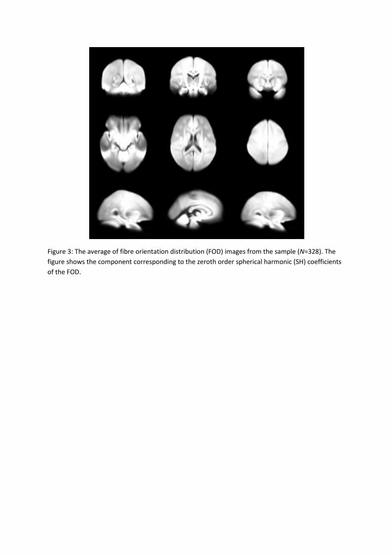
Figure 3: The average of fibre orientation distribution (FOD) images from the sample (N=328). The
figure shows the component corresponding to the zeroth order spherical harmonic (SH) coefficients
of the FOD.
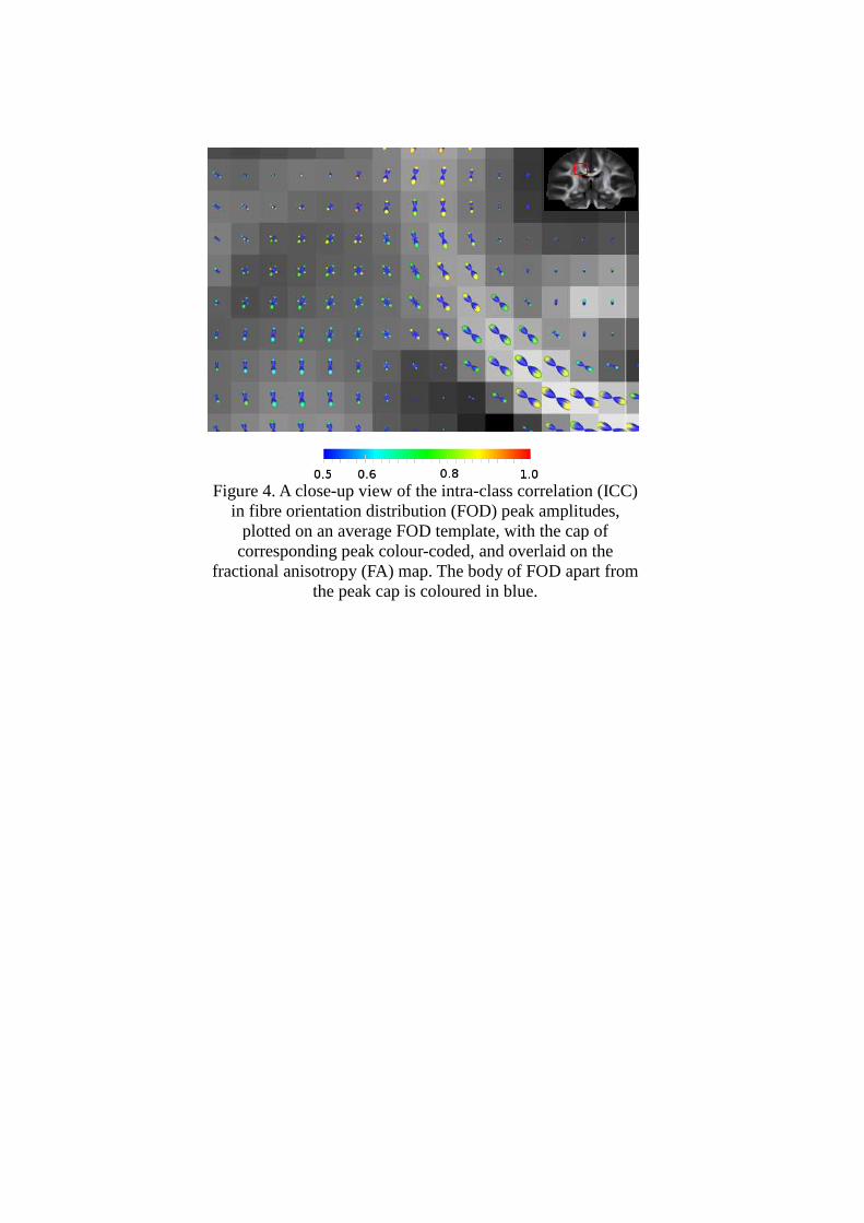
Figure 4. A closein fibre orientation distribution (FOD) peak amplitudes,
plotted on an average FOD template, with the cap of corresponding peak
fractional anisotropy (FA) map. The body of FOD apart from
Figure 4. A close-up view of the intra-class correlation (ICC) in fibre orientation distribution (FOD) peak amplitudes,
plotted on an average FOD template, with the cap of corresponding peak colour-coded, and overlaid on the
fractional anisotropy (FA) map. The body of FOD apart from the peak cap is coloured in blue.
class correlation (ICC)
fractional anisotropy (FA) map. The body of FOD apart from

Figure 5: Intraclass correlation (ICC) of the first peak in fibre orientation distribution (FOD)
measurement of diffusion MR in white matter region overlaid on the average fractional anisotropy
(FA) map.
Figure 6: Intraclass correlation (ICC) of the second peak in fibre orientation distribution (FOD)
measurement of diffusion MR in white matter region overlaid on the average fractional anisotropy
(FA) map. The second peak is identified by threshold with FOD > 0.1.
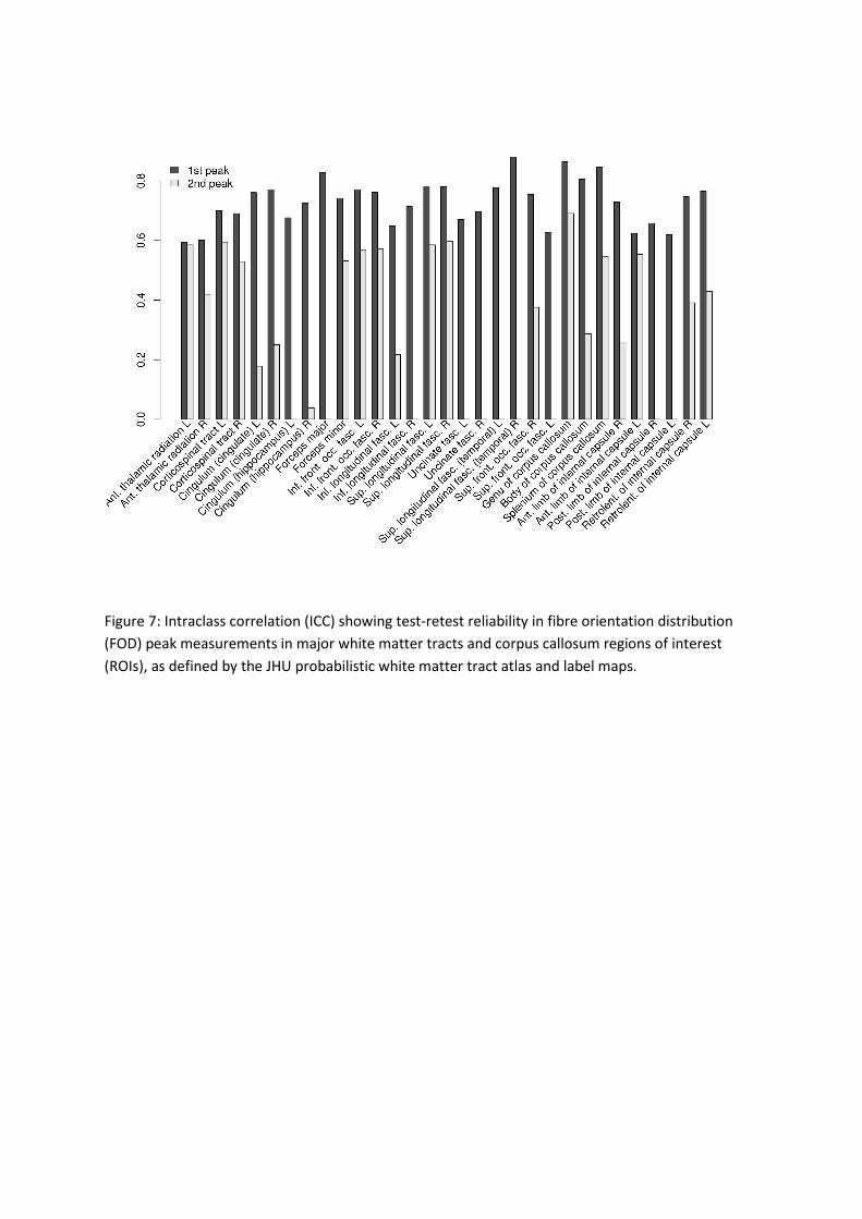
Figure 7: Intraclass correlation (ICC) showing test-retest reliability in fibre orientation distribution
(FOD) peak measurements in major white matter tracts and corpus callosum regions of interest
(ROIs), as defined by the JHU probabilistic white matter tract atlas and label maps.

Figure 8: Heritability of the first peak in fibre orientation distribution (FOD) measurement of
diffusion MR in white matter overlaid on the average fractional anisotropy (FA) map. From top to
bottom: intraclass correlation (ICC) in monozygotic (MZ) twins; ICC in dizygotic (DZ) twins;
heritability index h2; p-value of the additive genetic component by permutation test.
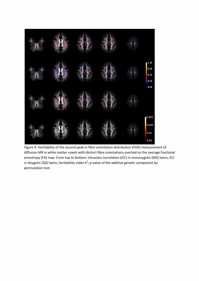
Figure 9: Heritability of the second peak in fibre orientation distribution (FOD) measurement of
diffusion MR in white matter voxels with distinct fibre orientations overlaid on the average fractional
anisotropy (FA) map. From top to bottom: intraclass correlation (ICC) in monozygotic (MZ) twins; ICC
in dizygotic (DZ) twins; heritability index h2; p-value of the additive genetic component by
permutation test.
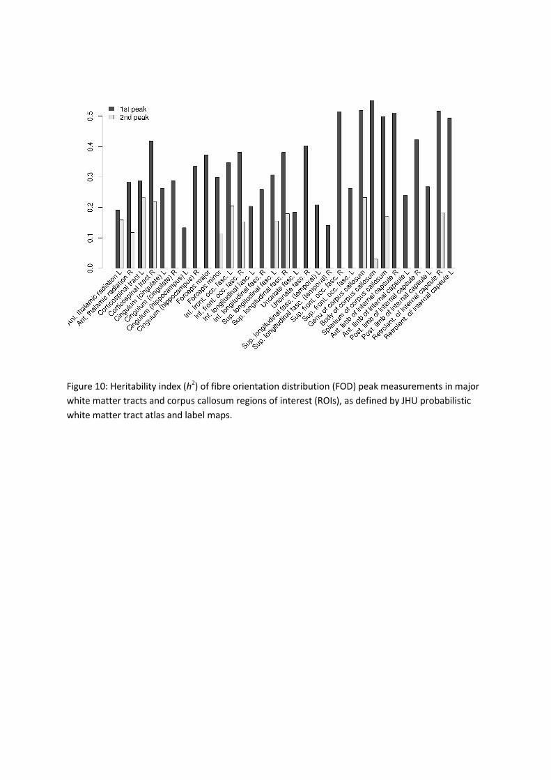
Figure 10: Heritability index (h2) of fibre orientation distribution (FOD) peak measurements in major
white matter tracts and corpus callosum regions of interest (ROIs), as defined by JHU probabilistic
white matter tract atlas and label maps.

Figure 11: Projections of tractograms produced using a probabilistic tractography algorithm on the
average fibre orientation distribution (FOD) map, overlaid on the average FA map. Top: all the tracts
with average intraclass correlation (ICC) of test-retest reliability ICC > 0.6; bottom: among the tracts
with reliability ICC > 0.6, the tracts with average heritability h2 > 0.45.
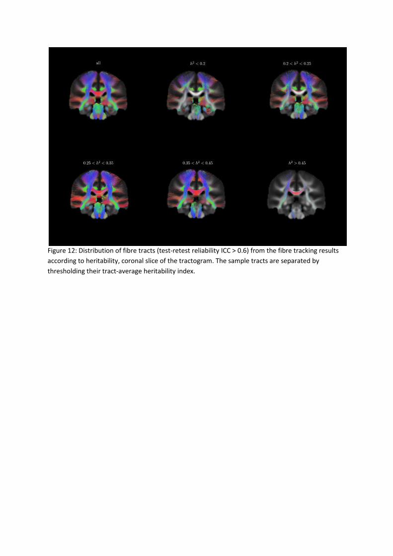
Figure 12: Distribution of fibre tracts (test-retest reliability ICC > 0.6) from the fibre tracking results
according to heritability, coronal slice of the tractogram. The sample tracts are separated by
thresholding their tract-average heritability index.
(a) mean h2 of fibre tracts connected to each cortical region
(b) percentage of fibre tracts with heritability greater than 0.3 among all the tracks connected to the
cortical region
(c) percentage of fibre tracts with heritability greater than 0.4 among all the tracks connected to the
cortical region Figure 13: Cortical regions associated with heritable white matter fibres. From top to bottom: (a)
mean heritability h2 of fibre tracks ending in each cortical regions; (b) percentage of tracks
connected to each cortical region with h2 > 0.3; (c) h
2 > 0.4.
Precentral gyrus (L)
Precentral gyrus (R)
Superior Frontal gyrus
(L)
Superior Frontal gyrus
(R)

Supplementary motor area (L)
Supplementary motor area (R)
Cingulate gyrus (L)
Figure 14: Bimodal distribution of heritability of fibre tracts ending in cortical regions (trimmed
average reliability ICC > 0.6). A minimum error threshold (Kittler and Illingworth, 1986) found by
fitting bimodal normal distribution (McLachlan and Peel, 2000) was used to separate the fibres with
different heritability.