invasive properties of murine squamous carcinoma cells...
TRANSCRIPT
[CANCER RESEARCH 60, 4070–4076, August 1, 2000]
Invasive Properties of Murine Squamous Carcinoma Cells: Secretion of Matrix-degrading Cathepsins Is Attributable to a Deficiency in the Mannose6-Phosphate/Insulin-like Growth Factor II Receptor1
Kim Lorenzo,2 Phuong Ton,2 Jason L. Clark, Sogue Coulibaly,3 and Lukas Mach4
Department of Biochemistry, University of Western Australia, Nedlands WA 6907, Australia [K. L., P. T., J. L. C., L. M.], and Zentrum fur Angewandte Genetik, Universita¨t furBodenkultur Wien, A-1190 Vienna, Austria [S. C., L. M.]
ABSTRACT
Penetration of basement membrane layers is a hallmark feature ofmetastatic tumor cells. The invasive propensity of murine SCC-VII squa-mous carcinoma cells is in part attributable to the extracellular action ofthe lysosomal cysteine proteinase cathepsin B. Although most noncancer-ous cells store this enzyme in the lysosomes, we found that SCC-VII cellsrelease a large fraction (42%) of their newly synthesized procathepsin Binto the culture medium. Procathepsins D and L, the precursors of othermajor lysosomal proteinases, are also secreted in significant amounts (24and 75%, respectively). In contrast, normal murine 3T3-L1 fibroblastsexocytose only minor amounts of their newly synthesized procathepsins B(10%), D (<1%), and L (16%). Western blotting analysis revealed thatSCC-VII cells are deficient in the 300 kDa mannose 6-phosphate/insulin-like growth factor-II receptor (M6P/IGF2R), a tumor suppressor with acentral role in the intracellular transport of lysosomal enzymes. Consist-ent with the absence of M6P/IGF2R, SCC-VII cells lack dense lysosomes,with the bulk of intracellular acid hydrolases residing in late endosomes/prelysosomes. On the other hand, the synthesis of the M6P recognitionmarker on lysosomal enzymes is not impaired in SCC-VII cells, because[33P]Pi is incorporated into the carbohydrate moieties of procathepsins B,D, and L. Furthermore, 69% of the phosphorylatedN-linked oligosaccha-rides synthesized by SCC-VII cells carry phosphomonoester groups andas such constitute high-affinity ligands for M6P receptors. SCC-VII cellsexpress the 46 kDa cation-dependent M6P receptor (MPR46), but intra-cellular retention of procathepsins B, D, and L is not affected by ammo-nium chloride and chloroquine, agents known to perturb the M6P recep-tor system. Our results indicate that failure to express the 300 kDaM6P/IGF2R may enhance the metastatic capacity of tumor cells by in-ducing the secretion of procathepsins B, D, and L.
INTRODUCTION
Malignant cancer cells display a distinct capacity to degrade extra-cellular matrix components, a feature indispensable for penetration ofbasement membranes and subsequent spreading to distant sites. Var-ious proteinases have been implicated in tumor invasion and metas-tasis, including the lysosomal enzymes cathepsins B, D, and L (1–3).In most noncancerous cells, the subcellular location of these cathep-sins is restricted to lysosomes. However, the proteinases are fre-quently redistributed in tumor cells to peripheral vesicles, promotingexocytosis of the enzymes (4). In addition, tumor cells secrete pre-cursor forms of cathepsins B, D, and L into the pericellular fluid,because of leakage in the biosynthetic transport of the latent proen-zymes to lysosomes. For cathepsin L, secretion was attributed to the
intrinsic low affinity of the proteinase to its sorting receptors (5). Oncereleased, self-activation of the individual proenzymes may occur (6).
Intracellular trafficking of cathepsins to lysosomes depends on thepresence of M6P5 in the carbohydrate moieties of the proteinases.These residues mediate binding to specific M6P receptors (7). Mutantcells with either impaired synthesis of the M6P recognition marker ora M6P receptor deficiency fail to retain their newly synthesizedlysosomal hydrolases (8). Two distinct M6P-binding proteins occur inmammalian cells, the 300 kDa M6P/IGF2R and the 46 kDa cation-dependent MPR46. Besides representing the main targeting receptorfor lysosomal enzymes, M6P/IGF2R binds IGF-II and TGF-b precur-sor (9). M6P/IGF2R participates in the degradation of IGF-II, a potentgrowth stimulant and mitogen that is often overproduced in tumors (9,10). Furthermore, the receptor facilitates activation of latent TGF-b, agrowth-suppressing cytokine (11). Because of this dual role in thecontrol of cellular growth, M6P/IGF2R is considered a tumor sup-pressor (10). Indeed, theM6P/IGF2Rgene is frequently mutated inliver, breast, and gastrointestinal cancers (12–14). Recently, directevidence for a growth-suppressive role of M6P/IGF2R in tumors hasbeen provided (15). However, the impact of M6P/IGF2R on tumorinvasion and metastasis has not yet been investigated.
We have shown previously that the invasive properties of murineSCC-VII squamous carcinoma cells are in part attributable to theextracellular action of the major lysosomal cysteine proteinase ca-thepsin B (16). In this report, we demonstrate that SCC-VII cells aredeficient in M6P/IGF2R, causing hypersecretion of cathepsin B andother lysosomal proteinases. Our results indicate that loss of M6P/IGF2R may promote the invasiveness of malignant tumor cells.
MATERIALS AND METHODS
Reagents.Tran[35S]label metabolic labeling reagent (.1000 Ci/mmol)was obtained from ICN Pharmaceuticals (Costa Mesa, CA). [3H]Mannose(10–20 Ci/mmol), [33P]Pi (.2500 Ci/mmol), UDP-[3H]galactose (5–20 Ci/mmol), unlabeled and14C-methylated molecular mass standards, Amplifyfluorographic reagent, Percoll, protein A-Sepharose 4B, Sephadex G-10, andEnhanced Chemiluminescence Western blotting reagents were obtained fromAmersham Pharmacia Biotech (Rainham, United Kingdom). Endoglu-cosaminidase H and peptideN-glycosidase F were from Roche Diagnostics(Mannheim, Germany). Calf intestinal alkaline phosphatase, chloroquine, HRP(type VI-A), 4-nitrophenyl-b-N-acetylglucosaminide, 4-nitrophenyl phos-phate, 3,39,5,59-tetramethylbenzidine, diethyl(2-hydroxypropyl)aminoethyl-Sephadex, leupeptin, E-64, sodiumb-glycerophosphate, BSA (fraction V), andchicken egg ovalbumin (grade V) were obtained from Sigma Chemical Co. (St.Louis, MO). All other chemicals were of reagent grade.
Antibodies. The production and characterization of a rabbit antiserumagainst purified human liver cathepsin B has been reported previously (17).The antiserum has been shown to cross-react with all forms of the murineenzyme (16). Rabbit antiserum against rat M6P/IGF2R was generously sup-plied by Dr. Thomas Braulke (Georg-August University Gottingen, Germany).
Received 11/2/99; accepted 5/30/00.The costs of publication of this article were defrayed in part by the payment of page
charges. This article must therefore be hereby markedadvertisementin accordance with18 U.S.C. Section 1734 solely to indicate this fact.
1 This work was supported in part by Australian Research Council Grants 04/15/412/170, 04/15/412/284, and 04/15/412/343 (to L. M.).
2 These authors contributed equally to this study.3 Present address: Department of Animal Housing, Baxter AG, A-2304 Orth, Austria.4 To whom requests for reprints should be addressed, at Zentrum fur Angewandte
Genetik, Universita¨t fur Bodenkultur Wien, Muthgasse 18, A-1190 Vienna, Austria.Phone: 43-1-36006-6360; Fax: 43-1-36006-6392; E-mail: [email protected].
5 The abbreviations used are: M6P, mannose 6-phosphate; IGF-II, insulin-like growthfactor II; IGF2R, IGF-II receptor; HRP, horseradish peroxidase; MPR46, 46-kDa cation-dependent M6P receptor; TGF, transforming growth factor; uPAR, urokinase-type plas-minogen activator receptor.
4070
Research. on June 23, 2018. © 2000 American Association for Cancercancerres.aacrjournals.org Downloaded from

Dr. Braulke also provided an anti-peptide antiserum against the COOH termi-nus of murine MPR46, with the kind permission of Dr. Annette Hille-Rehfeldfrom the same department. Antisera against mouse cathepsins D and L weredonated by Dr. John S. Mort (Shriner’s Hospital for Crippled Children,Montreal, Quebec, Canada) and Dr. Ann H. Erickson (University of NorthCarolina, Durham, NC), respectively.
Cell Culture. Murine SCC-VII squamous carcinoma cells were propagatedin Minimal Essential Medium supplemented with 10% fetal bovine serum, 2mM glutamine, 100mg/ml penicillin, and 100mg/ml streptomycin at 37°C asdescribed (16). Human HepG2 hepatoma cells and normal human GM5522skin fibroblasts were obtained from the American Type Culture Collection(Rockville, MD) and the Human Genetic Mutant Cell Repository (Camden,NJ), respectively. Both cell lines were cultured as outlined above. Murine3T3-L1 fibroblasts, supplied by Dr. David James (University of Queensland,Brisbane, Queensland, Australia), were maintained in DMEM supplementedwith 10% bovine calf serum, 2 mM glutamine, 100mg/ml penicillin, and 100mg/ml streptomycin at 37°C. Murine NIH 3T3 fibroblasts were obtained fromthe American Type Culture Collection and propagated in DMEM supple-mented with 10% fetal bovine serum, 2 mM glutamine, 100mg/ml penicillin,and 100mg/ml streptomycin at 37°C. All tissue culture reagents were pur-chased from Life Technologies, Inc. (Gaithersburg, MD).
Metabolic Labeling and Immunoprecipitation. Confluent cell monolay-ers were metabolically labeled for 1 h with [35S]methionine (100mCi/ml) or[33P]Pi (250mCi/ml) and were subsequently chased for 4 h as described (17).When added, NH4Cl (10 mM) and chloroquine (30mM) were present through-out the entire experimental procedure. Immunoprecipitation of antigens fromlabeled cell and medium extracts, followed by SDS-PAGE and fluorography,was performed as reported (17, 18). To determine the amount of label incor-porated into individual polypeptides, the corresponding gel areas were excisedand solubilized in 30% H2O2/0.25% NH3 prior to quantification by liquidscintillation counting (17). The raw data were corrected for the number ofmethionine residues present in each polypeptide. The complete sequences ofmouse procathepsins B, D, and L are available from the National Center ofBiotechnology Information.6
Endocytosis Experiments.Confluent layers of 3T3-L1 and SCC-VII cellswere incubated with NH4Cl-induced secretions of [35S]methionine-labeledHepG2 cells (33 106 cpm/ml) for 20 h at 37°C. Internalization of HepG2procathepsins B and D by the respective recipient cells was determined byimmunoprecipitation, followed by SDS-PAGE and fluorography as reported(18).
Subcellular Fractionation. HepG2- and SCC-VII cells were incubated incomplete culture medium containing 2 mg/ml HRP for 5 min at 37°C. The cellswere then washed prior to a 10-min chase in HRP-free medium to labelendosomes. Postnuclear supernatants were obtained and fractionated by Per-coll density gradient centrifugation as described (19). The activity of lysosomalb-N-acetylhexosaminidase was determined spectrophotometrically with 4-ni-trophenyl-b-N-acetylglucosaminide (17). Cathepsin B was assayed spectroflu-orometrically with benzyloxycarbonyl-arginyl-arginyl-7-amido-4-methylcou-marin (Bachem, Bubendorf, Switzerland) as substrate (6). The activity ofgalactosyltransferase, a Golgi enzyme, was measured with UDP-[3H]galactoseand chicken ovalbumin as reported by Romeet al. (20) with minor modifica-tions. Lysosomal acid phosphatase was assayed with 4-nitrophenyl phosphateaccording to Saftiget al. (21). HRP activity was determined at ambienttemperature in 0.1M sodium acetate buffer (pH 5.0), containing 50mg/ml3,39,5,59-tetramethylbenzidine and 0.01% H2O2. The reactions were stoppedwith 2 M sulfuric acid and analyzed by spectrophotometry at 450 nm. Thebuoyant density of each gradient fraction was determined gravimetrically,using calibrated glass constriction pipettes (19).
Western Blotting Analysis. Confluent cell monolayers were harvested anddisrupted essentially as described (17), and the homogenates were then cen-trifuged for 10 min at 10,0003 g. The soluble fractions were recovered, andthe membrane pellets were extracted with 20 mM sodium phosphate buffer (pH7.4), 150 mM NaCl, 0.5% (w/v) Triton X-100, 1mg/ml leupeptin, and 1mg/mlE-64 for 30 min at 4°C prior to recentrifuging. Soluble fractions and membraneextracts (100mg of protein each) were separated by SDS-PAGE and electro-phoretically transferred onto a nitrocellulose membrane (Hybond-C; Amer-
sham Pharmacia Biotech) as reported (18). The membrane was probed withrabbit antibodies to the cytoplasmic domain of murine MPR46. Bound immu-noglobulins were detected with HRP-conjugated goat anti-rabbit IgG immu-noglobulins (Accurate, Westbury, NY) and Enhanced ChemiluminescenceWestern blotting reagents (16).
Analysis of Phosphorylated Oligosaccharides.SCC-VII cells (83 106)were metabolically labeled for 8 h with [3H]mannose (1 mCi/ml) in 5 ml ofglucose-poor culture medium (1 mM glucose) containing 10 mM NH4Cl.Medium proteins were collected by precipitation with 0.5 g/ml ammoniumsulfate, redissolved in 2 ml of 20 mM sodium acetate buffer (pH 5.5), 5 mM
sodiumb-glycerophosphate, and dialyzed against the same buffer. The reten-tate was concentrated by ultrafiltration (10 kDa cutoff) and incubated with 5milliunits of endoglucosaminidase H as described (5).3H-labeled oligosaccha-rides thus released were isolated by ultrafiltration, desalted on a 1.53 7-cmcolumn of Sephadex G-10 eluted with water, and finally fractionated ondiethyl(2-hydroxypropyl)aminoethyl-Sephadex according to (5). To removephosphomonoester groups,3H-labeled oligosaccharides were treated with 1unit of alkaline phosphatase in 0.1M Tris/HCl buffer (pH 8.0) for 1 h at 37°C.Phosphodiester linkages were cleaved by incubation in 2M acetic acid for 2 hat 80°C.
Immunofluorescence Staining.SCC-VII cells and NIH 3T3 fibroblastsgrown on glass coverslips were fixed by incubation for 10 min in 4%paraformaldehyde in PBS. After blocking with PBS containing 2 mg/ml BSAfor 1 h, the cells were incubated for 1 h with rabbit anti-mouse cathepsin Dantiserum (diluted 1:50 in PBS containing 0.1% saponin). Nonimmune rabbitserum was used as a negative control. After a second blocking step in PBScontaining 0.1% saponin and 5% fetal bovine serum (1 h), bound primaryantibodies were detected by incubation for 1 h with fluorescein-conjugated,affinity-purified goat anti-rabbit immunoglobulin antibodies (Sigma) at a con-centration of 30mg/ml in PBS containing 0.1% saponin. All steps wereperformed at room temperature. The immunostained cells were examinedusing a Zeiss Axiovert 35 microscope with the appropriate filter combination.
Other Methods. Enzymatic deglycosylation of polypeptides was carriedout with endoglucosaminidase H and peptideN-glycosidase F as described (6,18). Sensitivity of protein-bound phosphate groups to alkaline phosphatase wastested according to (22). Total protein was determined by the Lowry methodwith the Bio-Rad DC Protein Assay kit (Bio-Rad, Richmond, CA) using BSAas a standard.
RESULTS
Biosynthesis of Cathepsin B in SCC-VII Cells and 3T3-L1Fibroblasts. To follow the biosynthesis of cathepsin B, SCC-VIIcells and control mouse 3T3-L1 fibroblasts were metabolically la-beled with [35S]methionine. Upon immunoprecipitation with an anti-serum to the purified enzyme, a protein of 42 kDa was detected inboth cell types, which corresponds to procathepsin B, the latentprecursor of the proteinase (not shown). When 3T3-L1 cells werechased, this precursor was found to be rapidly converted into maturesingle-chain cathepsin B (32 kDa), indicating delivery of the enzymeto lysosomes. In contrast to human cells (17, 18), the two-chain formof the proteinase was not produced, even after prolonged chaseperiods (not shown). Under normal conditions, only a small fraction(10%) of the newly synthesized proenzyme was secreted into theculture medium. Procathepsin B release was significantly stimulatedby the presence of 10 mM NH4Cl (22%), albeit weaker than in humanGM5522 fibroblasts (.60%; not shown). Similar results were ob-tained with 30mM chloroquine (not shown). Both lysosomotropicbases are known to raise the pH in the lumen of endosomes andlysosomes, which perturbs M6P receptor-mediated trafficking of acidhydrolases to these compartments. Furthermore, NH4Cl and chloro-quine inhibited procathepsin B processing in 3T3-L1 cells, which alsorequires an acidic environment (Fig. 1A).
In contrast to 3T3-L1 fibroblasts, SCC-VII cells released a substan-tial fraction (42%) of newly produced procathepsin B into the culturemedium. The fraction (58%) retained inside the cells was processed to6 Internet address: http://www.ncbi.nlm.nih.gov/.
4071
M6P/IGF2R AND CATHEPSIN B SECRETION BY TUMOR CELLS
Research. on June 23, 2018. © 2000 American Association for Cancercancerres.aacrjournals.org Downloaded from

the mature enzyme (Fig. 1A). The heterogeneity of mature SCC-VIIcathepsin B (32–36 kDa) was found to be attributable to differences inglycosylation, because removal ofN-linked oligosaccharides by treat-ment with peptideN-glycosidase F yielded a discrete 31 kDa band(Fig. 1B). Interestingly, procathepsin B secretion by SCC-VII cellswas not significantly enhanced by NH4Cl (44%). Similar results wereobtained with chloroquine (not shown). However, procathepsin Bmaturation was incomplete (86%) when SCC-VII cells were treatedwith either base, indicating that endosomal/lysosomal alkalinizationhad indeed occurred. Therefore, the residual retention of cathepsin Bby SCC-VII cells is apparently mediated by a pH-insensitive mech-anism that differs from the classic M6P receptor pathway.
Biosynthesis of Cathepsin L in SCC-VII Cells and 3T3-L1Fibroblasts. The most prominent mammalian lysosomal cysteineproteinase besides cathepsin B is cathepsin L. In 3T3-L1 cells, only16% of newly synthesized procathepsin L (37 kDa) was secreted into
the culture medium, with the remainder being intracellularly retained.Unlike cathepsin B, the retained procathepsin L was processed inthese cells via a single-chain intermediate (29 kDa) into the maturedouble-chain enzyme, as indicated by the appearance of its heavychain (20 kDa). NH4Cl substantially stimulated procathepsin L secre-tion (62%). As for cathepsin B, residual intracellular procathepsin Ldid not undergo proteolytic maturation in the presence of the base(Fig. 2A).
In contrast, SCC-VII cells secreted 75% of their newly madeprocathepsin L into the culture medium. Any proenzyme retained bythe cells was rapidly processed via the single-chain intermediate (29kDa) into mature two-chain cathepsin L. The presence of NH4Cl didnot significantly enhance procathepsin L release (78%). However,NH4Cl did inhibit the formation of the mature forms of the proteinaseto 69%, which confirms that NH4Cl is effective in elevating the pH oflysosomes in SCC-VII cells (Fig. 2A).
Our results demonstrate that SCC-VII cells and 3T3-L1 fibroblasts
Fig. 1. Biosynthesis of cathepsin B in SCC-VII cells and 3T3-L1 fibroblasts.A,confluent monolayers were metabolically labeled for 1 h with 100mCi/ml [35S]methi-onine and subsequently chased for 4 h in the absence (2) or continuous presence (1) ofNH4Cl as outlined in “Materials and Methods.” Cathepsin B was then immunoprecipitatedfrom equivalent amounts of cell (C) and medium (M) extracts and analyzed by SDS-PAGE and fluorography.proCB,procathepsin B;scCB,mature cathepsin B (single-chainform). The band labeled with anasteriskrepresents a polypeptide unrelated to cathepsinB that is unspecifically coprecipitated by the antiserum used in these studies. Thisexperiment was repeated three times with comparable results.B, cathepsin B was immu-noprecipitated from35S-labeled cell extracts as above and then incubated with (1) orwithout (2) 0.2 unit of peptideN-glycosidase F (PNGase) for 16 h at 37°C prior toanalysis by SDS-PAGE and fluorography. The migration positions of the14C-labeledmolecular mass standards BSA (69 kDa), chicken ovalbumin (46 kDa), bovine carbonicanhydrase (30 kDa), and chicken lysozyme (14 kDa) are indicated.
Fig. 2. Biosynthesis of cathepsins D and L in SCC-VII cells and 3T3-L1 fibroblasts.Confluent monolayers were metabolically labeled with [35S]methionine in the absence(2) or continuous presence (1) of NH4Cl as described in the legend of Fig. 1. CathepsinL (A) and cathepsin D (B) were then immunoprecipitated from equivalent amounts of cell(C) and medium (M) extracts and analyzed by SDS-PAGE and fluorography.A, proCL,procathepsin L;scCL, mature cathepsin L (single-chain form);tcCL, heavy-chain ofmature cathepsin L (two-chain form). The band labeled with anasterisk represents apolypeptide unrelated to cathepsin L that is unspecifically coprecipitated by the antiserumused in these studies.B, proCD, procathepsin D;scCD,mature cathepsin D (single-chainform). This experiment was repeated once with comparable results.
4072
M6P/IGF2R AND CATHEPSIN B SECRETION BY TUMOR CELLS
Research. on June 23, 2018. © 2000 American Association for Cancercancerres.aacrjournals.org Downloaded from

sort procathepsin L less efficiently than procathepsin B to lysosomes.This discrepancy may be attributable to the intrinsic low affinity ofprocathepsin L for M6P receptors (5) or indicate the involvement ofdistinct M6P-independent sorting receptors, with preference for ca-thepsin B over cathepsin L.
Biosynthesis of Cathepsin D in SCC-VII Cells and 3T3-L1Fibroblasts. Cathepsin D, the major aspartic proteinase in mamma-lian lysosomes, was found to be initially synthesized by SCC-VII cellsand 3T3-L1 fibroblasts as a latent 46 kDa precursor. 3T3-L1 cellsreleased,1% of newly made procathepsin D into the medium. Allproenzymes retained by the cells were converted to mature single-chain cathepsin D (44 kDa). Treatment with NH4Cl induced secretionof the latent proenzyme to some extent (21%), but most cathepsin Dremained inside the cells even in the presence of the lysosomotropicagent. In contrast to cathepsins B and L, proteolytic maturation ofintracellular procathepsin D in these cells was not significantly af-fected by NH4Cl (Fig. 2B).
SCC-VII cells secreted substantial amounts (24%) of their newlysynthesized procathepsin D. Any intracellularly retained proenzymewas processed into the mature single-chain form of the proteinase.Procathepsin D secretion was only slightly stimulated by NH4Cl(33%). As in 3T3-L1 cells, NH4Cl did not inhibit intracellular proca-thepsin D processing (Fig. 2B).
Our results indicate that SCC-VII cells and 3T3-L1 fibroblastspreferentially retain cathepsin D as compared with cathepsins B or L.This finding is consistent with an alternative, M6P-independent lyso-somal targeting mechanism for cathepsin D, as it has been reported forhuman breast cancer cells (23).
Phosphorylation of Lysosomal Enzymes in SCC-VII Cells.Be-cause SCC-VII cells hypersecrete, in addition to cathepsins B, D, andL, also the lysosomal markerb-N-acetylhexosaminidase (not shown),we investigated whether the cells are capable of forming M6P resi-dues on lysosomal enzymes. Secreted procathepsin B (42 kDa) wasimmunoprecipitated and then treated with endoglucosaminidase H totest for the presence of high-mannose-type, phosphorylatedN-linkedoligosaccharides (5). Treatment with endoglucosaminidase H resultedin the appearance of two novel polypeptides of 40 and 38 kDareflecting the loss of one and twoN-glycans, respectively. Completedeglycosylation of procathepsin B with peptideN-glycosidase F re-duced the apparent molecular mass of the protein to 36 kDa, consist-ent with the removal of threeN-linked oligosaccharide side chains.Similar results were obtained for procathepsin B synthesized in thepresence of NH4Cl. These data indicate that one or two of the threecarbohydrate side chains of procathepsin B contain M6P residues(Fig. 3A).
To directly establish phosphorylation of lysosomal enzymes, SCC-VII cells and 3T3-L1 fibroblasts were metabolically labeled with[33P]Pi in the presence of NH4Cl. Procathepsins D (46 kDa) and L (37kDa) recovered from the culture medium of either cell line containedsubstantial amounts of the radiolabel, as judged by comparison withthe radioactivity of the proenzymes isolated from cultures labeledwith [35S]methionine (Fig. 3B). Similar results were obtained forprocathepsin B (not shown).
When 33P-labeled procathepsin L was treated with peptideN-glycosidase F, all radioactivity associated with the protein was lost,demonstrating that the radiolabel had been indeed incorporated intothe carbohydrate moiety of the proenzyme. Incubation with peptideN-glycosidase F led to complete deglycosylation of procathepsin L,because the treatment shifted the apparent molecular mass of the35S-labeled cathepsin L precursor to 35 kDa. The33P-label of proca-thepsin L also proved sensitive to alkaline phosphatase (Fig. 3C). Thesame results were obtained for procathepsin B (not shown).
To comprehensively assess the status of phosphorylatedN-linked
oligosaccharides, SCC-VII cells were metabolically labeled with[3H]mannose.3H-Labeled glycoproteins secreted into the culture me-dium were isolated and treated with endoglucosaminidase H. Bymeans of anion-exchange chromatography, theN-glycans thus re-leased were found to consist of neutral sugars (51.9%) and oligosac-charides containing either one (9.8%) or two (4.2%) phosphodiesters,both one phosphomonoester and one phosphodiester (6.3%), or eitherone (5.4%) or two (20.1%) phosphomonoesters. The identity andcomposition of each fraction were verified by mild acid treatment(which converts phosphodiesters into phosphomonoesters) and alka-
Fig. 3. Phosphorylation of lysosomal proteinases in SCC-VII cells.A, SCC-VII cellswere metabolically labeled with [35S]methionine in the absence (2) or continuouspresence (1) of NH4Cl as described in the legend of Fig. 1. Procathepsin B wasimmunoprecipitated from the culture media and treated with (1) or without (2) 2milliunits of endoglucosaminidase H (Endo H) or 0.2 unit of peptideN-glycosidase F(PNGase) for 16 h at 37°C prior to analysis by SDS-PAGE and fluorography as in Fig.1. ProCB(3), fully glycosylated procathepsin B carrying threeN-linked oligosaccharideside chains;ProCB(0),completely deglycosylated procathepsin B.B, 3T3-L1 fibroblasts(1) and SCC-VII cells (2) were metabolically labeled for 1 h with 100mCi/ml [35S]me-thionine (35S) or 250mCi/ml [33P]Pi (33P) and subsequently chased for 4 h in thecontinuous presence of NH4Cl as outlined in “Materials and Methods.” Procathepsins Dand L were immunoprecipitated from the culture media and analyzed by SDS-PAGE andfluorography as in Fig. 1.C, procathepsin L secreted by33P- and35S-labeled SCC-VIIcells as described above was immunoprecipitated and treated with (1) or without (2) 0.2unit of peptideN-glycosidase F (PNGase) or 10 units of alkaline phosphatase (AlkPhos)for 16 h at 37°C prior to analysis by SDS-PAGE and fluorography, as outlined in thelegend of Fig. 1.
4073
M6P/IGF2R AND CATHEPSIN B SECRETION BY TUMOR CELLS
Research. on June 23, 2018. © 2000 American Association for Cancercancerres.aacrjournals.org Downloaded from
line phosphatase treatment (which cleaves exclusively linkages in-volving phosphomonoester groups). These results demonstrate that69% of the phosphorylatedN-linked oligosaccharides synthesized bySCC-VII cells contain phosphomonoester groups that serve as high-affinity ligands for M6P receptors (7). This is in good agreement withdata reported for human fibroblasts (22). Thus, it appears that phos-phorylation of lysosomal enzymes is fully functional in SCC-VIIcells.
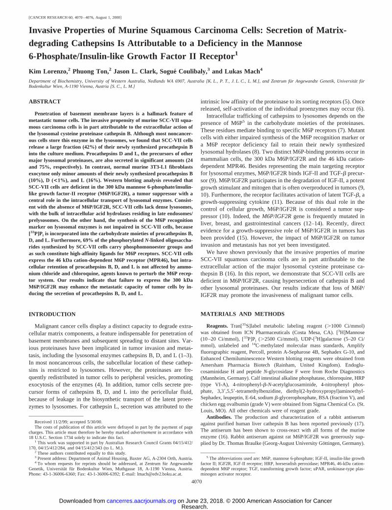
Expression of M6P Receptors in SCC-VII Cells.Mammaliancells usually synthesize both known M6P receptors, M6P/IGF2R andMPR46. We have investigated the expression of both receptors inSCC-VII cells and 3T3-L1 fibroblasts. Upon immunoblotting withantibodies against MPR46, a diffuse 45-kDa protein was detected inmembrane extracts of SCC-VII cells. A slightly smaller immunore-active polypeptide (43 kDa) was present in 3T3-L1 membranes. Noreaction was observed with the corresponding soluble protein frac-tions (Fig. 4A). It has been established that differences in glycosyla-tion may account for cell type-specific molecular forms of MPR46(24).
When extracts of 3T3-L1 fibroblasts labeled with [35S]methioninewere immunoprecipitated with antibodies against M6P/IGF2R, a
polypeptide of.250 kDa was detected. However, this protein was notpresent in extracts of SCC-VII cells. No cross-reactive polypeptideswere detectable in the culture medium of either cell line, ruling out thepossibility that shedding of cell surface-resident M6P/IGF2R wasresponsible for the absence of this protein in SCC-VII cell lysates(Fig. 4B).
Although both M6P receptors contribute to intracellular sorting oflysosomal enzymes, receptor-mediated uptake of exogenous M6P-containing ligands is exclusively mediated by M6P/IGF2R (7). Wehave shown previously that procathepsin B synthesized by humanhepatoma HepG2 cells is efficiently internalized by autologous recip-ient cells and human fibroblasts in a M6P/IGF2R-dependent manner(18). As a functional test for the presence of M6P/IGF2R, NH4Cl-induced secretions of HepG2 cells labeled with [35S]methionine wereoffered to unlabeled SCC-VII cells and 3T3-L1 fibroblasts for recep-tor-mediated endocytosis. Although procathepsin B was internalizedby 3T3-L1 cells in a M6P-inhibitable manner, no uptake was detect-able in SCC-VII cells. Identical results were obtained for procathepsinD (data not shown). We conclude that SCC-VII cells lack any endog-enous synthesis of M6P/IGF2R, which induces hypersecretion ofnewly synthesized lysosomal enzymes as well as impairs their recap-ture via receptor-mediated endocytosis.
The Formation of Mature Lysosomes Is Impaired in SCC-VIICells. Recent studies have shown that M6P/IGF2R plays a key role inthe biogenesis of lysosomes (8). To assess the status of lysosomes inSCC-VII cells, postnuclear organelles were separated by density-gradient centrifugation. When the distribution of cathepsin B in thegradient fractions was determined, a marked peak of enzyme activitywas observed at a buoyant density of 1.06 g/ml (Fig. 5A). The samedistribution was found for the lysosomal markerb-N-acetylhex-osaminidase and acid phosphatase, a lysosomal enzyme transported tothese compartments in a M6P-independent manner (7, 21). In contrast,the bulk of the cathepsin B (andb-N-acetylhexosaminidase) activityof control HepG2 cells was located in compartments with a density of.1.08 g/ml, as typical for mature lysosomes (Fig. 5B). However, thesubcellular distribution of lysosomal enzymes in SCC-VII cells over-laps significantly with the locations of the Golgi marker galactosyl-transferase and the endocytic tracer HRP (Fig. 5A), a feature remi-niscent of late endosomal/prelysosomal compartments. In fact, mutanthuman fibroblasts with a defect in the formation of dense lysosomesexhibited the same sedimentation pattern as observed for SCC-VIIcells (19).
In normal cells, lysosomes are usually located in the perinuclearregion (8, 19). Such a pattern was observed when control NIH 3T3fibroblasts were immunostained with anti-cathepsin D antibodies (Fig.6A). In contrast, immunocytochemical detection of cathepsin D inSCC-VII cells revealed numerous vesicles distributed throughout thecytoplasm (Fig. 6B). A similar subcellular distribution of lysosomalmarkers was observed in M6P/IGF2R-deficient murine fibroblasts,where the labeled structures were found to resemble late endocyticcompartments (8). Thus, SCC-VII “lysosomes” display at least somecharacteristics of late endosomes/prelysosomes, indicating impairedformation of mature lysosomes in these cells as a direct consequenceof the absence of M6P/IGF2R.
DISCUSSION
Our results demonstrate that SCC-VII cells secrete lysosomal pro-teinases attributable to the absence of functional M6P/IGF2R. Simi-larly, lack of M6P/IGF2R synthesis has been observed for the highlydeviated Morris 7777 hepatoma, a malignant rat tumor (24). Indeed,Morris 7777 hepatoma cells display enhanced secretion of procathep-sin D as compared with normal rat hepatocytes (25). Interestingly,
Fig. 4. Expression of M6P receptors in SCC-VII cells and 3T3-L1 fibroblasts.A,membrane extracts (M) and soluble fractions (S) of SCC-VII cells and 3T3-L1 fibroblasts(100 mg of protein each) were subjected to SDS-PAGE and Western blotting with amonospecific rabbit antiserum to murine MPR46 as outlined in “Materials and Methods.”The molecular mass standards used were BSA (66 kDa), chicken ovalbumin (45 kDa),bovine carbonic anhydrase (29 kDa), and bovineb-lactoglobulin (18 kDa).B, SCC-VIIcells (1) and 3T3-L1 fibroblasts (2) were metabolically labeled with [35S]methionine asdescribed in the legend of Fig. 1. M6P/IGF2R-related polypeptides were immunoprecipi-tated from cell (C) and medium (M) extracts and analyzed by SDS-PAGE and fluorog-raphy. The band labeled with anasteriskrepresents a protein unrelated to M6P/IGF2R thatis unspecifically coprecipitated by the antiserum used in these studies. The migrationpositions of the14C-labeled molecular mass standards rabbit myosin (205 kDa), rabbitphosphorylase b (97 kDa), and BSA (69 kDa) are indicated.
4074
M6P/IGF2R AND CATHEPSIN B SECRETION BY TUMOR CELLS
Research. on June 23, 2018. © 2000 American Association for Cancercancerres.aacrjournals.org Downloaded from

transformation of murine BALB/3T3 fibroblasts with Moloney mu-rine sarcoma virus triggers exocytosis of procathepsins B and L,concomitant with the loss of M6P/IGF2R activity (26). M6P/IGF2Rdeficiency has been also detected in leukemia cells, which secretelarge quantities of lysosomal hydrolases (27). Hence, the release ofmatrix-degrading cathepsins as a consequence of M6P/IGF2R ab-sence may represent a common feature of many tumor cells.
M6P/IGF2R plays a pivotal role in the formation of mature lyso-somes (7, 8). SCC-VII cells fail to synthesize the receptor and thuslack a functional set of these organelles. Consequently, these cellsstore lysosomal enzymes in compartments with properties reminiscentof late endosomes/prelysosomes. Similar results were obtained forother M6P/IGF2R-negative tumor cell lines, such as murine J774myeloid leukemia cells (28) and Morris 7777 hepatoma cells (29).Interestingly, cathepsin B was relocated to endosomal vesicles inhuman MCF-10A breast epithelial cells upon transfection with anoncogenic version of the c-Ha-rasgene (30). Furthermore, transfor-mation of mouse NIH 3T3 fibroblasts with Kirsten murine sarcomavirus leads to redistribution of cathepsin L and other lysosomalenzymes to endosomes/prelysosomes (31). Because both latter cell
types are not deficient in M6P/IGF2R,7 impaired formation of lyso-somes may also occur in M6P/IGF2R-positive tumor cells.
Despite the lack of M6P/IGF2R, SCC-VII cells retain a largeproportion of their newly synthesized lysosomal proteinases. Al-though the other known mammalian M6P receptor, MPR46 (8), ispresent in SCC-VII cells, the insensitivity of the intracellular transportof cathepsins B, D, and L to lysosomal alkalinization suggests thatM6P-independent sorting of lysosomal enzymes may take place inthese cells. Similarly, M6P-independent targeting mechanisms havebeen proposed for procathepsins D and L in normal and transformedmouse NIH 3T3 fibroblasts (32) and for procathepsin D in humanbreast cancer cells (23). Interestingly, procathepsin D transientlyassociates during its biosynthesis with prosaposin (33), a proteindelivered to lysosomes via interaction with the low-density lipopro-tein receptor-related protein (34). Thus, interaction of procathepsin Dwith endogenous prosaposin may account for its preferential retentionin SCC-VII cells as compared with cathepsins B and L.
It has been reported recently that M6P/IGF2R interacts with uPARthrough a domain distinct from its M6P- and IGF-II binding sites (35).In the absence of M6P/IGF2R, uPAR is not efficiently internalized,effectively increasing the number of surface binding sites for plas-minogen activators, which may cause enhanced focal proteolysis.Because SCC-VII cells produce plasminogen activators (16), deficientuPAR internalization could further add to the proteolytic load in thepericellular environment of the cells. Hence, M6P/IGF2R deficiencymay enhance the invasiveness of tumor cells by at least two means,elevated secretion of matrix-degrading cathepsins and increasedamounts of cell-surface plasminogen activators. Interestingly, uPARmay also promote tumor cell invasion in a protease-independentmanner through stimulation of integrin-mediated cell migration (36).
7 P. Ton and L. Mach, unpublished data.
Fig. 5. Characterization of SCC-VII lysosomes. Postnuclear supernatants of SCC-VII(A) and HepG2 (B) cells were subjected to Percoll density-gradient centrifugation asdescribed in “Materials and Methods.” Fractions (1 ml) were collected from the bottom ofthe tubes and analyzed for buoyant density (M), cathepsin B activity (E), and galacto-syltransferase activity (F). Enzyme activities are expressed as the percentage of the totalactivity recovered in the fractions of each gradient. InA, endocytosed HRP emerged as abroad peak in fractions 9–12. InB, HRP distribution was essentially the same as forgalactosyltransferase, with maximum activity in fraction 9.
Fig. 6. Subcellular localization of cathepsin D in SCC-VII cells and NIH 3T3fibroblasts. Control NIH 3T3 fibroblasts (A) and SCC-VII cells (B) were fixed andimmunostained with anti-cathepsin D antibodies as specified in “Materials and Methods.”The labeled compartments were visualized by fluorescence microscopy.Bars,10 mm.
4075
M6P/IGF2R AND CATHEPSIN B SECRETION BY TUMOR CELLS
Research. on June 23, 2018. © 2000 American Association for Cancercancerres.aacrjournals.org Downloaded from

The M6P/IGF2R gene is often inactivated in human and animaltumors. Rodents appear particularly susceptible becauseM6P/IGF2Rdisplays monoallelic expression in mice and rats, with the paternalallele being repressed. In contrast, this genomic imprinting ofM6P/IGF2R is a polymorphic trait in humans, with both alleles beingtranscribed in most humans. Thus, inactivation of theM6P/IGF2Rlocus generally requires two genetic events in humans but only one inmice (10). Interestingly, loss of oneM6P/IGF2Rallele is a commonfeature of human hepatocellular and breast cancers (12, 13).M6P/IGF2Rcontains several microsatellite sequences in its coding region,and deletions within the remainingM6P/IGF2Rallele arise frequentlyas a consequence of microsatellite instability (14). These mutationsgenerally cause frameshifts that result in premature termination oftranslation, giving rise to the synthesis of truncated, soluble receptors(12). However, M6P/IGF2R-related polypeptides were not detectablein the culture supernatants of SCC-VII cells, thus indicating that themolecular defect in theM6P/IGF2Rgene of these cells is probably notdirectly related to microsatellite instability.
M6P/IGF2R exerts at least two growth-suppressive functions: (a)the receptor accounts for the internalization and degradation of IGF-II,a powerful growth-stimulating factor; and (b) M6P/IGF2R bindslatent TGF-b1 and stimulates activation of the growth-suppressivecytokine by recruitment of plasminogen and uPAR-bound plasmino-gen activators (37). InM6P/IGF2R-mutant gastrointestinal tumors,IGF-II degradation and TGF-b1 activation are significantly reduced(38). Recently, transfection of wild-typeM6P/IGF2R cDNA intoM6P/IGF2R-mutant human SW48 colorectal carcinoma cells wasshown to suppress growth and induce apoptosis (15). Because ourresults indicate that M6P/IGF2R-negative tumor cells display anincreased potential to degrade extracellular matrix components, inac-tivation of the M6P/IGF2R gene may support growth as well asmetastasis of malignant cancers.
ACKNOWLEDGMENTS
We express our gratitude to Drs. Ann H. Erickson, Annette Hille-Rehfeld,Thomas Braulke, David James, and John S. Mort for providing antibodies andcell lines. We also thank Melinda Abas and Prof. Josef Glossl for criticalreading of the manuscript.
REFERENCES
1. Sloane, B. F., Moin, K., Krepela, E., and Rozhin, J. Cathepsin B and its endogenousinhibitors: the role in tumor malignancy. Cancer Metastasis Rev.,9: 333–352, 1990.
2. Rochefort, H., Capony, F., and Garcia, M. Cathepsin D: a protease involved in breastcancer metastasis. Cancer Metastasis Rev.,9: 321–331, 1990.
3. Kane, S. E., and Gottesman, M. M. The role of cathepsin L in malignant transfor-mation. Semin. Cancer Biol.,1: 127–136, 1990.
4. Rozhin, J., Sameni, M., Ziegler, G., and Sloane, B. F. Pericellular pH affectsdistribution and secretion of cathepsin B in malignant cells. Cancer Res.,54: 6517–6525, 1994.
5. Dong, J. M., and Sahagian, G. G. Basis for low affinity binding of a lysosomalcysteine protease to the cation-independent mannose 6-phosphate receptor. J. Biol.Chem.,265: 4210–4217, 1990.
6. Mach, L., Mort, J. S., and Glossl, J. Noncovalent complexes between the lysosomalproteinase cathepsin B and its propeptide account for stable, extracellular, highmolecular mass forms of the enzyme. J. Biol. Chem.,269: 13036–13040, 1994.
7. Kornfeld, S., and Mellman, I. The biogenesis of lysosomes. Annu. Rev. Cell Biol.,5:483–525, 1989.
8. Ludwig, T., Munier-Lehmann, H., Bauer, U., Hollinshead, M., Ovitt, C., Lobel, P.,and Hoflack, B. Differential sorting of lysosomal enzymes in mannose 6-phosphatereceptor-deficient fibroblasts. EMBO J.,13: 3430–3437, 1994.
9. Kornfeld, S. Structure and function of the mannose 6-phosphate/insulin-like growthfactor II receptors. Annu. Rev. Biochem.,61: 307–330, 1992.
10. De Souza, A. T., Yamada, T., Mills, J. J., and Jirtle, R. L. Imprinted genes in livercarcinogenesis. FASEB J.,11: 60–67, 1997.
11. Dennis, P. A., and Rifkin, D. B. Cellular activation of latent transforming growthfactorb requires binding to the cation-independent mannose 6-phosphate/insulin-likegrowth factor type II receptor. Proc. Natl. Acad. Sci. USA,88: 580–584, 1991.
12. De Souza, A. T., Hankins, G. R., Washington, M. K., Orton, T. C., and Jirtle, R. L.M6P/IGF2R gene is mutated in human hepatocellular carcinomas with loss ofheterozygosity. Nat. Genet.,11: 447–449, 1995.
13. Hankins, G. R., De Souza, A. T., Bentley, R. C., Patel, M. R., Marks, J. R., Iglehart,J. D., and Jirtle, R. L.M6P/IGF2 receptor: a candidate breast tumor suppressor gene.Oncogene,12: 2003–2009, 1996.
14. Souza, R. F., Appel, R., Yin, J., Wang, S., Smolinski, K. N., Abraham, J. M., Zou,T. T., Shi, Y. Q., Lei, J., Cottrell, J., Cymes, K., Biden, K., Simms, L., Leggett, B.,Lynch, P. M., Frazier, M., Powell, S. M., Harpaz, N., Sugimura, H., Young, J., andMeltzer, S. J. Microsatellite instability in the insulin-like growth factor II receptorgene in gastrointestinal tumours. Nat. Genet.,14: 255–257, 1996.
15. Souza, R. F., Wang, S., Thakar, M., Smolinski, K. N., Yin, J., Zou, T. T., Kong, D.,Abraham, J. M., Toretsky, J. A., and Meltzer, S. J. Expression of the wild-typeinsulin-like growth factor II receptor gene suppresses growth and causes death incolorectal carcinoma cells. Oncogene,18: 4063–4068, 1999.
16. Coulibaly, S., Schwihla, H., Abrahamson, M., Albini, A., Cerni, C., Clark, J. L., Ng,K. M., Katunuma, N., Schlappack, O., Glossl, J., and Mach, L. Modulation ofinvasive properties of murine squamous carcinoma cells by heterologous expressionof cathepsin B and cystatin C. Int. J. Cancer,83: 526–531, 1999.
17. Hanewinkel, H., Glossl, J., and Kresse, H. Biosynthesis of cathepsin B in culturednormal and I-cell fibroblasts. J. Biol. Chem.,262: 12351–12355, 1987.
18. Mach, L., Stuwe, K., Hagen, A., Ballaun, C., and Glossl, J. Proteolytic processing andglycosylation of cathepsin B. The role of the primary structure of the latent precursorand of the carbohydrate moiety for cell-type-specific molecular forms of the enzyme.Biochem. J.,282: 577–582, 1992.
19. Schmid, J. A., Mach, L., Paschke, E., and Glossl, J. Accumulation of sialic acid inendocytic compartments interferes with the formation of mature lysosomes. Impairedproteolytic processing of cathepsin B in fibroblasts of patients with lysosomal sialicacid storage disease. J. Biol. Chem.,274: 19063–19071, 1999.
20. Rome, L. H., Garvin, A. J., Allietta, M. M., and Neufeld, E. F. Two species oflysosomal organelles in cultured human fibroblasts. Cell,17: 143–153, 1979.
21. Saftig, P., Hartmann, D., Lullmann-Rauch, R., Wolff, J., Evers, M., Koster, A.,Hetman, M., von Figura, K., and Peters, C. Mice deficient in lysosomal acidphosphatase develop lysosomal storage in the kidney and central nervous system.J. Biol. Chem.,272: 18628–18635, 1997.
22. Isidoro, C., Radons, J., Baccino, F. M., and Hasilik, A. Suppression of the “uncov-ering” of mannose-6-phosphate residues in lysosomal enzymes in the presence ofNH4Cl. Eur. J. Biochem.,191: 591–597, 1990.
23. Capony, F., Braulke, T., Rougeot, C., Roux, S., Montcourrier, P., and Rochefort, H.Specific mannose-6-phosphate receptor-independent sorting of pro-cathepsin D inbreast cancer cells. Exp. Cell Res.,215: 154–163, 1994.
24. Stein, M., Braulke, T., Krentler, C., Hasilik, A., and von Figura, K. 46-kDa mannose6-phosphate-specific receptor: biosynthesis, processing, subcellular location and to-pology. Biol. Chem. Hoppe Seyler,368: 937–947, 1987.
25. Isidoro, C., Demoz, M., De Stefanis, D., Mainferme, F., Wattiaux, R., and Baccino,F. M. Altered intracellular processing and enhanced secretion of procathepsin D in ahighly deviated rat hepatoma. Int. J. Cancer,60: 61–64, 1995.
26. Achkar, C., Gong, Q. M., Frankfater, A., and Bajkowski, A. S. Differences intargeting and secretion of cathepsins B and L by BALB/3T3 fibroblasts and Moloneymurine sarcoma virus-transformed BALB/3T3 fibroblasts. J. Biol. Chem.,265:13650–13654, 1990.
27. Gabel, C. A., Goldberg, D. E., and Kornfeld, S. Identification and characterization ofcells deficient in the mannose 6-phosphate receptor: evidence for an alternate pathwayfor lysosomal enzyme targeting. Proc. Natl. Acad. Sci. USA,80: 775–779, 1983.
28. Tjelle, T. E., Brech, A., Juvet, L. K., Griffiths, G., and Berg, T. Isolation andcharacterization of early endosomes, late endosomes and terminal lysosomes: theirrole in protein degradation. J. Cell Sci.,109: 2905–2914, 1996.
29. Burge, V., Mainferme, F., and Wattiaux, R. Transient membrane association of theprecursors of cathepsin C during their transfer to lysosomes. Biochem. J.,275:797–800, 1991.
30. Sloane, B. F., Moin, K., Sameni, M., Tait, L. R., Rozhin, J., and Ziegler, G.Membrane association of cathepsin B can be induced by transfection of human breastepithelial cells with c-Ha-rasoncogene. J. Cell Sci.,107: 373–384, 1994.
31. Gal, S., Willingham, M. C., and Gottesman, M. M. Processing and lysosomallocalization of a glycoprotein whose secretion is transformation stimulated. J. CellBiol., 100: 535–544, 1985.
32. McIntyre, G. F., and Erickson, A. H. Procathepsins L and D are membrane-bound inacidic microsomal vesicles. J. Biol. Chem.,266: 15438–15445, 1991.
33. Zhu, Y., and Conner, G. E. Intermolecular association of lysosomal protein precursorsduring biosynthesis. J. Biol. Chem.,269: 3846–3851, 1994.
34. Hiesberger, T., Huttler, S., Rohlmann, A., Schneider, W., Sandhoff, K., and Herz,J. Cellular uptake of saposin (SAP) precursor and lysosomal delivery by the lowdensity lipoprotein receptor-related protein (LRP). EMBO J.,17: 4617–4625, 1998.
35. Nykjaer, A., Christensen, E. I., Vorum, H., Hager, H., Petersen, C. M., Roigaard, H.,Min, H. Y., Vilhardt, F., Møller, L. B., Kornfeld, S., and Gliemann, J. Mannose6-phosphate/insulin-like growth factor-II receptor targets the urokinase receptor tolysosomes via a novel binding interaction. J. Cell Biol.,141: 815–828, 1998.
36. Wei, Y., Lukashev, M., Simon, D. I., Bodary, S. C., Rosenberg, S., Doyle, M. V., andChapman, H. A. Regulation of integrin function by the urokinase receptor. Science(Washington DC),273: 1551–1555, 1996.
37. Godar, S., Horejsi, V., Weidle, U. H., Binder, B. R., Hansmann, C., and Stockinger,H. M6P/IGFII-receptor complexes urokinase receptor and plasminogen for activationof transforming growth factor-b1. Eur. J. Immunol.,29: 1004–1013, 1999.
38. Wang, S., Souza, R. F., Kong, D., Yin, J., Smolinski, K. N., Zou, T. T., Frank, T.,Young, J., Flanders, K. C., Sugimura, H., Abraham, J. M., and Meltzer, S. J. Deficienttransforming growth factor-b1 activation and excessive insulin-like growth factor II(IGFII) expression in IGFII receptor-mutant tumors. Cancer Res.,57: 2543–2546,1997.
4076
M6P/IGF2R AND CATHEPSIN B SECRETION BY TUMOR CELLS
Research. on June 23, 2018. © 2000 American Association for Cancercancerres.aacrjournals.org Downloaded from

2000;60:4070-4076. Cancer Res Kim Lorenzo, Phuong Ton, Jason L. Clark, et al. Factor II ReceptorDeficiency in the Mannose 6-Phosphate/Insulin-like GrowthSecretion of Matrix-degrading Cathepsins Is Attributable to a Invasive Properties of Murine Squamous Carcinoma Cells:
Updated version
http://cancerres.aacrjournals.org/content/60/15/4070
Access the most recent version of this article at:
Cited articles
http://cancerres.aacrjournals.org/content/60/15/4070.full#ref-list-1
This article cites 36 articles, 19 of which you can access for free at:
Citing articles
http://cancerres.aacrjournals.org/content/60/15/4070.full#related-urls
This article has been cited by 9 HighWire-hosted articles. Access the articles at:
E-mail alerts related to this article or journal.Sign up to receive free email-alerts
Subscriptions
Reprints and
To order reprints of this article or to subscribe to the journal, contact the AACR Publications
Permissions
Rightslink site. Click on "Request Permissions" which will take you to the Copyright Clearance Center's (CCC)
.http://cancerres.aacrjournals.org/content/60/15/4070To request permission to re-use all or part of this article, use this link
Research. on June 23, 2018. © 2000 American Association for Cancercancerres.aacrjournals.org Downloaded from