introduction: why the cytoskeleton is important what is the function of the system on the right?
TRANSCRIPT
Introduction: Why the Cytoskeleton Is Important
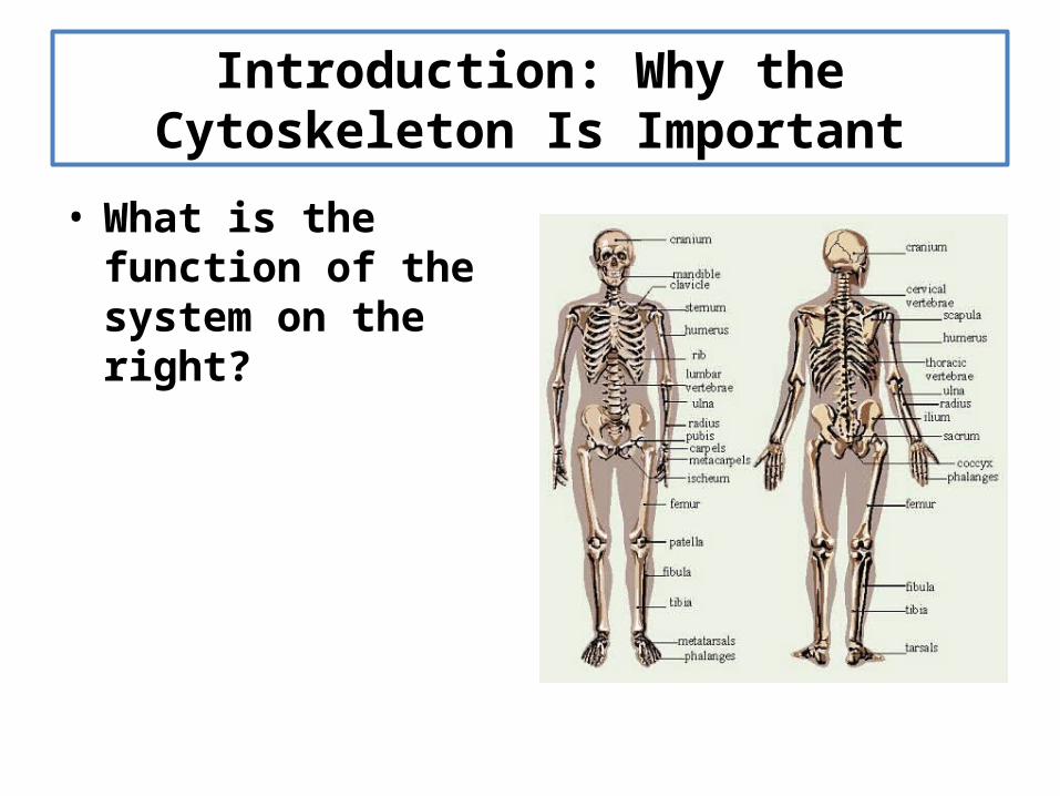
• What is the function of the system on the right?

Introduction: Why the Cytoskeleton Is Important
• What is the function of the system on the right?– Provides shape to the human
body– Gives the human body
structural integrity– A system by which other
structures can be attached
• The human skeleton is composed of bone– Contains Bone Cells– Contains Calcium– Contains Phosphorus

Introduction: Why the Cytoskeleton Is Important
• The cytoskeleton is a skeleton inside the cell
• Functions of the cytoskeleton– Provide shape to the cell– Gives the cell structural integrity– Place to attach organelles, or the
cell membrane– Can be used to move the cell– Can be used to move
molecules/organelles through the cell
• The cytoskeleton is composed of protein
Neurons
Human Epithelial Cells
Introduction: Types of Cytoskeletal Systems
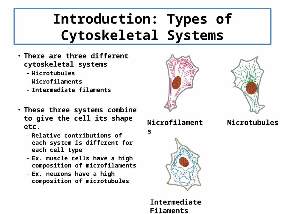
• There are three different cytoskeletal systems– Microtubules– Microfilaments– Intermediate filaments
• These three systems combine to give the cell its shape etc.– Relative contributions of each
system is different for each cell type
– Ex. muscle cells have a high composition of microfilaments
– Ex. neurons have a high composition of microtubules
MicrotubulesMicrofilaments
Intermediate Filaments
Introduction: Important Qualities of Cytoskeletal Systems
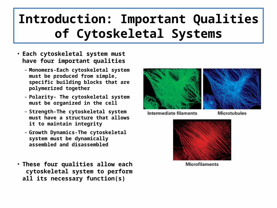
• Each cytoskeletal system must have four important qualities– Monomers-Each cytoskeletal system must
be produced from simple, specific building blocks that are polymerized together
– Polarity- The cytoskeletal system must be organized in the cell
– Strength-The cytoskeletal system must have a structure that allows it to maintain integrity
– Growth Dynamics-The cytoskeletal system must be dynamically assembled and disassembled
• These four qualities allow each cytoskeletal system to perform all its necessary function(s)
Microtubules: Introduction To Structure and Function
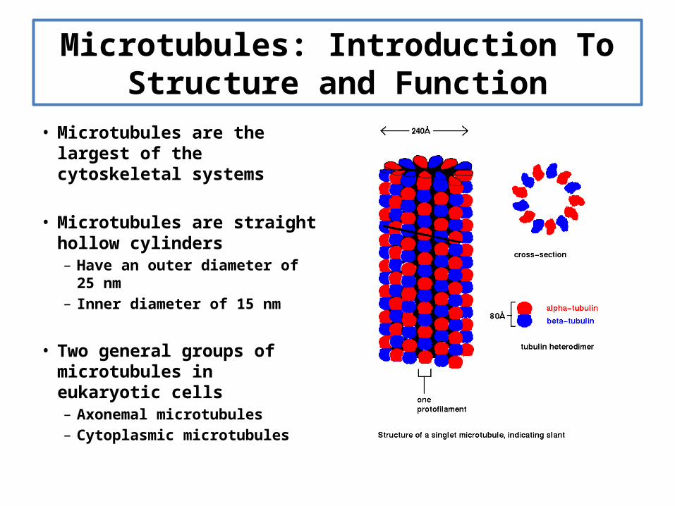
• Microtubules are the largest of the cytoskeletal systems
• Microtubules are straight hollow cylinders – Have an outer diameter of 25
nm – Inner diameter of 15 nm
• Two general groups of microtubules in eukaryotic cells– Axonemal microtubules– Cytoplasmic microtubules
Microtubules: Introduction To Structure and Function
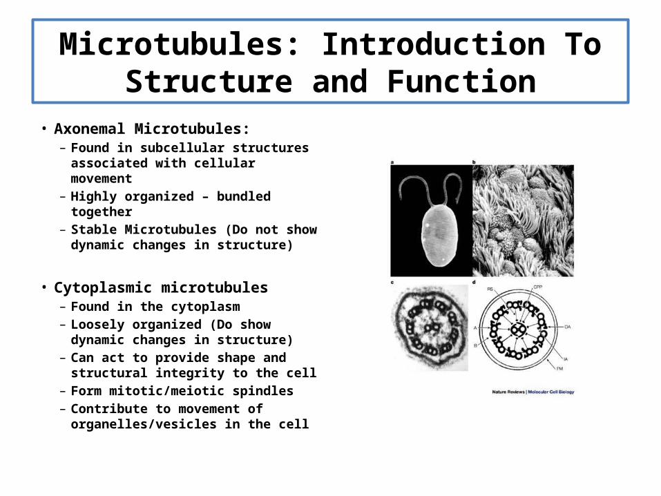
• Axonemal Microtubules:– Found in subcellular structures
associated with cellular movement– Highly organized – bundled together– Stable Microtubules (Do not show
dynamic changes in structure)
• Cytoplasmic microtubules– Found in the cytoplasm– Loosely organized (Do show dynamic
changes in structure)– Can act to provide shape and
structural integrity to the cell– Form mitotic/meiotic spindles– Contribute to movement of
organelles/vesicles in the cell
Microtubules: Building a Microtubule
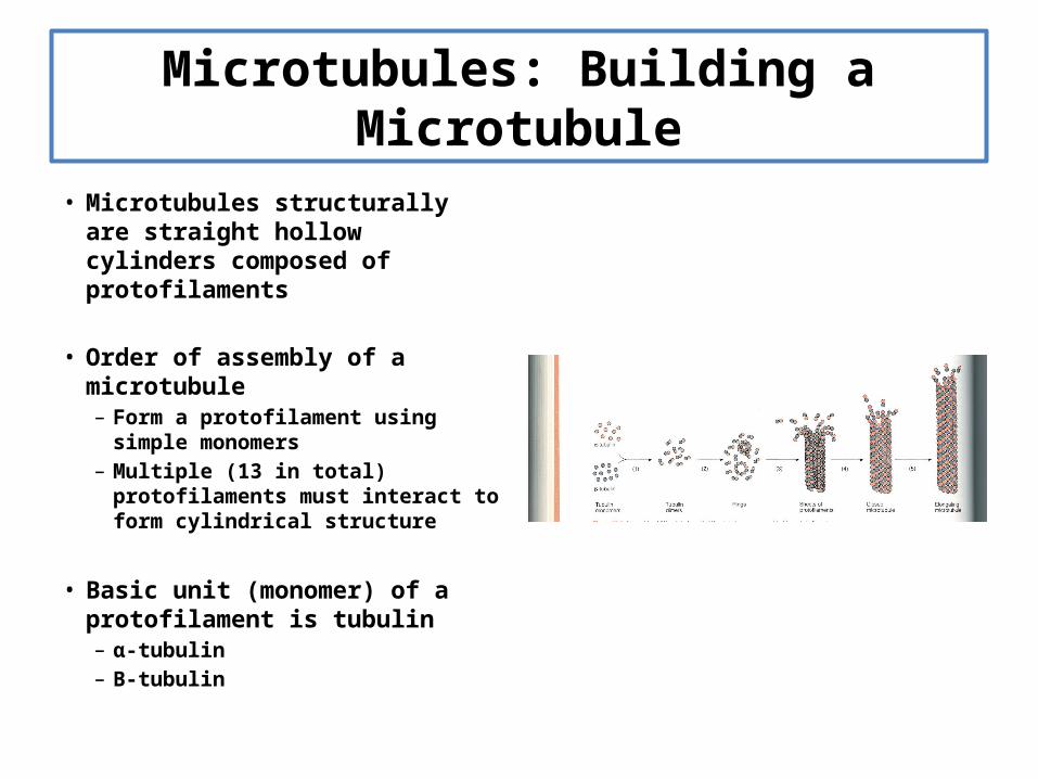
• Microtubules structurally are straight hollow cylinders composed of protofilaments
• Order of assembly of a microtubule– Form a protofilament using simple
monomers– Multiple (13 in total) protofilaments
must interact to form cylindrical structure
• Basic unit (monomer) of a protofilament is tubulin– α-tubulin– Β-tubulin
Microtubules: Building a Microtubule
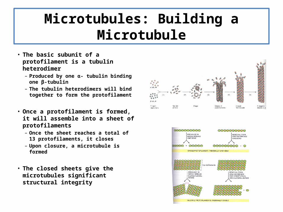
• The basic subunit of a protofilament is a tubulin heterodimer– Produced by one α- tubulin binding
one β-tubulin– The tubulin heterodimers will bind
together to form the protofilament
• Once a protofilament is formed, it will assemble into a sheet of protofilaments– Once the sheet reaches a total of 13
protofilaments, it closes– Upon closure, a microtubule is formed
• The closed sheets give the microtubules significant structural integrity
Microtubules: Microtubules Have Specific Polarity
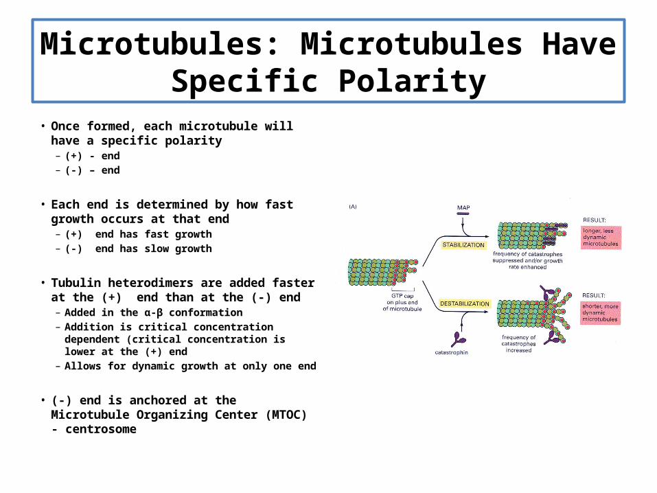
• Once formed, each microtubule will have a specific polarity– (+) - end– (-) – end
• Each end is determined by how fast growth occurs at that end– (+) end has fast growth– (-) end has slow growth
• Tubulin heterodimers are added faster at the (+) end than at the (-) end– Added in the α-β conformation– Addition is critical concentration dependent
(critical concentration is lower at the (+) end– Allows for dynamic growth at only one end
• (-) end is anchored at the Microtubule Organizing Center (MTOC) - centrosome
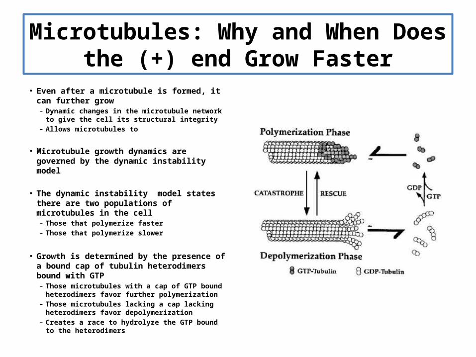
Microtubules: Why and When Does the (+) end Grow Faster
• Even after a microtubule is formed, it can further grow– Dynamic changes in the microtubule network to
give the cell its structural integrity– Allows microtubules to
• Microtubule growth dynamics are governed by the dynamic instability model
• The dynamic instability model states there are two populations of microtubules in the cell– Those that polymerize faster– Those that polymerize slower
• Growth is determined by the presence of a bound cap of tubulin heterodimers bound with GTP– Those microtubules with a cap of GTP bound
heterodimers favor further polymerization– Those microtubules lacking a cap lacking
heterodimers favor depolymerization– Creates a race to hydrolyze the GTP bound to the
heterodimers

Microtubules: Microtubule Binding Proteins Allow For Fast Growth When Forming Spindle
• Microtubules can be bound by two types of associated proteins which allow for fast assembly and disassembly– MAPs (Microtubule Associated Proteins)– Catastrophins
• MAPs promote fast growth– Bind microtubules that are forming
spindles early in mitosis– Stabilize the GTP cap
• Catastrophins promote fast disassembly– Allow for disassembly of spindle as the
chromosomes are pulled to opposite sides of the cell during mitosis
– Destabilize the GTP cap
Microtubules: A Target For Cancer Therapy
• Cancer results from cells losing proper cell cycle controls
• Divide uncontrollably
• Eventually form a tumor
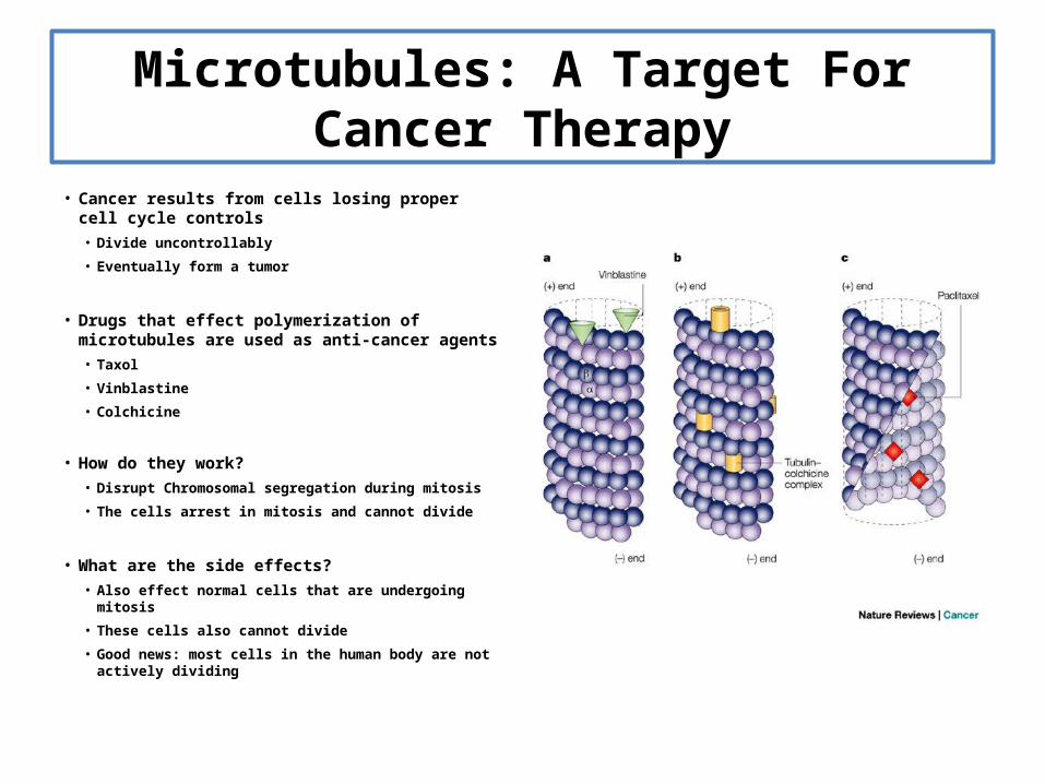
• Drugs that effect polymerization of microtubules are used as anti-cancer agents
• Taxol
• Vinblastine
• Colchicine
• How do they work?• Disrupt Chromosomal segregation during mitosis
• The cells arrest in mitosis and cannot divide
• What are the side effects?• Also effect normal cells that are undergoing mitosis
• These cells also cannot divide
• Good news: most cells in the human body are not actively dividing
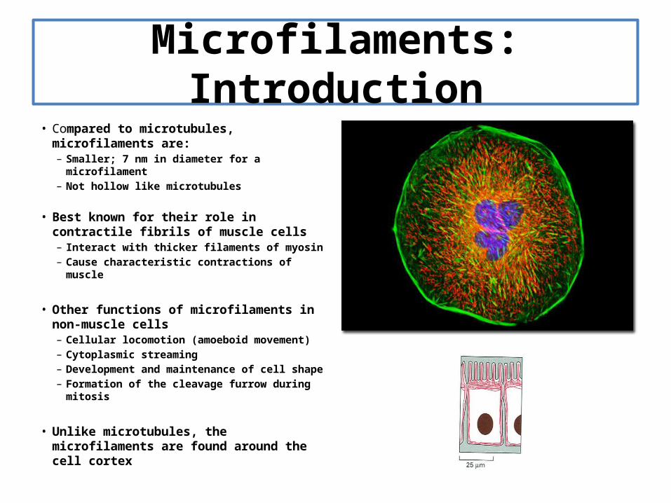
Microfilaments: Introduction• Compared to microtubules, microfilaments
are:– Smaller; 7 nm in diameter for a
microfilament– Not hollow like microtubules
• Best known for their role in contractile fibrils of muscle cells– Interact with thicker filaments of myosin– Cause characteristic contractions of muscle
• Other functions of microfilaments in non-muscle cells– Cellular locomotion (amoeboid movement)– Cytoplasmic streaming– Development and maintenance of cell shape– Formation of the cleavage furrow during
mitosis
• Unlike microtubules, the microfilaments are found around the cell cortex
Microfilaments: Building a Microfilament
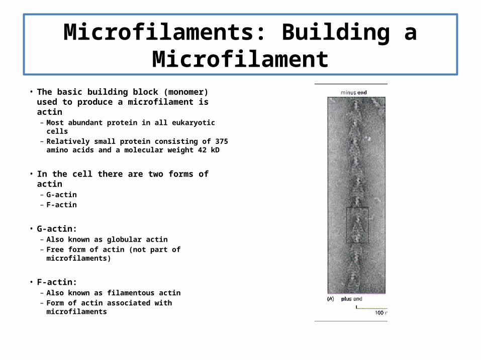
• The basic building block (monomer) used to produce a microfilament is actin– Most abundant protein in all eukaryotic cells– Relatively small protein consisting of 375
amino acids and a molecular weight 42 kD
• In the cell there are two forms of actin– G-actin – F-actin
• G-actin:– Also known as globular actin– Free form of actin (not part of
microfilaments)
• F-actin:– Also known as filamentous actin– Form of actin associated with microfilaments

Microfilaments: Building a Microfilament
• G-actin monomers must polymerize to form F-actin microfilaments
• A G-actin monomer is not symmetrical– The resulting F-actin microfilament is not
symmetrical– The resulting F-actin microfilament will have
polarity
• Just like a microtubule, an F-actin microfilament has polarity– (+) end– (-) end
• Growth of an F-actin microfilament occurs at the (+) end– Growth is supported when the (+) end when
capped with G-actin monomers bound to ATP– Disassembly is supported at the (-) end, which is
capped with G-actin bound to ADP– Creates a treadmilling effect– Other proteins can cap the (+) and (-) ends to
completely halt growth (very common)

Microfilaments: Building a Microfilament
• G-actin-ATP
• F-actin-ATP
• F-actin-ADP
• G-actin-ADP
• G-actin-ATP
• Pi
• G-actin Monomer • F-actin Polymer
• ATP
• ADP
• Microfilaments, like Microtubles, have polarity-They polymerize at the (+) end and depolymerize at the (-) end
• (+)
• (-)

G-actin
ATP
ADP
(-) end(+) end
F-actin
Microfilaments Have A Structure That Allows For It To Function In Determining Cell Shape and Yet Be Dynamic For Cell Motility
Monomers of G-actin Polymerize into Double Stranded F-actin Polymers