introduction to x-ray fluorescence xrf
TRANSCRIPT

BRUKER ADVANCED X-RAY SOLUTIONS
____________________________________________________
GUIDE TO XRF BASICS
____________________________________________________

This guide was first published in West Germany under the title “Introduction to X-ray
Fluorescence Analysis (XRF).”
© 2000 - 2006 Bruker AXS GmbH, Karlruhe, West Germany. All rights reserved.
Authors: Dr. Reinhold Schlotz, freelance writer and applications scientist, Bruker AXS
Dr. Stefan Uhlig, International Sales Manager, Bruker AXS
Bruker AXS GmbH Östliche Rheinbrückenstr. 49 D-76187 Karlsruhe Germany Tel: (+49) (721) 595-2888 Fax: (+49) (721) 595-4587 Email: [email protected] Web: www.bruker-axs.de
Bruker AXS Inc. 5465 East Cheryl Parkway Madison, WI 53711-5373 USA Tel: +1 (800) 234-XRAY Fax: +1 (608) 276-3006 Email: [email protected] Web: www.bruker-axs.com

Introduction to X-Ray Fluorescence (XRF) Table of Contents
i
Introduction to X-ray Fluorescence (XRF)
Table of Contents
1. Fundamental Principles........................................................................................1 1.1 Electromagnetic Radiation, Quanta ........................................................................................1
1.1.1 The Origin of X-rays..........................................................................................................2 1.1.2 Bohr's Atomic Model .........................................................................................................2 1.1.3 Characteristic Radiation ...................................................................................................3
1.2 Nomenclature ............................................................................................................................3 1.3 Generating the Characteristic Radiation ................................................................................4
1.3.1 X-ray Tubes, Bremsspektrum...........................................................................................5 1.3.2 Tube Types, the Generator ..............................................................................................6
1.3.2.1 Side-window Tubes ................................................................................................6 1.3.2.2 End-window Tubes .................................................................................................7 1.3.2.3 Generator ...............................................................................................................7
1.4 Excitation of Characteristic Radiation in Sample Material ...................................................8 1.4.1 Layer Thickness, Saturation Thickness..........................................................................10 1.4.2 Secondary Enhancement ...............................................................................................11
1.5 Tube-spectrum Scattering at the Sample Material ..............................................................11 1.6 X-ray Detectors .......................................................................................................................12
1.6.1 Pulse Height Spectrum...................................................................................................12 1.6.2 Gas Proportional Counter ...............................................................................................12 1.6.3 Scintillation Counter........................................................................................................14
1.7 Pulse Height Analysis (PHA) .................................................................................................14 1.7.1 Pulse Height Distribution ................................................................................................14 1.7.2 The Counter Plateau.......................................................................................................16
1.8 Diffraction in Crystals.............................................................................................................17 1.8.1 Interference.....................................................................................................................17 1.8.2 Diffraction........................................................................................................................18 1.8.3 X-ray Diffraction From a Crystal Lattice, Bragg's Equation ............................................19 1.8.4 Reflections of Higher Orders ..........................................................................................21 1.8.5 Crystal Types..................................................................................................................22 1.8.6 Dispersion, Line Separation ...........................................................................................24 1.8.7 Standard Types, Multilayers ...........................................................................................24 1.8.8 Special Crystals ..............................................................................................................26 1.8.9 Curved Crystals ..............................................................................................................31
2. Instrumentation ...................................................................................................33 2.1 Multichannel Spectrometers..................................................................................................33
2.1.1 Scanners for Multichannel Spectrometers......................................................................34 2.2 Sequential Spectrometers......................................................................................................35 2.3 Incident Beam Components ..................................................................................................37

Table of Contents Introduction to X-Ray Fluorescence (XRF)
ii
2.3.1 The End-window Tube and Generator ...........................................................................37 2.3.2 The Primary Beam Filter.................................................................................................37 2.3.3 Sample Cups, the Cup Aperture.....................................................................................39
2.4 Emitted Beam Components...................................................................................................39 2.4.1 The Vacuum Seal ...........................................................................................................39 2.4.2 Collimator Masks ............................................................................................................39 2.4.3 Collimators, the Soller Slit ..............................................................................................39 2.4.4 The Crystal Changer.......................................................................................................40 2.4.5 The Flow Counter ...........................................................................................................40 2.4.6 The Sealed Proportional Counter ...................................................................................41 2.4.7 The Scintillation Counter.................................................................................................42
2.5 Electronic Pulse Processing .................................................................................................42 2.5.1 The Discriminator............................................................................................................42 2.5.2 Main Amplifier, Sine Amplifier.........................................................................................42 2.5.3 Dead Time Correction.....................................................................................................42 2.5.4 Line-shift Correction........................................................................................................44
3. Sample Preparation Techniques........................................................................45 3.1 Introduction .............................................................................................................................45 3.2 Preparation of Solid Samples................................................................................................48
3.2 1 Metals .............................................................................................................................48 3.2.2 Pressed Pellets...............................................................................................................48 3.2.3 Fused Beads...................................................................................................................49
3.3 Preparation of Liquid Samples..............................................................................................50 3.4 Preparation of Filter Samples................................................................................................50
Appendix A – Sources of Standard Samples........................................................51
Appendix B – Supplementary Literature ...............................................................53 Books .............................................................................................................................................53 Tables.............................................................................................................................................53
Index.........................................................................................................................55

Introduction to X-Ray Fluorescence (XRF) Fundamental Principles
1
1. Fundamental Principles
1.1 Electromagnetic Radiation, Quanta
From a physical point of view, X-rays are of the same nature as visible light. Visible light can be described as electromagnetic wave radiation whose variety of colors (e.g. the colors of the rainbow) we interpret as different wavelengths. The wavelengths of electromagnetic radiation reach from the kilometer range of radio waves up to the picometer range (10-12 m) of hard gamma radiation (Table 1).
Table 1: Energy and wavelength ranges of electromagnetic radiation
Energy range (keV) Wavelength range Name
< 10-7 cm to km Radio waves (short, medium, long waves)
< 10-3 µm to cm Microwaves
< 10-3 µm to mm Infra-red
0.0017 – 0.0033 380 to 750 nm Visible light
0.0033 – 0.1 10 to 380 nm Ultra-violet
0.11 – 100 0.01 to 12 nm X-rays
10 – 5000 0.0002 to 0.12 nm Gamma radiation In the following text, the unit nanometer (nm = 10-9 m) is used for the wavelength, λλλλ (= Lambda), and the unit kiloelectronvolts (keV) for energy, E.
Comment In literature the unit Angström (Å) is often stated for the wavelength:
1 Å = 0.1 nm = 10-10 m
The following relationship (conversion formula) exists between the units E (keV) and λ (nm):
)(
24.1)(
nmkeVE
λ= or
)(
24.1)(
keVEnm =λ
The X-ray fluorescence analysis records the following range of energy or wavelengths:
E = 0.11 – 60 keV
λ = 11.3 – 0.02 nm
Apart from the wave properties, light also has the properties of particles. This is expressed by the term “photon”. In the following we will be using the term quanta or X-ray quanta for this. The radiation intensity is the number of X-ray quanta that are emitted or measured per unit of time. We use the number of X-ray quanta measured per second, cps (= counts per second) or kcps (= kilocounts per second) as the unit of intensity.
Fundamental Principles Introduction to X-Ray Fluorescence (XRF)
2
1.1.1 The Origin of X-rays
Electromagnetic radiation can occur whenever electrically charged particles, particularly electrons, lose energy as a result of a change in their state of motion, e.g. upon deceleration, changing direction or moving to a lower energy level in the atomic shell. The deceleration of electrons and the transition from an energy level in the atomic shell to a lower one play an important part in the creation of X-rays in the field of X-ray analysis. To understand the processes in the atomic shell we must take a look at the Bohr's atomic model.
1.1.2 Bohr's Atomic Model
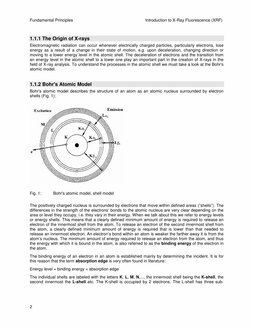
Bohr's atomic model describes the structure of an atom as an atomic nucleus surrounded by electron shells (Fig. 1):
Fig. 1: Bohr's atomic model, shell model
The positively charged nucleus is surrounded by electrons that move within defined areas (”shells”). The differences in the strength of the electrons‘ bonds to the atomic nucleus are very clear depending on the area or level they occupy, i.e. they vary in their energy. When we talk about this we refer to energy levels or energy shells. This means that a clearly defined minimum amount of energy is required to release an electron of the innermost shell from the atom. To release an electron of the second innermost shell from the atom, a clearly defined minimum amount of energy is required that is lower than that needed to release an innermost electron. An electron’s bond within an atom is weaker the farther away it is from the atom’s nucleus. The minimum amount of energy required to release an electron from the atom, and thus the energy with which it is bound in the atom, is also referred to as the binding energy of the electron in the atom.
The binding energy of an electron in an atom is established mainly by determining the incident. It is for this reason that the term absorption edge is very often found in literature:
Energy level = binding energy = absorption edge
The individual shells are labeled with the letters K, L, M, N,..., the innermost shell being the K-shell, the second innermost the L-shell etc. The K-shell is occupied by 2 electrons. The L-shell has three sub-

Introduction to X-Ray Fluorescence (XRF) Fundamental Principles
3
levels and can contain up to 8 electrons. The M-shell has five sub-levels and can contain up to 18 electrons.
1.1.3 Characteristic Radiation
Every element is clearly defined by its atomic number Z in the periodic table of elements or by the number of its electrons in a neutral state. The binding energies or the energy levels in every element are different and characteristic for every element as a result of the varying number of electrons (negative charges) or the number Z of the positive charges in the atomic nucleus (= atomic number).
If an electron of an inner shell is now separated from the atom by the irradiation of energy, an electron from a higher shell falls into this resultant “hole” which releases an amount of energy equivalent to the difference between the energy levels involved.
The energy being released can be either emitted in the form of an X-ray or transferred to another atomic shell electron (Auger effect). The probability of an X-ray resulting from this process is called the fluorescence yield ωωωω. This depends on the element’s atomic number and the shell in which the “hole”
occurred. ωωωω is very low for light elements (approx. 10-4 for boron) and almost reaches a value of 1 for the K-shell of heavier elements (e.g. uranium).
However, since the energy or wavelength of the X-ray is very characteristic for the element from which it is emitted; such radiation is called characteristic X-rays.
This provides the basis for determining chemical elements with the aid of X-ray fluorescence analysis.
1.2 Nomenclature
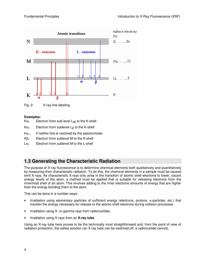
The energy of an X-ray corresponds to the difference in energy of the energy levels concerned. K-radiation is the term given to the radiation released when replenishing the K-shell, L-radiation to that released when replenishing the L-shell etc. (Fig. 2).
Also needed for the full labeling of the emitted X-ray line is the information telling us which shell the electron filling the “hole” comes from. The Greek letters α, β, χ, ... are used for this with the numbering 1, 2, 3, ... to differentiate between the various shells and sub-levels.
Fundamental Principles Introduction to X-Ray Fluorescence (XRF)
4
Fig. 2: X-ray line labeling
Examples:
Kα1 Electron from sub-level LIII to the K-shell
Kα2 Electron from sublevel LII to the K-shell
Kα1,2 if neither line is resolved by the spectrometer
Kβ1 Electron from sublevel M to the K-shell
Lα1 Electron from sublevel M to the L-shell
1.3 Generating the Characteristic Radiation
The purpose of X-ray fluorescence is to determine chemical elements both qualitatively and quantitatively by measuring their characteristic radiation. To do this, the chemical elements in a sample must be caused emit X-rays. As characteristic X-rays only arise in the transition of atomic shell electrons to lower, vacant energy levels of the atom, a method must be applied that is suitable for releasing electrons from the innermost shell of an atom. This involves adding to the inner electrons amounts of energy that are higher than the energy bonding them to the atom.
This can be done in a number ways:
• Irradiation using elementary particles of sufficient energy (electrons, protons, α-particles, etc.) that transfer the energy necessary for release to the atomic shell electrons during collision processes
• Irradiation using X- or gamma rays from radionuclides
• Irradiation using X-rays from an X-ray tube
Using an X-ray tube here proves to be the technically most straightforward and, from the point of view of radiation protection, the safest solution (an X-ray tube can be switched off, a radionuclide cannot).

Introduction to X-Ray Fluorescence (XRF) Fundamental Principles
5
1.3.1 X-ray Tubes, Bremsspektrum
In an X-ray tube, electrons are accelerated in an electrical field and shot against a target material where they are decelerated. The technical means of achieving this is to apply high voltage between a heated cathode (e.g. a filament) and a suitable anode material. Electrons emanate from the heated cathode material and are accelerated towards the anode by the applied high voltage. There they strike the anode material and lose their energy through deceleration. Only a small proportion of their energy loss (approx. 1-2%, depending on the anode material) is radiated in the form of X-rays. The greatest amount of energy contributes to heating up the anode material. Consequently the anode has to be cooled which is achieved by connection to a water-cooling system.
The proportion of the electron energy loss emitted in the form of an X-ray can be between zero and the maximum energy that the electron has acquired as a result of the acceleration in the electrical field. If 30 kV (kilovolt) are applied between the anode and cathode, the electrons acquire 30 keV from passing through this voltage (kiloelectronvolts) (Definition: 1 eV = the energy that an electron acquires when passing through a potential of 1 Volt).
A maximum X-ray energy of 30 keV can be acquired from deceleration in the anode material, i.e. the distribution of the energies of numerous X-rays is between zero and the maximum energy. If the intensity of this type of X-ray is applied depending on the energy, the result is the Bremsspektrum (= continuum) of the tube.
Fig. 3: A Bremsspektrum (continuum) with characteristic radiation of the anode material

Fundamental Principles Introduction to X-Ray Fluorescence (XRF)
6
In addition to the Bremsspektrum, an X-ray tube of course emits the characteristic radiation of the anode material as well, which is of major importance for X-ray fluorescence analysis (Fig. 3).
1.3.2 Tube Types, the Generator
All X-ray tubes work on the same principle: accelerating electrons in an electrical field and decelerating them in a suitable anode material. The region of the electron beam in which this takes place must be evacuated in order to prevent collisions with gas molecules. Hence there is a vacuum within the housing. The X-rays escape from the housing at a special point that is particularly transparent with a thin beryllium window.
The main differences between tube types are in the polarity of the anode and cathode and the arrangement of the exit window. The two most significant types are the end-window tubes and the side-window tubes.
1.3.2.1 Side-window Tubes
In side-window tubes, a negative high voltage is applied to the cathode. The electrons emanate from the heated cathode and are accelerated in the direction of the anode. The anode is set on zero voltage and thus has no difference in potential to the surrounding housing material and the laterally mounted beryllium exit window (Fig. 4).
Fig. 4: The principle of the side-window tube
For physical reasons, a proportion of the electrons are always scattered on the surface of the anode. The extent to which these backscattering electrons arise depends, among other factors, on the anode material and can be as much as 40%. In the side-window tube, these backscattering electrons contribute to the heating up of the surrounding material, especially the exit window. As a consequence, the exit window must withstand high levels of thermal stress and cannot be selected with just any thickness. The minimum usable thickness of a beryllium window for side-window tubes is 300 µm. This causes an excessively high absorption of the low-energy characteristic L radiation of the anode material in the exit window and thus a restriction of the excitation of lighter elements in a sample.

Introduction to X-Ray Fluorescence (XRF) Fundamental Principles
7
1.3.2.2 End-window Tubes
The distinguishing feature of the end-window tubes is that the anode has a positive high voltage and the beryllium exit window is located on the front end of the housing (Fig. 5).
Fig. 5: The principle of the end-window tube
The cathode is set around the anode in a ring (annular cathode) and is set at zero voltage. The electrons emanate from the heated cathode and are accelerated towards the electrical field lines on the anode. Due to the fact that there is a difference in potential between the positively charged anode and the surrounding material, including the beryllium window, the backscattering electrons are guided back to the anode and thus do not contribute to the rise in the exit window’s temperature. The beryllium window remains “cold” and can therefore be thinner than in side-window tubes. Windows are used with a thickness of 125 µm and 75 µm. This provides a prerequisite for exciting light elements with the characteristic L radiation of the anode material (e.g. rhodium).
Due to the high voltage applied, non-conductive, deionized water must be used for cooling. Instruments with end-window tubes are therefore equipped with a closed, internal circulation system containing deionized water that cools the tube head as well.
End-window tubes have been implemented by all renowned manufacturers of wavelength dispersive X-ray fluorescence spectrometers since the early 1980‘s.
1.3.2.3 Generator
Current and high voltage for the X-ray tubes as well as the heating current for the cathode are produced in a so-called X-ray generator. The generators available today supply a maximum tube current of 170 mA and a maximum high voltage of 60 kV at a maximum output of 4 kW, i.e. current and voltage must be selected in such a way that 4 kW is not exceeded. The architecture of modern control electronics and

Fundamental Principles Introduction to X-Ray Fluorescence (XRF)
8
software ensures that damage to the tube resulting from maladjustment is impossible. The reason for restricting the maximum excitation power to 1 kW is so that cooling with external coolant can be eliminated, which simplifies installation requirements.
1.4 Excitation of Characteristic Radiation in Sample Material
The Bremsspektrum and the characteristic radiation of the X-ray tube’s anode material are used to excite the characteristic radiation of the elements in the sample material. It is very important to know that an element in the sample can only be made to emit X-rays when the energy of the incident X-ray quanta is higher than the binding energy (absorption edge) of the element’s inner electrons. If the sample is irradiated with a tube high-voltage of 20 kV, the maximum energy of the quanta emitted from the tube is 20 keV. Hence, it is impossible, for example, to excite the K radiation of the elements that have an atomic number Z > 43 as their K binding energy is greater than 20 keV. Excitation of the K radiation of heavier elements is achieved with a generator setting of 60 kV.
All spectrometer manufacturers use rhodium (Rh) as the standard anode material because the characteristic energies of this element are simultaneously suitable for exciting both heavy and light elements.
Energies and wavelengths of rhodium’s characteristic lines, and the heaviest element that can be excited with the appropriate line in each case, are listed in Table 2.
Table 2: Rhodium’s characteristic lines
Line Energy Wavelength Heaviest element
Rh Ka1 20.214 keV 0.0613 nm Molybdenum (Mo)
Rh Ka2 20.072 keV 0.0617 nm Molybdenum (Mo)
Rh Ka1 22.721 keV 0.0546 nm Ruthenium (Ru)
Rh La1,2 2.694 keV 0.4601 nm Sulphur (S)
Rh La1 2.834 keV 0.4374 nm Chlorine (Cl)
The following can be extracted from Table 2:
• The K lines of the heavy elements from rhodium to tantalum (Ta) can, in principle, only be excited with the Bremsspektrum of the rhodium tube because the energy of the rhodium lines is insufficient to do it. A generator setting of 60 kV is recommended for such cases.
• Elements up to molybdenum are excited by the Rh K radiation. The Rh-Kβ1 radiation can even excite the element ruthenium but it is of lower intensity than the K-alpha radiation.
• The light elements up to sulphur are excited very effectively by the Rh L radiation.
• The Rh-Lβ1 radiation can excite the element chlorine but is of a lower intensity. The available intensity of the Rh L radiation depends on the thickness of the tube’s beryllium exit window.
Instead of rhodium, other elements can be used as an anode material for special applications. Tungsten (W) and gold (Au) are particularly suitable for exciting heavier elements with the Bremsspektrum. Chromium (Cr) is often used in side-window tubes for exciting lighter elements. Molybdenum (Mo) is frequently used for the interference-free measurement of rhodium and, for example, cadmium.
The use of the rhodium end-window tube as a “universal tube” is justified because the light elements can be excited far more effectively with the Rh L radiation than with the K radiation of a chromium anode.

Introduction to X-Ray Fluorescence (XRF) Fundamental Principles
9
Moreover, instrument technology is so advanced nowadays that measuring rhodium itself (or cadmium) presents no problem.
Absorption, the Mass Attenuation Coefficient Passing through matter weakens the intensity of X-rays. The degree of this weakening depends on both the radiation energy and the chemical composition of the absorbing material (e.g. the sample). Heavier elements absorb better than light ones: 1 mm of lead absorbs practically all of the higher-energy radiation occurring during X-ray fluorescence, whereas 1 mm of polypropylene is more or less permeable to X-rays. Low-energy X-ray quanta are absorbed more readily than quanta with higher energy (= short wavelengths): the quanta emitted by the element boron, for example, have a very low energy of 0.185 keV (= 67 nm) and are almost completely absorbed by even 6 µm of polypropylene foil.
If an X-ray with quanta of energy E and an intensity of Io pass through a layer of material, e.g. 1 mm sheet of pure iron (Fe), the ray emerging from behind the iron layer will only be left with the intensity I < Io as a result of the absorption. The relationship between I and Io after the transition through the layer thickness x is described by the law of absorption:
xeII
µ−= 0
µ = linear absorption coefficient
The linear absorption coefficient has the dimension [1/cm] and is dependent on the energy or the wavelength of the X-ray quanta and the special density ρ (in [g/cm3]) of the material that was passed through.
If the iron sheet in the above example is replaced by a 1 mm layer of iron powder, the absorption is less because the density of the absorber is lower. Therefore, it is not the linear absorption coefficient that is specific to the absorptive properties of the element iron, but the coefficient applicable to the density ρ of the material that was passed through
µ / ρ = mass attenuation coefficient
The mass attenuation coefficient has the dimension [cm2/g] and only depends on the atomic number of the absorber element and the energy, or wavelength, of the X-ray quanta.
Fig. 6 illustrates the schematic progression of the mass attenuation coefficients depending on the energy or wavelength.

Fundamental Principles Introduction to X-Ray Fluorescence (XRF)
10
Fig. 6: Schematic progression of the mass attenuation coefficient of energy or wavelength
As shown in Fig. 6:
• The overall progression of the coefficient decreases as energy increases, i.e. the higher the energy of the X-ray quanta, the less they are absorbed.
• The rapid changes in the mass attenuation coefficient reveal the binding energies of the electrons in the appropriate shells. If an X-ray quanta has a level of energy that is equivalent to the binding energy of an atomic shell electron in an appropriate shell, it is then able to transfer all its energy to this electron and displace it from the atom. In this case, absorption increases sharply. Quanta whose energy is only slightly below the absorption edge are absorbed far less readily.
Example: The K radiation of iron (Fe) is absorbed less by its neighboring element manganese (Mn) than by the element chromium (Cr) as Fe Kα1,2 is below the absorption edge of Mn but above that of Cr.
1.4.1 Layer Thickness, Saturation Thickness
The more readily the radiation of an element in the sample material is absorbed, the smaller is the layer of the sample from which the measurable radiation comes. A K-alpha quantum from the element molybdenum (Mo Kα1, 17.5 keV) has a far greater chance of being measured at a depth of 0.5 mm from the analysis surface of a steel sample than a quantum from carbon (C Kα1,2, 0.282 keV). As a consequence, a specific layer thickness is analyzed for each element, which depends on the specific energy of the used element line. The analysis of very light elements e.g. in solids (such as Be, B, C, N and O) is comparable with a plain surface analysis as their radiation originates from a few atomic layers. Practically all the radiation from deeper layers is fully absorbed within the sample.
A sample is referred to as being infinitely thick for a radiation component if it is sufficiently thick to completely absorb the radiation from the rear. Thus, a 1mm thick sample of cement is infinitely thick for Fe Kα1,2 radiation as the radiation from the rear of the sample is almost fully absorbed in the sample material. The thickness of a sample that is sufficient to absorb the radiation of an element line to a high degree (e.g. 90%) is called the saturation thickness.
Caution is advised with sample materials that are composed of light elements such as liquids or plastics (hydrocarbons). Here, for the high-energy radiation of heavier elements, high saturation thicknesses that

Introduction to X-Ray Fluorescence (XRF) Fundamental Principles
11
cannot be used in practice (e.g. 10 cm) are easily attainable. Hence, where these material groups are concerned, it must be ensured that identical sample quantities are used for quantitative analysis as the measured intensity may depend on the thickness of the sample.
Applying liquid sample materials to filter paper is a method of almost completely preventing the effects of absorption. The term for this is infinitely thin samples.
Nowadays, the calculation of those layer thicknesses in defined samples that contribute to the analysis is integrated into modern software packages.
1.4.2 Secondary Enhancement
Secondary enhancement, i.e. those X-ray quanta that are produced as a result of the effect of the sample elements‘ absorbed radiation, is closely linked to produced X-rays‘ absorption in the sample.
Example:
A Si Kα1 quantum is produced in a sample by the effect of an X-ray tube’s radiation. Inside the sample, it can be absorbed again by transferring its energy to an Al K electron. This can then emit an X-ray quantum itself. The silicon radiation thus contributes to the X-ray emission of the aluminum. This is referred to as secondary enhancement (Fig. 7).
In quantitative analyses, the effects of absorption and secondary enhancement may have to be corrected. Modern software packages offer a selection of correction models (matrix correction or inter-element correction) for this purpose.
Fig. 7: Secondary enhancement
1.5 Tube-spectrum Scattering at the Sample Material
The purpose of X-ray fluorescence spectrometry is the qualitative and quantitative determination of the elements in a sample by measuring their characteristic radiation. As the sample is exposed to a beam of X-ray quanta from a tube, a proportion of these X-rays also reach the detector in the form of radiation background as a result of physical scattering processes. While the scattered Bremsstrahlung proportion generally produces a continuous background, the scattered characteristic radiation of the anode material contributes towards the line spectrum. Besides the lines of elements from the sample, the anode material’s lines and the scattered Bremsspektrum usually appear as well as a background.
The intensity of the scattering depends on the composition of the sample: for samples that are mainly composed of light elements (light matrix), the proportion of scattered radiation is high. In samples composed mainly of heavy elements (heavy matrix), the scattered proportion is relatively low.

Fundamental Principles Introduction to X-Ray Fluorescence (XRF)
12
Background and characteristic scattering can be very effectively reduced by inserting a suitable absorption material between tube and sample.
There are two types of scattering whose physical scattering processes differ from each other and are referred to in literature as follows:
Rayleigh scattering = elastic scattering = classic scattering
Compton scattering = inelastic scattering
We will use these terms from now on and elaborate upon the effects of scattered characteristic radiation of the anode material.
Rayleigh scattering The Rh quanta coming from the tube change their direction in the sample material without losing energy and can thus enter the detector and be measured. The peaks of the anode material (e.g. rhodium) appear in the line spectrum. If the element rhodium in the sample material is to be analyzed using an Rh tube then the characteristic radiation coming from the tube must be absorbed by a primary beam filter before it reaches the sample (refer to Fig. 2).
Compton scattering The Rh quanta coming from the tube strike the sample elements‘ electrons. In this process, some of a quantum’s energy is transferred to an electron. The X-ray quantum therefore loses energy. The intensity of the quanta scattered by the Compton effect depends, among other factors, on the tube radiation’s angle of incidence to the sample and on the take-off angle of the radiation in the spectrometer. As these angle settings are fixed in a spectrometer, a somewhat wider peak appears on the low-energy side of the appropriate Rh peak. These peaks are called “Compton peaks.”
1.6 X-ray Detectors
1.6.1 Pulse Height Spectrum
When measuring X-rays, use is made of their ability to ionize atoms and molecules, i.e. to displace electrons from their bonds by energy transference. In suitable detector materials, pulses whose strengths are proportional to the energy of the respective X-ray quanta are produced by the effect of X-rays. The information about the X-ray quanta's energy is contained in the registration of the pulse height. The number of X-ray quanta per unit of time, e.g. pulses per second (cps = counts per second, kcps = kilocounts per second), is called their intensity and contains in a first approximation the information about the concentration of the emitting elements in the sample. Two main types of detectors are used in wavelength dispersive X-ray fluorescence spectrometers: the gas proportional counter and the scintillation counter.
The way these quantum counters function is described below.
1.6.2 Gas Proportional Counter
The gas proportional counter comprises a cylindrical metallic tube in the middle of which a thin wire (counting wire) is mounted. This tube is filled with a suitable gas (e.g. Ar + 10% CH4). A positive high voltage (+U) is applied to the wire. The tube has a lateral aperture or window that is sealed with a material permeable to X-ray quanta (Fig. 8).

Introduction to X-Ray Fluorescence (XRF) Fundamental Principles
13
Fig. 8: A gas proportional counter
An X-ray quantum penetrates the window into the counter’s gas chamber where it is absorbed by ionizing the gas atoms and molecules. The resultant positive ions move to the cathode (the metallic tube), the free electrons to the anode (the wire). The number of electron-ion pairs created is proportional to the energy of the X-ray quantum. To produce an electron-ion pair, approx. 0.03 keV are necessary, i.e. the radiation of the element boron (0.185 keV) produces approx. 6 pairs and the K-alpha radiation of molybdenum (17.5 keV) produces approx. 583 pairs. Due to the cylinder-geometry arrangement, the primary electrons created in this way “see” an increasing electrical field on route to the wire. The high voltage in the counting tube is now set so high that the electrons can obtain enough energy from the electrical field in the vicinity of the wire to ionize additional gas particles. An individual electron can thus create up to 10,000 secondary electron-ion pairs.
The secondary ions moving towards the cathode produce a measurable signal. Without this process of gas amplification, signals from boron, for example, with 6 or molybdenum with 583 pairs of charges would not be able to be measured, as they would not be sufficiently discernible from the electronic “noise.” Gas amplification is adjustable via high voltage in the counting tube and is set higher for measuring boron than for measuring molybdenum. The subsequent pulse electronics supply pulses of voltage whose height depends, among other factors, on the energy of the X-ray quanta.
There are two models of gas proportional counters: the flow counter (FC) and the sealed proportional counter (PC). The flow counter is connected to a continuous supply of counting gas (Ar + 10% CH4) and has the advantage of being able to be equipped with a very thin window (< 0.6 µm). The FC is therefore also suitable for measuring the very light elements and is very stable. The proportional counter, on the other hand, has a closed volume and requires a thick window normally made of beryllium. The absorption in this "thick" beryllium window prevents the measurement of the very light elements (Be to Na).
Since innovative, highly transparent organic materials have been in use, there has now been success in developing sealed proportional counters that are just as sensitive to the very light elements (Be to Na) as flow counters are.

Fundamental Principles Introduction to X-Ray Fluorescence (XRF)
14
1.6.3 Scintillation Counter
The scintillation counter, “SC,” used in XRF comprises a sodium iodide crystal in which thallium atoms are homogeneously distributed “NaI(Tl).” The density of the crystal is sufficiently high to absorb all the XRF high-energy quanta. The energy of the pervading X-ray quanta is transferred step by step to the crystal atoms that then radiate light and cumulatively produce a flash. The amount of light in this scintillation flash is proportional to the energy that the X-ray quantum has passed to the crystal. The resulting light strikes a photocathode from which electrons can be detached very easily. These electrons are accelerated in a photomultiplier and, within an arrangement of dynodes, produce secondary electrons giving a measurable signal once they have become a veritable “avalanche” (Fig. 9). The height of the pulse of voltage produced is, as in the case of the gas proportional counter, proportional to the energy of the detected X-ray quantum.
Fig. 9: Scintillation counter including photomultiplier
1.7 Pulse Height Analysis (PHA) 1.7.1 Pulse Height Distribution
If the number of measured pulses (intensity) dependent on the pulse height is displayed in a graph, we have the “pulse height spectrum.” Synonymous terms are: “pulse height analysis” or “pulse height distribution.” Since the height of the pulses of voltage is proportional to the X-ray quanta's energy, it is also referred to as the energy spectrum of the counter (Fig. 10a and 10b). The pulse height is given in volts, scale divisions are in “%” (and can be stated in keV after appropriate calibration). The “%” scale is defined in such a way (SPECTRAplus) that the peak to be analyzed appears at 100%.

Introduction to X-Ray Fluorescence (XRF) Fundamental Principles
15
Fig. 10a: Pulse height distribution (S) Gas proportional counter
Fig. 10b: Pulse height distribution (Fe) Scintillation counter
If argon is used as the counting-gas component in a gas proportional counter (flow counter or sealed proportional counter), an additional peak, the escape peak (Fig. 11a), appears when X-ray energies are irradiated that are higher than the absorption edge of argon.

Fundamental Principles Introduction to X-Ray Fluorescence (XRF)
16
Fig. 11a Pulse-height distribution (Fe) with escape peak
The escape peak arises as follows:
The incident X-ray quantum passes its energy to the counting gas thereby displacing a K electron from an argon atom. The Ar atom can now emit an Ar Kα1,2 X-ray quantum with an energy of 3 keV. If this Ar fluorescence escapes from the counter, then only the incident energy minus 3 keV remains for the measured signal. A second peak, the escape peak that is always 3 keV below the incident energy, appears in the pulse height distribution. In Fig. 10a, no escape peak appears because the incident energy of sulphur radiation (S Kα1,2) is lower than the absorption edge of argon.
When using other counting gases (Ne, Kr, Xe) instead of argon, the escape peaks appear with an energy difference below the incident energy that is equivalent to the appropriate emitted fluorescence radiation (Kr, Xe). Using neon as the counting-gas component produces no recognizable escape peak because the Ne K-radiation, with an energy of 0.85 keV, is almost completely absorbed in the counter. Also, the energy difference from the incident energy of 0.85 keV and the fluorescence yield are very small.
1.7.2 The Counter Plateau
Every counter has a high-voltage area within which it can be optimally adapted to the appropriate application (operating range). It has already been mentioned that the gas amplification must be set somewhat higher for measuring light elements than for the K-radiation of heavier elements by changing the high voltage of the gas proportional counter. The high-voltage area that can be used for the application is called the "plateau" of the counter. This applies for the gas counter as well as for the scintillation counter with an integrated photomultiplier. Generally, the counter plateau is determined by irradiating X-ray energy typical for the application into the counter and measuring the intensity under increasing high voltage.

Introduction to X-Ray Fluorescence (XRF) Fundamental Principles
17
Fig. 11b illustrates the example of a counter plateau for a gas proportional counter with Ar + 10% CH4 as counting gas and Fe Kα1 as the radiation source (Fig. 11a). The number of pulses has been applied whose pulse height (Volt) exceeds a lower electronic discriminator threshold (e.g. 100 mV). If the high voltage is too low, the electrical field strength is not sufficient for producing gas amplification; the pulse heights are too low to pass the threshold.
If the high voltage is increased in increments, at first the pulses produced by the Fe K-peak will exceed the discriminator threshold’s voltage height and be registered. If the power is increased further, the escape peak will pass the threshold, too. So, by increasing the counter high-voltage the radiation source’s peaks are pushed over the discriminator threshold.
After a steep increase in intensity, a relatively flat high-voltage area takes shape. This is the counter’s plateau or operating range. At the end of the plateau, the intensity increases sharply again due to counter pulses that do not primarily originate from the incident source. No measurements are to be taken in this area. Fig. 11b shows a form of plateau that occurs as a result of the integral measurement of all pulses over the discriminator threshold. If the pulses are pushed over a discriminator window with a lower and upper threshold, the intensity drops once more as the peaks are pushed out of the window again.
Fig. 11b: A gas proportional counter plateau
1.8 Diffraction in Crystals
1.8.1 Interference
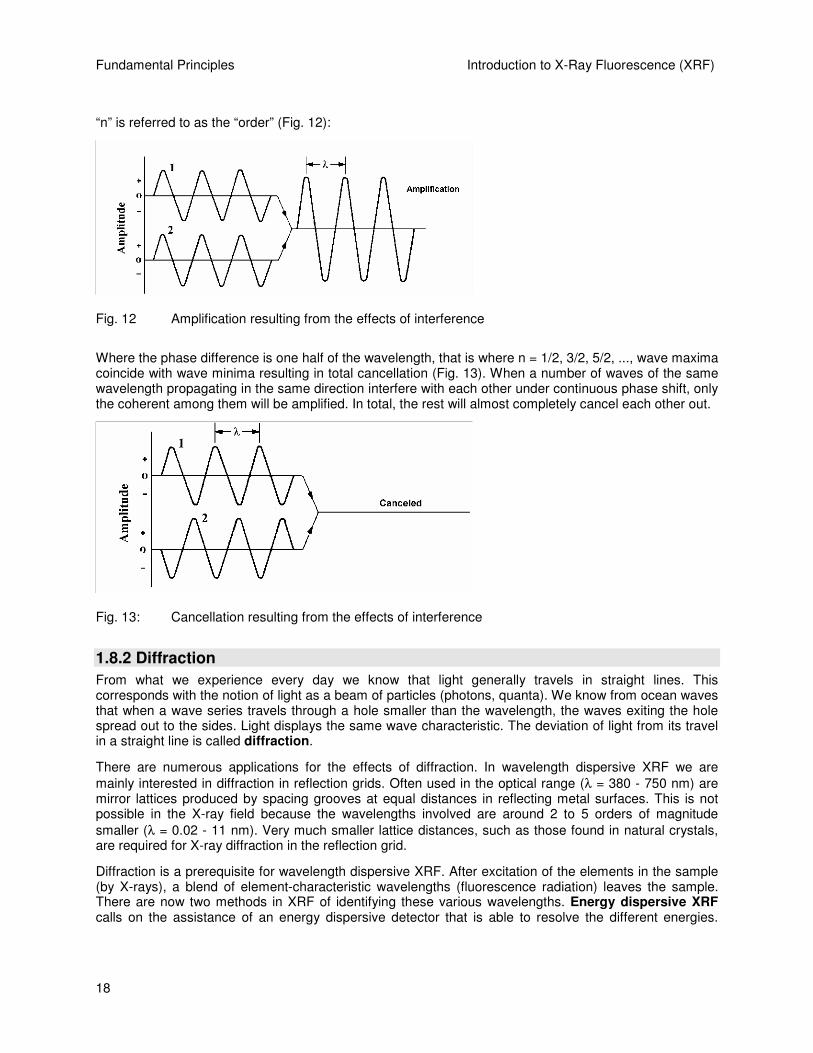
Electromagnetic radiation displays interference and diffraction effects due to the nature of its waves. “Interference” is the property of waves to overlap each other and, under certain circumstances, to cancel out or amplify each other.
Amplification takes place when waves of identical wavelength have zero phase difference (coherence), i.e. when "wave maxima" and "wave minima" overlap in such a way that minima meet minima and maxima meet maxima. This is precisely the case when the phase difference ∆λ∆λ∆λ∆λ is zero or a multiple of the wavelength λλλλ, i.e.:
λλ n=∆ n = 0, 1, 2, ...
Fundamental Principles Introduction to X-Ray Fluorescence (XRF)
18
“n” is referred to as the “order” (Fig. 12):
Fig. 12 Amplification resulting from the effects of interference
Where the phase difference is one half of the wavelength, that is where n = 1/2, 3/2, 5/2, ..., wave maxima coincide with wave minima resulting in total cancellation (Fig. 13). When a number of waves of the same wavelength propagating in the same direction interfere with each other under continuous phase shift, only the coherent among them will be amplified. In total, the rest will almost completely cancel each other out.
Fig. 13: Cancellation resulting from the effects of interference
1.8.2 Diffraction
From what we experience every day we know that light generally travels in straight lines. This corresponds with the notion of light as a beam of particles (photons, quanta). We know from ocean waves that when a wave series travels through a hole smaller than the wavelength, the waves exiting the hole spread out to the sides. Light displays the same wave characteristic. The deviation of light from its travel in a straight line is called diffraction.
There are numerous applications for the effects of diffraction. In wavelength dispersive XRF we are mainly interested in diffraction in reflection grids. Often used in the optical range (λ = 380 - 750 nm) are mirror lattices produced by spacing grooves at equal distances in reflecting metal surfaces. This is not possible in the X-ray field because the wavelengths involved are around 2 to 5 orders of magnitude smaller (λ = 0.02 - 11 nm). Very much smaller lattice distances, such as those found in natural crystals, are required for X-ray diffraction in the reflection grid.
Diffraction is a prerequisite for wavelength dispersive XRF. After excitation of the elements in the sample (by X-rays), a blend of element-characteristic wavelengths (fluorescence radiation) leaves the sample. There are now two methods in XRF of identifying these various wavelengths. Energy dispersive XRF calls on the assistance of an energy dispersive detector that is able to resolve the different energies.

Introduction to X-Ray Fluorescence (XRF) Fundamental Principles
19
Wavelength dispersive XRF utilizes diffraction effects of crystal to separate the various wavelengths. The detector subsequently determines the intensity of a particular wavelength. The procedure will be covered in detail in the following sections.
1.8.3 X-ray Diffraction From a Crystal Lattice, Bragg's Equation
Crystals consist of a periodic arrangement of atoms or molecules that form a crystal lattice. In such an arrangement of atoms you generally find numerous planes running in different directions through the lattice points (atoms, molecules), and not only horizontally and vertically but also diagonally. These are called lattice planes. All of the planes parallel to a lattice plane are also lattice planes and are a set distance apart from each other. This distance is called the lattice plane distance “d.”
When parallel X-rays strike a pair of parallel lattice planes, every atom within the planes acts as a scattering centre and emits a secondary wave. All of the secondary waves combine to form a reflected wave. The same occurs on the parallel lattice planes for only very little of the X-ray wave is absorbed within the lattice plane distance, d. All these reflected waves interfere with each other. If the amplification condition "phase difference = a whole multiple of wavelengths" (∆λ = nλ) is not precisely met, the reflected wave will interfere such that cancellation occurs. All that remains is the wavelength for which the amplification condition is precisely met. For a defined wavelength and a defined lattice plane distance, this is only given with a specific angle, the Bragg angle (Fig. 14).
Fig. 14 Bragg's Law
Under amplification conditions, parallel, coherent X-ray light (rays 1, 2) falls on a crystal with a lattice plane distanced d and is scattered below the angle θ (rays 1', 2'). The proportion of the beam that is scattered on the second plane has a phase difference of 'ACB' to the proportion of the beam that was scattered at the first plane. Following the definition of sine:
θsin''=
d
AC or θsin'' dAC =
The phase difference 'ACB' is twice that, so:
θsin2'' dACB =
The amplification condition is fulfilled when the phase difference is a whole multiple of the wavelength λ, so:
λnACB =''

Fundamental Principles Introduction to X-Ray Fluorescence (XRF)
20
This results in Bragg's Law:
nλλλλ=2d sinθθθθ Bragg's equation
n = 1, 2, 3 ... Reflection order
Fig. 15a: 1st order reflection: λ = 2d sin θ1
Fig. 15b: 2nd order reflection: 2λ = 2d sin θ2

Introduction to X-Ray Fluorescence (XRF) Fundamental Principles
21
Fig. 15c: 3rd order reflection: 3λ = 2d sin θ3
Figures 15a, b and c illustrate Bragg's Law for the reflection orders n = 1, 2, 3.
On the basis of Bragg's Law, by measuring the angle θ, you can determine either the wavelength λ, and thus chemical elements, if the lattice plane distance d is known or, if the wavelength λ is known, the lattice plane distance d and thus the crystalline structure.
This provides the basis for two measuring techniques for the quantitative and qualitative determination of chemical elements and crystalline structures, depending on whether the wavelength λ or the 2d-value is identified by measuring the angle θ (Table 3):
Table 3: Wavelength dispersive X-ray techniques
Known Sought Measured Method Instrument type
d λ θ X-ray fluorescence Spectrometer λ d θ X-ray diffraction Diffractometer
In X-ray diffraction (XRD) the sample is excited with monochromatic radiation of a known wavelength (λ) in order to evaluate the lattice plane distances as per Bragg's equation.
In XRF, the d-value of the analyzer crystal is known and we can solve Bragg's equation for the element-characteristic wavelength (λ).
1.8.4 Reflections of Higher Orders
Figures 15a-c illustrate the reflections of the 1st, 2nd, and 3rd order of one wavelength through the different angles θ1, θ2, and θ3. Here, the total reflection is made up of the various reflection orders (1, 2, ... n). The higher the reflection order, the lower the intensity of the reflected proportion of radiation. How great the maximum detectable order is depends on the wavelength, the type of crystal used and the angular range of the spectrometer.

Fundamental Principles Introduction to X-Ray Fluorescence (XRF)
22
It can be seen from Bragg's equation that the product of reflection order 'n = 1, 2, ...' and wavelength 'λ' for greater orders, and shorter wavelengths 'λ* < λ' that satisfy the condition 'λ* = λ/n', give the same result.
Accordingly, radiation with one half, one third, one quarter etc. of the appropriate wavelength (using the same type of crystal) is reflected through an identical angle 'θθθθ':
1λ = 2(λ/2) = 3(λ/3) = 4(λ/4) = ...
As the radiation with one half of the wavelength has twice the energy, the radiation with one third of the wavelength three times the energy etc., peaks of twice, three times the energy etc. can occur in the pulse height spectrum (energy spectrum) as long as appropriate radiation sources (elements) exist (Fig. 16).
Fig. 16 shows the pulse height distribution of the flow counter using the example of the element hafnium (Hf) in a sample with a high proportion of zircon. The Zr Kα1 peak has twice the energy of the Hf Lα1 peak and appears, when the Hf Lα1 peak is set, at the same angle in the pulse height spectrum.
Fig. 16: 2nd order reflection (n=2)
1.8.5 Crystal Types
The wavelength dispersive X-ray fluorescence technique can detect every element above the atomic number 4 (Be). The wavelengths cover the range of values of four magnitudes: 0.01 - 11.3 nm. As the angle θ can theoretically only be between 0° and 90° (in practice 2° to 75°), 'sin θ' only accepts values between 0 and +1. When Bragg's equation is applied:
1sin2
0 += ππ θλ
d
n
Introduction to X-Ray Fluorescence (XRF) Fundamental Principles
23
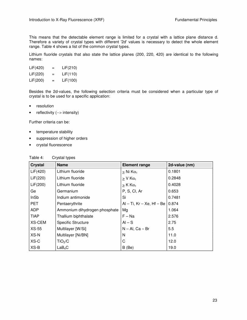
This means that the detectable element range is limited for a crystal with a lattice plane distance d. Therefore a variety of crystal types with different '2d' values is necessary to detect the whole element range. Table 4 shows a list of the common crystal types.
Lithium fluoride crystals that also state the lattice planes (200, 220, 420) are identical to the following names:
LiF(420) = LiF(210)
LiF(220) = LiF(110)
LiF(200) = LiF(100)
Besides the 2d-values, the following selection criteria must be considered when a particular type of crystal is to be used for a specific application:
• resolution
• reflectivity (--> intensity)
Further criteria can be:
• temperature stability
• suppression of higher orders
• crystal fluorescence
Table 4: Crystal types
Crystal Name Element range 2d-value (nm)
LiF(420) Lithium fluoride > Ni Kα1 0.1801
LiF(220) Lithium fluoride > V Kα1 0.2848
LiF(200) Lithium fluoride > K Kα1 0.4028
Ge Germanium P, S, Cl, Ar 0.653
InSb Indium antimonide Si 0.7481
PET Pentaerythrite Al – Ti, Kr – Xe, Hf – Be 0.874
ADP Ammonium dihydrogen phosphate Mg 1.064
TlAP Thallium biphthalate F – Na 2.576
XS-CEM Specific Structure Al – S 2.75
XS-55 Multilayer [W/Si] N – Al, Ca – Br 5.5
XS-N Multilayer [Ni/BN] N 11.0
XS-C TiO2/C C 12.0
XS-B LaB4C B (Be) 19.0

Fundamental Principles Introduction to X-Ray Fluorescence (XRF)
24
1.8.6 Dispersion, Line Separation
The extent of the change in angle ∆θ upon changing the wavelength by the amount ∆λ (thus: ∆θ/∆λ) is called “dispersion.” The greater the dispersion, the better the separation of two adjacent or overlapping peaks. Resolution is determined by the dispersion as well as by surface quality and purity of the crystal.
Mathematically, dispersion can be obtained from the differentiation of the Bragg equation:
θλ
θ
cos2d
n
d
d=
It can be seen from this equation that dispersion (or peak separation) increases as the lattice plane distance d decreases.
Examples:
The 2θ-values of the Kα1 peaks of vanadium (V) and chromium (Cr) are farther apart when measuring with LiF(220) than when measuring with LiF(200).
The 2θ−values of the Kα1 peaks of sulphur (S) and phosphorus (P) are farther apart when measuring with the Ge crystal than when measuring with the PET crystal.
Table 5: Explanatory details for dispersion
Crystal type 2d-value (nm) 2θθθθ (El1) (degrees) 2θθθθ (El2) (degrees) Difference (degrees)
LiF(220) 0.2848 107.11 (Cr) 123.17 (V) 16.06
LiF(200) 0.4028 69.34 (Cr) 76.92 (V) 7.58
Ge 0.653 110.69 (S) 141.03 (P) 30.34
PET 0.874 75.85 (S) 89.56 (P) 13.71
The following describes the characteristics of the individual crystal types divided into “standard crystals,” “multilayer crystals” and “special crystals.”
1.8.7 Standard Types, Multilayers
LiF(200), LiF(220), LiF(420)
LiF crystal types exist in a variety of lattice planes (200, 220, 420, etc.). In the sequence (200) --> (220) --> (420), resolution increases and reflectivity decreases (Fig. 17).

Introduction to X-Ray Fluorescence (XRF) Fundamental Principles
25
Fig. 17: Intensities of the crystals LiF(220) and LiF(420) in relation to LiF(200). (Intensity LiF(200) = 1)
LiF(200): A universally usable crystal for the element range atomic number 19 (K) onwards; high reflectivity, high sensitivity.
LiF(220): Lower reflectivity than LiF(200) but higher resolution; can be used for the element range atomic number 23 (V) onwards; particularly suitable for better peak separation where peaks overlap.
Examples of the application of the LiF(220) for reducing peak overlaps:
Cr Kα1,2 - V Kβ1
Mn Kα1,2 - Cr Kβ1
U Lα1 - Rb Kα1,2
LiF(420): One of the special crystals; can be used for the element range atomic number 28 (Ni or Co Kβ1) onwards; best resolution but low reflectivity.
Figure 17 shows a reflectivity of only 10% of that of LiF(200) for LiF(420) in the energy range around 10 keV.
PET: A universal crystal for the elements Al to Ti (K-peaks), Kr to Xe (L-peaks) and Hf to Bi (M-peaks).
ATTENTION
The PET is the crystal with the greatest heat-expansion coefficients, i.e. temperature fluctuations are most noticeable here.

Fundamental Principles Introduction to X-Ray Fluorescence (XRF)
26
Multilayers XS-55, XS-N, XS-C, XS-B Multilayers are not natural crystals but artificially produced “layer analyzers.” The lattice plane distances d are produced by applying thin layers of two materials in alternation onto a substrate (Fig. 18). Multilayers are characterized by high reflectivity and a somewhat reduced resolution. For the analysis of light elements the multilayer technique presents an almost revolutionary improvement for numerous applications in comparison to natural crystals with large lattice plane distances.
Fig. 18: Diffraction in the layers of a multilayer crystal
XS-55: The most commonly used multilayer with a 2d-value of 5.5 nm for analyzing the elements N to Al and Ca to Br; standard application for measuring the elements F, Na and Mg.
1.8.8 Special Crystals
The term “special crystals” refers to crystal types and multilayers that are not used universally but are employed in special applications.
LiF(420): See, for example, “standard types” description of the LiF crystals (200, 220 and 420).
Ge: A very good crystal for measuring the elements S, P and Cl. In comparison to PET, Ge is characterized by a higher dispersion and a more stable temperature behavior. Ge suppresses the peaks of the 2nd and 4th order, in particular.
Ge is especially suitable for differentiating between sulphide and sulphate, such as in samples of cement.
Introduction to X-Ray Fluorescence (XRF) Fundamental Principles
27
ADP: In practice, ADP is only used for the analysis of Mg and has a higher resolution with a significantly lower reflectivity compared to the multilayer XS-55. ADP is therefore required where interference peaks can occur such as in the case of low Mg concentrations in an Al matrix.
TlAP: TlAP has high resolution but low reflectivity and is recommended for analyzing F and Na if the resolution of XS-55 is insufficient (e.g. where Na is overlapped by the Zn-L peaks in Zn-rich samples).
DANGER
Disadvantages are the limited lifetime, toxicity, and high price.
InSb: InSb is a very good crystal for analyzing Si in traces as well as in higher concentrations (e.g. glass). It replaces PET and is used wherever high precision and great stability is required. The disadvantages are the limited use (only Si) and the high price.
XS-N: XS-N is a multilayer with a 2d-value of 11.0 nm, specially optimized for nitrogen.
XS-C: XS-C is a multilayer with a 2d value of 12.0 nm, specially optimized for carbon.
XS-B: XS-B is a multilayer with a 2d-value of 19.0 nm, specially optimized for boron and is equally suitable for the analysis of beryllium.
Which multilayer crystal is the most suitable for analyzing the very light elements? Fig. 19a shows that XS-B is the best crystal for analyzing boron (B), naturally with a corresponding coarse collimator (at least 2° opening). A compromise for analyzing boron is the XS-160 crystal when carbon (C) also needs to be measured with the same crystal.
For the analysis of carbon (C) the XS-C crystal provides a sharper peak and a better ratio of peak/background intensities, which means that better sensitivities can be achieved (Fig. 19b). To apply the XS-55 for analyzing carbon should be exceptional in case of having no XS-C or XS-160. Only very high concentrations (several tens of percent) of carbon can be determined with XS-C. In case of determining carbon with XS-55 using a “standardless“ precalibrated XRF routine, remember to select a very slow scanning speed (long measuring time) for carbon or to select the peak/background measurement mode.
Nitrogen (N) is best analyzed using XS-N. If needed, XS-55 can also be applied (Fig. 19c). XS-B, XS-C and XS -160 are not suitable for analyzing nitrogen.
Oxygen (O) and all “heavier“ light elements have to be analyzed with XS-55, which gives the best resolution and the best peak/background ratio (Fig. 19d).

Fundamental Principles Introduction to X-Ray Fluorescence (XRF)
28
B KA1,2 in BN (OVOs; 2,0°; 20kV/50mA)
05 (#) - B - - - - - -
Operations: Import [004]Immediate Measurement - Crystal: OVO-N - 2Th.0: 54,000 - 2T h.1: 94,000 - Coll imator: 2.00 degr. - Filter: none - Mask: 34 mm - kV: 20 - mA: 50 - Step Size: 0,200 - T ime.: 1,000Operations: Import [003]Immediate Measurement - Crystal: OVO-C - 2Th.0: 49,800 - 2T h.1: 89,800 - Coll imator: 2.00 degr. - Filter: none - Mask: 34 mm - kV: 20 - mA: 50 - Step Size: 0,200 - T ime.: 1,000Operations: Import [002]Immediate Measurement - Crystal: OVO-160 - 2T h.0: 29,800 - 2T h.1: 69,800 - Coll imator: 2.00 degr. - Filter: none - Mask: 34 mm - kV: 20 - mA: 50 - Step Size: 0,200 - T ime.: 1,000Operations: Import [001]Immediate Measurement - Crystal: OVO-B - 2Th.0: 18,900 - 2T h.1: 58,900 - Coll imator: 2.00 degr. - Filter: none - Mask: 34 mm - kV: 20 - mA: 50 - Step Size: 0,200 - T ime.: 1,000
Lin
(KC
ps)
0
0,1
0,2
0,3
0,4
0,5
0,6
0,7
0,8
0,9
1,0
1,1
1,2
1,3
1,4
1,5
1,6
1,7
1,8
1,9
2,0
2,1
2,2
2,3
2,4
2,5
2,6
2,7
2,8
2,9
3,0
3,1
3,2
3,3
3,4
3,5
3,6
3,7
3,8
3,9
4,0
Lin
(KC
ps)
0
0,1
0,2
0,3
0,4
0,5
0,6
0,7
0,8
0,9
1,0
1,1
1,2
1,3
1,4
1,5
1,6
1,7
1,8
1,9
2,0
2,1
2,2
2,3
2,4
2,5
2,6
2,7
2,8
2,9
3,0
3,1
3,2
3,3
3,4
3,5
3,6
3,7
3,8
3,9
4,0
SqE - Scale7,025 7,03 7,04 7,05 7,06 7,07 7,08 7,09 7,10
SqE - Scale
7,025 7,03 7,04 7,05 7,06 7,07 7,08 7,09 7,10
OVO-B
OVO-160
OVO-C
OVO-N
Fig. 19a: XS-B is the best multilayer crystal for analyzing boron (B).

Introduction to X-Ray Fluorescence (XRF) Fundamental Principles
29
C KA1,2 in Graphite (OVOs; 1,0°; 20kv/50mA)
06 (#) - C - - - - - - Operations: Import [005]Immediate Measurement - Crystal: OVO-55 - 2Th.0: 99,400 - 2Th.1: 119,400 - Collimator: 1.00 degr. - Filter: none - Mask: 34 mm - kV: 20 - mA: 50 - Step Size: 0,100 - T ime.: 1,000
Operations: Import [004]Immediate Measurement - Crystal: OVO-B - 2Th.0: 15,300 - 2Th.1: 35,400 - Coll imator: 1.00 degr. - Filter: none - Mask: 34 mm - kV: 20 - mA: 50 - Step Size: 0,100 - T ime.: 1,000Operations: Import [003]Immediate Measurement - Crystal: OVO-160 - 2Th.0: 22,300 - 2T h.1: 42,400 - Collimator: 1.00 degr. - Filter: none - Mask: 34 mm - kV: 20 - mA: 50 - Step Size: 0,100 - T ime.: 1,000Operations: Import [002]Immediate Measurement - Crystal: OVO-N - 2Th.0: 36,800 - 2Th.1: 56,900 - Coll imator: 1.00 degr. - Filter: none - Mask: 34 mm - kV: 20 - mA: 50 - Step Size: 0,100 - T ime.: 1,000Operations: Import [001]Immediate Measurement - Crystal: OVO-C - 2Th.0: 34,400 - 2Th.1: 54,400 - Coll imator: 1.00 degr. - Filter: none - Mask: 34 mm - kV: 20 - mA: 50 - Step Size: 0,100 - T ime.: 1,000
Lin
(KC
ps)
0
1
2
3
4
5
6
7
8
9
10
11
12
13
14
15
16
17
18
19
20
21
22
23
24
25
26
27
28
29
30
31
32
33
34
35
36
37
38
39
40
Lin
(KC
ps)
0
1
2
3
4
5
6
7
8
9
10
11
12
13
14
15
16
17
18
19
20
21
22
23
24
25
26
27
28
29
30
31
32
33
34
35
36
37
38
39
40
SqE - Scale6,90 6,91 6,92 6,93 6,94 6,95 6,96 6,97 6,98 6,99 7,00 7,01 7,02 7,03
SqE - Scale
6,90 6,91 6,92 6,93 6,94 6,95 6,96 6,97 6,98 6,99 7,00 7,01 7,02 7,03
OVO-160
OVO-C
OVO-N
OVO-55
OVO-B
Fig. 19b: The XS-C multilayer crystal is suitable for the determination of carbon.

Fundamental Principles Introduction to X-Ray Fluorescence (XRF)
30
N KA1,2 in NOPS (OVOs; 1,0°; 20kV/50mA)
07 (#) - N - - - - - - Operations: Import [005]Immediate Measurement - Crystal: OVO-55 - 2Th.0: 60,400 - 2Th.1: 80,400 - Col limator: 1.00 degr. - Fil ter: none - Mask: 34 mm - kV: 20 - mA: 50 - Step Size: 0,100 - T ime.: 1,000
Operations: Import [004]Immediate Measurement - Crystal: OVO-B - 2Th.0: 14,052 - 2Th.1: 27,852 - Coll imator: 1.00 degr. - Filter: none - Mask: 34 mm - kV: 20 - mA: 50 - Step Size: 0,100 - T ime.: 1,000Operations: Import [003]Immediate Measurement - Crystal: OVO-C - 2Th.0: 20,900 - 2Th.1: 41,000 - Coll imator: 1.00 degr. - Filter: none - Mask: 34 mm - kV: 20 - mA: 50 - Step Size: 0,100 - T ime.: 1,000Operations: Import [002]Immediate Measurement - Crystal: OVO-160 - 2Th.0: 14,052 - 2T h.1: 32,652 - Collimator: 1.00 degr. - Filter: none - Mask: 34 mm - kV: 20 - mA: 50 - Step Size: 0,100 - T ime.: 1,000Operations: Import [001]Immediate Measurement - Crystal: OVO-N - 2Th.0: 22,600 - 2Th.1: 42,700 - Coll imator: 1.00 degr. - Filter: none - Mask: 34 mm - kV: 20 - mA: 50 - Step Size: 0,100 - T ime.: 1,000
Sqr
(K
Cps
)
0
0,001
0,01
0,1
1
0,2
0,3
0,4
0,5
0,6
2
3
Sqr
(K
Cps
)
0
0,001
0,01
0,1
1
0,2
0,3
0,4
0,5
0,6
2
3
SqE - Scale6,837 6,84 6,85 6,86 6,87 6,88 6,89 6,90 6,91
SqE - Scale
6,837 6,84 6,85 6,86 6,87 6,88 6,89 6,90 6,91
OVO-N
OVO-55
OVO-B
OVO-C
OVO-160
Fig. 19c: Nitrogen (N) is best analyzed using XS-N.

Introduction to X-Ray Fluorescence (XRF) Fundamental Principles
31
O KA1,2 in NOPS (OVOs; 0,46°; 20kV/50mA)
08 (#) - O - - - - - -
Operations: Import [004]Immediate Measurement - Crystal: OVO-C - 2Th.0: 18,300 - 2Th.1: 27,592 - Coll imator: 0.46 degr. - Filter: none - Mask: 34 mm - kV: 20 - mA: 50 - Step Size: 0,046 - T ime.: 1,000Operations: Import [003]Immediate Measurement - Crystal: OVO-N - 2Th.0: 19,600 - 2Th.1: 28,892 - Coll imator: 0.46 degr. - Filter: none - Mask: 34 mm - kV: 20 - mA: 50 - Step Size: 0,046 - T ime.: 1,300Operations: Import [002]Immediate Measurement - Crystal: OVO-160 - 2Th.0: 14,025 - 2T h.1: 21,477 - Collimator: 0.46 degr. - Filter: none - Mask: 34 mm - kV: 20 - mA: 50 - Step Size: 0,046 - T ime.: 1,000Operations: Import [001]Immediate Measurement - Crystal: OVO-55 - 2Th.0: 46,400 - 2Th.1: 55,692 - Col limator: 0.46 degr. - Filter: none - Mask: 34 mm - kV: 20 - mA: 50 - Step Size: 0,046 - T ime.: 1,000
Lin
(KC
ps)
0
0,1
0,2
0,3
0,4
0,5
0,6
0,7
0,8
0,9
1,0
1,1
1,2
1,3
1,4
1,5
1,6
1,7
1,8
1,9
2,0
2,1
2,2
2,3
2,4
2,5
2,6
2,7
2,8
2,9
3,0
3,1
3,2
3,3
3,4
3,5
3,6
3,7
3,8
3,9
4,0
Lin
(KC
ps)
0
0,1
0,2
0,3
0,4
0,5
0,6
0,7
0,8
0,9
1,0
1,1
1,2
1,3
1,4
1,5
1,6
1,7
1,8
1,9
2,0
2,1
2,2
2,3
2,4
2,5
2,6
2,7
2,8
2,9
3,0
3,1
3,2
3,3
3,4
3,5
3,6
3,7
3,8
3,9
4,0
SqE - Scale6,712 6,72 6,73 6,74 6,75 6,76 6,77 6,78 6,79 6,80 6,81 6,82
SqE - Scale
6,712 6,72 6,73 6,74 6,75 6,76 6,77 6,78 6,79 6,80 6,81 6,82
OVO-C
OVO-160
OVO-N
OVO-55
Fig. 19d: Oxygen (O) and all “heavier“ light elements have to be analyzed with XS-55.
1.8.9 Curved Crystals
Whereas flat crystals are used in sequence spectrometers, multichannel spectrometers principally employ curved crystals.
The curvature of the crystals is selected in such a way that by applying slit optics the X-ray entrance slit is focused by the curved crystal onto the exit slit. This allows higher intensities in a space-saving geometric arrangement.
Different types of crystal curvature are used for focusing. The most commonly used are the curvatures that follow a logarithmic spiral (Fig. 20a) and the Johansson curvature (including grinding) (Fig. 20b).

Fundamental Principles Introduction to X-Ray Fluorescence (XRF)
32
Fig. 20a: Logarithmic spiral curvature Fig. 20b: Johansson curvature

Introduction to X-Ray Fluorescence (XRF) Instrumentation
33
2. Instrumentation
This chapter explains the instrumentation in Bruker AXS X-ray fluorescence spectrometers. The first three sections contain brief summaries of multichannel X-ray spectrometers. The fourth section deals in detail with the technology of sequential spectrometers.
2.1 Multichannel Spectrometers
The MRS multichannel spectrometer can measure up to 28 elements simultaneously. A multichannel spectrometer is always required when short measurement times are needed to analyze large numbers of elements, or when high sample throughput (e.g. 600 samples per day) is needed for industrial quality and process control.
An individual measuring channel, comprising crystal, detector and electronics module, must be installed for each element line. As there are limited possibilities for the geometric arrangement of 28 channels in close proximity to the sample, so-called monochromators with slit-optics are used. A monochromator is an arrangement of an entry slit, curved focusing crystal and an exit slit (Figs. 21 and 22). The crystals are curved in a logarithmic spiral and focus the desired wavelength of the beam passing through the entry slit onto the exit slit. The detector is located behind the exit slit. Scintillation counters or gas proportional counters are used depending on the element line. Flow counters and sealed proportional counters can be used as gas proportional counters. Sealed proportional counters can be equipped with a 25-µm Be or a Super-High Transmission (SHT) window. The 25-µm Be window is used for the elements Al to Fe. The SHT window is used for Be to Mg.
All monochromators are located in a large vacuum chamber. The beam is applied from above. The fixed channels are used exclusively for quantitative analyses. A scanner can be employed for qualitative analysis.
As all elements are measured simultaneously, a generator setting (kV/mA) must be selected that provides the best compromise for all the elements to be measured. The measurement time depends on statistical accuracy requirements of the element with the lowest intensity and is typically between 20 and 60 seconds. No background positions can be measured because the monochromators are at a fixed location in the angle for the corresponding line.
When measuring trace and major elements simultaneously, the generator is normally set so that the trace elements can be measured with the highest possible intensity. This means that the major elements are usually of such high intensity that the detectors cannot process them. For cases such as these, the MRS can be fitted with absorbers (attenuators) for the major elements. Absorbers sufficiently reduce the intensities of major elements so that they then lie in the operational range of the detectors.

Instrumentation Introduction to X-Ray Fluorescence (XRF)
34
Fig. 21: Beam path in multichannel spectrometers
Fig. 22: Monochromator with absorber and flow counter
2.1.1 Scanners for Multichannel Spectrometers
In addition to fixed channels, a scanner can be installed in the vacuum chamber of multichannel spectrometers. The scanner is a “moveable channel” enabling sequential coverage of a large element range. As only a single curved crystal (LiF(200) or PET) is fitted and the scanner's 2θ angular range is limited (30 - 120 degrees), several elements in the periodic table must be measured in the second reflection order. A flow counter or a sealed proportional counter serves as a detector.
The scanner works on the physical principle of the Rowland Circle, in which the crystal and detector move in such a way that the entry slit, crystal and exit slit lie on a fixed-radius circle that changes in position (Fig. 23).
The scanner can be used for qualitative as well as quantitative analyses.

Introduction to X-Ray Fluorescence (XRF) Instrumentation
35
Fig. 23: The Rowland Circle scanner principle
2.2 Sequential Spectrometers
The heart of the SRS 3x00, S4 EXPLORER, S4 PIONEER and S8 TIGER sequential spectrometers is a high-precision goniometer with two independent stepper motors for separate θ/2θ drive.
Several microprocessors control and monitor the functions and processes inside the spectrometer. A master processor coordinates the internal flow of information and communicates with the external analysis computer. Having its own service interface allows the master processor to be remotely diagnosed by Bruker AXS Service Engineers, without their being able to access proprietary data in the analysis computer. This concept enables rapid location and diagnosis of operational issues.
The various measuring parameters are set exclusively via the analysis computer’s software and provide the user with great flexibility.
Besides extending the adjustment parameters of the primary beam filter, the crystal and collimator changer beyond those possible on the SRS 303, the detector high-voltages, too, are set via the analysis computer.
The separate θ/2θ goniometer drive with two independent stepper motors allows precise θ/2θ−angle alignment via the analysis computer’s software.
The flow counter is situated inside the spectrometer chamber and has an angle range of 2° to 148°. Located behind the flow counter and outside the chamber, separated by a 0.1 mm Al foil, is the scintillation counter with an angle range of 2° to 110°. Both detectors can be used individually or in tandem. In tandem operation, the intensity in the flow counter is measured as well as the radiation that passes through the flow counter and the radiation that is absorbed by the scintillation counter.
Tandem operation was excluded from the S4 EXPLORER and the S4 PIONEER sequential spectrometers to save space. In addition, the scintillation counter is located in the spectrometer chamber next to the proportional counting tube.

Instrumentation Introduction to X-Ray Fluorescence (XRF)
36
Integrating temperature measurement points allows this stability-relevant factor to be checked in the instrument. Furthermore, the temperature of the water in the internal deionized cooling system is kept constant.
An optional protractable/retractable foil screen can be installed between the sample chamber and the spectrometer chamber for measuring, for example, liquids in a He atmosphere.
Fig. 24 shows the beam path and adjustable parameters of the S4 EXPLORER, S4 PIONEER, and S8 TIGER.
A flexible, modifiable sample changer with a robot arm that moves in the directions X and Y allows fully automatic transport of samples to the instrument’s entry position, including:
• sample cups with a grab
• bare samples with a suction unit
• steel rings with a magnetic holder
The SRS 3000 was the first X-ray spectrometer with an X-Y sample magazine, thereby setting the standard for all subsequent developments. An internal 2-sample changer enables rapid processing of samples in stack operation without time loss through transporting samples. While one sample is being measured, the next is being inserted into the pre-vacuum chamber.
Fig. 24 Beam path in the S4 EXPLORER, S4 PIONEER and S8 TIGER
Problem-free docking onto a conveyor belt allows easy integration of S4 spectrometers into an automated environment.
The function and possible settings for the various parameters will now be described in the order they are encountered by the beam propagating from the tube to the scintillation counter.
X-ray Tube
Primary Beam Filters
Sample Vacuum Seal
Collimators
Analyzer Crystals
Scintillation Counter
Proportional Counter
Introduction to X-Ray Fluorescence (XRF) Instrumentation
37
2.3 Incident Beam Components
2.3.1 The End-window Tube and Generator
The X-ray tube and generator are designed for a permanent output of 4 kW at a maximum high voltage of 60 kV and a maximum tube current of 100 mA or 150 mA. The combination of high voltage and tube current must not exceed 4 kW, e.g. at 4 kW maximum:
• 27 kV, 150 mA = 4.05 kW
• 30 kV, 134 mA = 4.02 kW
• 40 kV, 100 mA = 4.00 kW
• 50 kV, 80 mA = 4.00 kW
• 60 kV, 67 mA = 4.02 kW
The minimum setting is 20 kV, 5 mA for a power of 0.1 kW.
NOTE
The control and analysis software SPECTRA AT, SPECTRA 3000, and SPECTRAplus checks the settings and prevents the maximum permissible values from being exceeded.
Rhodium is used as the standard anode material. The light elements Be to Cl are effectively excited by the Rh-L beam’s high transmission rate through the tube’s 75-µm Be window. The characteristic Rh-K radiation excites the elements up to Mo (Ru) (see Table 2). The elements from Rh onwards are excited by the Bremsstrahlung’s high-energy “tail.”
Four-kW tubes with other anode materials (e.g. Mo, W, Au, Cr) can be used for special applications.
2.3.2 The Primary Beam Filter
The primary beam filter is seated on a changer for 10 positions (including vacant positions) and is equipped with a selection of absorber foils. It is located between the tube and the sample, filters out undesirable or interfering components of the tube radiation for certain applications and improves the signal-to-noise ratio. Al and Cu foils, for example, are used as absorbers in a variety of thicknesses. Primary beam filters can be selected to suit individual requirements when purchasing the instrument.
When measuring Rh K with the Rh tube, the characteristic Rh radiation coming from the tube must be filtered out because it would otherwise be measured as a result of elastic scattering on the sample. By using a 0.2-mm Cu filter, the characteristic Rh tube radiation is largely absorbed prior to reaching the sample. The measurement must be taken with a tube high-voltage of 60 kV as the Rh in the sample is only excited by the high-energy Bremsstrahlung.
Fig. 25 illustrates the tube spectrum acting on the sample without a primary beam filter and tube high-voltage of 60 kV.
Fig. 26 shows the reduction in Rh radiation scattering on a plant sample using different primary beam filters made of Cu or Al.

Instrumentation Introduction to X-Ray Fluorescence (XRF)
38
Fig. 25: Cd and Rh peaks without a copper primary beam filter
Fig. 26: Cd and Rh peaks with a 0.2- or 0.3-mm copper primary-beam filter
Fig. 27: The effect of the aluminum primary beam filter for optimizing the peak/background ratio

Introduction to X-Ray Fluorescence (XRF) Instrumentation
39
When analyzing a sample of very pure graphite, peaks of the elements Cr, Fe, Ni and Cu can occur in the 2θ-spectrum although the sample contains none of these elements. The Cu peak originates from the excitation of the collimator material that mainly consists of copper. The Cr, Fe and Ni peaks are called “spectral impurities” of the tube. If the elements Cr, Fe and Ni are to be measured as traces, it may be advantageous to use the 0.2-mm Al filter to absorb these components.
2.3.3 Sample Cups, the Cup Aperture
In the S4 EXPLORER and S4 PIONEER, the sample to be measured is first transported into the sample changer’s pre-vacuum chamber and then into the measuring position where it rotates at up to 30 revolutions per minute, depending on the application, to even out sample inhomogeneity. The collimator mask’s optimized screening allows steel apertures to be used in the majority of cases. Other aperture diameters and materials are available upon request.
Depending on the type of sample magazine, samples have to be placed in the cups either manually (magazine with grab) or automatically. Automatic sample magazines include those designed for “bare samples” (magazine with sucker) or those designed for steel rings (magazine with magnet). When using thin filters for measurements, an anti-background scattering cup must be used to eliminate the fixing plate radiation.
2.4 Emitted Beam Components
2.4.1 The Vacuum Seal
When measuring liquids or loose powder samples in a helium atmosphere, the vacuum can be maintained in the spectrometer volume by inserting a thin separating, or sealing, foil between the sample chamber and the spectrometer chamber. This causes the separating foil to absorb less radiation than would be the case if the spectrometer chamber were filled with helium.
For alternate measurements of samples in a vacuum and in helium, the smaller sluicing volume reduces helium consumption and considerably speeds up the change from one operating mode to another.
2.4.2 Collimator Masks
Collimator masks are situated between the sample and collimator and serve the purpose of cutting out the radiation coming from the edge of the cup aperture. The size of the mask is generally adapted to match the cup aperture being used. The SRS 3x00’s changer has 4 positions; the SRS 300 and 303 are equipped with a 3-position changer. The S4 EXPLORER, S4 PIONEER and S8 TIGER employ collimator masks that are set very close to the sample and therefore optimally screen the sample cup aperture.
2.4.3 Collimators, the Soller Slit
Collimators consist of a row of parallel slats. They are used to select a parallel beam of X-rays coming from the sample and striking the crystal. The spaces between the slats determine the degree of parallelism and thus the angle resolution of the collimator.
Sequential spectrometers are fitted with a 4-position changer. Besides the standard collimators with aperture angles of 0.15°, 0.23° and 0.46°, two additional collimators can be installed to optimize measurements for a particular application. A 0.77° collimator is available for high-resolution measurements (e.g. with LiF(420)). Collimators with a low resolution (e.g. 1.5° to 2.0°) are advantageous for light elements such as Be, B and C since the XS multilayer crystal’s angle resolution is limited. Using a

Instrumentation Introduction to X-Ray Fluorescence (XRF)
40
low-resolution collimator increases the intensity significantly. This enables intensity to be increased without a loss in angle resolution when analyzing the light elements (Fig. 28).
Fig. 28: Example of the influence of collimator resolution on the intensity of a light element
2.4.4 The Crystal Changer
The sequential spectrometer’s crystal changer can hold up to 8 crystals and can be custom-equipped to suit the requirements of specific applications.
2.4.5 The Flow Counter
The flow counter is located inside the vacuum chamber and has an entrance window made of a thin, coated foil with a thickness of 0.6 µm or 0.3 µm.
The thick entrance window allows optimum measurement of the light elements Be to Na. Fig. 29 illustrates the permeability for a variety of counting tube foils that were used in the SRS 200 and 300. It can be seen from the transmission curve that the permeability of the 1-µm polypropylene foil for Na is around twice that of the 2-µm Makrofol foil.

Introduction to X-Ray Fluorescence (XRF) Instrumentation
41
ATTENTION
As plastic foils have a high proportion of carbon, the absorption of nitrogen radiation close to the C absorption edge is very high. This means that even the 1 µm foil only has about 10% permeability for N. For this reason, the measured intensities for N are relatively low. The newer 0.6 µm and especially the 0.3 µm foils are more permeable to N radiation.
Generally, Ar + 10% CH4, also known as “P10 gas,” is used as the counting gas. The flow of counting gas is held constant in the instrument as a fluctuating counting-gas density in the counting tube would cause fluctuations in the absorption depth as well as fluctuations in the gas amplification and thus in the position of the peaks in the pulse height spectrum too (refer to Fig. 8).
The high voltage at the counting wire is set higher for light elements (XS Multilayer crystals) than for measuring the K-radiation (LiF crystals) of medium and heavy elements. In sequential spectrometers the detector high voltage is set separately for each element range and thus for each installed crystal (see also Fig. 8).
2.4.6 The Sealed Proportional Counter
The S4 EXPLORER, S4 PIONEER and S8 TIGER utilize a sealed proportional counter, thus enabling even the very light elements (Be – Na) to be determined as effectively as with a flow counter. The basis for this is the Super High Transmission (SHT) window material.
Fig. 29: X-ray transmission for various counting-tube foils

Instrumentation Introduction to X-Ray Fluorescence (XRF)
42
2.4.7 The Scintillation Counter
The scintillation counter is positioned behind the flow counter outside the vacuum chamber. The radiation measured inside it must pass through the flow counter, a 0.1-mm thick vacuum-chamber sealing foil and a 0.2-mm Be entrance window. It therefore makes sense to use the scintillation counter for energies above 4.5 keV (Cr Kα1) as lower energies are absorbed mainly in the flow counter (see Fig. 9).
The scintillation counter’s angle range is 4° to 110° (SRS 30x: 4° to 90°).
In the S4 EXPLORER, S4 PIONEER and S8 TIGER, the scintillation counter is directly beside the proportional counter in the spectrometer chamber and can be moved between 0° and 115°.
2.5 Electronic Pulse Processing
The pulses produced in the detectors by X-rays are processed and counted by subsequent electronic processing. The flow counter’s signals are electronically amplified in a preamplifier, shaped and further processed as voltage pulses in a main amplifier (sine amplifier) and discriminator. After the photomultiplier, the scintillation counter’s signals are fed directly into a main amplifier and discriminator.
2.5.1 The Discriminator
Depending on the application, higher order peaks or other sources of interference appear in the pulse height distribution, i.e. the detector’s energy spectrum with different levels of energy (see also Figs. 10a, 10b and 11a). A discriminator window is used to set a lower and an upper pulse-height threshold. Only the pulse heights that lie within these limits are counted. In this way, higher order peaks or interference radiation with pulse heights beyond the window are suppressed (see Fig. 16). Discriminating undesirable pulses reduces the background.
2.5.2 Main Amplifier, Sine Amplifier
After the preamplifier (flow counter and proportional counter), or photomultiplier (scintillation counter), the pulses are further enhanced electronically in a main amplifier. As the detectors' high voltages are set separately for each crystal, the electronic additional amplification must also be made dependent on the crystal used.
X-ray energies, for example, of 3.3 keV to 30 keV (potassium to iodine) are detectable with LiF(200). To have each one of the set peaks in the pulse height spectrum always appear in the same place (i.e. have identical pulse height), the electronic amplification must be linked to the goniometer’s angle setting. This is achieved by making the main amplifier’s amplification factor V for the appropriate crystal (2d value) and the selected reflection order (n) dependent on the sine of the adjustment angle:
V ∼ sin θ
This ensures that a discriminator window, once set for a crystal, will be applicable for all detectable energies. A main amplifier coupled in this way is called a sine amplifier.
2.5.3 Dead Time Correction
Spectrometer electronics need a certain amount of time to process a pulse, during which no other pulse can be registered. This period is called counter channel dead time for an individual pulse. As the pulse formation is different for the flow counter and the scintillation counter, the dead times (typically 300 to 400 ns) are also different for each detector. Total dead time is the result of an individual pulse multiplied by

Introduction to X-Ray Fluorescence (XRF) Instrumentation
43
the pulse rate. As the measured pulses occur statistically distributed over time, the proportion of pulses occurring during the processing period of a previously registered pulse depends on the intensity of the radiation, i.e. the total dead time increases due to the increase of the intensity. This results in a non-linear rise of the measured intensity with the intensity irradiated in the detector. The greater the incident intensity, the greater the losses during measurement. Fig. 30 illustrates how the dead time is dependent on the increasing incident intensity (increasing generator current). The curve flattens out distinctly at high measured intensities.
A correction of the measured intensities is necessary to produce a linear relationship between incident and measured intensities. A dead time correction can be made in the analysis computer. Fig. 31 shows the dead time corrected measurement points. A useful correction can be obtained in tandem operation up to an incident intensity of approximately 1,200 kcps per detector. Greater intensities are not worthwhile.
For routine operation it is recommended not to exceed an intensity of approximately 400 kcps per detector.
Fig. 30: The dead time effect
Fig. 31: Dead time corrected readings

Instrumentation Introduction to X-Ray Fluorescence (XRF)
44
2.5.4 Line-shift Correction
Line-shift correction is only important for the flow counter and the proportional counter at high intensities. Line shifts are noticeable when a peak in the pulse height spectrum shifts to lower values at high counting rates. The reason for this is that high counting rates in the detector volume between the flow counter’s cathode and counting wire build up a space charge that causes a temporary reduction of the effective high voltage and thus the reduction of gas amplification.
Shifts of pulse heights to lower values are automatically corrected by the electronics. The correction can be switched on and off in the software (SPECTRA 3000 and SPECTRAplus).

Introduction to X-ray Fluorescence (XRF) Sample Preparation Techniques
45
3. Sample Preparation Techniques
3.1 Introduction
X-ray fluorescence (XRF) analysis is a fast, non-destructive and environmentally friendly analysis method with very high accuracy and reproducibility. All elements of the periodic table from beryllium to californium can be measured qualitatively, semi-quantitatively and quantitatively in powders, solids and liquids. Concentrations of up to 100% are analyzed directly, without any dilution, with reproducibilities better than ±0.1%. Typical limits of detection are from 0.1 to 10 ppm. Most modern X-ray spectrometers with modular sample changers enable fast, flexible sample handling and adaptation to customer-specific automation processes.
XRF samples can be solids such as glass, ceramic, metal, rock, coal, or plastic. They can also be liquids, like petrol, oil, paint, solutions, blood or even wine. With an XRF spectrometer both very small concentrations of very few ppm and very high concentrations of up to 100% can be analyzed directly without any dilution process. Based on its simple and fast sample preparation requirements, XRF analysis is a universal analysis method, that has been widely accepted in the fields of research and industrial process control. XRF is particularly effective for complex environmental analysis and for production and quality control of intermediate and end products.
The quality of sample preparation for XRF analysis is at least as important as the quality of measurements.
An ideal sample is prepared so that it is:
• representative of the material
• homogeneous
• thick enough to meet the requirements of an infinitely thick sample
• without surface irregularities
• composed of small enough particles for the wavelengths to be measured
With XRF it is not necessary to bring solid samples into solution and then dispose of solution residues, as is the case with all wet-chemical methods. The main prerequisite for exact and reproducible analysis is a plain, homogeneous and clean analysis surface. For analysis of very light elements, e.g. beryllium, boron and carbon, the fluorescence radiation to be analyzed originates from a layer whose thickness is only a few atom layers to a few tenths of micrometer and which strongly depends on the sample material (Table 1). Careful sample preparation is therefore extremely important for analysis of light elements.

Sample Preparation Techniques Introduction to X-ray Fluorescence (XRF)
46
Table 1: Layer thickness (h, in µm), from which 90% of the fluorescence radiation originates
Sample matrix Graphite Glass Iron Lead
Line h_(90%, µm) h_(90%, µm) h_(90%, µm) h_(90%, µm)
U Lα1 28000 1735 154 22.4
Pb Lβ1 22200 1398 125 63.9
Hg Lα1 10750 709 65.6 34.9
W Lα1 6289 429 40.9 22.4
Ce Lβ1 1484 113 96.1 6.72
Ba Lα1 893 71.3 61.3 4.4
Sn Lα1 399 44.8 30.2 3.34
Cd Kα1 144600 8197 701 77.3
Mo Kα1 60580 3600 314 36.7
Zr Kα1 44130 2668 235 28.9
Sr Kα1 31620 1947 173 24.6
Br Kα1,2 18580 1183 106 55.1
As Kβ1 17773 1132 102 53
Zn Kα1,2 6861 466 44.1 24
Cu Kα1,2 5512 380 36.4 20
Ni Kα1,2 4394 307 29.8 16.6
Fe Kα1,2 2720 196 164 11.1
Mn Kα1,2 2110 155 131 9.01
Cr Kα1,2 1619 122 104 7.23
Ti Kα1,2 920 73.3 63 4.52
Ca Kα1,2 495 54.3 36.5 3.41
K Kα1,2 355 40.2 27.2 3.04
Cl Kα1,2 172 20.9 14.3 2.19
S Kα1,2 116 14.8 10.1 4.83
Si Kα1,2 48.9 16.1 4.69 2.47
Al Kα1,2 31.8 10.5 3.05 1.7
Mg Kα1,2 20 7.08 1.92 1.13
Na Kα1,2 12 5.56 1.15 0.728
F Kα1,2 3.7 1.71 0.356 0.262
O Kα1,2 1.85 2.50 0.178 0.143
N Kα1,2 0.831 1.11 0.08018 0.07133
C Kα1,2 13.6 0.424 0.03108 0.03124
B Kα1,2 4.19 0.134 0.01002 0.01166

Introduction to X-ray Fluorescence (XRF) Sample Preparation Techniques
47
Massive materials, such as rocks, soils, and slags have to be crushed, e.g. in a crushing device. The crushed material is afterwards very finely pulverized in a vibrating disk mill or planetary ball mill in order to be suitable for analysis. The grain size should then be less than 50 µm. In earlier times, in the cement industry, milling in suspension was preferred. Nowadays, in fully automatic sample preparation, it is dry milled using milling and binding agents in order to reach grain sizes smaller than 15 µm. The smaller and more equally grain-sized the sample, the more homogeneous the pressed powder sample will be. Through pressing under 10 to 20 t with pressing times of 5 to 10 seconds, the sample powder is pressed into aluminum cups, steel rings or directly with or without a binding agent. Rotating the sample holder in the spectrometer (at 30 r/min) compensates for any inhomogeneities of the sample.
Important for XRF analysis of powder samples is the homogeneity and the fineness of the sample powder, not only in the case of very light elements, but also for analysis of Si in samples containing quartz. Reproducibility of the samples is also very important, as XRF analysis is a comparing analysis method. This means that all unknown samples measured in comparison to a calibration curve should show the same grain size distribution as the standard samples used for calibration.
With high requirements for sample homogeneity, fused beads are produced from the sample powder at 1100 to 1200°C. Melting of the sample powder is supported by melting agents (e.g. lithium tetraborate) and, if necessary, by oxidation agents, which are mixed into the sample in different ratios. In earlier times, the melting method was preferred because of the decreased influence of the matrix effects due to the dilution of the sample matrix with the “lighter” melting agents. Nowadays very efficient PC programs are available for calculation of the correction coefficients (“theoretical alphas”) for matrix correction. These programs are also successfully used for pressed pellets.
For this reason, only the requirement for a very high analytical accuracy now speaks against quicker and more inexpensive pressing of sample powder. With careful sample preparation (milling), sufficient accuracy even with pressed pellets can often be reached.
Since X-ray spectrometry is essentially a comparative method of analysis, it is vital that all standards and unknown samples are presented to the spectrometer in a reproducible and identical manner. Any method of sample preparation must give specimens which are reproducible and which, for a certain calibration range, have similar physical properties including mass absorption coefficient, density and particle size. In addition, the sample preparation method must be rapid and inexpensive and must not introduce significant systematic errors, such as the introduction of trace elements from contaminants in a diluent.

Sample Preparation Techniques Introduction to X-ray Fluorescence (XRF)
48
3.2 Preparation of Solid Samples
3.2 1 Metals
Preparation of metal samples must be simple, rapid and reproducible. Usually, metal samples are prepared as solid disks by conventional methods of machining: cutting, milling and polishing. Grinding is used in the case of hard alloys and brittle materials such as ceramics.
The best polishing operations require very fine abrasives to produce the scratch-free surface necessary for most analyses, and a mirror-like surface if the sample is to be analyzed for light elements. The surface finish is of prime importance because polishing striations give rise to the so-called shielding effect, which results in a decrease in fluorescence intensities. As expected, the decrease in intensity is more important for lighter elements when the primary radiation is perpendicular to the striations and weaker when they are parallel to them. Modern spectrometers are equipped with spinning sample holders to smooth out the influence of sample orientation, resulting in observed intensities on samples and standards that are reproducible.
However, the shielding effect may still be present; sample rotation will compensate for it only if the magnitude of the effect is the same for standards and samples. Striations therefore must be of the same size and the compositions of the standards and samples must be similar (same effective wavelength).
In practice, striation depths of 100 µm are acceptable for elements with characteristic lines of short wavelengths, but striations deeper than few µm may significantly impair the accuracy of Si, Al and Mg measurements.
Very fine grits of Al2O3, SiC, B6C (80 to 120 grits) are commonly used to obtain the desired surface finish for most metals (Fe, Ni and Co bases).
Mechanical polishing may be undesirable for soft, malleable, multiphase alloys because of smearing of the softer components. The intensities of the elements in softer phases increase while those of the harder phases decrease. In such cases, special precautions must be taken during milling and final polishing of metals with Pb, Cu, Al, Zn or Sn bases.
Polishing may be a source of contamination since currently used abrasives, SiC and Al2O3, contain two elements that are often determined in commercial alloys. Sample surface cleaning may be necessary to remove contamination as well as grease and handling residue.
3.2.2 Pressed Pellets
Since powders are not affected by particle size limitations, the quickest and simplest method of sample preparation is to press the powders directly into pellets of equal density, with or without the use of a binder. In general, provided that the powder particles are less than about 50 µm in diameter, the sample will pelletize at 10 to 30 t. Where the self-binding properties of the powder are poor, higher pressure may have to be employed or in extreme cases a binder may have to be used. It is sometimes necessary to add a binder before pelletizing and the choice of the binding agent must be made with care. The binder must be free from significant contaminant elements and must have low absorption. It must also be stable under vacuum and irradiation conditions and it must not introduce significant interelement interferences. Of the large number of binding agents that have been successfully employed, probably the most useful are wax and ethyl cellulose.
The analysis of powders is invariably more complex than that of metal samples because, in addition to interelement interferences and macroscale heterogeneity, particle size effects are also important. Although inhomogeneity and particle size can often be minimized by grinding and pelletizing at high pressure, often the effects cannot be completely removed because the harder compounds present in a

Introduction to X-ray Fluorescence (XRF) Sample Preparation Techniques
49
particular matrix are not broken down. These effects produce systematic errors in the analysis of specific type of material, such as siliceous compounds in slags, sinters and certain minerals.
Analytical data for longer wavelengths will sometimes be improved if a finely ground powder is compacted at higher pressures (say up to 30 t). A 40-ton press should be therefore considered if light element analysis is required in pressed powder samples. A good quality die set is required to produce good quality pressed powder samples. Powders can be pressed into aluminum cups or steel rings. Alternatively boric acid backing can be used, or free pressing if a binder is used.
Sample preparation with the binding agent Moviol
Preparation of the solution Over a mild heat mix 100 ml aqua dest. with 2 g Moviol flakes. Stir the solution for approx. 15 to 20 min and, afterwards, transfer to a container to cool. If necessary, allow the undissolved parts to settle (to reduce cloudiness) and draw off the clear liquid. Transfer the solution to a PE dropper bottle and label it. The solution is now usable and can be kept for approx. 2 years.
Use of the binding agent A prerequisite is a finely ground sample powder. Depending on the sample type, three drops of Moviol will be used for 5 to 7 g samples, for example to be used with Polysius rings, aluminum rings or “freely” pressed 30 mm samples. For “freely” pressed 40 mm samples (9 to 10 g), start with 4 drops.
Gently grind the sample containing Moviol using a mortar and pestle, so that no clumps are visible. Afterwards, transfer the sample to a press. If the sample doesn’t bind firmly, then the amount of Moviol needs to be raised (quartzite or very quartz-rich samples may need up to 50 drops / 10 g). Due to the volume of 150 ml = 1 drop, the concentration of C, H and O can be neglected.
User Tips For TiO2 and other “pasty” substances, it is helpful to cover the surface of the sample with a thick Mylar foil before pressing. A standard 12-µm foil is successful for this purpose. The use of the foil keeps the sample from sticking to the plunger of the press and ensures an even smoother surface.
In the case of smaller amounts of a sample substance, one can prepare the sample by using the sandwich method:
Sample material
Boric acid
The aforementioned sample will be pressed onto approximately two spatulas full of boric acid. The use of non-backscattering boric acid is recommended. For XRF applications, it is necessary to make the sample layer on the boric acid so thick that the sample appears to be infinitely thick. As a general rule, the thickness should be at least 1.5 mm.
For samples containing higher amounts of Moviol, the sample should be dried before measurement with the XRF analyzer, otherwise chamber evacuation time takes too long.
3.2.3 Fused Beads
The dissolution or decomposition of a portion of the sample by a flux and fusion into a homogeneous glass eliminates particle size and mineralogical effects entirely. The fusion technique also has additional advantages:
• Possibility of high or low specimen dilution for the purpose of decreasing matrix effects

Sample Preparation Techniques Introduction to X-ray Fluorescence (XRF)
50
• Possibility of adding compounds such as heavy absorbers or internal standards to decrease or compensate for matrix effects
• Possibility of preparing standards of desired composition
The fusion procedure consists of heating a mixture of sample and flux at high temperatures (800 to 1200°C) so that the flux melts and the sample dissolves. The overall composition and cooling conditions must be such that the end product after cooling is a one-phase glass.
Heating of the sample-flux mixture is usually done in platinum alloy crucibles, but graphite may also be used when conditions permit.
The more frequently used fluxes are borates, namely sodium tetraborate, lithium tetraborate and lithium metaborate. A mixture of these fluxes is sometimes more effective in certain cases.
3.3 Preparation of Liquid Samples
Provided that the liquid sample to be analyzed is single phase and relatively involatile, it represents an ideal form for presentation to the X-ray spectrometer. A special sample cup (liquid sample holder) and helium path instrument must be used for measurement. The liquid phase is particularly convenient since it offers a very simple means for the preparation of standards and most matrix interferences can be successfully overcome by introducing the sample into a liquid solution. Although the majority of matrix interferences can be removed by the solution technique, the process of dealing with a liquid rather than a solid can itself present special problems which, in turn, can limit the usefulness of the technique.
For example, the introduction of a substance into a solution inevitably means dilution and this, combined with the need for a support window in the sample cell, plus the extra background arising from scatter by the low atomic number matrix, invariably leads to a loss of sensitivity, particularly for longer wavelengths (greater than 2.5 Å). Problems can also arise from variations in the thickness and/or composition of the sample support film. The most commonly used types of film are 4 to 6 µm Spectrolene and 2.5 to 6.5 µm Mylar.
The process of introducing a sample into a solution can be tedious and difficulties sometimes arise where a substance tends to precipitate during analysis. This itself may be due to the limited solubility of the compound or to the photochemical action of the X-rays causing decomposition. In addition, systematic variations in intensity can frequently be traced to the formation of air bubbles on the cell windows following the local heating of the sample. Despite these problems, the liquid solution technique represents a very versatile method of sample handling in that it can remove nearly all matrix effects to the extent that accuracies obtainable with solution methods approach very closely the ultimate precision of any particular X-ray spectrometer.
3.4 Preparation of Filter Samples
Where the concentration of an element in a sample is too low to allow analysis by one of the methods already described, work-up techniques must be used in order to bring the concentration within the detection range of the spectrometer. Concentration methods can be employed where sufficiently large quantities of sample are available. For example, gases, air or water that are contaminated with solid particles can be treated very simply by drawing the gases, air or water through a filter disk followed by direct analysis of the disk in a vacuum environment. Concentration can sometimes be effected simply by evaporating the solution straight onto confined spot filter paper.

Introduction to X-ray Fluorescence (XRF) Appendix A – Sources of Standard Samples
51
Appendix A – Sources of Standard Samples
ALCOA European sales: Alcoa of Great Britain Ltd. Droitwich, Worcestershire, P.O. Box 15 England ALCOA Spectrochemical Standards for Analysis of Aluminum and its Alloys Aluminum Company of America Alcoa Laboratories Alcoa Center, Pennsylvania 15069 USA ASI Analytical Services, Inc. P.O. Box 7895 The Woodlands, TX 77387 USA Tel: (281) 419-9229 BAS Bureau of Analyzed Samples Ltd. Newham Hall, Newsby Middlesbrough, Cleveland, TS8 9EA England BNF Analytical Reference Materials Grove Laboratories Denchworth Road Wantage, Oxon, OX12 9BJ England Breitländer Eichproben und Labormaterial GmbH Postfach 8046 D-59035 Hamm Germany CANMET Canadian Certified Reference Materials 555 Booth Street Ottawa, Ontario, K1A OG1 Canada

Appendix A – Sources of Standard Samples Introduction to X-ray Fluorescence (XRF)
52
IRMM European Commission Directorate-General Joint Research Centre Institute for Reference Materials and Measurements Reference Materials Unit Attn. reference materials sales Retieseweg 111 B-2440 Geel Belgium MBH MBH Analytical Ltd. Certified Reference Materials Holland House, Queens Road, Barnet, Herts EN5 4DJ England NBS U.S Department of Commerce National Bureau of Standards Washington, DC 20234, USA Tel.: (301) 921-2045

Introduction to X-ray Fluorescence (XRF) Appendix B – Supplementary Literature
53
Appendix B – Supplementary Literature
Books
Bennet, Harry; Oliver, Graham J. XRF Analysis of Ceramics, Minerals and Allied Materials. John Wiley & Sons, 1992. Bertin, Eugene P. Introduction to X-Ray Spectrometric Analysis. Plenum Press, New York - London, 1978. Birks, L.S. X-Ray Spectrochemical Analysis. Second Edition, Interscience Publishers, New York, 1969. Blokhin, M.A. Methods of X-Ray Spectroscopic Research. Pergamon Press, New York, 1965. Burke, Victor E.; Jenkins. Ron; Smith, Deane K. (Eds.). A Practical Guide for the Preparation of Specimens for X-ray Fluorescence and X-ray Diffraction Analysis. Wiley-VCH, 1998. Jenkins, Ron. An Introduction to X-Ray Spectrometry. Heyden, London - New York - Rheine, 1980. Jenkins, Ron. X-ray Fluorescence Spectrometry. Second Edition, John Wiley & Sons, Inc. 1999. Jenkins, Ron; De Vries, Johan Louis. Practical X-Ray Spectrometry. MacMillan, London, 1976. Van Grieken, Rene E.; Markowicz, Andrzej A. (Eds.). Handbook of X-ray Spectrometry: Methods and Techniques. Second Edition, Marcel Dekker Inc., New York, 2002.
Tables
Leroux, J.; Thinh, T. Ph. Revised Tables of X-Ray Mass Attenuation Coefficients. Corporation Scientifique Claisse Inc., Quebec, 1977. Theisen R.; Vollath, D. Tables of X-Ray Mass Attenuation Coefficients. Verlag Stahleisen m.b.H. Düsseldorf, 1967. American Society for Testing and Materials. ASTM Data Series DS37A: X-Ray Absorption Wavelengths and Two-Theta Tables. Second Edition, Heyden, London - New York - Rheine, 1973.


55
Index
A
absorption...........................................................9 absorption edge .................................................2 Angström (Å) ......................................................1 anode .................................................................6 anode material ...................................................5 atomic number ...................................................3 atomic shell ........................................................2 Auger effect........................................................3
B
backscattering electrons ....................................6 binding energy....................................................2 Bohr's atomic model...........................................2 Bragg's equation ..............................................19 Bremsspektrum ..................................................5
C
cathode...............................................................6 characteristic radiation .......................................3
of the elements in the sample material...........8 collimator masks ..............................................39 Collimators .......................................................39 Compton scattering ..........................................12 counter plateau ................................................16 crystal changer .................................................40 crystal types .....................................................22 cup aperture .....................................................39 curved crystals .................................................31
D
dead time correction.........................................42 detectors...........................................................12 diffraction..........................................................17 discriminator .....................................................42 Dispersion ........................................................24
E
electromagnetic radiation............................... 1, 2 electron shells .................................................... 2 electronic pulse processing................................... 41 end-window tube.......................................... 7, 37 energy levels...................................................... 2 energy shells...................................................... 2 exit window ........................................................ 6
F
flow counter...................................................... 40 fluorescence yield .............................................. 3
G
gamma radiation ................................................ 1 generator...................................................... 7, 37
H
high voltage........................................................ 7
I
intensity.............................................................. 2 interference ...................................................... 17
K
kcps.................................................................... 2 kiloelectronvolts
keV ................................................................. 1
L
layer thickness ................................................. 10 LiF(200), LiF(220), LiF(420)............................. 24 line separation.................................................. 24 line-shift correction........................................... 43
Index Introduction to X-ray Fluorescence (XRF)
56
M
main amplifier ...................................................42 mass attenuation coefficient...................................9 multichannel spectrometer
scanner .........................................................34 multichannel spectrometer
MRS..............................................................33 multilayer
XS-55, XS-N, XS-C, XS-B ............................26 multilayers ........................................................24
N
Nomenclature.....................................................3
O
output .................................................................7
P
photon ................................................................1 primary beam filter ...........................................37 proportional counter
sealed ...........................................................41 pulse height analysis (PHA).............................14 pulse height distribution ...................................14 pulse height spectrum ......................................12
Q
quanta ................................................................1
R
radiation intensity ...............................................1 Rayleigh scattering...........................................12 Reflection
higher order ..................................................21
S
Sample cups ....................................................39 saturation thickness .........................................10 scintillation counter...........................................41 Secondary enhancement .................................11
sequential spectrometers SRS 3X00 and S4 ........................................ 35
side-window tube ............................................... 6 sine amplifier.................................................... 42 Soller slit .......................................................... 39 Sources of standard samples .......................... 51 special crystals
InSb .............................................................. 27 special crystals................................................. 26
ADP .............................................................. 27 Ge................................................................. 26 LiF(420) ........................................................ 26 TIAP ............................................................. 27
special crystals XS-N............................................................. 27
special crystals XS-C............................................................. 27
special crystals XS-B ............................................................. 27
standard samples sources......................................................... 51
standard types ................................................. 24 sub-levels........................................................... 3 Supplementary literature.................................. 53
T
tube current........................................................ 7 Tube types ......................................................... 6 Tube-spectrum scattering
at the sample material .................................... 11
V
vacuum seal..................................................... 39
W
wavelengths ....................................................... 1
X
X-ray fluorescence spectrometer instrumentation............................................. 33
X-ray generator .................................................. 7 X-ray quanta ...................................................... 1 X-ray tube ...................................................... 4, 5

57