introduction to hybridoma technologies for antibody...
TRANSCRIPT
Introduction to Hybridoma Technologies for Antibody Discovery

2
About the Authors Brian Zabel, Ph.D. received BS degrees in Biology and Mathematics from the Massachusetts Institute of Technology in 1997 and a PhD in Immunology from Stanford University in 2004. Dr. Zabel holds nine US patents and has published more than 50 peer-reviewed research articles in high impact journals such as the Journal of Experimental Medicine. Dr. Zabel has special expertise in hybridoma-based monoclonal antibody discovery, with demonstrated success against challenging targets. At LakePharma, Dr. Zabel leads a scientific team of hybridoma experts in the discovery of therapeutic and diagnostic antibodies.
Guillaume Trusz received his B.S. in Molecular, Cell, and Developmental Biology from the University of California, Los Angeles (UCLA) in 2015 and his M.S. in Biomedical Imaging from the University of California, San Francisco (UCSF) in 2018. Prior to joining the Discovery Immunology Group at LakePharma, Guillaume contributed to various academic and industry related research projects pertaining to small molecules, nanoparticles, as well as biosimilars.
Table of ContentsHistory ..........................................................................................3
The Origin of Monoclonal Antibodies............................3
Evolution of Antibody Technologies ..............................4
Antibody Structure ...................................................................5
In vivo Humoral Immune Response ...................................6
Approved Monoclonal Antibodies ......................................7
About LakePharma LakePharma is a leading US-based contract research, development, and manufacturing organization (CRDMO). LakePharma offers comprehensive, end-to-end integrated solutions for antibody discovery, development, and manufacturing.
Hybridoma Technology ....................................................... 10
Conventional Hybridoma Overview ........................... 11
Human Antibody-Producing Rodents ....................... 12
LakePharma’s PentaMice™ Platform .......................... 13
B Cell : Myeloma Cell Fusion ......................................... 14
Hybridoma Selection ....................................................... 15
Discovery Immunology at LakePharma ........................ 16
For more information visit hybridoma.com
• Hybridoma platform for in vivo discovery
• In vitro phage and yeast display platforms
• Antibody functional characterization
• Epitope binning & mapping
• Humanization• Affinity maturation
& measurement• Sequence liability
identification• Therapeutic
developability assessment
• Transient antibody production using TunaCHO™ platform
• Stable cell line development using proprietary CHO-GSN™
• Process development
• GMP manufacturing• Tech transfer• Biorepository
Discovery Engineering Development Manufacturing
3
History
The Origin of Monoclonal Antibodies
In the early 1970s, the field of antibody research was restricted by an inability to generate, isolate and purify single antibodies of a known specificity. On one hand, immortal myeloma cell lines were known to produce monoclonal antibodies or antibody fragments, though of unknown specificity. On the other hand, Norman Klinman and others had developed methods for cloning primary B cells that produced single antibodies of known specificity but were limited by low mAb yield and short cell lifespan. César Milstein’s lab (Medical Research Council Laboratory of Molecular Biology, Cambridge, UK) had been studying the origin of antibody diversity for a number of years and at the time was using the technique of cell:cell fusion to study the potential role of allelic exclusion in antibody expression in myeloma cells.
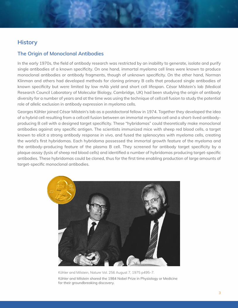
Georges Köhler joined César Milstein’s lab as a postdoctoral fellow in 1974. Together they developed the idea of a hybrid cell resulting from a cell:cell fusion between an immortal myeloma cell and a short-lived antibody-producing B cell with a designed target specificity. These “hybridomas” could theoretically make monoclonal antibodies against any specific antigen. The scientists immunized mice with sheep red blood cells, a target known to elicit a strong antibody response in vivo, and fused the splenocytes with myeloma cells, creating the world’s first hybridomas. Each hybridoma possessed the immortal growth feature of the myeloma and the antibody-producing feature of the plasma B cell. They screened for antibody target specificity by a plaque assay (lysis of sheep red blood cells) and identified a number of hybridomas producing target-specific antibodies. These hybridomas could be cloned, thus for the first time enabling production of large amounts of target-specific monoclonal antibodies.
Köhler and Milstein, Nature Vol. 256 August 7, 1975 p495–7.
Köhler and Milstein shared the 1984 Nobel Prize in Physiology or Medicine for their groundbreaking discovery.

4
— 1975 While working at Cambridge University,
Georges Köhler and César Milstein concretize the idea of fusing myeloma cells with B cells, resulting in the creation of synkaryon cells, later named hybridomas, that have the capabilities of secreting antibodies of a single specificity.
— 1976 Susumu Tonegawa elucidates the
rearrangement of immunoglobulin genes and demonstrates the genetic mechanism that results in antibody diversity.
— 1977 The Food and Drug Administration (FDA) approves first home
pregnancy test which uses antibodies specific for the human chorionic gonadotropin (hCG) hormone.
— 1986 The FDA approves the first therapeutic
monoclonal antibody: muromonab-CD3 for prevention of kidney transplant rejection.
— 1992 The FDA approves the first diagnostic monoclonal antibody:
indium-111 satumomab pendetide, targeting the tumor-associated glycoprotein 72 (TAG-72), for the detection and imaging of colorectal and ovarian tumors.
— 1992–1995 James Allison and Tasuku Honjo
independently discover the first cancer checkpoint inhibitors: programmed cell death-1 (PD-1) and cytotoxic T-lymphocyte-associated protein-4 (CTLA-4).
— 1998 The FDA approves the first cancer “immunotherapy”:
trastazumab for human epidermal growth factor receptor (HER2) overexpressing breast cancer.
— 2006 The FDA approves the first fully human
monoclonal antibody derived from a transgenic mouse: panitumumab for the treatment of epidermal growth factor-receptor (EGFR) positive colorectal cancer.
— 2020 Combined world-wide sales of monoclonal antibody-based
therapies is estimated to reach $150 billion.
— 1796 British country doctor Edward Jenner uses
cowpox to inoculate patients and protect them against smallpox. Today, Jenner is recognized as the “Father of Immunology” for his contributions to the field of vaccination.
— 1890 Emil von Behring and Shibasaburo Kitasato
show that serum from infected animals can be used to treat as well as prevent infection in other animals. Eventually their idea is carried out in humans and used to treat pediatric cases of diphtheria.
— 1900 Paul Ehrlich introduces his side-chain theory:
cells can express a variety of “side-chains” that can be released into the bloodstream and act as antitoxins or antikörpers (antibodies).
— 1914–1918 Horse serum therapy is expanded on a grand scale to combat
tetanus during World War I; thousands of soldiers are presumed to have survived because of it.
— 1945 Following Karl Landsteiner’s discovery of the
ABO blood group system as well as rhesus factors, Robin Coombs develops the Coombs test which detects pre-existing antibodies to rhesus factors in the blood.
— 1947 During the completion of her doctorate, Astrid Fagraeus
demonstrates that plasma cells (mature B cells) are responsible for the production of antibodies.
— 1957 Frank Macfarlane Burnet proposes his clonal selection theory,
explaining how lymphocytes respond in the presence of antigens and how each lymphocyte produces antibodies with a single specificity.
— 1965 After working with children suffering from
Wiskott-Aldrich syndrome, Max Cooper and his collaborators discover the bursa of Fabricius (an organ exclusively found in birds) as the organ generating antibody producing B cells.
— 1969 Gerald Edelman describes the structure of an antibody protein.
History
Evolution of Antibody Technologies

5
Antibody Structure
Along with the double helix of DNA, the distinctive Y-shape of an antibody is one of the most recognized structures in biology and perhaps all of science. There are five classes of antibodies in humans and rodents defined by their respective immunoglobulin (Ig) heavy chains: IgG, IgM, IgD, IgA, and IgE. Here we focus on IgG, as it is one of the most abundant proteins in human serum (10-20% of total plasma protein), comprises 70-85% of the total immunoglobulin pool, has the longest plasma half-life (20-24 days), and is the most common format used in antibody-based therapeutics. IgG (~150 kD) comprises four peptide chains: two identical heavy chains and two identical light chains connected by disulfide (S-S) bonds. Each heavy chain in IgG comprise ~450 residues. There are four IgG subclasses in humans (IgG1, IgG2, IgG3, and IgG4), five in mice (IgG1, IgG2a, IgG2b, IgG2c, IgG3), and four in rats (IgG1, IgG2a, IgG2b, IgG2c). Each light chain in an IgG comprises ~215 residues, and there are two light chain subclasses in humans, mice, and rat: kappa (κ) and lambda (λ).
The two identical complementarity-determining regions (CDRs) at the upper tips of the Y-structure make up the antigen recognition surface for the antibody. There are 3 CDRs in the heavy chain and light chain (CDR1, CDR2, and CDR3), and in most cases antigen binding requires contributions from both heavy and light chain CDRs. Furthermore, in most cases the CDR3 region in the heavy chain is the key determinant of antigen recognition specificity. The CDRs are contained within the fragment variable (Fv) domain (~25 kD): the variable heavy (VH) and variable light (VL) chain regions make up the Fv domain. The antigen-binding fragment (Fab, ~50 kD) domain includes the entire light chain (VL and constant light (CL)), the VH and the first constant domain of the heavy chain (CH1). Monomeric Fabs can be generated by digesting an antibody with papain protease. A divalent F(ab’)2 fragment (~100 kD) can be generated by digesting an antibody with Immunoglobulin-degrading enzyme from Streptococcus pyrogenes (IdeS) or by using pepsin peptidase. The fragment crystallizable (Fc, ~50 kD) region forms the base of the Y-structure and comprise the CH2 and CH3 domains of the heavy chain (HC). The Fc region binds to Fc receptors and complement proteins, and is often modified for therapeutic purposes, as in antibody-drug conjugates (ADCs) wherein the Fc is coupled to cytotoxic molecules.
Antibody Structure Human Gene Segment Location
Heavy Chain Chromosome 14
Light Chain (κ) Chromosome 2
Light Chain (λ) Chromosome 22

6
In vivo Humoral Immune Response
Terminally differentiated B cells capable of secreting high affinity antibodies (aka plasma cells) arise following an extensive process of cell differentiation and activation that begins with hematopoietic stem cells (HSCs)in the bone marrow. Antigen-independent B cell development occurs in five discrete stages that are coupled with the sequential somatic recombination of the variable (V), diversity (D), and joining (J) gene segments. Antigen-dependent B cell development takes place initially in the bone marrow and then in the periphery.
B Cell Development Pathway
Memory B cell – a subset of B cells survive as long-lived memory B cells, which recirculate and can rapidly respond to reintroduced antigen and differentiate into antibody-producing plasma cells.
Hematopoietic stem cell – heavy chain and light chain genes are in germline configuration.
Early pro-B cell – heavy chain undergoes D–J gene rearrangement.
Late pro-B cell – heavy chain undergoes V–DJ rearrangement.
Large pre-B cell – transient surface expression of IgM heavy chain with invariant pseudo light chain (pre-B cell receptor). Successful cell surface expression of pre-B cell receptor triggers allelic exclusion to prevent rearrangement of the second allele and also initiates pre-B cell proliferation, which results in different light chains matched with the same heavy chain in different daughter cells. Dividing cells are larger than resting cells, hence the name large pre-B cell.
Small pre-B cell – light chain undergoes V–J rearrangement. Individual cells first attempt κ chain rearrangement. If rearrangement of both κ alleles is unsuccessful, cells attempt λ chain rearrangement.
Immature B cell – successfully rearranged heavy and light chains are expressed as IgM on the cell surface. At this stage the cells are highly sensitive to antigen binding: if they bind self-antigen in the bone marrow they die (negative selection).
Mature naïve B cell – if the immature B cell survives, it matures in the bone marrow or spleen and expresses surface IgD and IgM, made from alternatively spliced transcripts. Mature naïve B cells recirculate between blood, spleen, peripheral lymph nodes and other secondary lymphoid tissues (e.g. Peyer’s Patches in the gut) in search of cognate antigen.
B Lymphoblast – antigen binding leads to alternative splicing to secrete Ig. The location of antigen encounter triggers isotype switching (gut microenvironmental signals lead to IgA, whereas lymph node signals lead to IgG). Somatic hypermutation is triggered in germinal centers in the follicles of secondary lymphoid tissues. CD4+ T cell help in germinal centers, by means of antigen presentation via major histocompatibility complex (MHC) class II molecules, drives in vivo affinity maturation.
Plasma cell – continuous antibody secretion, estimated at 2,000 IgM or 15,000 IgG molecules per second per cell. Alternative splicing yields some membrane-bound Ig, but most is secreted. Most plasma cells are incapable of proliferation, with their protein-synthesizing machinery dedicated entirely to making antibody. Although many die after several days, some survive in the bone marrow for months or years and continue to secrete antibodies into the blood.

7
Approved Monoclonal Antibodies
Whether it’s a pioneering antibody like OKT3, or an effective and financially attainable biosimilar, each approved antibody plays an important role in the quest to improve patient health. By our estimation, as of January 2020, 100 originator (as opposed to biosimilar) monoclonal antibodies have been approved in the US (FDA) and/or Europe (EMA) since OKT3 in 1986. These antibodies cover a wide range of indications and worldwide antibody sales are expected to reach nearly $150 billion in 2020*. The current rate of approval (last 5 years) by the FDA is 7.6 antibodies per year. It’s important to note that of all of the monoclonal antibodies approved by the FDA, the vast majority (roughly 88%) were discovered via hybridoma-based technologies (with a small but growing portion from transgenic human antibody-producing animals). That’s quite a track record for what many consider to be an “ageing” technology!
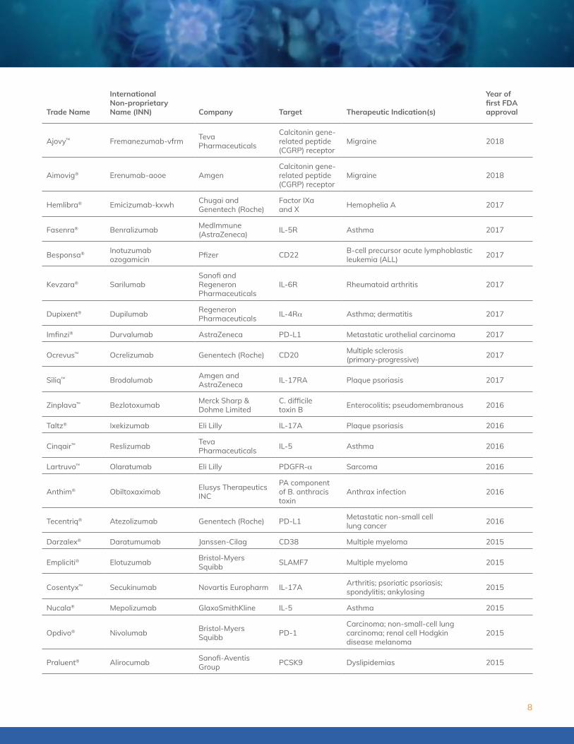
The table below provides a list of all of the hybridoma-derived monoclonal antibodies that have been granted FDA approval in the last 10 years.
Trade Name
International Non-proprietary Name (INN) Company Target Therapeutic Indication(s)
Year of first FDA approval
Padcev™ Enfortumab vedotin-ejfv
Astellas Pharma and Seattle Genetics Nectin-4 Locally advanced or metastatic
urothelial cancer (mUC) 2019
Adakveo® Crizanlizumab-tmca Novartis P-selectin Sickle cell disease 2019
Beovu® Brolucizumab-dbll Novartis VEGF-A Macular degeneration 2019
Polivy™ Polatuzumab vedotin-piiq Genentech (Roche) CD79b Diffuse large B-cell lymphoma
(DLBCL) 2019
Skyrizi™ Risankizumab-rzaa Abbvie IL-23 Plaque psoriasis 2019
Evenity® Romosozumab-aqqg Amgen and UCB Pharma Sclerostin Osteoporosis 2019
Ultomiris™ Ravulizumab-cwvz Alexion Pharmaceuticals
Complement protein C5
Paroxysmal nocturnal hemoglobinuria (PNH) 2018
Ilumya™ Tildrakizumab-asmn Sun Pharma IL23 (p19) Plaque psoriasis 2018
Trogarzo® Ibalizumab-uiyk Theratechnologies Inc CD4 Human immunodeficiency virus-1
(HIV-1) 2018
Libtayo® Cemiplimab-rwlc Sanofi PD-1 Advanced/metastatic cutaneous squamous cell carcinoma (CSCC) 2018
Poteligeo® Mogamulizumab-kpkc Kyowa Hakko Kirin CC chemokine receptor 4 (CCR4)
Mycosis fungoides (MF) or Sézary syndrome (SS) 2018
Crysvita® Burosumab-twzaUltragenyx Pharmaceuticals and Kyowa Kirin
FGF-23 X-linked hypophosphatemia (XLH) 2018
Emgality® Galcanezumab-gnlm Eli LillyCalcitonin gene-related peptide (CGRP) receptor
Migraine 2018
* Lu et al. (2020). Development of Therapeutic Antibodies for the Treatment of Diseases. J Biomed Sci. 2020 Jan2:27(1):1 doi: 10.1186/s12929-019-0592-z
8
Trade Name
International Non-proprietary Name (INN) Company Target Therapeutic Indication(s)
Year of first FDA approval
Ajovy™ Fremanezumab-vfrm Teva Pharmaceuticals
Calcitonin gene-related peptide (CGRP) receptor
Migraine 2018
Aimovig® Erenumab-aooe Amgen Calcitonin gene-related peptide (CGRP) receptor
Migraine 2018
Hemlibra® Emicizumab-kxwh Chugai and Genentech (Roche)
Factor IXa and X Hemophelia A 2017
Fasenra® Benralizumab MedImmune (AstraZeneca) IL-5R Asthma 2017
Besponsa® Inotuzumab ozogamicin Pfizer CD22 B-cell precursor acute lymphoblastic
leukemia (ALL) 2017
Kevzara® SarilumabSanofi and Regeneron Pharmaceuticals
IL-6R Rheumatoid arthritis 2017
Dupixent® Dupilumab Regeneron Pharmaceuticals IL-4Rα Asthma; dermatitis 2017
Imfinzi® Durvalumab AstraZeneca PD-L1 Metastatic urothelial carcinoma 2017
Ocrevus™ Ocrelizumab Genentech (Roche) CD20 Multiple sclerosis (primary-progressive) 2017
Siliq™ Brodalumab Amgen and AstraZeneca IL-17RA Plaque psoriasis 2017
Zinplava™ Bezlotoxumab Merck Sharp & Dohme Limited
C. difficile toxin B Enterocolitis; pseudomembranous 2016
Taltz® Ixekizumab Eli Lilly IL-17A Plaque psoriasis 2016
Cinqair™ Reslizumab Teva Pharmaceuticals IL-5 Asthma 2016
Lartruvo™ Olaratumab Eli Lilly PDGFR-α Sarcoma 2016
Anthim® Obiltoxaximab Elusys Therapeutics INC
PA component of B. anthracis toxin
Anthrax infection 2016
Tecentriq® Atezolizumab Genentech (Roche) PD-L1 Metastatic non-small cell lung cancer 2016
Darzalex® Daratumumab Janssen-Cilag CD38 Multiple myeloma 2015
Empliciti® Elotuzumab Bristol-Myers Squibb SLAMF7 Multiple myeloma 2015
Cosentyx™ Secukinumab Novartis Europharm IL-17A Arthritis; psoriatic psoriasis; spondylitis; ankylosing 2015
Nucala® Mepolizumab GlaxoSmithKline IL-5 Asthma 2015
Opdivo® Nivolumab Bristol-Myers Squibb PD-1
Carcinoma; non-small-cell lung carcinoma; renal cell Hodgkin disease melanoma
2015
Praluent® Alirocumab Sanofi-Aventis Group PCSK9 Dyslipidemias 2015

9
Trade Name
International Non-proprietary Name (INN) Company Target Therapeutic Indication(s)
Year of first FDA approval
Praxbind® IdarucizumabBoehringer Ingelheim International GmbH
DabigatranHemorrhage (inactivating oral anticoagulant dabigatran; dabigatran etexilate prodrug)
2015
Repatha® Evolocumab Amgen LDL-C / PCSK9 Dyslipidemias; hypercholesterolemia 2015
Unituxin® Dinutuximab United Therapeutics Europe GD2 Neuroblastoma 2015
Entyvio® Vedolizumab Takeda Pharma Integrin-α4β7 Colitis; ulcerative Crohn’s disease 2014
Blincyto® Blinatumomab Amgen Europe CD19 Precursor cell lymphoblastic leukemia-lymphoma 2014
Keytruda® Pembrolizumab Merck Sharp & Dohme Limited PD-1
Melanoma, non-small cell lung cancer (NSCLC), small cell lung cancer (SCLC), head and neck squamous cell cancer (HNSCC), Hodgkin lymphoma, urothelial carcinoma, GI tract cancers
2014
Sylvant® Siltuximab Janssen-Cilag International cCLB8 Giant lymph node hyperplasia,
Castleman’s disease 2014
Lemtrada® Alemtuzumab Sanofi CD52 Multiple sclerosis (relapsing-remitting) 2014
Kadcyla® Trastuzumab emtansine Genentech (Roche) HER2 Breast Cancer 2013
Gazyva® Obinutuzumab Genentech (Roche) CD20 Chronic Lymphocytic leukemia (CLL) 2013
Perjeta® Pertuzumab Genentech (Roche) HER2 Breast Cancer 2012
Adcetris® Brentuximab vendotin MMAE Seattle Genetics CD30
Hodgkin lymphoma (HL), systemic anaplastic large cell lymphoma (ALCL)
2011
Yervoy® Ipilimumab Bristol-Myers Squibb CTLA-4 Melanoma 2011
Prolia®/ Xgeva® Denosumab Amgen RANKLOsteoporosis/ Prevention of skeletal-related events (SREs) in patients with bone metastases from solid tumors
2010/ 2011
Actemra® Tocilizumab Chugai (Roche) IL-6 receptor Rheumatoid arthritis 2010
Marketing Approval Agencies:
Approval by the US Food and Drug Administration (FDA) grants marketing authorization in all 50 US states as well as other US territories and possessions.
Approval by the European Medicines Agency (EMA) grants marketing authorization in all European Union (EU) and European Economic Area (EEA)-European Free Trade Association (EFTA) states (which includes Iceland, Liechtenstein, and Norway).
What will you target next?

10
Conventional Hybridoma Overview
Human Antibody-Producing Rodents
LakePharma’s PentaMice™ Platform
Hybridoma Technology
B Cell : Myeloma Cell Fusion
Hybridoma Selection

11
Hybridoma Technology
Conventional Hybridoma Overview
Hybridoma technology utilizes a wide variety of experimental procedures to yield antigen-specific monoclonal antibody (mAb)-producing immortal hybridoma clones.
Conventional hybridoma technology utilizes in vivo immunizations, cell:cell fusion, specialized cell culture conditions, and various screening techniques to yield antigen-specific monoclonal antibody (mAb)-producing immortal hybridoma clones. The concept is essentially unchanged from Köhler and Milstein’s original approach reported in 1975. Animals (typically rodents) are immunized with the target antigen, splenocytes are fused with myeloma partners and grown in HAT media to select for hybridomas, and the hybridoma supernatants are screened for target reactivity. Single cell cloning is performed to yield monoclonal antibody-producing hybridomas.
Rodents are typically immunized with antigen and adjuvant to generate a strong in vivo B cell antibody response.
The spleen is harvested and single cell suspensions of splenocytes are generated.
Splenocytes are fused with myeloma partners to generate hybridomas.
The fusion mixture is plated into 96-well plates where HAT culture media drives selection of hybridomas vs. unfused myelomas or B cells.
The plates are screened for antigen-specific antibodies. Cells in positive wells are expanded, and single cell hybridoma clones are generated from the parental hybridomas.
The clonal hybridoma is expanded for production of antigen-specific mAbs.
Splenocyte Myeloma
+
Cell Fusion
Hybrid Cell

12
Hybridoma Technology
Human Antibody-Producing Rodents
Multiple innovative platforms are currently available for antibody discovery using genetically modified mice that express fully-human heavy and light chain variable regions. These animals can generate diverse repertoires of in vivo affinity-matured antibodies with intrinsic drug-like properties necessary for successful development, including high potency, specificity, solubility, and manufacturability. A key advantage is that the fully-human variable regions have a low risk of immunogenicity, thus mitigating efficacy-killing anti-drug immune responses as a fully-human antibody format.
Ligand OmniMouse®
Harbour H2L2 Mouse™ Trianni Mouse® Alloy
Gx Mouse™Ablexis AlivaMab Mouse®
Light Chain Hu κHu λ Hu κ Hu κ
Hu λHu κHu λ
Hu κ Hu κ Hu λHu λ
Heavy ChainFc
RatIgG1IgG2bIgG2c
RatIgG1IgG2bIgG2c
MouseIgG1IgG2bIgG2cIgG3
Not disclosed MouseIgG
Heavy Chain Repertoire(no. V Gene Segments)) 44 18 44 40+ Not disclosed
Light Chain Repertoire(no. V Gene Segments)
20 κ15 λ 11 κ 39 κ
38 λ19+ κ22+ λ Not disclosed
Parental Strain(s) C57Bl/6SJL
C57Bl/6FVB129
C57Bl/6 C57Bl/6BALB/c Not disclosed
MHC Haplotype(s) H-2bH-2s
H-2bH-2q H-2b H-2b
H-2d Not disclosed
References
Lowitz J, Lin G, Somera J, Santibanez-Vargas L, Vo C, Rodriguez E, Nguyen B, Trang M, Nichols J, Kenney J. Optimization of Therapeutic Discovery Strategies for Human Antibody Transgenic Animal Platforms. Poster presented at PEGS; 2019 Apr. 8-12; Boston, MA.
Maximizing the AlivaMab Mouse Advantage. Retrieved from https://alivamab.com/alivamab-mouse.

13
Hybridoma Technology
LakePharma’s PentaMice™ Platform
A Proprietary Set of Wildtype Mice Designed to Achieve Maximum Plasma Titers in Hybridoma Campaigns
Conventional immunization approaches utilized in hybridoma-based antibody discovery campaigns typically use one or two common wildtype (WT) mouse strains (e.g. Balb/c or C57Bl/6). LakePharma’s scientists have noted through a course of over 100 campaigns that this approach likely limits the identification of high-quality antibodies to just those target antigens that are efficiently processed and presented by a restricted major histocompatibility complex (MHC) repertoire that is distinct for each WT strain.
LakePharma’s PentaMice™ Platform is a royalty-free set of mice comprising 5 WT strains that cover 9 distinct MHC haplotypes. A total of 10 mice (2 mice of each strain) are included in each set to achieve maximum plasma titers, thus boosting the opportunity to generate high-quality antibodies in vivo.
The Concept Behind the PentaMice Platform
ELIS
A R
LU
Dilution
Mouse 1 (Strain 1)
Mouse 2 (Strain 1)
Mouse 3 (Strain 2)
Mouse 4 (Strain 2)
Antigen
CD4+ T cell
Activated B cell
MHC II/Antigen
T Cell Receptor
Costimulatory Molecules
CytokinesClonal Expansion
Antibody-Secreting Plasma Cells
Peptide 1Peptide 2
MHC Class II Haplotype
PentaMice Strain
IAk, IAg7, IEk
k x g7 d x u b x s q x v Mixed
IAd, IAu, IEd, IEu IAb, IAs IAv, IAq, IEv IAmixed,IEmixed
1 Plasma titers are highly predictive of antibody discovery success. Based on LakePharma’s experience, there is often a strong strain-dependent difference in plasma titers for most targets.
2 High plasma titers require T cell help, and one of the requirements for effective T cell activation is recognition of cognate antigens presented by the MHC. Only certain peptides are effectively presented by certain MHC.
3 MHCs are highly polymorphic. LakePharma’s scientists continue to garner evidence to support the hypothesis that this polymorphism drives strain-dependent differences in plasma titers. Hence, the PentaMice Platform is designed to cover a wide range of MHC haplotypes to enable effective T cell help.Two different peptide binding profiles are shown as examples.
Peptide 1 is efficiently presented by most MHC II. Peptide 2 is only efficiently presented by IAg7.

14
Hybridoma Technology
B Cell : Myeloma Cell Fusion
Although cell fusion may seem a little odd at first, it is important to remember that it is quite common in the natural world. Such is the case with gamete fusion in the formation of a one-cell embryo, as well as later in development with the fusion of myocytes resulting in a myotube or skeletal muscle cell syncytium. Nevertheless, when it comes to hybridomas, B cells and myeloma cells make for an unlikely fusion pair, hence the need for an external catalyst. Köhler and Milstein harnessed the power of the Sendai virus, a murine respiratory tract virus. Once infected, a cell would be more prone to fuse with an adjacent cell via fusogens or fusion proteins. We have come a long way since the inception of the hybridoma cell, and nowadays the two most common techniques include polyethylene glycol (PEG) – mediated fusion and electrofusion.
PEG-Mediated Fusion Electrofusion
Procedure Specifics
PEG is a one-step fusion process that consists of placing a mixture of cells into a flask containing PEG solution and shaking vigorously. Over time PEG dehydrates the cell membranes by making water molecules between cells thermodynamically unfavorable, ultimately promoting membrane fusion.
Electrofusion is achieved by applying carefully controlled pulses of electricity to a suspended cell mixture. Cells are first aligned by dielectrophoresis (resulting from an alternating current – AC – field). Cells are then exposed to direct current – DC – pulses, stimulating electroporation, bringing the cell membranes into a permeable and fusogenic state.
Duration Short (minutes) Short (minutes); DC pulses range from µs to ns.
Fusion Efficiency
Very low (1:10,000) Low (1:1,000)
Complexity Low (PEG remains the simplest hybridoma fusion technique currently in use)
Medium (the procedure requires the use of high-grade fusion chambers)
Cost $ $ $
Hybridomas are formed when a B cell is fused with a myeloma cell.
An AC field polarizes the cells and induces their linear “pearl chain” alignment. A DC pulse then triggers cell fusion.
Both methods rely extensively on cell-to-cell contact as well as the quality of the cells used. Although PEG fusions are more economically advantageous, results vary. Head-to-head comparisons have demonstrated that electrofusions not only had a greater number of fusion events but the resulting hybridomas grew more vigorously. Within minutes following the fusion event, intermingling of surface proteins and restructuring of the two separate cytoskeletons into one occurs, with nuclei fusion ensuing shortly after. When it comes to electrofusion, the number, duration, and frequency of pulses is optimized so as to preserve cell viability and increase fusion efficiency, rendering it the favored method for LakePharma’s hybridoma-based antibody discovery!

15
Hybridoma Technology
Hybridoma Selection
Cell division requires an adequate supply of free nucleotides for deoxyribonucleic acid (DNA) replication. Nucleic acids are generated either via:
• De novo DNA synthesis – which is dependent on the activity of dihydrofolate reductase to generatepurine nucleotides (GMP, IMP, AMP) and thymidylate;
• Salvage pathway synthesis – which requires exogenous hypoxanthine and thymidine and the enzymeshypoxanthine-guanine phosphoribosyltransferase (HGPRT) and thymidine kinase (TK).
Aminopterin is a dihydrofolate reductase inhibitor: treatment of cells with aminopterin prevents de novo DNA synthesis, and in the absence of exogenous hypoxanthine and thymidine to supply the salvage pathway, the cells will die. A mutation in HGPRT disables the salvage pathway, which is lethal in aminopterin-treated cells even if they are cultured with exogenous hypoxanthine and thymidine. The identification of myleoma cells with mutations in HGPRT is straightforward. The HGPRT gene is on the X-chromosome, and due to X-linked inactivation a single mutation is all that is needed to result in the loss of HGPRT. Thus, specific cell culture conditions (HAT media: Hypoxanthine, Aminopterin, Thymidine) + a myeloma partner with a HGPRT mutation enables the selection and survival of hybridomas (B cell:myeloma hybrids) vs. unfused myeloma cells or B cells. The key concept is that the hybridoma receives a functional HGPRT gene from the primary B cell, thus enabling the survival of hybridomas vs. myeloma cells. While primary B cells can survive for a time in culture and produce antibody, they will eventually die without any additional selection, leaving hybridomas as the only living cell in the culture wells. A subset of hybridomas express antibody genes derived from their parental B cell contributor, and single cell cloning yields clonal hybridomas that express monoclonal antibodies.
APRT, adenine phosphoribosyltransferase; PRPP, 5-Phosphoribosyl-1-pyrophosphate.
* Modified from Hnasko et al, “Hybridoma Technology”, Methods in Molecular Biology 2015.
Introduction to Hybridoma Technologies for Antibody Discovery

Goals Aligned
Target Analysis
Biofunction QC
of Immunogen
Materials Ready
Readiness QC
Goals Aligned
Target Analysis
Biofunction QC
of Immunogen
Materials Ready
Readiness QC
With a success rate of 98%, LakePharma’s hybridoma platform has a proven track record in effectively identifying novel therapeutic or diagnostic monoclonal antibodies. The hybridoma team is ready to partner with you on your antibody quest.
Hybridoma Discovery Immunology – Chain of Discovery™ Series
Complete campaigns incorporate LakePharma’s unique suite of integrated services including:• Target analysis and immunogen production
• Immunization, hybridoma generation, and single cell cloning
• Variable region sequencing and purified mAb production/characterization
Discovery Immunology mAbs can seamlessly transition to downstream GXP production, enabling a one-stop shop from discovery to development.
• Target analysis andantigen design
• Goal-oriented proposal development
• Antigen biofunction QC
ImmunizationsUpfront DueDiligence
• Rapid immunization protocols
• DNA, protein, peptide,and cell-basedimmunizations
• In-life titer checks enablereal-time optimizationof immunizations, andselection of animals withoptimal titers
Target the Future Today!
16Introduction to Antibody Libraries for Display-Based Antibody Discovery
HybridomaScreens
• Hybridoma generation by electrofusion
• 384-well plate highthroughput screens(ELISA or multiplex FACS)
• Data Master Files forcandidate selection
mAb Screens
• Hybridoma single cellcloning and variableregion sequencing
• mAb characterization, affinity binding EC50(ELISA or FACS),biofunction IC50 or EC50(customized functionalassays such as ligandblocking & receptorinternalization)
Learn More at https://www.hybridoma.com/
Goals Aligned
Target Analysis
Biofunction QC
of Immunogen
Materials Ready
Readiness QC
Goals Aligned
Target Analysis
Biofunction QC
of Immunogen
Materials Ready
Readiness QC

Tel. 650-288-4891Tel. 888-406-5658 (Toll-free)Fax 888-635-3618
Corporate Headquarters201 Industrial RoadSan Carlos, CA 94070
Email [email protected] www.lakepharma.com
©2020 LakePharma, Inc. The trademarks used herein are the property of LakePharma, Inc. or their respective owners. 140A 4/20
Contact Us