functional single-cell hybridoma screening using droplet ...into an unrelated hybridoma cell...
TRANSCRIPT

Functional single-cell hybridoma screeningusing droplet-based microfluidicsBachir El Debsa,b,c, Ramesh Utharalac, Irina V. Balyasnikovad, Andrew D. Griffithsa,b, and Christoph A. Mertenc,1
aInstitut de Science et d’Ingénierie Supramoléculaires, Université de Strasbourg, 8 allée Gaspard Monge, 67083 Strasbourg Cedex, France; bCentreNational de la Recherche Scientifique, Unité Mixte de Recherche 7006, 8 allée Gaspard Monge, 67083 Strasbourg Cedex, France; cGenome Biology Unit,European Molecular Biology Laboratory, Meyerhofstrasse 1, 69117 Heidelberg, Germany; and dThe Brain Tumor Center, University of Chicago,5841 South Maryland Avenue (MC 3026), Chicago, IL 60637
Edited by David A. Weitz, Harvard University, Cambridge, MA, and approved June 12, 2012 (received for review March 21, 2012)
Monoclonal antibodies can specifically bind or even inhibit drugtargets and have hence become the fastest growing class of humantherapeutics. Although they can be screened for binding affinitiesat very high throughput using systems such as phage display,screening for functional properties (e.g., the inhibition of a drugtarget) is much more challenging. Typically these screens requirethe generation of immortalized hybridoma cells, as well as clonalexpansion in microtiter plates over several weeks, and the numberof clones that can be assayed is typically no more than a few thou-sand. We present here a microfluidic platform allowing the func-tional screening of up to 300,000 individual hybridoma cell cloneswithin less than a day. This approach should also be applicableto nonimmortalized primary B-cells, as no cell proliferation is re-quired: Individual cells are encapsulated into aqueous microdro-plets and assayed directly for the release of antibodies inhibitinga drug target based on fluorescence. We used this system to per-form a model screen for antibodies that inhibit angiotensin con-verting enzyme 1, a target for hypertension and congestive heartfailure drugs. When cells expressing these antibodies were spikedinto an unrelated hybridoma cell population in a ratio of 1∶10,000we observed a 9,400-fold enrichment after fluorescence activateddroplet sorting. A wide variance in antibody expression levels atthe single-cell level within a single hybridoma line was observedand high expressors could be successfully sorted and recultivated.
single-cell screening ∣ high-throughput screening ∣ cell-based assay ∣monoclonal antibody ∣ angiotensin converting enzyme 1
Antibodies are powerful research and diagnostic tools andhave proven to be potent therapeutics against infectious,
autoimmune, and neoplastic diseases. Indeed, the number oftherapeutic monoclonal antibodies (mABs) reaching the marketis increasing exponentially (1) and the global monoclonal anti-body market for therapeutic use was $38 billion in 2009 (2).Monoclonal antibodies can be screened very efficiently for bind-ing using phage display and related technologies (3). However,binding alone is not sufficient; therapeutic antibodies must alsomodulate (typically inhibit) the activity of the target whereasthese methods select only for binding to a drug target and not forinhibition of its function. To overcome this limitation, functionalantibody screens are typically carried out using hybridoma celltechnology (4). In this approach, laboratory animals are immu-nized with the antigen of interest before antibody-releasingB-cells are isolated from spleen. These B-cells are then renderedimmortal by fusion with myeloma cells, diluted to generate mi-crotitre plate wells containing single cells and expanded to formclonal populations. Subsequently, the supernatant of each popu-lation can be tested to screen for the desired activity. However,the need for clonal cell expansion (to obtain detectable concen-trations of antibodies), and hence cell immortalization, typicallylimits the number of clones that can be screened to no more thana few thousand. Improved techniques have been described facil-itating the screening of >105 clones in <12 h, based on, for ex-ample, antigen-based microarrays (5), or compartmentalization
of individual cells in 0.1–1 nL lithographically fabricated micro-wells (6). Still, these methods can only be used to screen for bind-ing activity and do not allow functional assays.
Droplet-based microfluidics (7) holds great potential for func-tional high-throughput screening at the single-cell level. In thesesystems, cells are encapsulated into aqueous droplets surroundedby an immiscible carrier phase (e.g., fluorinated oil) (8). Eachdroplet serves as a miniaturized assay vessel of picoliter-nanolitervolume, and up to several hundred droplets can be generatedper second. Furthermore, fluorescence assays and fluorescence-activated droplet sorting (FADS) can be carried out at a similarthroughput (9). FADS is similar to fluorescence-activated cellsorting (FACS), but is not limited to sorting based on intracellularor cell-surface markers: With FADS the entire microvessels aresorted rather than cells, allowing screening of secreted proteins(such as antibodies) as well. Typically, all components of a fluor-escence assay are added directly during encapsulation, or at alater time point upon fusion with a second droplet species hostingthe assay reagents. Subsequently, the droplets pass through a laserbeam, the emitted light is measured, and droplets with particularfluorescence intensities are diverted into a collection channel usingelectric fields (7). Although these steps have been demonstratedindividually for small droplets (approximately 30 μm), an inte-grated chip combining all required modules in a single platform,as well as fusion and sorting modules allowing droplets big enoughfor the cultivation of mammalian cells (approximately 100 μm indiameter) to be manipulated, have not previously been described.In consequence, single mammalian cells have been analyzed indroplets (8) and it had also been shown that detectable antibodyconcentrations can be obtained from individually encapsulatedhybridoma cells (10), but the sorting of encapsulated mammaliancells based on a functional screen has not yet been achieved. Wepresent here a fully integrated system that can overcome this lim-itation and demonstrate the screening and sorting of hybridomacells for the release of antibodies inhibiting angiotensin convertingenzyme 1 (ACE-1; Fig. 1A).
ACE-1 plays a key role in the regulation of blood pressure andthe development of vascular pathology and remodeling. It is atype I integral membrane protein which is converted into a solu-ble circulating form by membrane protein secretases (11). ACE-1has two catalytic domains, the so-called N- and C-domain, bothhaving the capacity to hydrolyze the same peptides (angiotensin Iand bradykinin). However, the two catalytic sites have differentsubstrate specificities and catalytic properties as well as different
Author contributions: B.E.D., A.D.G., and C.A.M. designed research; B.E.D., R.U., andC.A.M. performed research; B.E.D., I.V.B., and C.A.M. contributed new reagents/analytictools; B.E.D., I.V.B., A.D.G., and C.A.M. analyzed data; and B.E.D., I.V.B., A.D.G., and C.A.M.wrote the paper.
Conflict of interest statement: C.A.M and A.D.G are inventors on patent applicationsincluding some of the ideas described in this manuscript.
This article is a PNAS Direct Submission.1To whom correspondence should be addressed. E-mail: [email protected].
This article contains supporting information online at www.pnas.org/lookup/suppl/doi:10.1073/pnas.1204514109/-/DCSupplemental.
11570–11575 ∣ PNAS ∣ July 17, 2012 ∣ vol. 109 ∣ no. 29 www.pnas.org/cgi/doi/10.1073/pnas.1204514109
Dow
nloa
ded
by g
uest
on
Apr
il 24
, 202
0

affinities for competitive inhibitors (12, 13). Monoclonal antibo-dies against this enzyme have been used in structural and func-tional studies of ACE-1 (14, 15) and mAbs against ACE-1 haveboth diagnostic and therapeutic potential: ACE-1 is a target fordrugs to treat hypertension and congestive heart failure (16).Here we demonstrate the enrichment of hybridoma cells secret-ing the ACE-1 inhibitory mAb 4E3 (14, 15) from a large excess ofunrelated hybridoma cells.
ResultsDesign of the Microfluidic Platform. We developed here an inte-grated microfluidic system consisting of a previously describeddrop maker for the encapsulation of cells into droplets with avolume of 660 pL (8) (Fig. 1C) and a unique device for the mani-pulation of these droplets subsequent to an off-chip incubationperiod (drops are collected outside the microfluidic device in aseparate syringe). During this incubation period detectableconcentrations of antibodies are released from encapsulatedhybridoma cells into the droplets (Fig. 1A). In the next step, thesyringe in which the droplets were incubated is used to reinjectthem into a second microfluidic chip (Fig. 1D). This second chipallows the addition of further substrates by droplet fusion, as wellas incubation and sorting based on fluorescence. The dropletfusion module (Fig. S1 and Movie S1) is a further developmentof a pillar-induced passive fusion device (17), into which we in-tegrated a second drop maker: Large droplets (660 pL) hostingsingle hybridoma cells slow down in the pillar chamber (due tothe drainage of oil) and are brought into contact with small(25 pL volume) droplets containing a custom-made FRET pep-tide (Fig. 1B) mediating a green fluorescence signal upon hydro-lysis by recombinant ACE-1. These 25 pL droplets are generated
in the absence of any surfactant (amphiphilic compounds usuallyadded to stabilize the droplets and to reduce their adherence tothe channel walls; also referred to as wetting) and stop whenreaching the pillar chamber due to wetting and the drainage ofoil. Furthermore, they fuse spontaneously with any incominglarge droplets, even if these are stabilized with surfactant (as re-quired to allow off-chip incubation). A high level of synchroniza-tion between the small and the large droplets can be achieved,resulting in the reliable addition of specific amounts of substrates.
Efficiency of the Droplet Fusion Module. To analyze the fusion effi-ciency at different flow rates, we generated small (25 pL) andlarge (660 pL) droplets on the same chip and labeled them withdifferent fluorescein concentrations. Subsequently, we varied theaqueous flow rates of both types of droplets and monitored thefusion efficiency by video analysis and fluorescence measurements.Precise 1∶1 fusion events were obtained for a range of differentflow rates at frequencies of up to 50 Hz and with an efficiencyof more than 99% (Fig. S2A, regime I and Movie S1). Failure ofthe fusion process occurred only if either the frequency of the smalldroplets was too high (resulting in the release of nonfused smalldroplets from the pillar chamber; Fig. S2A, regime II), or if thefrequency of the large droplets was very high in comparison to thatof the small droplets (Fig. S2A, regime III), in which case the largedroplets passed the small droplets without fusion.
Screening of a Heterogeneous Hybridoma Cell Population. Next, weperformed a model screen. For this purpose we stained 4E3-hybridoma cells, producing an mAB inhibiting ACE-1, with cal-cein-red/orange and diluted them in a 1∶75 excess of nonstainedhybridoma cells releasing an unrelated antibody [Elec-403 anti-
Fig. 1. Microfluidic setup. (A) A mixed population of hybridoma cells either expressing the ACE-1 inhibitory antibody 4E3 or the noninhibitory antibodyElec-403 is encapsulated into droplets together with recombinant ACE-1. (B) Fluorogenic ACE-1 substrate. The ACE-1 cleavage site is indicated by an arrow.(C) Microfluidic chip for cell encapsulation. (D) Integrated microfluidic chip for the reinjection of droplets after a 6 h off-chip incubation period. Dropletshosting cells are fused with droplets containing the fluorogenic ACE-1 substrate and subsequently incubated for 30 min in a delay line. The final sortingmodule allows specific collection of droplets with low fluorescence intensity (indicating inhibition of ACE-1).
Debs et al. PNAS ∣ July 17, 2012 ∣ vol. 109 ∣ no. 29 ∣ 11571
APP
LIED
BIOLO
GICAL
SCIENCE
SAPP
LIED
PHYS
ICAL
SCIENCE
S
Dow
nloa
ded
by g
uest
on
Apr
il 24
, 202
0

body inhibiting Electrophorus electricus acetylcholinesterase (18)].This cell suspension was subsequently encapsulated into 660 pLdroplets together with recombinant ACE-1. The average numberof cells per droplet was approximately 0.3, as measured by videoanalysis of the cell encapsulation process (1,000 droplets in total)(Movie S2: 65.7% empty drops; 29.5% drops with single cells;4.8% drops with more than one cell). These results are in goodagreement with previous studies showing that the number of cellsper droplet follows a Poisson distribution when encapsulatinghuman cell lines in this device (8). These experiments also de-monstrated that adherent as well as suspension cells showed aviability of 90% and above during the first two days in drops ofthe same volume.
After encapsulating hybridoma cells, we incubated the result-ing emulsion for 6 h off-chip to obtain significant antibodyconcentrations (around 20 μg∕mL). Longer incubation times re-sulted in even higher 4E3 antibody concentrations (>30 μg∕mL;Fig. S3A), but turned out to be incompatible with the downstreamassay for ACE-1 activity: We observed that with increasing incu-bation times the supernatants showed higher levels of unspecificconversion of the ACE-1 FRET substrate (Fig. S3A and B). Fol-lowing off-chip incubation, the droplets were reinjected into thesecond device (Fig. 1D and Movie S3), fused with droplets con-taining the fluorogenic ACE-1 substrate and incubated in a delayline for another 30 min (to facilitate generation of the fluorescentproduct). Finally, the droplets were analyzed and sorted, trig-gered on fluorescence (19) (Fig. 1D and Movie S4). When thegreen fluorescence intensity was plotted against the droplet width[used to measure droplet coalescence (8)], three populationswere observed (Fig. 2A). One main population showing stronggreen fluorescence corresponded to droplets in which no inhibi-tion of ACE-1 occurred. Another very sharp, but much smallerpopulation exhibiting almost no green fluorescence signal corre-sponded to droplets that did not obtain any fluorogenic substrate(due to failed fusion). In between these populations a third popu-lation of droplets with intermediate green fluorescence signalscould be observed. This last population corresponds to dropletsin which inhibition of ACE-1 occurred and was characterized bya high variance in green fluorescence. As with FACS, we set gatesfor different green fluorescence intensities (Fig. 2A) and collectedthe corresponding droplets separately. After breaking the emul-sion, the recovered cells were analyzed for calcein-red/orangestaining. Whereas before the sort only 3% of the mixed cell popu-
lation were calcein-red/orange-positive (corresponding to 4E3-expressing cells), this value increased to approximately 83% forcells recovered from droplets with intermediate fluorescencesignals (Fig. 2B). In contrast, the cell populations recovered fromdroplets with high fluorescence intensity (indicating no inhibitionof ACE-1) included only 0.8–6% stained cells. The presence ofsome stained cells in the noninhibited population is not surprising,as dead 4E3 hybridoma cells or cells expressing only low levels of4E3 antibodies inevitably end up in this population, too.
Biochemical Characterization of Cell Culture Supernatants fromRecovered Sorted Cells. Next, we repeated the experiment usingmultiple narrow gates for the sorting of droplets with intermedi-ate fluorescence intensity (indicating ACE-1 inhibition) andanalyzed the secretion of 4E3 antibody from cultured recoveredcells, as well as the ACE-1-inhibitory activity of the supernatants(Fig. 3). Compared to the nonsorted population, cultures of cellsrecovered from droplets within all of these gates showed stronglyincreased yields of 4E3 antibody (up to 12.5-fold), as determinedby ELISA. In contrast, cultures of cells recovered from the twoother droplet populations (nonfused droplets and droplets withhigh fluorescence intensity) did not show elevated levels of 4E3antibody. Analysis of cell culture supernatants for ACE-1 inhibi-tion showed similar results: Whereas supernatants from the non-sorted population did not significantly decrease ACE-1 activity,supernatants of cells recovered from droplets with intermediatefluorescence showed a strong inhibitory effect (approximately50% decrease in ACE-1 activity). In fact, some of these nonpur-ified supernatants lowered ACE-1 activity even more than pur-ified antibodies (200 μg∕mL) from the 4E3 cell line.
Mimicking the Selection of Individual Clones from Large Heteroge-neous Populations.To mimic the selection of individual hybridomacell clones from large heterogeneous populations, we repeatedthe experiments using higher dilutions of the 4E3 hybridoma cells(4E3 and Elec-403 hybridoma cells in ratios of 1∶1;000 and1∶10;000) and additionally performed clonal expansion of indi-vidually sorted cells. We again stained the 4E3 hybridoma cellsprior to the sort to allow direct measurement of the sorting effi-ciency. The scatter plot of the fluorescence signals of drops con-taining these cell mixtures versus the width showed similar resultscompared to the 1∶75 cell mixture (Figs. 2 and 3). Because of themuch lower absolute number of 4E3 cells we set only two gates(1∶1;000 sample) or one gate (1∶10;000 sample) for the collec-tion of droplets showing decreased fluorescence signals (indicat-ing ACE-1 inhibition), plus an additional gate for the main highfluorescence droplet population (Fig. 4).
The number of stained hybridoma cells recovered from theinhibited population indicated an enrichment factor of 700-foldfor the 1∶1;000 mixture: Before sorting only 0.11% of the mixedcell population were calcein-red/orange-positive (correspondingto 4E3-expressing cells), whereas after the sorting approximately78% of the cells recovered from droplets with decreased fluores-cence signals were calcein-red/orange positive. An even higherenrichment factor of around 9,400-fold was achieved for the1∶10;000 mixture for which the percentage of stained 4E3 hybri-doma cells increased from 0.01% before sorting to 94% aftersorting. This higher enrichment factor is consistent with the factthat the main source of false positives is the cocompartmentali-zation of two cells (one positive and one negative) in the samedroplet. With a Poisson distribution of cells in droplets, the maxi-mally achievable enrichment factor inversely correlates with boththe initial ratio of positive to negative cells (ε0) and the averagenumber of cells per droplet (λ) (9).
Interestingly the percentage of stained hybridoma cells iso-lated from the two nonoverlapping gates of the inhibited popula-tion in the 1∶1;000mixture was highly similar (75% in gate A and78% in gate B), indicating that the higher inhibition of ACE-1
Fig. 2. Sorting of 4E3 hybridoma cells mixed with a 75-fold excess of unre-lated control cells. A population of calcein-red/orange-stained hybridoma cellsexpressing 4E3 antibody and nonstained hybridoma cells expressing Elec-403antibody was mixed in a 1∶75 ratio and sorted. The fluorescence signals (greenchannel corresponding to ACE-1 activity; x axis) of the drops at the sortingjunction were plotted against the droplet width (y axis) and gates for the col-lection of droplets with specific fluorescence intensity were set (green and redrectangles). The relative frequency of all events is color coded as indicated onthe right. Bright field (Top) and red/orange fluorescence (Bottom) imagesshows the nonsorted cell population as well as cells recovered from dropletswithin the specific gates. The percentage of calcein-red/orange-stained hybri-doma cells is indicated.
11572 ∣ www.pnas.org/cgi/doi/10.1073/pnas.1204514109 Debs et al.
Dow
nloa
ded
by g
uest
on
Apr
il 24
, 202
0

activity in drops from gate A was not a consequence of an in-creased enrichment of 4E3 hybridoma cells. To further assess thispoint, we broke the pools of drops sorted using the different gatesto directly measure the concentrations of 4E3 antibodies andtotal IgG levels in the drops (Fig. S4A). This analysis revealed ahigher concentration of 4E3 antibodies in drops from gate Acompared to gate B (300 μg∕mL versus 200 μg∕mL), thus indi-cating that the difference in the inhibition rate of ACE-1 resulted
from an altered production rate of the 4E3 antibody inhibitingACE-1. As expected, drops from gate C contained almost unde-tectable amounts of 4E3 antibody (0.3 μg∕mL) as well as loweramounts of total IgG (30 μg∕mL) than drops from gate A and B.In parallel, cells were recovered directly from the pool of brokendrops sorted using each gate and 20 single cells were individuallypipetted into 96-well plates (one cell per well). After expansionfor 14 days, 500 cells for each well were seeded again in fresh
Fig. 3. Biochemical characterization of supernatants from recovered cells after sorting a 1∶75 mixture of 4E3 and Elec-403 hybridoma cells. (A) The fluor-escence signals (green channel corresponding to ACE-1 activity; x axis) of the drops at the sorting junction were plotted against the droplet width (y axis). Therelative frequency of all events is color coded as indicated on the right. Cells were recovered from droplets within gates for specific fluorescence intensities(colored rectangles labeled with capital letters), expanded and characterized biochemically (B and C). The percentage of total droplets in each gate is indicated.(B) Concentration of 4E3 antibody in the supernatant of the recovered, expanded, cells determined by ELISA. Error bars correspond to �1 standard deviation.(C) Activity of recombinant ACE-1 in the presence of the corresponding cell culture supernatants. Control samples refer to samples without ACE-1 (negative) orwithout antibodies (positive).
Fig. 4. Selection of individual hybridoma cell clones from large heterogeneous populations. Mixed populations of calcein-red/orange-stained hybridoma cellsexpressing 4E3 antibody and nonstained hybridoma cells expressing Elec-403 antibody in ratios of 1∶1;000 (A) and 1∶10;000 (C) were sorted. The fluorescencesignals (green channel corresponding to ACE-1 activity; x axis) of the drops at the sorting junction were plotted against the droplet width (y axis) and gates forthe collection of droplets with specific fluorescence intensity were set (green and red rectangles). The percentage of total droplets in each gate is indicated. Therelative frequency of all events is color coded as indicated on the right. (B andD) Bright field (Top) and red/orange fluorescence (Bottom) images of the 1∶1;000and 1∶10;000 cell dilutions before the sort and after recovery from droplets within the specific gates. The percentage of calcein-red/orange-stained hybridomacells is indicated.
Debs et al. PNAS ∣ July 17, 2012 ∣ vol. 109 ∣ no. 29 ∣ 11573
APP
LIED
BIOLO
GICAL
SCIENCE
SAPP
LIED
PHYS
ICAL
SCIENCE
S
Dow
nloa
ded
by g
uest
on
Apr
il 24
, 202
0
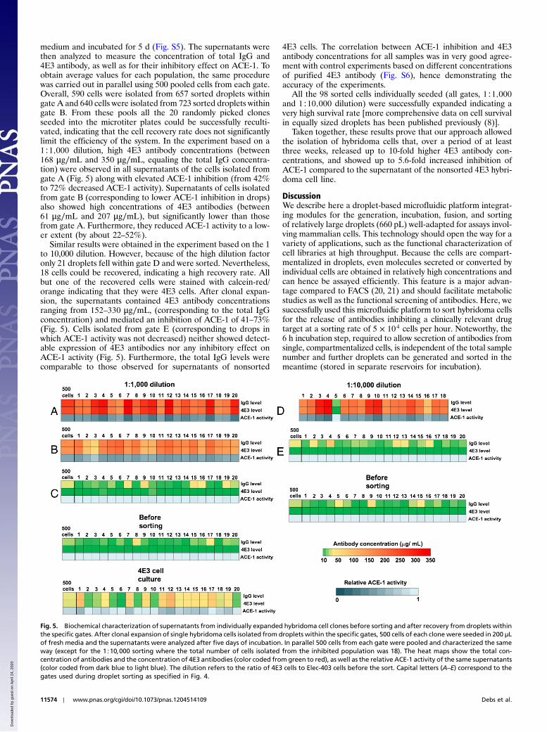
medium and incubated for 5 d (Fig. S5). The supernatants werethen analyzed to measure the concentration of total IgG and4E3 antibody, as well as for their inhibitory effect on ACE-1. Toobtain average values for each population, the same procedurewas carried out in parallel using 500 pooled cells from each gate.Overall, 590 cells were isolated from 657 sorted droplets withingate A and 640 cells were isolated from 723 sorted droplets withingate B. From these pools all the 20 randomly picked clonesseeded into the microtiter plates could be successfully reculti-vated, indicating that the cell recovery rate does not significantlylimit the efficiency of the system. In the experiment based on a1∶1;000 dilution, high 4E3 antibody concentrations (between168 μg∕mL and 350 μg∕mL, equaling the total IgG concentra-tion) were observed in all supernatants of the cells isolated fromgate A (Fig. 5) along with elevated ACE-1 inhibition (from 42%to 72% decreased ACE-1 activity). Supernatants of cells isolatedfrom gate B (corresponding to lower ACE-1 inhibition in drops)also showed high concentrations of 4E3 antibodies (between61 μg∕mL and 207 μg∕mL), but significantly lower than thosefrom gate A. Furthermore, they reduced ACE-1 activity to a low-er extent (by about 22–52%).
Similar results were obtained in the experiment based on the 1to 10,000 dilution. However, because of the high dilution factoronly 21 droplets fell within gate D and were sorted. Nevertheless,18 cells could be recovered, indicating a high recovery rate. Allbut one of the recovered cells were stained with calcein-red/orange indicating that they were 4E3 cells. After clonal expan-sion, the supernatants contained 4E3 antibody concentrationsranging from 152–330 μg∕mL, (corresponding to the total IgGconcentration) and mediated an inhibition of ACE-1 of 41–73%(Fig. 5). Cells isolated from gate E (corresponding to drops inwhich ACE-1 activity was not decreased) neither showed detect-able expression of 4E3 antibodies nor any inhibitory effect onACE-1 activity (Fig. 5). Furthermore, the total IgG levels werecomparable to those observed for supernatants of nonsorted
4E3 cells. The correlation between ACE-1 inhibition and 4E3antibody concentrations for all samples was in very good agree-ment with control experiments based on different concentrationsof purified 4E3 antibody (Fig. S6), hence demonstrating theaccuracy of the experiments.
All the 98 sorted cells individually seeded (all gates, 1∶1;000and 1∶10;000 dilution) were successfully expanded indicating avery high survival rate [more comprehensive data on cell survivalin equally sized droplets has been published previously (8)].
Taken together, these results prove that our approach allowedthe isolation of hybridoma cells that, over a period of at leastthree weeks, released up to 10-fold higher 4E3 antibody con-centrations, and showed up to 5.6-fold increased inhibition ofACE-1 compared to the supernatant of the nonsorted 4E3 hybri-doma cell line.
DiscussionWe describe here a droplet-based microfluidic platform integrat-ing modules for the generation, incubation, fusion, and sortingof relatively large droplets (660 pL) well-adapted for assays invol-ving mammalian cells. This technology should open the way for avariety of applications, such as the functional characterization ofcell libraries at high throughput. Because the cells are compart-mentalized in droplets, even molecules secreted or converted byindividual cells are obtained in relatively high concentrations andcan hence be assayed efficiently. This feature is a major advan-tage compared to FACS (20, 21) and should facilitate metabolicstudies as well as the functional screening of antibodies. Here, wesuccessfully used this microfluidic platform to sort hybridoma cellsfor the release of antibodies inhibiting a clinically relevant drugtarget at a sorting rate of 5 × 10⁴ cells per hour. Noteworthy, the6 h incubation step, required to allow secretion of antibodies fromsingle, compartmentalized cells, is independent of the total samplenumber and further droplets can be generated and sorted in themeantime (stored in separate reservoirs for incubation).
Fig. 5. Biochemical characterization of supernatants from individually expanded hybridoma cell clones before sorting and after recovery from droplets withinthe specific gates. After clonal expansion of single hybridoma cells isolated from droplets within the specific gates, 500 cells of each clone were seeded in 200 μLof fresh media and the supernatants were analyzed after five days of incubation. In parallel 500 cells from each gate were pooled and characterized the sameway (except for the 1∶10;000 sorting where the total number of cells isolated from the inhibited population was 18). The heat maps show the total con-centration of antibodies and the concentration of 4E3 antibodies (color coded from green to red), as well as the relative ACE-1 activity of the same supernatants(color coded from dark blue to light blue). The dilution refers to the ratio of 4E3 cells to Elec-403 cells before the sort. Capital letters (A–E) correspond to thegates used during droplet sorting as specified in Fig. 4.
11574 ∣ www.pnas.org/cgi/doi/10.1073/pnas.1204514109 Debs et al.
Dow
nloa
ded
by g
uest
on
Apr
il 24
, 202
0

Performing the screens on the single-cell level revealed a widevariance in antibody expression levels within a single hybridomaline and cells could be efficiently sorted and recovered based onthe level of ACE-1 inhibitory activity. In fact, the antibody secre-tion rate of 4E3 hybridoma cells (releasing ACE-1 inhibiting anti-bodies) recovered from droplets with low fluorescence intensity,indicating efficient ACE-1 inhibition, was significantly higherthan that of the unsorted 4E3 cell line. ELISA data indicated ap-proximately eightfold higher concentrations of the anti-ACE-1antibody on average and up to 10-fold higher concentrations forsome individual clones. It is well-known that point mutations andchromosome rearrangements frequently occur in hybridoma cellpopulations and change the expression level of individual cells(22). Over time, this effect can result in a percentage of nonanti-body producing cells (within a hybridoma cell population derivedfrom one and the same clone) between 40% and 85% (22) andillustrates the need for frequent single-cell sorting of existinghybridoma cell lines. As demonstrated here, our approach allowsthe specific selection and expansion of cells releasing higheramounts of antibodies (over a period of at least three weeks)compared to the nonsorted hybridoma cell population. More-over, the sensitivity of the system should even facilitate the func-tional screening of large cell libraries subsequent to immu-nization experiments, because we successfully demonstrated theselection and expansion of individual positive cells (releasingantibodies with desired properties) from a 10,000-fold excessof negative cells. As no cell proliferation is required, this techni-que could also open the way to directly screen nonimmortalizedprimary B-cells or plasma cells, which might be particularly usefulfor the cloning of antibodies from human donors, such as diseasesurvivors expressing therapeutically relevant antibodies of un-known identity. For example, the immune system of HIV-infectedindividuals sometimes evolves HIV-neutralizing antibodies, buttheir identification and characterization is still difficult and verytime consuming (23). Just recently, a method for the selection ofprimary B-cells releasing antibodies binding influenza virus he-magglutinin A has been developed in a microtiter plate formatand even enabled the identification of a neutralizing antibody(24). However, the screen itself was still based on binding activ-ities and did not allow the direct selection for functional proper-ties. In contrast, the approach described here can overcome thislimitation, even though distinguishing between highly inhibitoryantibodies secreted at low concentrations and less inhibitory anti-
bodies secreted at high concentrations might be difficult. None-theless the results obtained here clearly show an up to 9,400-foldenrichment of cells expressing antibodies with desired propertiesusing fluorescence activated droplet sorting. The false positivesobserved occasionally are most likely due to the co-encapsulationof a negative and a positive cell into the same droplet and canbe reduced by starting with a lower cell density during compart-mentalization (at the price of a lower overall throughput) (19).Alternatively hydrodynamic cell encapsulation modules (25–27)allowing the specific generation of droplets hosting single cellscould be used. False positives can also be ruled out during down-stream biochemical characterizations of sorted cells, at whichstage truly quantitative data on the inhibitory potency of thereleased antibodies can be obtained. Compared to conventionalapproaches our system also requires fewer cells and should allowscreening of a much larger fraction of the immune repertoire: In aconventional hybridoma experiment, typically no more than afew thousands of hybridoma clones are screened which representsonly a tiny fraction (approximately 1∕104) of the available anti-body repertoire in a mouse, and an even smaller fraction (approx-imately 1∕106) of the available human repertoire. Hence thetechnique described here the selection of antibodies against lessimmunogenic, but functionally more relevant, epitopes.
Materials and MethodsHeterogeneous hybridoma cell populations [4E3 and Elec-403 (13–15, 18)cells in a ratio of 1∶75; 1∶1;000 or 1∶10; 000] were encapsulated into 660 pLdrops at a density of 1.25 × 10⁶ cells∕mL together with 1.6 ng∕mL ACE-1(R&D Systems). The resulting emulsion was incubated off-chip for 6 h at37 °C under a 5% CO2 atmosphere, followed by reinjection into the inte-grated microfluidic chip (Fig. 1D), where drops containing the hybridomacells were fused with a second drop species containing a fluorogenic ACE-1substrate (Fig. 1B). Drops showing low fluorescence intensities (indicating alow ACE-1 activity) were sorted by applying an electrical field viaembedded electrodes adjacent to the channels. Sorted drops were brokenby adding an equal volume of 1H, 1H, 2H, 2H-Perfluoro-1-octanol (Aldrich)and subsequently hybridoma cells were isolated and seeded into 96-wellplates (Fig. S5). After expansion, the supernatants were characterized fortheir antibody concentration and ACE-1 inhibiting activity. For detailedexperimental procedures, see SI Materials and Methods.
ACKNOWLEDGMENTS. We thank Christophe Créminon for kindly providingElec-403 hybridoma cells and Alan Sawyer for valuable comments on themanuscript.
1. Nelson AL, Dhimolea E, Reichert JM (2010) Development trends for human monoclo-nal antibody therapeutics. Nat Rev Drug Discov 9:767–774.
2. Walsh G (2010) Biopharmaceutical benchmarks 2010. Nat Biotechnol 28:917–924.3. Hoogenboom HR (2005) Selecting and screening recombinant antibody libraries. Nat
Biotechnol 23:1105–1116.4. Karsunke XY, et al. (2011) Screening and characterization of new monoclonal anti-
benzo[a]pyrene antibodies using automated flow-through microarray technology.J Immunol Methods 371:81–90.
5. Sawyer A, et al. (2005) High throughput production of mouse monoclonal antibodiesusing antigen microarrays. Proteomics 5:4070–4081.
6. Ogunniyi AO, Story CM, Papa E, Guillen E, Love JC (2009) Screening individual hybri-domas by microengraving to discover monoclonal antibodies. Nat Protoc 4:767–782.
7. Theberge AB, et al. (2010) Microdroplets in microfluidics: An evolving platform fordiscoveries in chemistry and biology. Angew Chem Int Ed Engl 49:5846–5868.
8. Clausell-Tormos J, et al. (2008) Droplet-based microfluidic platforms for the encapsu-lation and screening of mammalian cells and multicellular organisms. Chem Biol15:427–437.
9. Baret JC, et al. (2009) Fluorescence-activated droplet sorting (FADS): Efficient micro-fluidic cell sorting based on enzymatic activity. Lab Chip 9:1850–1858.
10. Koster S, et al. (2008) Drop-based microfluidic devices for encapsulation of single cells.Lab Chip 8:1110–1115.
11. Parvathy S, et al. (1997) Angiotensin-converting enzyme secretase is inhibited by zincmetalloprotease inhibitors and requires its substrate to be inserted in a lipid bilayer.Biochem J 327:37–43.
12. Wei L, Clauser E, Alhenc-Gelas F, Corvol P (1992) The two homologous domains ofhuman angiotensin I-converting enzyme interact differently with competitive inhibi-tors. J Biol Chem 267:13398–13405.
13. Danilov S, et al. (1994) Structure-function analysis of angiotensin I-converting enzymeusing monoclonal antibodies. Selective inhibition of the amino-terminal active site.J Biol Chem 269:26806–26814.
14. Skirgello OE, et al. (2006) Inhibitory antibodies to human angiotensin-converting en-zyme: Fine epitope mapping and mechanism of action. Biochemistry 45:4831–4847.
15. Naperova IA, et al. (2008) [Characteristics of monoclonal antibody binding with the Cdomain of human angiotensin converting enzyme]. Bioorg Khim 34:358–364.
16. Zaman MA, Oparil S, Calhoun DA (2002) Drugs targeting the renin-angiotensin-aldos-terone system. Nat Rev Drug Discov 1:621–636.
17. Niu X, Gulati S, Edel JB, deMello AJ (2008) Pillar-induced droplet merging in microflui-dic circuits. Lab Chip 8:1837–1841.
18. Remy MH, Frobert Y, Grassi J (1995) Characterization of monoclonal antibodiesthat strongly inhibit Electrophorus electricus acetylcholinesterase. Eur J Biochem231:651–658.
19. Baret JC, et al. (2009) Fluorescence-activated droplet sorting (FADS): Efficient micro-fluidic cell sorting based on enzymatic activity. Lab Chip 9:1850–1858.
20. Pierzchalski A,Mittag A, Tarnok A (2011) Introduction A: Recent advances in cytometryinstrumentation, probes, and methods—review. Methods Cell Biol 102:1–21.
21. Mouquet H, et al. (2011) Memory B cell antibodies to HIV-1 gp140 cloned fromindividuals infected with clade A and B viruses. PLoS One 6:e24078.
22. Kromenaker SJ, Srienc F (1994) Stability of producer hybridoma cell lines after cellsorting: A case study. Biotechnol Prog 10:299–307.
23. Pietzsch J, et al. (2010) Anti-gp41 antibodies cloned from HIV-infected patients withbroadly neutralizing serologic activity. J Virol 84:5032–5042.
24. Corti D, et al. (2011) A neutralizing antibody selected from plasma cells that binds togroup 1 and group 2 influenza A hemagglutinins. Science 333:850–856.
25. Chabert M, Viovy JL (2008) Microfluidic high-throughput encapsulation and hydrody-namic self-sorting of single cells. Proc Natl Acad Sci USA 105:3191–3196.
26. Edd JF, et al. (2008) Controlled encapsulation of single-cells into monodisperse pico-litre drops. Lab Chip 8:1262–1264.
27. Abate AR, Chen CH, Agresti JJ, Weitz DA (2009) Beating Poisson encapsulation statis-tics using close-packed ordering. Lab Chip 9:2628–2631.
Debs et al. PNAS ∣ July 17, 2012 ∣ vol. 109 ∣ no. 29 ∣ 11575
APP
LIED
BIOLO
GICAL
SCIENCE
SAPP
LIED
PHYS
ICAL
SCIENCE
S
Dow
nloa
ded
by g
uest
on
Apr
il 24
, 202
0