introduction to advanced trauma life support atls · blunt abdominal trauma with hypotension &...
TRANSCRIPT

Introduction toAdvanced Trauma Life Support
ATLS
Objectives
● Concepts of primary & secondary survey● Priorities & Life threatening conditions● Clinical & Surgical skills
Basic knowledge
● Rapid assessment● Resuscitate & Stabilize (Prioritize)● Patient's needs & facility's capabilities● Appropriate transfer● Optimum care

Initial Assessment & Management
● Preparation (Prehospital - Hospital)● Triage● Primary survey (ABCDE)● Resuscitation● Adjuncts to primary survey & resuscitation● ->

Initial Assessment & Management
● Secondary survey● Adjuncts to the secondary survey● Postresuscitation monitoring● Definitive care

Primary Survey
● Treatment priorities● A: Airway maintenance + C-spine protection● B: Breathing & Ventilation● C: Circulation & Hemorrhage control● D: Disability – Neuro● E: Exposure / Environment control

A
● Airway– Patency / Obstruction– Severe head injury -> Definitive airway
Airway: Patency
● Maxillofacial trauma● Neck trauma● Laryngeal trauma (Hoarseness, Subcutaneous
emphysema, Palpable fracture)

A
● C-spine protection– Multiple system trauma– Altered level of consciousness– Blunt injury above clavicle– Manual in-line stabilization

A: Nexus
● Midline cervical tenderness● Altered level of consciousness● Evidence of intoxication● Neurologic abnormality● Presence of painful distracting injury

A
● Trauma patient is dynamic● Repeated assessment

A: Resuscitation
● Jaw thust / Chin lift / Head tilt● Naso / Oropharyngeal airway● Combitube, LMA● Definitive airway (Cuff in trachea)
– Oro / Naso tracheal intubation– Surgical cricothyroidotomy

Endotracheal intubation
● Indication– Provide patent airway– Deliver supplemental oxygen– Support ventilation– Prevent aspiration
Endotracheal intubation
● Decision– Apnea (orotracheal)– Cannot maintain patent airway– Protect aspiration / vomitus– Impending compromise airway– Closed head injury required assisted ventilation– Inadequate oxygenation

Surgical Airway
● Cricothyroidotomy / Tracheostomy
● Indication– Unable to intubate (severe maxillofacial injury,
failed intubation)● Contraindication
– Airway transection

B: Breathing

B: Life Threatening Conditions
● Tension pneumothorax● Flail chest with pulmonary contusion● Massive Hemothorax● Open pneumothorax● Cardiac tamponade

Thoracic Trauma: Primary survey
● Looking, Palpation, Percussion, Listening– Tension pneumothorax– Open pneumothorax (sucking chest wound)– Flail chest– Massive hemothorax– Cardiac tamponade

Thoracic Trauma: Primary survey
● Tension pneumothorax– Chest pain, Respiratory distress, Tachycardia,
Hypotension, Tracheal deviation, Absent breath sound, Neck vein distension
– Immediate decompression● Needle thoracostomy● Intercostal drainage

Thoracic Trauma: Primary survey
● Open pneumothorax (sucking chest wound)– > 2/3 of tracheal diameter– 3 sided dressing– Chest tube insertion

Open Chest Wound: 3-Sided Dressing

Thoracic Trauma: Primary survey
● Flail chest– >2 ribs fractures in 2 or more places– Paradoxical chest wall movement– Adequate ventilation– Reexpand lungs: Intubation

Thoracic Trauma: Primary survey
● Massive hemothorax– >1500 cc of blood (1/3 of blood volume) in chest
cavity– IV resuscitation– Chest tube– Thoracotomy
● >1500 cc immediately● 200 cc/h for 2-4 h
Thoracic Trauma: Primary survey
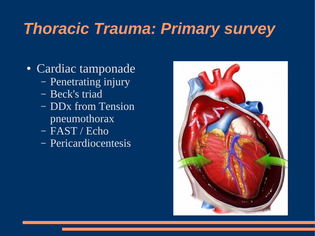
● Cardiac tamponade– Penetrating injury– Beck's triad– DDx from Tension
pneumothorax– FAST / Echo– Pericardiocentesis

B: Resuscitation
● Supplemental oxygen● Tension pneumothorax decompression

C: Circulation & Hemorrhage control
● Circulation – Blood volume & Cardiac output● Level of consciousness● Skin color● Pulse

C
● Hemorrhage control - External hemorrhage– Manual pressure– Splinting– Tourniquet– Hemostats

C: Resuscitation
● 2 large-caliber IV catheter● “warm” NSS, RLS● Blood● Control bleeding
– Direct pressure– Operative control
● Vasopressors
Shock
● Inadequate tissue perfusion / oxygenation● Hemorrhagic / Non-hemorrhagic

Hemorrhagic shock
● Most common cause of shock in trauma● External vs Internal hemorrhage● Blood volume = 7% of BW● Rx: Volume replacement● Shock Classification

Hemorrhagic shock classification
● Class I– 15% blood loss– P < 100– BP normal– PP normal– RR 14-20– Urine output >30 cc/h– Mental status: Slightly anxious
Hemorrhagic shock classification
● Class II– 15-30% blood loss– P > 100– BP Normal– PP decreased– RR 20-30– Urine output 20-30 cc/h– Mental status: mildly anxious

Hemorrhagic shock classification
● Class III– 30-40% blood loss– P >120– BP decreased– PP decreased– RR 30-40– Urine output 5-15 cc/h– Mental status: confused

Hemorrhagic shock classification
● Class IV– >40% blood loss– P >140– BP decreased– PP decreased– RR > 35– Urine output ---– Mental status: confused / lethargic
Fluid replacement
● Class I, II: Crystalloid● Class III, IV: Crystalloid, Blood● Initial fluid therapy
– 1-2 L for adult– 20 cc/kg for children
● “3-for-1” rule– 1 cc blood loss = 3 cc crystalloid replacement

Response to fluid resuscitation
● Rapid response– <20% blood loss– Cross-match, Surgical consultation
● Transient response– 20-40% blood loss– On going blood loss– Blood transfusion, Surgical intervention

Response to fluid resuscitation
● No response– Immediate operative intervention

Non-hemorrhagic shock
● Cardiogenic shock● Tension pneumothorax● Neurogenic shock● Septic shock

Cardiogenic shock
● Cardiac contusion● Cardiac tamponade: “Beck's triad”
– Tachycardia– Muffled heart sound– Distended neck vein
● Echo / FAST

Cardiac Tamponade
● Penetrating injury● Beck's triad● DDx from Tension pneumothorax● FAST / Echo● Rx: Pericardiocentesis
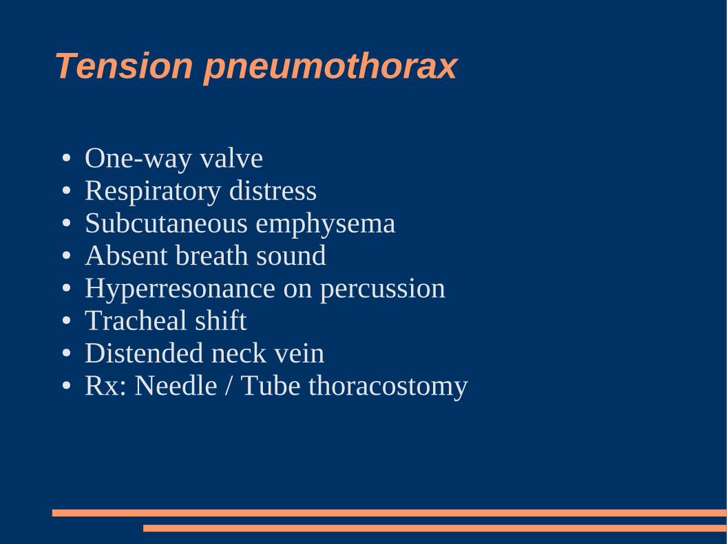
Tension pneumothorax
● One-way valve● Respiratory distress● Subcutaneous emphysema● Absent breath sound● Hyperresonance on percussion● Tracheal shift● Distended neck vein● Rx: Needle / Tube thoracostomy
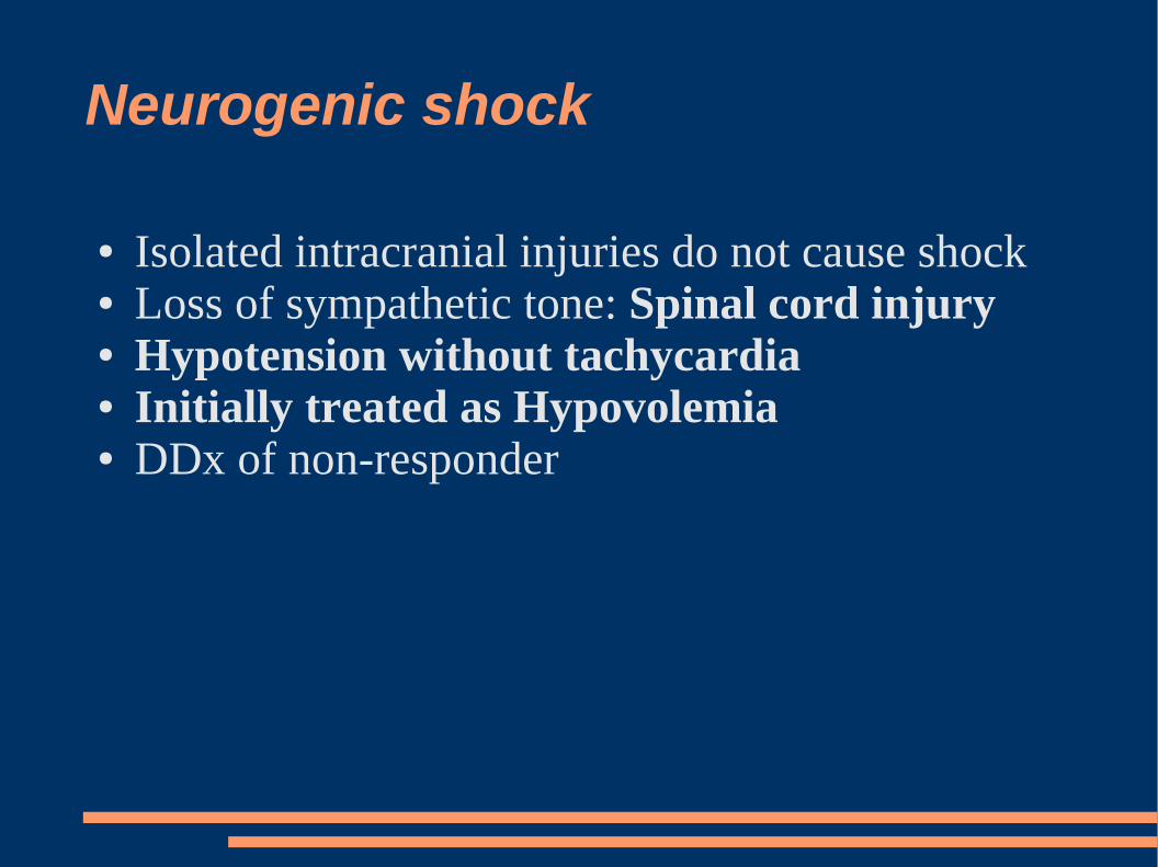
Neurogenic shock
● Isolated intracranial injuries do not cause shock● Loss of sympathetic tone: Spinal cord injury● Hypotension without tachycardia● Initially treated as Hypovolemia● DDx of non-responder

D
● Neurological status– Level of consciousness (AVPU / GCS)– Pupil size & Light reaction– Lateralizing sign– Spinal cord injury level

D
● A: Alert● V: Verbal command● P: Painful stimuli● U: Unresponsive
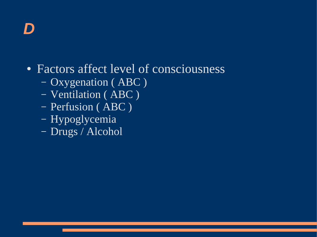
D
● Factors affect level of consciousness– Oxygenation ( ABC )– Ventilation ( ABC )– Perfusion ( ABC )– Hypoglycemia– Drugs / Alcohol

D
● Reevaluation

E
● Uncloth patient● Logroll patient● Prevent hypothermia
– Warm blanket– Warm IV fluid

E
● Rectal examination– Sphinctor tone– Position of prostate (high-riding?) = urethral injury– Gross blood (penetrating abdominal injury)– Pelvic fractures

Primary survey: Adjuncts
● Monitor● Diagnosis
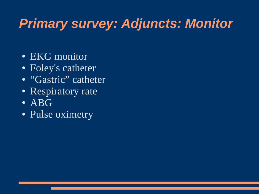
Primary survey: Adjuncts: Monitor
● EKG monitor● Foley's catheter● “Gastric” catheter● Respiratory rate● ABG● Pulse oximetry

Primary survey: Adjuncts: Diagnosis
● CXR, Pelvis AP, Lateral C-spine● DPL, FAST● Should not interrupt
resuscitation process

Foley's catheter
● Contraindicated in Urethral injury● Suspected urethral injury
– Inability to void– Unstable pelvic fracture– Blood at meatus– Scrotal hematoma– Perineal ecchymoses– High-riding prostate
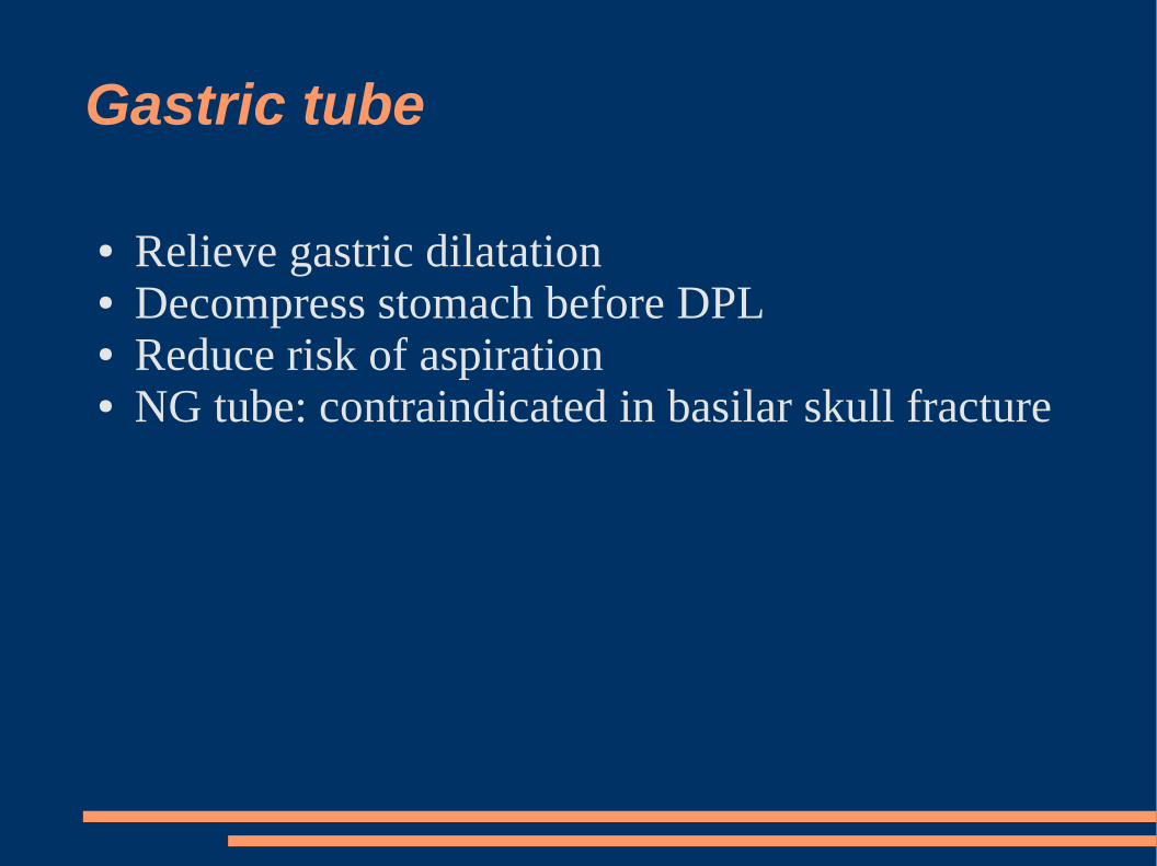
Gastric tube
● Relieve gastric dilatation● Decompress stomach before DPL● Reduce risk of aspiration● NG tube: contraindicated in basilar skull fracture

Secondary Survey
● Not begin until primary survey is completed● History (AMPLE)● Head-to-toe evaluation● GCS● X-rays

Secondary Survey: Adjuncts
● Specialized diagnostic tests (CT, US, scope)● Should not be performed until hemodynamic
stabilization

Secondary Survey
● History: AMPLE– A: Allergies– M: Medications– P: Past illnesses / Pregnancy– L: Last meal– E: Events

Secondary Survey
● Physical examination● Head-to-toe examination
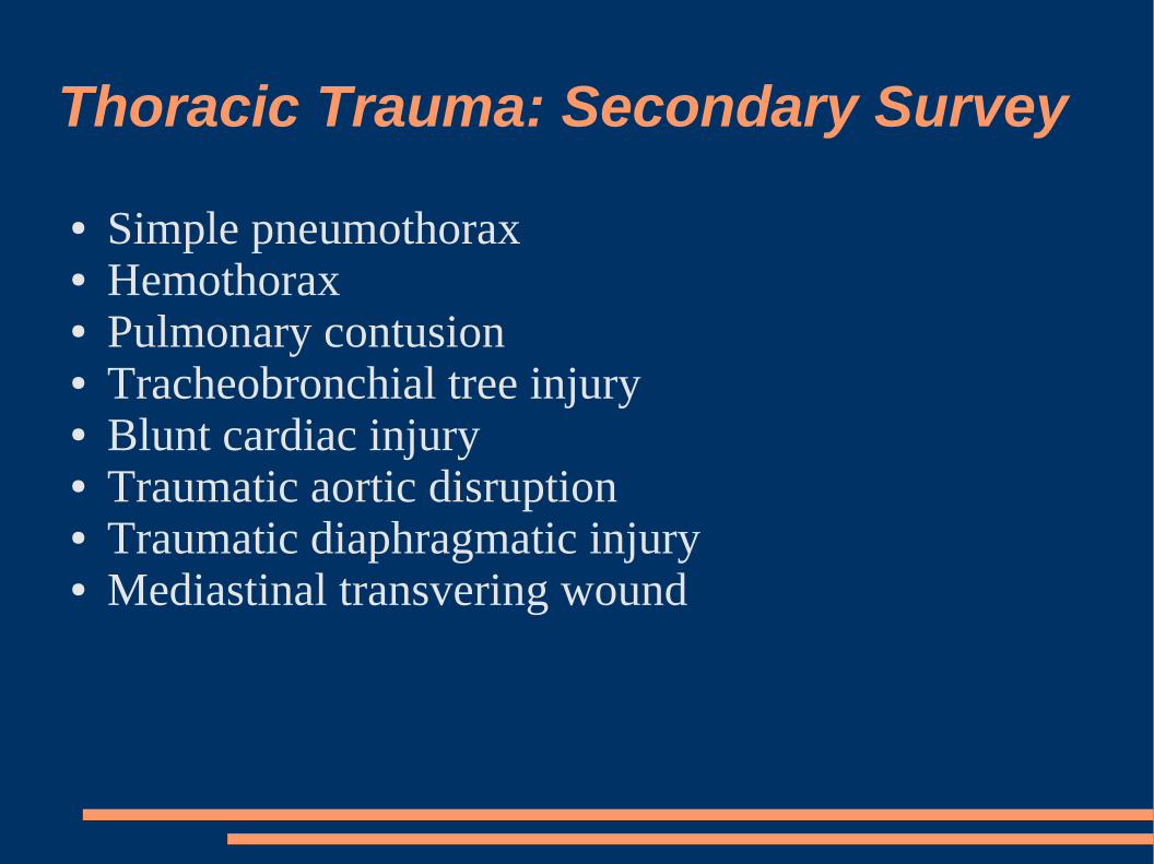
Thoracic Trauma: Secondary Survey
● Simple pneumothorax● Hemothorax● Pulmonary contusion● Tracheobronchial tree injury● Blunt cardiac injury● Traumatic aortic disruption● Traumatic diaphragmatic injury● Mediastinal transvering wound

Abdominal Trauma

Abdominal Trauma
● External anatomy– Anterion– Flank– Back

Abdominal Trauma
● Internal anatomy– Peritoneal cavity– Pelvic cavity– Retroperitoneal space

Abdominal Trauma
● Mechanism of injury– Blunt– Penetrating

Abdominal Trauma: Assessment
● History● Physical Exam
– Inspection, Auscultation, Percussion, Palpation– Evaluation of penetrating wound– Pelvic stability– Penile, Perineal, Rectal exam– Vaginal, Gluteal exam

Celiotomy: Indications
● Blunt abdominal trauma with hypotension & evidence of intraperitoneal bleeding
● Blunt abdominal trauma with positive DPL or FAST
● Hypotension with penetrating abdominal wound● GSW traversing the peritoneal cavity / visceral /
vascular retroperitoneum● Evisceration
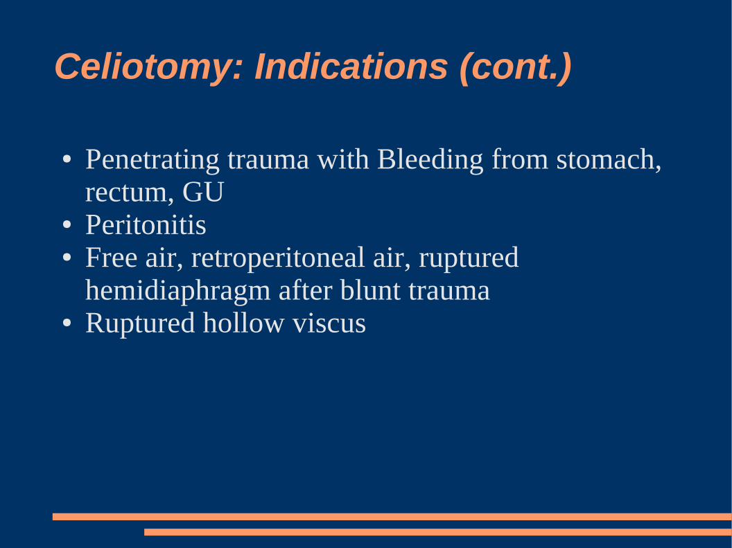
Celiotomy: Indications (cont.)
● Penetrating trauma with Bleeding from stomach, rectum, GU
● Peritonitis● Free air, retroperitoneal air, ruptured
hemidiaphragm after blunt trauma● Ruptured hollow viscus

Diagnostic Studies
● Diagnostic peritoneal lavage: DPL● FAST● CT scan● Urethrography, Cystography, IVP

Diagnostic Peritoneal Lavage:DPL
● Indications– Altered level of conscious / Spinal cord injury– Injury to adjacent structures– Equivocal physical exam– Prolonged loss of contact with patient– Lap-belt sign

Diagnostic Peritoneal Lavage:DPL
● Contraindications– Existing indication for celiotomy
● Relative contraindications– Previous abdominal operations– Morbid obesity– Advanced cirrhosis– Coagulopathy

Diagnostic Peritoneal Lavage:DPL
● 1 L of LRS● Fluid return: >30% of infused volume● Positive Interpretation (blunt abdominal injury):
– Gross blood > 10 cc– RBC >100,000 /mm3– WBC > 500 /mm3– Food particles– Gram stain +ve

Head injury

Head Injury
● Classification– Mechanism (Blunt, Penetrating)– Severity (mild, moderate, severe)– Morphology (Skull fractures, Intracranial)

Head Injury: Severity
● Mild: GCS 13-15● Moderate: GCS 9-12● Severe: GCS 3-8
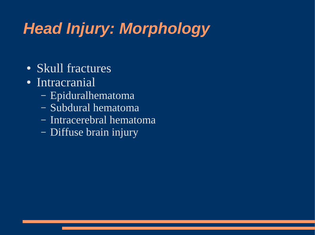
Head Injury: Morphology
● Skull fractures● Intracranial
– Epiduralhematoma– Subdural hematoma– Intracerebral hematoma– Diffuse brain injury

Skull fractures
● Cranium● Maxillofacial● Basilar skull fractures

Basilar skull fracture
● Raccoon's eyes● Battle's sign● CSF rhinorrhea / otorrhea
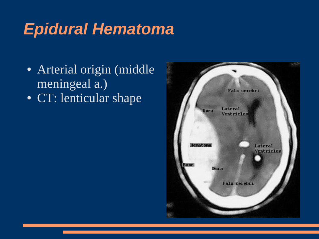
Epidural Hematoma
● Arterial origin (middle meningeal a.)
● CT: lenticular shape

Subdural Hematoma
● Venous origin● CT: Crescent shape

Intracerebral Hematoma
● Brain laceration

Head Injury: Management
● Mild HI (GCS 13-15)– Observe– CT:
● Lost of conscious > 5 min● Amnesia● Severe headache● Focal neurological deficit

Head Injury: Management
● Moderate HI (GCS 9-12)– CT brain– Admit observe neurosigns– F/U CT brain 12-24 h

Head Injury: Management
● Severe HI (GCS < 9)– Prompt diagnosis & treatment– Don't delay patient transfer to obtain CT scan
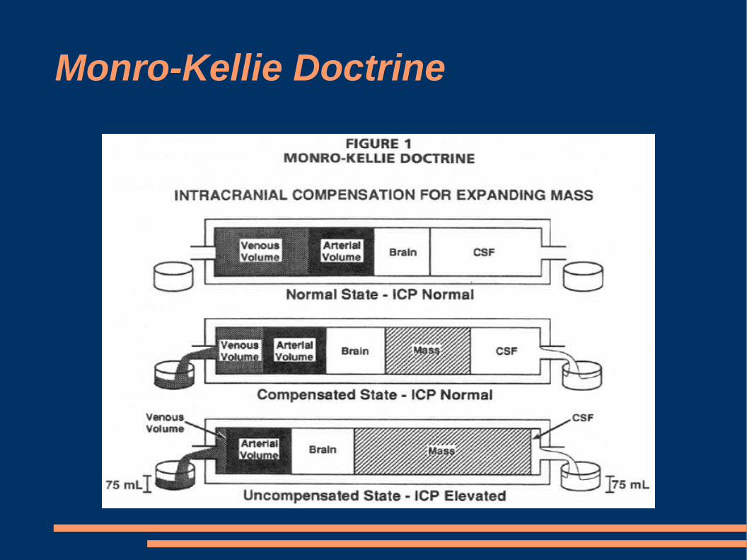
Monro-Kellie Doctrine
Brain resuscitation
● Maintain adequate– Cerebral Perfusion Pressure (CPP)– Oxygenation– Normocapnia

Cerebral Perfusion Pressure
● CPP = MAP – ICP– MAP = Mean Arterial Pressure– ICP = Intracranial Pressure

Cerebral Perfusion Pressure
● CPP = MAP – ICP– MAP = Mean Arterial Pressure
● Stabilize Vital signs● IV fluids
– ICP = Intracranial Pressure● Hyperventilation (limited usage)● Mannitol (1g/kg)● Furosemide
Brain resuscitation
● Oxygenation– Oxygen supplement– Anticonvulsants
● Normocapnia– Hyperventilation -> CO
2 -> Cerebral vasoconstriction
-> CPP

Conclusions
● Initial Assessment (Primary survey, Secondary survey)
● Adjuncts● Priority: Life threatening first● Knowledge & Skills for specific conditions● DOs & DON'Ts

Q?

http://www.slideshare.net/narenthorn/introduction-to-atls-presentation/