intravenous fluosol in the treatment ofacute myocardial...
TRANSCRIPT

114
Intravenous Fluosol in the Treatmentof Acute Myocardial Infarction
Results of the Thrombolysis and Angioplasty in MyocardialInfarction 9 Trial
Thomas C. Wall, MD; Robert M. Califf, MD; James Blankenship, MD; J. David Talley, MD;Mark Tannenbaum, MD; Markus Schwaiger, MD; Gerald Gacioch, MD; Mark D. Cohen, MD;Mark Sanz, MD; Jeffrey D. Leimberger, PhD; Eric J. Topol, MD; the TAMI 9 Research Group*
Background This study was performed to determine thesafety and potential efficacy of an intravenous perfluorochem-ical emulsion (Fluosol) as an adjunct reperfusion therapyaimed at preventing reperfusion injury for patients with acutemyocardial infarction.Methods and Results Patients (430) were randomized in a
prospective open-labeled study, 213 to receive Fluosol and 217to receive no Fluosol, along with 100 mg of tissue-typeplasminogen activator given over 3 hours. Major end pointsincluded global ejection fraction, regional wall motion analysis,infarct size as measured by tomographic thallium imaging, anda composite clinical outcome measure. Baseline patient andangiographic characteristics were similar in the two groups. Nosignificant difference in global ejection fraction (52% withoutFluosol, 51% with Fluosol) or regional wall motion (-2.4SD/chord with Fluosol, -2.2 SD/chord without Fluosol) wasdemonstrated in patients receiving Fluosol versus those notreceiving Fluosol, nor was there a significant difference inthallium infarct size. Although Fluosol-treated patients withanterior infarction had an insignificantly lower mean infarctsize (18.7% of the left ventricle) compared with patients with
anterior infarction not treated with Fluosol (21.2% of leftventricle), this trend was not evident in the median infarct sizevalues (22% versus 17%), left ventricular ejection fractionvalues (46% without Fluosol, 47% with Fluosol), or regionalwall motion (-2.5 SD/chord in both groups). Rates of deathand stroke were no different in the two groups; however,patients who received Fluosol experienced less recurrentischemia. Patients receiving intravenous Fluosol had moretransient congestive heart failure and pulmonary edema, per-haps because of necessary fluid administration. There was nodifference in hemorrhagic complications between the twostudy groups.
Conclusions When given with a thrombolytic agent, Fluosolwas not associated with improvement in ventricular systolicfunction, reduction in thallium infarct size, or overall clinicaloutcome. Fluosol was, however, associated with a reduction inischemic complications and with an increase in pulmonary edemaand congestive heart failure. (Cirulaton. 1994;90'.114-120.)Key Words * myocardial infarction * ventricles * Fluosol
* radioisotopes
T he amount of myocardial damage is the primarydeterminant of survival and functional statusafter acute myocardial infarction (MI).' Al-
though reperfusion reduces infarct size, improves sys-tolic left ventricular (LV) function, leads to lowerventricular volumes, reduces symptomatic congestiveheart failure, and improves mortality, the magnitude ofthis effect has been less substantial than initiallyhoped.23 Efforts to improve the amount of myocardialsalvage have focused on earlier reperfusion, more com-plete perfusion, and prevention of infarct vesselreocclusion.
Received February 25, 1994; revision accepted March 3, 1994.From Duke University Medical Center, Durham, NC (T.C.W.,
R.M.C., J.D.L.); Geisinger Medical Center, Danville, Pa (J.B.);University of Louisville (Ky) (J.D.T.); Mercy Hospital, DesMoines, Iowa (M.T.); Rochester General Hospital, Rochester, NY(G.G.); St. Francis Hospital, Beech Grove, Ind (M.D.C.); St.Patrick Hospital, Missoula, Mont (M. Sanz); Cleveland Clinic,Cleveland Ohio (E.J.T.); Poliklinik der Technischen UniversitatMunchen, Munich, Germany (M. Schwaiger).
Correspondence to Robert M. Califf, M.D., Box 31123, DukeUniversity Medical Center, Durham, NC 27710.*A full list of participating sites is included in the
Acknowledgments.© 1994 American Heart Association, Inc.
Another approach to improving reperfusion therapyarose from animal studies indicating that the amount ofmyocardial salvage after reperfusion may be limited byanatomic and metabolic consequences of reperfusion.4,5This phenomenon, referred to as reperfusion injury, hasbeen limited in animal models by Fluosol, a perfluoro-chemical emulsion. These studies have reported that theintracoronary administration of oxygenated Fluosol re-sulted in smaller infarct size and improved systolic LVfunction.6-8 Supportive evidence of beneficial effects, interms of both preservation of the microvasculatureand more substantial salvage of myocardium, has beendeveloped. In one study of 26 patients,9 the intracor-onary administration of Fluosol in the setting of directangioplasty for anterior MI produced a marked im-provement in global and regional LV function assessedby contrast ventriculography and a reduction in tomo-graphic thallium-estimated infarct size. Mechanismsthat have been raised as the pathophysiological basisfor reperfusion injury include leukocyte release of freeradicals and other toxic metabolites at the time ofreperfusion and destruction of microvasculature.10Fluosol has been demonstrated to be a potent inhibi-tor of white cell chemotaxis and activation."
by guest on June 16, 2018http://circ.ahajournals.org/
Dow
nloaded from
Wall et al Fluosol Treatment in MI 115
The Thrombolysis and Angioplasty in MyocardialInfarction (TAMI) 9 study was designed to determinewhether intravenous Fluosol in conjunction with throm-bolytic therapy would reduce myocardial infarct size asestimated by thallium scintigraphy or improve systolicLV function and provide preliminary evidence of clini-cal effect.
MethodsPatient EnrollmentTen university and large cardiac referral centers partici-
pated in this trial (see "Acknowledgments"). The protocol wasapproved by the institutional review board at all participatingsites. Patients were identified and enrolled in the study in theemergency room setting. Inclusion criteria included symptomsconsistent with an acute MI of <6 hours' duration unrespon-sive to sublingual nitroglycerin; age >18 and <75 years of age;ST-segment elevation of at least 0.1 mV in at least two of sixprecordial leads, two of three inferior leads, or in both leads Iand aVL; and ability and willingness to give informed consentto participate in this study. Exclusion criteria included treat-ment with Fluosol within the previous 6 months, ongoing renaldialysis, clinically significant liver disease, chronic obstructivepulmonary disease, other serious advanced illnesses likely tolimit life expectancy, prolonged cardiopulmonary resuscitation(>10 minutes) within the previous 2 weeks, other standardcontraindications to thrombolytic therapy, previous coronaryartery bypass graft surgery, previous Q-wave infarction, andcardiogenic shock.
TreatmentOnce inclusion and exclusion criteria were met, patients
were randomized by telephone by the Coordinating Center atDuke University Medical Center. All patients were initiallytreated with 324 mg of chewable aspirin, intravenous heparin,and 100 mg of intravenous tissue-type plasminogen activator(TPA) over 3 hours. In addition, patients received intravenousatenolol unless they had atrioventricular block, systolic bloodpressure <100 mm Hg, heart rate <60 beats per minute,bibasilar rales, or a significant history of bronchospasm. Intra-venous morphine, nitroglycerin, and atropine also were used atthe discretion of the investigator.
After the initiation of the above therapy, patients wererandomized to receive intravenous Fluosol or no Fluosol.Because of previous concern with hypotension in caninestudies, intravenous Fluosol was administered at the rate of 1mL/min for the first 5 minutes, 5 mL/min for the next 5minutes, and 20 mL/min until a total of 15 mL/kg had beenadministered. The rate was reduced in the presence of any signof pulmonary congestion or edema; 77% of patients receivedthe full dose. All patients continued to receive intravenousheparin at a rate of 1000 U/h to maintain the partial throm-boplastin time at 1.5 to 2 times the control value. An attemptwas made to administer oxygen at 100% concentration (6 to 10L/min) for 8 hours after randomization to maximize theoxygen carried by the study agent; this goal was achieved in89% of Fluosol-treated patients and in 86% of controlpatients.
Cardiac CatheterizationAn effort was made to perform cardiac catheterization,
including coronary angiography and left ventriculography, 5 to14 days after the administration of thrombolytic therapy. Leftventriculograms of adequate quality were obtained within theprespecified 5- to 14-day window in 312 (73%) of the patientsenrolled in the trial. An additional 32 left ventriculogramswere obtained before 5 days because of recurrent ischemia,and 6 studies were completed after 14 days, giving a total of81% of randomized patients with studies suitable for analysis.Of the remaining patients, 34 studies were done but were of
inadequate quality, 18 were not done because the patientsdied, 8 were medically contraindicated, 8 were not obtaineddue to patient preference, and 12 were not obtained for otherreasons. All cineangiograms were evaluated independently atthe core angiographic laboratory at the Cleveland Clinic.Quantitative analysis included TIMI perfusion grade,12 visualpercent stenosis of the infarct-related artery, and global LVfunction as determined by the area-length method,13 whichwas expressed as global ejection fraction. Regional wall motionof the infarct and noninfarct zones was evaluated quantita-tively and expressed as SD/chord using the centerline chordmethod.14 Technically inadequate studies due to inadequatecontrast or frequent ventricular extrasystoles were not in-cluded in the analysis. All efficacy analyses were performedtwice: in the primary analysis, only patients with studies withinthe 5- to 14-day window were used; then an analysis was doneusing all patients with technically adequate studies. Since thetreatment comparisons did not differ with either analysisstrategy, only the results for the analysis of 5- to 14-day studiesare presented.
Thallium ScintigraphyInfarct size was also measured by tomographic thallium
imaging 5 to 14 days after thrombolytic therapy. All thalliumscans were evaluated independently at the core nuclear labo-ratory at the University of Michigan. An effort was made toexercise all patients using the standard Bruce protocol to asymptom-limited end point unless the patient was incapable, inwhich case pharmacological stress with dipyridamole or aden-osine was used. An injection of 3 mCi IV of 20`TI was given atpeak exercise or after pharmacological stress. After the injec-tion, exercise was maintained for at least 60 seconds. Imagingwas started 15 minutes after cessation of exercise. Three to 5hours after exercise, the resting image was acquired afterinjection of an additional 1 mCi of 1Tl. In both cases, 32images were acquired on an 84x 84 matrix, with care taken tominimize motion artifact.
After the images were translated into digital images, theywere stored on floppy disk and sent to the Thallium CoreLaboratory. Because the data were collected on multiple-imaging systems, a Siemens Microdelta computer and softwaredeveloped by Sudbury Inc were used to convert the data intoone format. Care was taken to label the disks so that thetreatment assignment of the patient was unknown during theprocessing and analysis of the data.
All tomographic images were analyzed using the "bull's-eye" approach, in which peak `'Tl activity was profiled along60 radii extending from the center of the left ventricle. A polarmap of the left ventricle then was constructed using thehistograms of cross-sectional and sagittal images. Relativetracer distribution was color coded to provide normalizedratios compared with maximal activity and with a database ofage-matched normal subjects. After the polar map was dividedinto typical vascular territories defined from a database ofpatients with single-vessel disease, the estimate of infarct sizewas made based on the percentage of vascular territories of theleft ventricle with regional activity below 2.5 SD of normal.
Resting tomographic thallium studies were technically ade-quate and within the 5- to 14-day window in 258 patients(60%). An additional 67 studies were done outside the 5- to14-day window, for a total of 325 technically adequate studies(76%). Of the remaining patients, 17 studies were not donebecause the patients died, 16 were technically inadequate, 10were done with planar images only, 6 could not be donebecause of the patient's weight, 6 were not done because ofmedical contraindications, and 50 were not done for otherreasons. As with the left ventriculograms, analyses for efficacyend points were repeated, including and excluding studiescompleted within the prespecified 5- to 14-day window. Sincethe results were no different in terms of the treatment com-
by guest on June 16, 2018http://circ.ahajournals.org/
Dow
nloaded from
116 Circulation Vol 90, No 1 July 1994
parison, results are presented only for patients with studieswithin the time window.
Hemorrhagic MeasurementsBleeding observed during the hospital stay was defined as
follows: mild if it was of no clinical consequence, did notrequire transfusion, or resulted in a total blood loss of <250mL; moderate if 250 to 500 mL of blood loss was observed;severe if any of the following events occurred: >500 mL ofblood loss necessitating a transfusion, intracranial bleeding, orgastrointestinal or other external or internal bleeding causinghypotension and requiring emergency transfusion. The site ofbleeding, baseline and nadir hematocrit, and packed red bloodcell transfusion were recorded for all patients.
Clinical End PointsA composite clinical end point was constructed before the
study based on a composite of end points considered mostimportant by a survey of cardiologists.15 This end pointconsisted of death from any cause, stroke, nonfatal reinfarc-tion, emergency revascularization, development of new heartfailure or pulmonary edema, or significant recurrent ischemia.All of these end points were carefully monitored during andafter the study enrollment because the study was not blinded.Stroke was defined as a new focal neurological deficit lasting atleast 24 hours or documented to correspond to a finding atcomputed tomographic or magnetic resonance imaging if thepatient died before 24 hours. Strokes were further subclassi-fied as hemorrhagic or nonhemorrhagic.16 New heart failurewas defined as the development of new signs and symptoms ofpulmonary congestion documented by chest radiograph ifavailable or evidence of inadequate perfusion (hypotension >1hour or requirement for intravenous inotropic medication tomaintain a systolic blood pressure >90 mm Hg) persisting for>1 hour in the setting of adequate filling pressure. Because ofthe subjective nature of this end point, each case was reviewedin detail by an independent study monitor blinded to treat-ment collecting information on physical exam, chest radio-graph, medical intervention, and hemodynamic findings. Re-current ischemia was defined as symptoms compatible withmyocardial ischemia associated with new ST-segment or
T-wave changes on the electrocardiogram. These cases were
also reviewed independently by a study monitor.
Statistical MethodsRandomization by the Coordinating Center at Duke Uni-
versity was completed in blocks within each study site. Casereport forms were completed by the clinical research nurse
coordinators and reviewed by the principal investigators be-fore submission to the Coordinating Center. The data wereverified independently by study monitors through review of theclinical records. The Data and Safety Monitoring Board metregularly during the study to review the clinical outcome of thepatient population.
Continuous baseline and outcome variables are summarizedby the median and interquartile range (25th to 75th percen-
tiles), and discrete variables are expressed as percentages;95% confidence intervals are calculated for major clinicaloutcomes. The statistical significance of the effect of thetreatment on continuous variables was assessed by use of theWilcoxon rank-sum test. The likelihood ratio X2 statistic was
used to examine treatment effects for discrete variables. If lowevent rates precluded the use of a x2 statistic, Fisher's exacttest was used.The sample size was calculated to provide adequate num-
bers of imaging studies to detect at least a 4-unit improvementin left ventricular ejection fraction. A total of 122 patients withcompleted studies per group provides 80% power with an a of0.05, assuming an SD of the measurement of 11 units. Simi-larly, the study had adequate power (80%) to detect a 33%reduction in thallium infarct size with a total sample size of 210
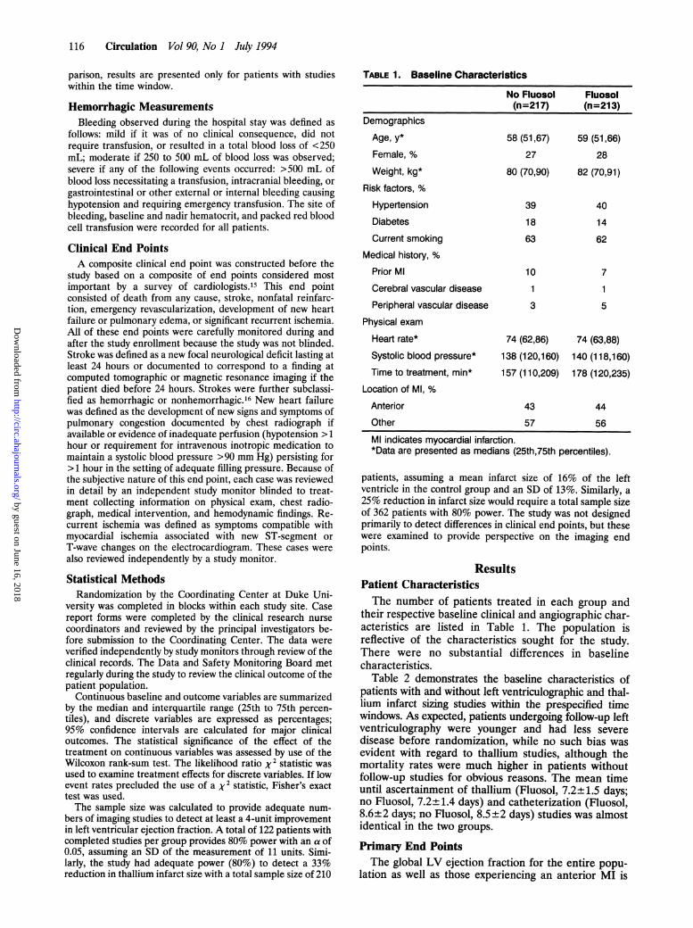
TABLE 1. Baseline Characteristics
No Fluosol Fluosol(n=217) (n=213)
Demographics
Age, y* 58 (51,67) 59 (51,66)Female, % 27 28
Weight, kg* 80 (70,90) 82 (70,91)Risk factors, %
Hypertension 39 40
Diabetes 18 14
Current smoking 63 62
Medical history, %Prior Ml 10 7
Cerebral vascular disease 1 1
Peripheral vascular disease 3 5
Physical exam
Heart rate* 74 (62,86) 74 (63,88)Systolic blood pressure* 138 (120,160) 140 (118,160)Time to treatment, min* 157 (110,209) 178 (120,235)
Location of Ml, %Anterior 43 44
Other 57 56
Ml indicates myocardial infarction.*Data are presented as medians (25th,75th percentiles).
patients, assuming a mean infarct size of 16% of the leftventricle in the control group and an SD of 13%. Similarly, a25% reduction in infarct size would require a total sample sizeof 362 patients with 80% power. The study was not designedprimarily to detect differences in clinical end points, but thesewere examined to provide perspective on the imaging endpoints.
ResultsPatient CharacteristicsThe number of patients treated in each group and
their respective baseline clinical and angiographic char-acteristics are listed in Table 1. The population isreflective of the characteristics sought for the study.There were no substantial differences in baselinecharacteristics.
Table 2 demonstrates the baseline characteristics ofpatients with and without left ventriculographic and thal-lium infarct sizing studies within the prespecified timewindows. As expected, patients undergoing follow-up leftventriculography were younger and had less severedisease before randomization, while no such bias wasevident with regard to thallium studies, although themortality rates were much higher in patients withoutfollow-up studies for obvious reasons. The mean timeuntil ascertainment of thallium (Fluosol, 7.2±1.5 days;no Fluosol, 7.2±1.4 days) and catheterization (Fluosol,8.6±2 days; no Fluosol, 8.5±2 days) studies was almostidentical in the two groups.
Primary End PointsThe global LV ejection fraction for the entire popu-
lation as well as those experiencing an anterior MI is
by guest on June 16, 2018http://circ.ahajournals.org/
Dow
nloaded from

Wall et al Fluosol Treatment in MI 117
TABLE 2. Baseline Characteristics According to End Point Ascertainment
Ejection No EjectionThallium No Thallium Fraction Fraction Both Neither(n=250) (n=180) (n=314) (n=116) (n=203) (n=69)
Age, y 58 (50,66) 59 (52,68) 58 (50,66) 61 (52,70) 58 (50,66) 62 (54,71)
Female, % 26 29 25 34 24 32
Weight, kg 82 (70,91) 80 (70,91) 80 (70,90) 81 (70,94) 80 (69,90) 77 (69,91)
Infarct location, %
Anterior 42 43 43 40 43 47
Other 58 57 57 60 57 53
Heart rate, bpm 72 (61,85) 75 (65,89) 73 (62,88) 76 (64,86) 72 (61,85) 76 (64,90)
Blood pressure,mm Hg 140 (120,160) 137 (118,158) 139 (120,160) 139 (118,160) 138 (118,160) 130 (111,153)Hospital course, %
Death 0.4 10.6 1.0 14.7 0.5 24.6
Reinfarction 2.8 3.9 2.9 4.3 3.0 5.8
Stroke 0.4 5.0 0.6 6.9 0.5 11.6
bpm indicates beats per minute.Numbers represent percentages for discrete variables and medians (25th,75th percentiles) for continuous variables.
shown in Fig 1. No significant difference was seenbetween patients receiving and not receiving Fluosol.Infarct zone regional wall motion for all patients and forthose with anterior MI is shown in Fig 2. Again, nosignificant difference was seen between patients receiv-ing Fluosol and those not receiving Fluosol. Statisticaladjustments for time from chest pain onset to treatmentand for time from treatment to left ventriculography didnot alter the results of the treatment comparison.
Thallium infarct size, measured as the percentage ofLV damage, is shown in Fig 3 for the entire populationas well as for those with anterior MI. No significantdifference was noted after Fluosol compared with noFluosol for the entire population. A trend towardsmaller mean infarct size was seen in the Fluosolpatients with anterior MI, but the median infarct sizewas larger in this group and no such trend was observedin measures of global or regional LV function.The coronary angiographic outcomes are demon-
strated in Table 3. No differences were observed in theproportion of patients with patent infarct-related arter-ies or in the TIMI grades at the time of the first cardiaccatheterization.
80
3X 40-
,,, 20
0
NoFluosol
NoFluosolFluosol
61 U
44 43
The frequency of clinical outcomes and 95% confi-dence intervals are displayed in Table 4. Although theprimary composite end point was not significantly dif-ferent between the two groups, a definite increase inheart failure and a trend toward higher death andstroke rates were seen in the Fluosol group, while areduction in recurrent ischemia and a tendency for lessreinfarction were also evident.The frequency of secondary clinical outcomes is
displayed in Table 5. No significant differences wereobserved in any of the major outcomes reflecting ar-rhythmia, hypotension, or secondary organ dysfunction.A significant increase in findings consistent with pulmo-nary congestion was present in the Fluosol group,although these findings resolved within 2 to 3 days anddid not lead to an increase in the need for mechanicalventilation. Conversely, a substantial decrease in find-ings of recurrent ischemia was present in the Fluosolgroup.The relation between Fluosol administration and
selected hematologic measures is demonstrated in Ta-
Fluosol.3.
so 55
39 37
0a
0)
-2
-1
Entire Populationu.
Anterior Ml
NoFluosol Fluosol42 42
433
-1A
Entire Population
NoFluo~o Fluo
*32 43
-I1-1i
Anterior Ml
FIG 1. Bar graph demonstrates the median and intraquartileranges as well as the mean values for ejection fraction in theentire population and for the population with anterior myocardialinfarction (Ml).
FIG 2. Bar graph demonstrates the median and intraquartileranges as well as the mean value for infarct zone function in theentire population and for patients with anterior myocardial infarc-tion (Ml).
by guest on June 16, 2018http://circ.ahajournals.org/
Dow
nloaded from

118 Circulation Vol 90, No 1 July 1994
Nopi~so Fluso
23 23
14.2 15
4.U.5
Entire Population
NoF h Ruosol32A
21.7
17A
54
Anterior Ml
FIG 3. Bar graph demonstrates the median and intraquartileranges and the mean value for thallium-estimated infarct size forthe entire population and for patients with anterior myocardialinfarction (Ml).
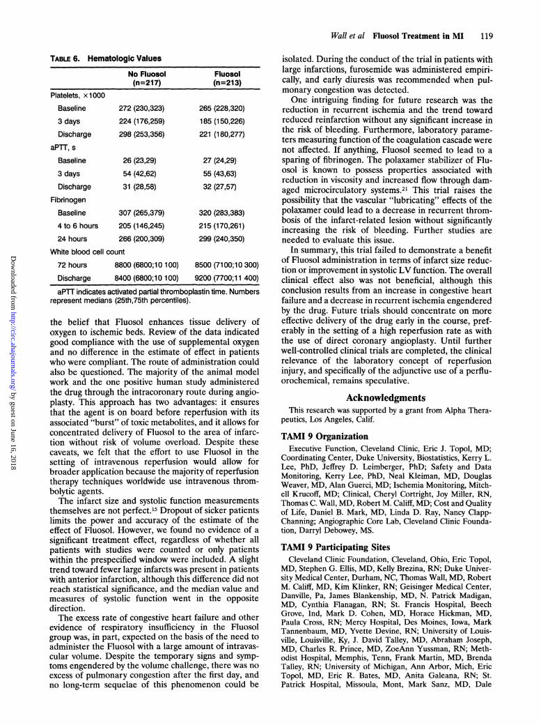
ble 6. Fluosol administration was associated with signif-icant preservation of fibrinogen, a lower platelet count,and no difference in leukocyte count. Measures ofanticoagulation, the activated partial thromboplastintime and the prothrombin time, were not affected.
DiscussionThe major finding of this study is that, despite prom-
ising data in several animal models, intravenous Fluosoldid not result in reduction in infarct size or improve-ment in LV systolic function when used in conjunctionwith intravenous TPA. Fluosol administration was asso-ciated with an increase in non-life-threatening pulmo-nary edema, but these negative effects were offset by areduction in the rate of recurrent ischemic events.While these findings are disappointing, the heterogene-ity of the clinical end point findings and the discordancefrom the animal model data suggest the need for furtherresearch.The baseline characteristics of the population re-
flected the careful selection of patients who mightbenefit without the confounding effects of prior infarc-tion on measurements of ventricular function or infarctsize. On the other hand, because of known problemswith the administration of intravascular volume re-quired with the Fluosol, patients with very large infarc-tions and hemodynamic compromise could not be in-cluded in the study. The time from symptom onset totreatment was fairly long, but when patients reportingearly with anterior infarctions treated within 3 hourswere evaluated separately, no effect of Fluosol could bedetected.The reason for the failure of the pharmacological
strategy to produce a positive effect on the primary endpoint is a matter of speculation. One possibility is thatreperfusion injury is a laboratory phenomenon that is
TABLE 3. Angiographic Outcomes
No Fluosol FluosolTIMI Grade, % (n=159) (n=146)0 10 101 4 52 17 173 69 68
TIMI indicates Thrombolysis In Myocardial Infarction.
TABLE 4. Clinical Outcomes
No Fluosol FluosolDeath, % 3.7 (1.2,6.2) 5.6 (2.6, 8.6)
Stroke, %
Hemorrhagic
Other
Reinfarction, %
0.9 (0.4, 2.2) 2.4 (0.3, 4.4)
0.0 1.9 (0.1, 3.7)
4.2 (1.5, 6.9) 2.4 (0.4, 4.4)
CHF/pulmonary edema, % 31 (24.8, 37.2) 45 (38.3, 51.7)*
Recurrent ischemia, % 11 (6.8,15.2) 6 (2.8, 9.2)t
Numbers represent percentage of patients with event and95% confidence intervals.
*P=.004; tP=.039.
not clinically relevant because the damage is limited toareas of myocardium that are destined to die eventuallyanyway.'7 Other randomized trials of agents touted toprevent reperfusion injury (superoxide dismutase orprostacyclin) also reported no effect on LV function inthe setting of angioplasty'8 or thrombolytic therapy19 forthe treatment of acute MI. A second possibility is thatthe particular regimen used was not effective. The dosethat was chosen was based on weight-adjusted effectiveanimal model doses.20 The protocol required supple-mental oxygen administration to all patients because of
TABLE 5. Secondary Clinical Outcomes
No Fluosol Fluosol(n=217) (n=213)
Arrhythmias, %
20 or 30 atrioventricular block 8.3 8.0
Sustained ventricular tachycardia 8.3 7.0
Ventricular fibrillation 7.4 8.0
Atrial fibrillation/flutter 8.8 13.2
Severe sinus bradycardia 24.4 23.9
Hemodynamics, %
Sustained hypotension 18.9 20.2
Killip class II or IIl 39.6 45.5
Other cardiovascular, %Acute septal rupture 0 0.5
Acute mitral regurgitation 0.5 2.4
Pericarditis 2.3 1.4
Coma, % 1.0 1.4Renal failure, % 3.7 5.2
Respiratory failure, % 8.3 6.6
Bleeding
Patients transfused, % 14 14
Hematocrit, %Baseline 43 (40,46) 43 (41,46)
3 days 37 (33,41) 38 (35,41)
Discharge 38 (33,41) 39 (34,42)
Index 2.7 (1.7,4.2) 2.7 (1.9,4.4)
Numbers represent percentages for discrete variables andmedians (25th,75th percentiles) for continuous variables.
40
2
CR0b
c
30
20
10
0
by guest on June 16, 2018http://circ.ahajournals.org/
Dow
nloaded from
Wall et al Fluosol Treatment in MI 119
TABLE 6. Hematologic Values
No Fluosol Fluosol(n=217) (n=213)
Platelets, x1000
Baseline 272 (230,323) 265 (228,320)
3 days 224 (176,259) 185 (150,226)Discharge 298 (253,356) 221 (180,277)
aPTT, s
Baseline 26 (23,29) 27 (24,29)3 days 54 (42,62) 55 (43,63)
Discharge 31 (28,58) 32 (27,57)
Fibrinogen
Baseline 307 (265,379) 320 (283,383)
4 to 6 hours 205 (146,245) 215 (170,261)
24 hours 266 (200,309) 299 (240,350)
White blood cell count
72 hours 8800 (6800;10 100) 8500 (71 00;10 300)
Discharge 8400 (6800;10 100) 9200 (7700;1 1 400)
aPTT indicates activated partial thromboplastin time. Numbersrepresent medians (25th,75th percentiles).
the belief that Fluosol enhances tissue delivery ofoxygen to ischemic beds. Review of the data indicatedgood compliance with the use of supplemental oxygenand no difference in the estimate of effect in patientswho were compliant. The route of administration couldalso be questioned. The majority of the animal modelwork and the one positive human study administeredthe drug through the intracoronary route during angio-plasty. This approach has two advantages: it ensuresthat the agent is on board before reperfusion with itsassociated "burst" of toxic metabolites, and it allows forconcentrated delivery of Fluosol to the area of infarc-tion without risk of volume overload. Despite thesecaveats, we felt that the effort to use Fluosol in thesetting of intravenous reperfusion would allow forbroader application because the majority of reperfusiontherapy techniques worldwide use intravenous throm-bolytic agents.The infarct size and systolic function measurements
themselves are not perfect.15 Dropout of sicker patientslimits the power and accuracy of the estimate of theeffect of Fluosol. However, we found no evidence of asignificant treatment effect, regardless of whether allpatients with studies were counted or only patientswithin the prespecified window were included. A slighttrend toward fewer large infarcts was present in patientswith anterior infarction, although this difference did notreach statistical significance, and the median value andmeasures of systolic function went in the oppositedirection.The excess rate of congestive heart failure and other
evidence of respiratory insufficiency in the Fluosolgroup was, in part, expected on the basis of the need toadminister the Fluosol with a large amount of intravas-cular volume. Despite the temporary signs and symp-toms engendered by the volume challenge, there was noexcess of pulmonary congestion after the first day, andno long-term sequelae of this phenomenon could be
isolated. During the conduct of the trial in patients withlarge infarctions, furosemide was administered empiri-cally, and early diuresis was recommended when pul-monary congestion was detected.One intriguing finding for future research was the
reduction in recurrent ischemia and the trend towardreduced reinfarction without any significant increase inthe risk of bleeding. Furthermore, laboratory parame-ters measuring function of the coagulation cascade werenot affected. If anything, Fluosol seemed to lead to asparing of fibrinogen. The polaxamer stabilizer of Flu-osol is known to possess properties associated withreduction in viscosity and increased flow through dam-aged microcirculatory systems.21 This trial raises thepossibility that the vascular "lubricating" effects of thepolaxamer could lead to a decrease in recurrent throm-bosis of the infarct-related lesion without significantlyincreasing the risk of bleeding. Further studies areneeded to evaluate this issue.
In summary, this trial failed to demonstrate a benefitof Fluosol administration in terms of infarct size reduc-tion or improvement in systolic LV function. The overallclinical effect also was not beneficial, although thisconclusion results from an increase in congestive heartfailure and a decrease in recurrent ischemia engenderedby the drug. Future trials should concentrate on moreeffective delivery of the drug early in the course, pref-erably in the setting of a high reperfusion rate as withthe use of direct coronary angioplasty. Until furtherwell-controlled clinical trials are completed, the clinicalrelevance of the laboratory concept of reperfusioninjury, and specifically of the adjunctive use of a perflu-orochemical, remains speculative.
AcknowledgmentsThis research was supported by a grant from Alpha Thera-
peutics, Los Angeles, Calif.
TAMI 9 OrganizationExecutive Function, Cleveland Clinic, Eric J. Topol, MD;
Coordinating Center, Duke University, Biostatistics, Kerry L.Lee, PhD, Jeffrey D. Leimberger, PhD; Safety and DataMonitoring, Kerry Lee, PhD, Neal Kleiman, MD, DouglasWeaver, MD, Alan Guerci, MD; Ischemia Monitoring, Mitch-ell Krucoff, MD; Clinical, Cheryl Cortright, Joy Miller, RN,Thomas C. Wall, MD, Robert M. Califf, MD; Cost and Qualityof Life, Daniel B. Mark, MD, Linda D. Ray, Nancy Clapp-Channing; Angiographic Core Lab, Cleveland Clinic Founda-tion, Darryl Debowey, MS.
TAMI 9 Participating SitesCleveland Clinic Foundation, Cleveland, Ohio, Eric Topol,
MD, Stephen G. Ellis, MD, Kelly Brezina, RN; Duke Univer-sity Medical Center, Durham, NC, Thomas Wall, MD, RobertM. Califf, MD, Kim Klinker, RN; Geisinger Medical Center,Danville, Pa, James Blankenship, MD, N. Patrick Madigan,MD, Cynthia Flanagan, RN; St. Francis Hospital, BeechGrove, Ind, Mark D. Cohen, MD, Horace Hickman, MD,Paula Cross, RN; Mercy Hospital, Des Moines, Iowa, MarkTannenbaum, MD, Yvette Devine, RN; University of Louis-ville, Louisville, Ky, J. David Talley, MD, Abraham Joseph,MD, Charles R. Prince, MD, ZoeAnn Yussman, RN; Meth-odist Hospital, Memphis, Tenn, Frank Martin, MD, BrendaTalley, RN; University of Michigan, Ann Arbor, Mich, EricTopol, MD, Eric R. Bates, MD, Anita Galeana, RN; St.Patrick Hospital, Missoula, Mont, Mark Sanz, MD, Dale
by guest on June 16, 2018http://circ.ahajournals.org/
Dow
nloaded from

120 Circulation Vol 90, No 1 July 1994
Mayer, RN; Rochester General Hospital, Rochester, NY,Gerald Gacioch, MD, Valerie Chiodo, RN.
Duke University Collaborating CentersAlamance County Hospital, Burlington, NC, James Strick-
land, MD; Alamance Memorial Hospital, Burlington, NC,Stuart Schnider, MD; Granville Medical Center, Oxford, NC,Stephen Ertischeck, MD, John Anderson, MD; Person CountyMemorial Hospital, Roxboro, NC, Thomas Long, MD; South-eastern General Hospital, Lumberton, NC, John Hoekstra,MD; Richmond Memorial Hospital, Rockingham, NC, MoosaHajisheikh, MD; Halifax-South Boston Community Hospital,South Boston, Va, Richard Goulah, MD; Wilson MemorialHospital, Wilson, NC, Mark Leithe, MD.
References1. Jennings R, Reimer K. Factors involved in salvaging ischemic
myocardium: effect of reperfusion of arterial blood. Circulation.1983;68(suppl I):I-25. Abstract.
2. Califf R, Topol E, Gersh B. From myocardial salvage to patientsalvage in acute myocardial infarction: the role of reperfusiontherapy. JAm Coll Cardiol. 1989;14:1382-1388.
3. Braunwald E. Myocardial perfusion, limitation of infarct size,reduction of left ventricular dysfunction and improved survival:should the paradigm be expanded? Circulation. 1989;79:441-444.
4. Braunwald E, Kloner R. Myocardial reperfusion: a double-edgedsword? J Clin Invest. 1985;76:1713-1719.
5. Forman MB, Virmani R, Puett DW. Mechanisms and therapy ofmyocardial reperfusion injury. Circulation. 1990;81(suppl IV):IV-69-IV-78.
6. Hirooka Y, Kudo H, Suzuki A. The effect of Fluosol-DA on theexperimental myocardial infarction. Proceedings of the FourthInternational Symposium on Perfluorochemical Blood Substitutes.Amsterdam, the Netherlands: Excerpta Medica; 1978:285-293.
7. Nunn G, Dance G, Peters J, Cohn L. Effect of fluorocarbonexchange transfusion on myocardial infarction size in dogs. Am JCardioL 1983;52:203-205.
8. Forman M, Bingham S, Kopelman H. Reduction of infarct sizewith intracoronary perfluorochemical in a canine preparation ofreperfusion. Circulation. 1985;71:1060-1068.
9. Forman MB, Perry JM, Wilson BH, Verani MS, Kaplan PR, ShawlFA, Friesinger GC. Demonstration of myocardial reperfusion injuryin humans: results of a pilot study utilizing acute coronary angio-plasty with perfluorochemical in anterior myocardial infarction.JAm Coll CardioL 1991;18:911-918.
10. Virmani R, Forman MB, Kolodgie FD. Myocardial reperfusioninjury: histopathological effects of perfluorochemical. Circulation.1990;81(suppl IV):IV-57-IV-68.
11. Bajaj A, Cobb M, Virmani R, Gay JC, Light RT, Forman MB.Limitation of myocardial reperfusion injury by intravenous perflu-orochemicals: role of neutrophil activation. Circulation. 1989;79:645-656.
12. TIMI Study Group. The Thrombolysis in Myocardial Infarctiontrial. N EnglJ Med. 1985;313:342-347.
13. Sandler H, Dodge HT. The use of single plane angiocardiogramsfor the calculation of left ventricular volume in man. Am Heart J.1968;75:325-334.
14. Sheehan FH, Bolson EL, Dodge HT, Mathey DG, Schofer J, WooHW. Advantages and applications of the centerline method forcharacterizing regional ventricular function. Circulation. 1986;74:293-305.
15. Califf RM, Harrelson-Woodlief L, Topol EJ. Left ventricularejection fraction may not be useful as an end point of thrombolytictherapy comparative trials. Circulation. 1990;82:1847-1853.
16. The GUSTO Investigators. An international randomized trialcomparing four thrombolytic regimens consisting of tissue plas-minogen activator, streptokinase, or both for acute myocardialinfarction. N Engl J Med. 1993;329:673-682.
17. Jennings RB, Reimer KA. Lethal reperfusion injury: fact or fancy?In Parratt JR, ed. Myocardial Response to Acute Injury. London,England: Macmillan Press Ltd; 1992:17-34.
18. Flaherty JT, Topol EJ, Pitt B, Gruber JW, Rothbaum DA, HeuserRR, George BS, Burwell LR, Kereiakes DJ, Deitchman D, Gus-tafson N, Brinker JA, Becker LC, Mancini GBJ, Werns SW.Recombinant human superoxide dismutase (h-SOD) fails toimprove recovery of ventricular function in patients undergoingcoronary angioplasty for acute myocardial infarction. Circulation.1994;89:1982-1991.
19. Topol EJ, Ellis SG, Califf RM, George BS, Stump DC, Bates ER,Nabel EG, Walton JA, Candela RJ, Lee KL, Kline EM, Pitt B, theTAMI 4 Study Group. Combined tissue-type plasminogenactivator and prostacyclin therapy for acute myocardial infarction.JAm Coll Cardiol. 1989;14:877-884.
20. Harrison JK, Califf RM, Woodlief L, Kereiakes D, George BS,Stack RS, Ellis SG, Lee KL, O'Neill W, Topol EJ, the TAMI StudyGroup. Systolic left ventricular function therapy for acute myo-cardial infarction: an analysis of determinants of improvement.Circulation. 1993;87:1531-1541.
21. Forman MB, Ingram DA, Murray JJ. Role of perfluorochemicalemulsions in the treatment of myocardial reperfusion injury. AmHeart J. 1992;124:1347-1357.
by guest on June 16, 2018http://circ.ahajournals.org/
Dow
nloaded from

D Cohen, M Sanz and J D LeimbergerT C Wall, R M Califf, J Blankenship, J D Talley, M Tannenbaum, M Schwaiger, G Gacioch, M
Group.Thrombolysis and Angioplasty in Myocardial Infarction 9 Trial. TAMI 9 Research Intravenous Fluosol in the treatment of acute myocardial infarction. Results of the
Print ISSN: 0009-7322. Online ISSN: 1524-4539 Copyright © 1994 American Heart Association, Inc. All rights reserved.
is published by the American Heart Association, 7272 Greenville Avenue, Dallas, TX 75231Circulation doi: 10.1161/01.CIR.90.1.114
1994;90:114-120Circulation.
http://circ.ahajournals.org/content/90/1/114World Wide Web at:
The online version of this article, along with updated information and services, is located on the
http://circ.ahajournals.org//subscriptions/
is online at: Circulation Information about subscribing to Subscriptions:
http://www.lww.com/reprints Information about reprints can be found online at: Reprints:
document. Permissions and Rights Question and Answer this process is available in the
click Request Permissions in the middle column of the Web page under Services. Further information aboutOffice. Once the online version of the published article for which permission is being requested is located,
can be obtained via RightsLink, a service of the Copyright Clearance Center, not the EditorialCirculationin Requests for permissions to reproduce figures, tables, or portions of articles originally publishedPermissions:
by guest on June 16, 2018http://circ.ahajournals.org/
Dow
nloaded from