intrauterine growth restriction
TRANSCRIPT
Intrauterine Growth RestrictionIntrauterine Growth Restriction(IUGR)(IUGR)
Dr. M.C. BansalDr. M.C. Bansal
Definition
• Foetuses of birth weight less than 10th percentile of those born at same gestational age
or
two standard deviations below the population mean are considered growth restricted.
« small for gestational age »(SGA) fetuses, all of which may not necessarily growth restricted as many of these may be just constitutionally small and not at risk of any adverse outcome.)
• « Therefore the term IUGR should more strictly refer to foetuses that are small for gestational age and display other signs of chronic hypoxia or failure to thrive. »
Incidence
• Approximately 3-5% of all pregnancies.
Normal Fœtal Growth• Fœtal growth depends on two components:
– Genetic Potential – Substrate supply
The genetic potential is derived from both parents and is mediated through growth factors such as insulin-like growth factor.
Adequate substrate supply is essential to achieve genetic potential. This is derived from placenta which is dependent upon the uterine and placental vascularity

A comparision between normal and IUGR babies.
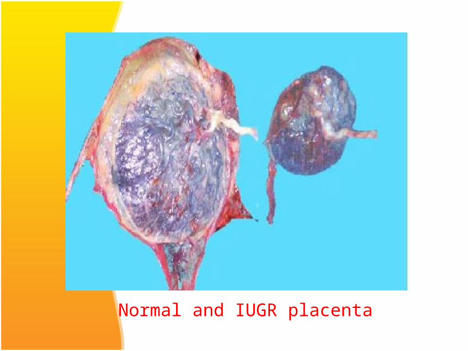
Normal and IUGR placenta

Normal Fetal Growth
• Normal fetal growth is characterized by cellular hyperplasia followed by hyperplasia and hypertrophy and lastly byhypertrophy alone.
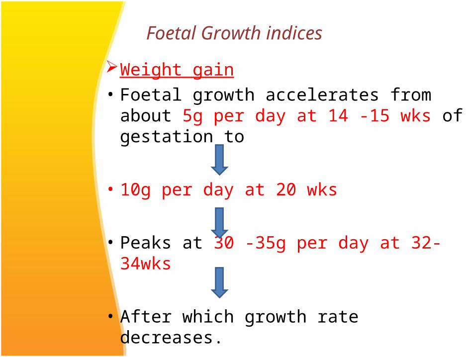
Weight gain• Foetal growth accelerates from about
5g per day at 14 -15 wks of gestation to
• 10g per day at 20 wks
• Peaks at 30 -35g per day at 32-34wks
• After which growth rate decreases.
Foetal Growth indices
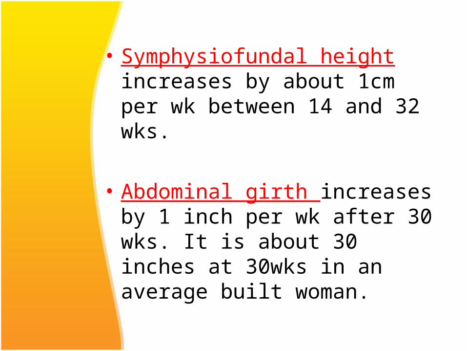
• Symphysiofundal height increases by about 1cm per wk between 14 and 32 wks.
• Abdominal girth increases by 1 inch per wk after 30 wks. It is about 30 inches at 30wks in an average built woman.
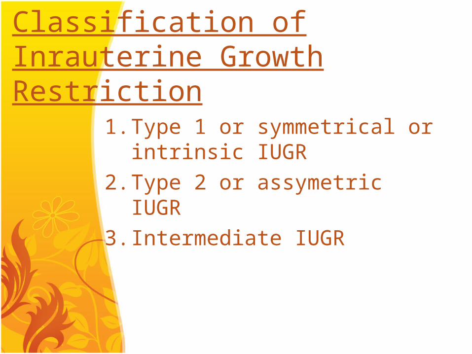
Classification of Inrauterine Growth Restriction
1. Type 1 or symmetrical or intrinsic IUGR
2. Type 2 or assymetric IUGR3. Intermediate IUGR
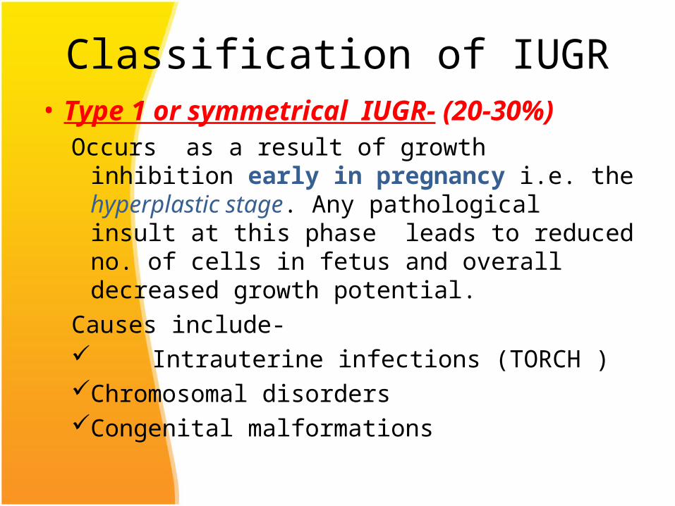
Classification of IUGR• Type 1 or symmetrical IUGR- (20-30%)
Occurs as a result of growth inhibition early in pregnancy i.e. the hyperplastic stage. Any pathological insult at this phase leads to reduced no. of cells in fetus and overall decreased growth potential.
Causes include- Intrauterine infections (TORCH )Chromosomal disordersCongenital malformations

All parameters(head and abdo circumference,
length and weight) are below 10th percentile for gestational age, hence normal ponderal index (birth weight/ht3).
Type 2 or asymmetric IUGR (70-80%)Occurs as a result of restriction of nutrient
supply in utero i.e. uteroplacental insufficiency.
It is usually associated with maternal diseases like:-
– Chronic hypertension– Renal disease– Vasculopathies
• The onset of growth restriction occurs usually after 28 wks of gestation i.e. in the stage of hypertrophy. The fetus has near normal total no. of cells but cell size is reduced.
• There is brain sparing effect so that the head growth remains normal but the abdominal girth slows down.
• The Ponderal index is low.• This asymmetry results from redistribution of
fetal cardiac output with increased flow to brain and heart at the expense of reduced splanchnic circulation.
• Liver size is reduced because of diminished glycogen stores.
• In case of severe placental insufficiency the head growth may also be affected.This type of growth restriction leads to decreased amniotic fluid, chronic hypoxia and may result in fetal death.
Intermediate IUGR (5-10% of all growth
restricted fetuses)• It is a combination of type 1 and type 2.• Fetal growth restriction occurs during
intermediate phase of growth affecting both hyperplasia and hypertrophy, resulting in decrease in cell no. as well as size.
• Causes includeChronic HTLupus nephritisMaternal vascular diseases that are severe and
have onset in early 2nd trimester
• IUGR may also be classified simply as Early onset (onset before 32
weeks) Late onset (onset after 32
weeks)
Aetiology• IUGR is a manifestation of fetal, maternal and
placental disorders that affect fetal growth.
A.Fetal Causes1. Chromosomal Disorders-
usually result in early onset IUGR. Trisomies 13, 18, 21 contribute to 5% of IUGR cases Autosomal deletions Ring chromosomes Sex chromosome disorders are frequently lethal,
fetuses that survive may have growth restriction (Turner Syndrome)
Fetal causes contd..
2. Congenital Infections:• The growth potential of fetus may be severely
impaired by intrauterine infections.• The timing of infection is crucial as the resultant
effects depends on the phase of organogenesis.• Viruses- rubella, CMV, varicella and HIV
rubella is the most embryotoxic virus, it cause capillary endothelial damage during organogenesis and impairs fetal growth.
CMV causes cytolysis and localized necrosis in fetus.
• Protozoa- like malaria, toxoplasma, trypanosoma have also been associated with growth restriction.
Fetal causes contd..3. Structural Anomalies-
All major structural defects involving CNS,CVS,GIT, Genitourinary and musculoskeletal system are associated with increased risk of fetal growth restriction.If growth restriction is associated with polyhydramnios, the incidence of structural anomaly is substantially increased.
Fetal causes contd..
4. Genetic Causes- Maternal genes have greater influence on fetal growth.
Inborn errors of metabolism like agenesis of pancreas, congenital lipodystrophy, galactosemia, phenylketonuria also result in growth restriction of fetus.
B. Placental causes
• Placenta is the sole channel for nutrition and oxygen supply to the fetus.Single umblical arteryabnormal placental implantationvelamentous umblical cord insertionbilobed placentaplacental haemangiomas have all been associated
with fetal growth restriction
C. Maternal Causes
1. Maternal Characteristics:those contributing to IUGR are- Extremes of maternal age Grandmultiparity History of IUGR in previous pregnancy Low maternal weight gain in pregnancy
2. Maternal diseases:Uteroplacental insufficiency resulting from medical complications like Hypertension Renal disease Autoimmune disease Hyperthyroidism Long term insulin dependent diabetes

Maternal causes contd..• Smoking- active or passive, especially during third
trimester is important cause of IUGR. Nicotine has vasoconstrctive effect on the maternal circulation and leads to formaton of toxic metabolites in fetus.
• Alchohol and Drugs- Alchohol crosses the placenta freely. It acts as a cellular poison reducing fetal growth potential.• Cocaine and opiates are potent vasoconstrictors. • Warfarin, anticonvulsants and antineoplastic agents are
also implicated in growth restriction

• Thrombophilias- antiphospholipid antibody syndrome and other thrombophilias leading to placental thrombosis and impaired trophoblastic function.
• Nutritional Deficiency- poor intake -inflammatory bowel
disease
Complications of IUGRPerinatal mortality and morbidity of IUGR infants is 3-20 times greater than normal infants.
• Antepartum period- increased incidence of--still births-oligohydramniosIUGR is found in 52% of unexplained stillbirths.
• During labour- higher incidence of--meconium aspiration-fetal distress-intrapartum fetal death
Complications of IUGR contd..
• Neonatal period- increased incidence of--Hypoxic ischemic encephalopathy-Persistent fetal circulation insufficiency
They have difficulty in temperature regulation because of absent brown fat and small body mass relative to surface area.
Lack of glycogen stores may predispose to hypoglycemiaChronic intrauterine hypoxia may lead to polycythemia,
necrotizing enterocolitis, other metabolic abnormalities.
Complications cont..
• Childhood- increases mortality from--infectious diseases-congenital anomalies
Incidence of cerebral palsy are 4-6 times higher.Subtle impairment of cognitive performance and
educational underachievement.• Long term complications- increased risk of
coronary heart disease, hypertension, type II diabetes mellitus, dyslipidaemia and stroke.
Diagnosis of IUGR
Identifying mothers at risk:Poor maternal nutrition Poor BMI at conceptionPre-eclampsiaRenal disordersDiseases causes vascular insufficiencyInfections (TORCH)Poor maternal wt. gain during pregnancy
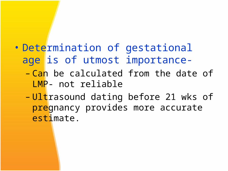
• Determination of gestational age is of utmost importance-– Can be calculated from the date of LMP- not
reliable– Ultrasound dating before 21 wks of pregnancy
provides more accurate estimate.
Diagnosis of IUGR
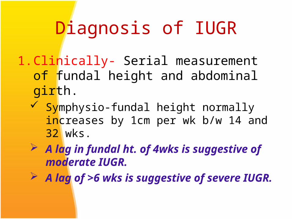
1. Clinically- Serial measurement of fundal height and abdominal girth. Symphysio-fundal height normally increases by
1cm per wk b/w 14 and 32 wks. A lag in fundal ht. of 4wks is suggestive of
moderate IUGR. A lag of >6 wks is suggestive of severe IUGR.
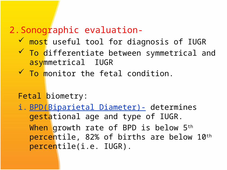
2. Sonographic evaluation- most useful tool for diagnosis of IUGR To differentiate between symmetrical and
asymmetrical IUGR To monitor the fetal condition.
Fetal biometry:i. BPD(Biparietal Diameter)- determines gestational age
and type of IUGR.When growth rate of BPD is below 5th percentile, 82% of births are below 10th percentile(i.e. IUGR).
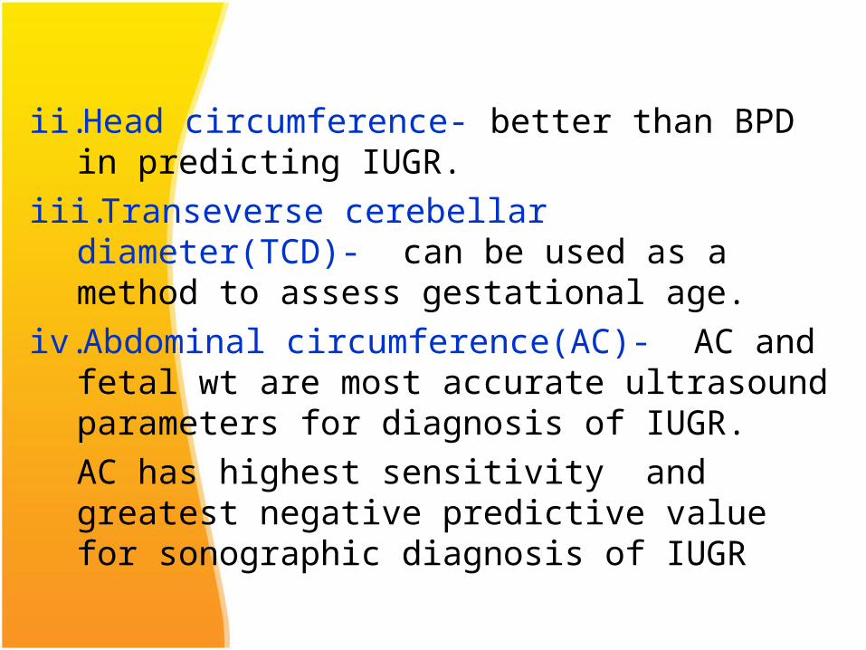
ii. Head circumference- better than BPD in predicting IUGR.
iii. Transeverse cerebellar diameter(TCD)- can be used as a method to assess gestational age.
iv. Abdominal circumference(AC)- AC and fetal wt are most accurate ultrasound parameters for diagnosis of IUGR. AC has highest sensitivity and greatest negative predictive value for sonographic diagnosis of IUGR
An increase in fetal AC of less than 10 mm in 14 days has sensitivity of 85% and specificity of 74% for identification of IUGR.
iv. Measurement ratios- there are some age independent ratios to detect IUGR.HC/AC: decreases linearly from 16 to 20 wks of gestation.
HC/AC >2 SD above mean is predictive of IUGR.FL/AC: normal value ranges from 22 + 2% in the second
half of pregnancy. Ratio above 23.5% is considered abnormal.
Placental Morphology: Acceleration of placental maturation may occur with IUGR and PIH.(Placenta is graded from grade 0 to grade III)
Amniotic fluid volume: type 2 IUGR is usually associated with oligohydramnios.Amniotic fluid index(AFI) between 8 and 25 is
normal.
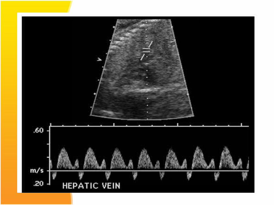
3. Doppler Ultrasonography: doppler flow studies are important adjuncts to fetal biometry in
identifying the IUGR fetuses at risk of adverse outcome.Most widely used arterial indices are :Pulsatility index (PI): Systolic end diastolic peak
velocity / time averaged maximum velocityResistance Index(RI): Systolic end diastolic peak
velocity/ systolic peak velocitySystolic to diastolic ratio(S/D): Systolic peak velocity /
diastolic peak velocity
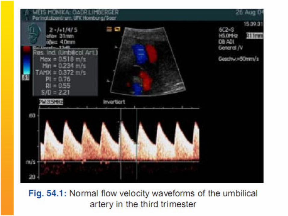
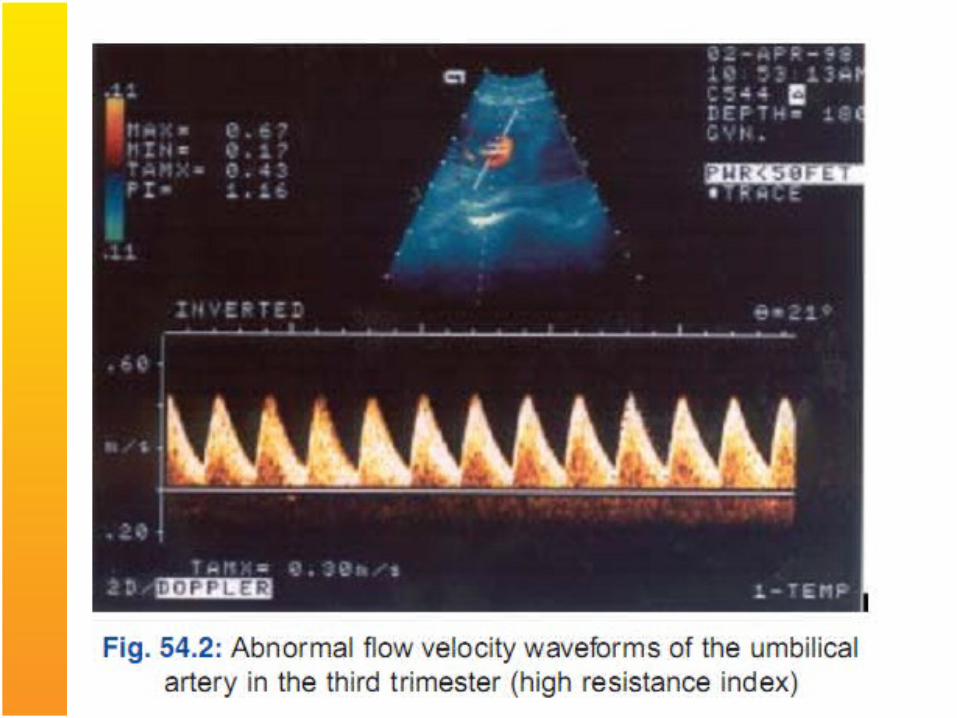
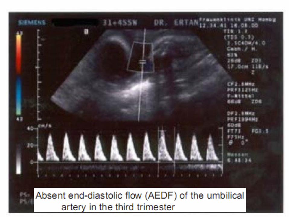
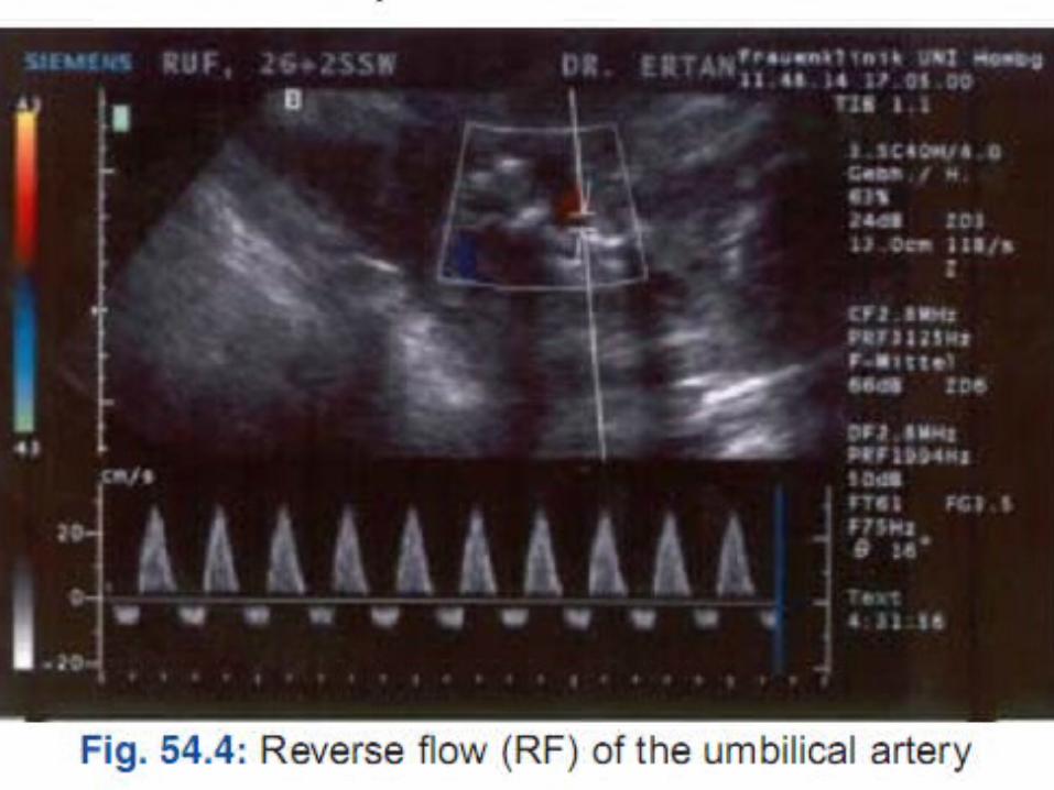
Umblical Artery doppler- In IUGR there is increased umblical artery resistance (increased S/D ratio), absent end diastolic flow and finally reversed end diastolic flow.Perinatal mortality rate increases significantly in fetuses with absent end diastolic flow (9-41%) and reversed end diastolic flow (33-73%) in umblical artery.

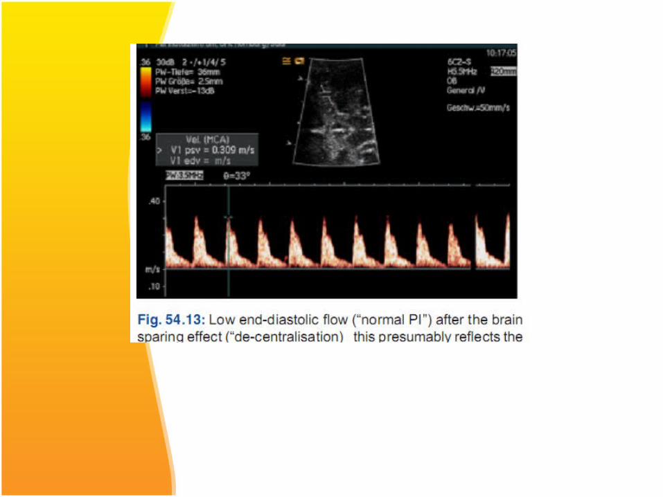
Middle cerebral artery doppler- in a normal fetus has relatively little flow during diastole. Increased resistance to blood flow in placenta results in redistribution of cardiac output to favour cardiac and cerebral circulations leading to increased flow in the diastolic phase with decreased S/D ratio.
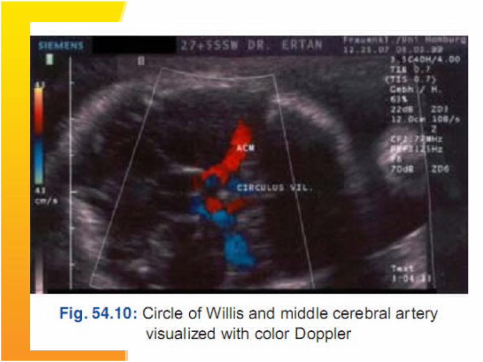
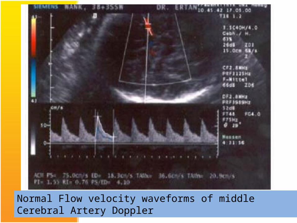
Normal Flow velocity waveforms of middle Cerebral Artery Doppler
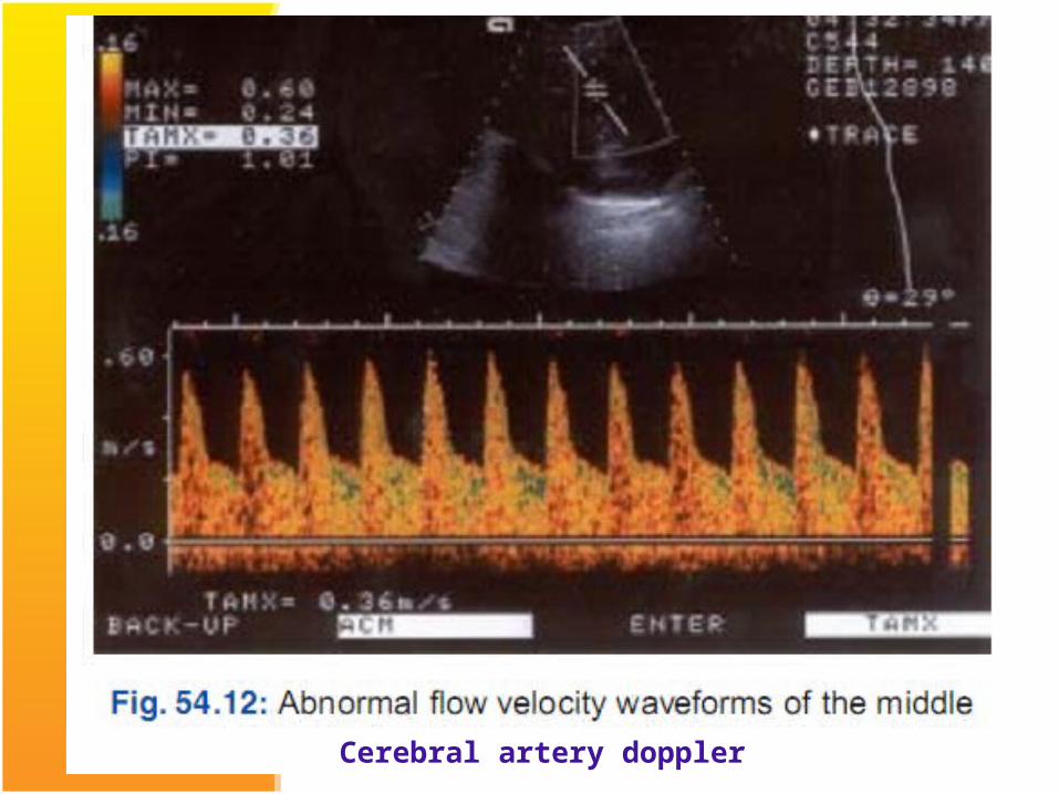
Cerebral artery doppler
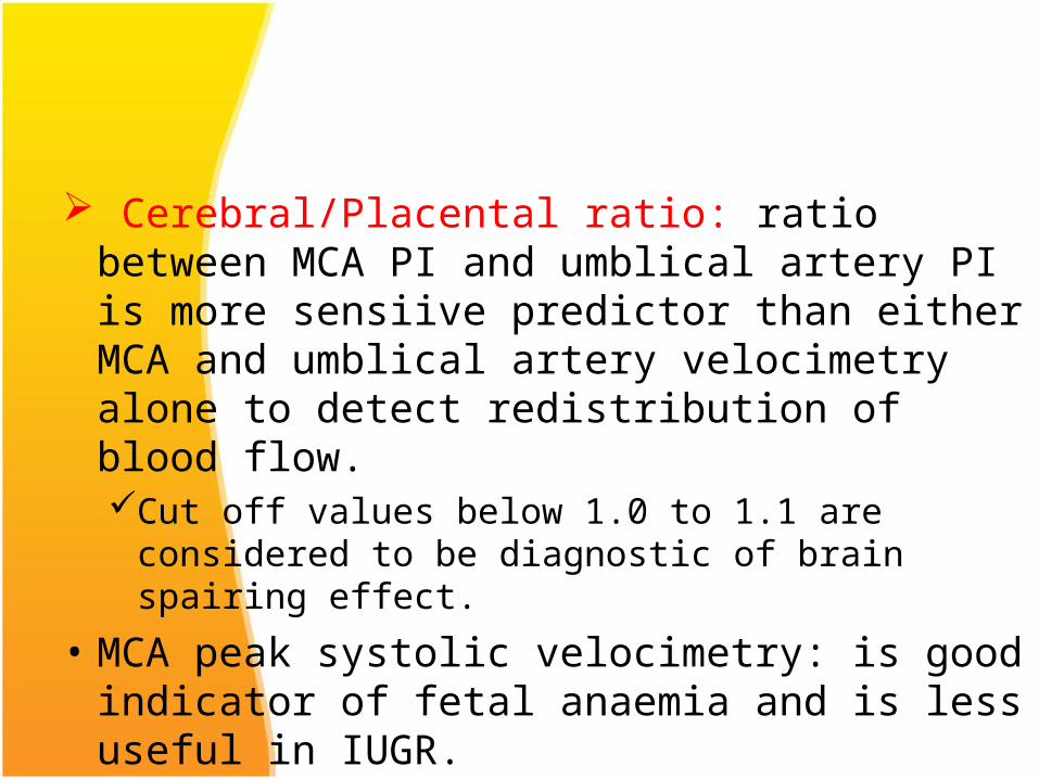
Cerebral/Placental ratio: ratio between MCA PI and umblical artery PI is more sensiive predictor than either MCA and umblical artery velocimetry alone to detect redistribution of blood flow. Cut off values below 1.0 to 1.1 are considered to be
diagnostic of brain spairing effect.• MCA peak systolic velocimetry: is good indicator
of fetal anaemia and is less useful in IUGR.

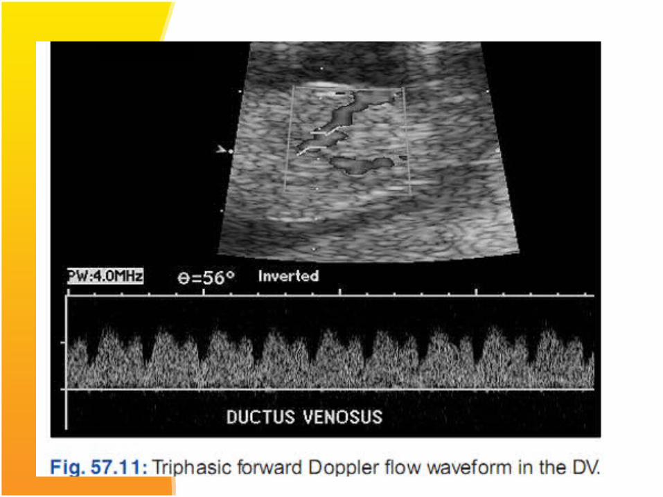
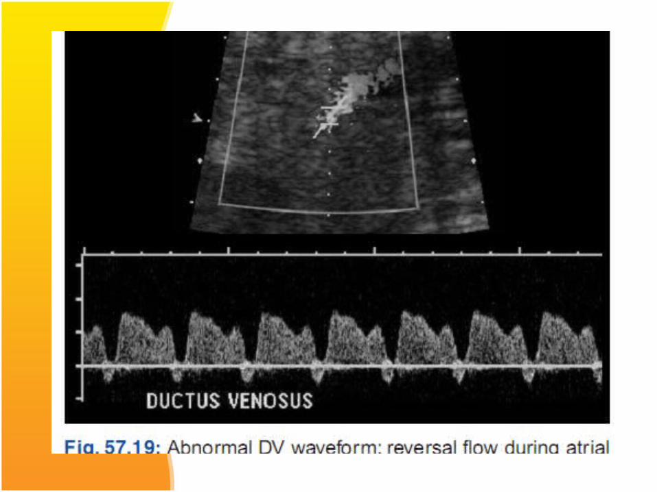
Ductus venosus doppler: Perinatal mortality in growth restricted fetuses has been found to be significantly worse when abnormalities in fetal venous circulation are detected.In the normal fetus, flow in the ductus venosus is
forwards , moving towards the heart during entire cardiac cycle.
When circulatory compensation of the fetus fails, the ductus venosus waveform shows absent or reverse blood flow during atrial conraction. Perinatal mortality being 63-100%.
Therefore it is recommended that fetus should be delivered before the development of absent of reversed blood flow of DV.
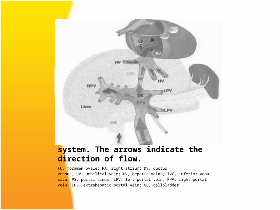
: Representation of fetal umbilical and hepatic venous system. The arrows indicate the direction of flow. FO, foramen ovale; RA, right atrium; DV, ductusvenous; UV, umbilical vein; HV, hepatic veins; IVC, inferior venacava; PS, portal sinus; LPV, left portal vein; RPV, right portalvein; EPV, extrahepatic portal vein; GB, gallbladder


MANAGEMENT
Principles:1. Identify the cause of growth restriction.2. Treat the cause if found.3. General management
MANAGEMENT First step is to identify the aetiology of IUGR:-Maternal history pertaining to the risk factors of
IUGR.Clinical examination- maternal habitus, height,
weight, BP etc.Laboratory investigations- Hb, blood sugar, renal
function tests, serology for TORCHSpecific investigations for thrombophilias in pts with history suggestive of early onset growth restriction.
Fetal evaluation: thorough ultrasound for growth restriction, amniotic fluid, congenital anomalies and doppler evaluation
Management cont..
Treatment of underlying cause: such as hypertension, cessation of smoking, protein energy supplementation in poorly nourished and underweight women.
General Management: Bed rest in left lateral position to increase
uteroplacental blood flow Maternal nutritional supplementation with high
caloric and protein diets, antioxidents, haematinics and omega 3 fatty acids, arginine .
Maternal oxygen therapy: Adminitration of 55% oxygen at a rate of 8L/min round the clock has shown decreased perinatal mortality rate.
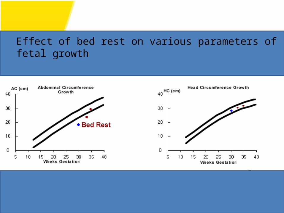
Effect of bed rest on various parameters of fetal growth

Management cont..
Pharmacological therapy:Aspirin in low doses(1-2 mg/kg body wt.), betamimetics etc have been tried but all have failed to show any significant difference in incidence of IUGR.
Thus there is no form of therapy currently available which can reverse IUGR, the only intervention possible in most cases is delivery.
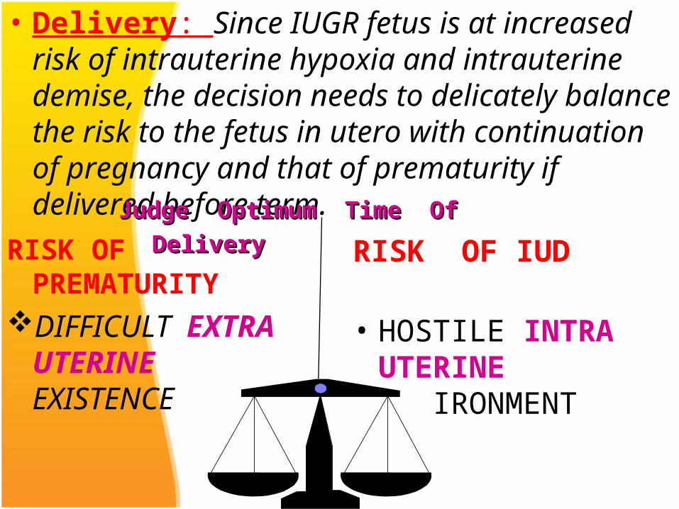
• Delivery: Since IUGR fetus is at increased risk of intrauterine hypoxia and intrauterine demise, the decision needs to delicately balance the risk to the fetus in utero with continuation of pregnancy and that of prematurity if delivered before term.
RISK OF PREMATURITYDIFFICULT EXTRA
UTERINE EXISTENCE
RISK OF IUD
• HOSTILE INTRA UTERINE ENVIRONMENT
Judge Optimum Time Of DeliveryJudge Optimum Time Of Delivery
• The optimum timing of delivery is determined by gestational age, underlying aetiology, possibility of extrauterine survival and fetal condition.
• Strict fetal surveillance is needed to monitor fetal well being and to detect signs of fetal compromise
Fetal Surveillance
1. Daily fetal movement score2. Non stress test(NST)3. Biophysical profile(BPP)4. Amniotic fluid index(AFI)5. Growth parameters6. Doppler studies
Sonography is usually repeated every 2 wks.
Management cont..
• Role of steroids:Antenatal glucocorticoid administration reduces the incidence of respiratory distress syndrome, intraventricular hemorrhage and death in IUGR fetusus weighing less than 1500gm.
Mode of Delivery Fetuses with significant IUGR should be preferably
delivered in well equiped centres which can provide intrapartum continuous fetal heart monitoring , fetal blood sampling and expert neonatal care.
Vaginal delivery: can be allowed as long as there is no obstetric indication for caesarian section and fetal heart rate is normal.
• Fetuses with major anomaly incompatible with life should also be delivered vaginally.

Caesarian section: In all cases of IUGR with features of acidosis caesarian section should be done without trial of labour. These include:Repetitive late decelerationspoor biophysical profilereversal of end diastolic flow in umblical arteryabnormal venous dopplerblood gas analysis showing acidic pH on
cordocentesis.
ConclusionOne of the primary aims of antenatal care is to
identify fetuses which show a significant growth lag, since they are at a high risk of hypoxic complications in the perinatal period.
Management options are limited to close fetal monitoring and termination of pregnancy balancing the risk of prematurity and that of intrauterine demise.