intrauterine growth restriction intrauterine growth restriction (iugr) by definition occurs when the...
TRANSCRIPT
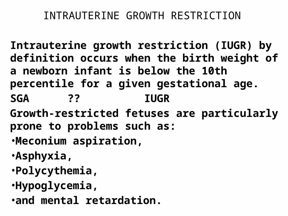
INTRAUTERINE GROWTH RESTRICTION Intrauterine growth restriction (IUGR) by definition occurs when the birth weight of a newborn infant is below the 10th percentile for a given gestational age. SGA ?? IUGR Growth-restricted fetuses are particularly prone to problems such as:•Meconium aspiration, •Asphyxia, •Polycythemia, •Hypoglycemia, •and mental retardation.
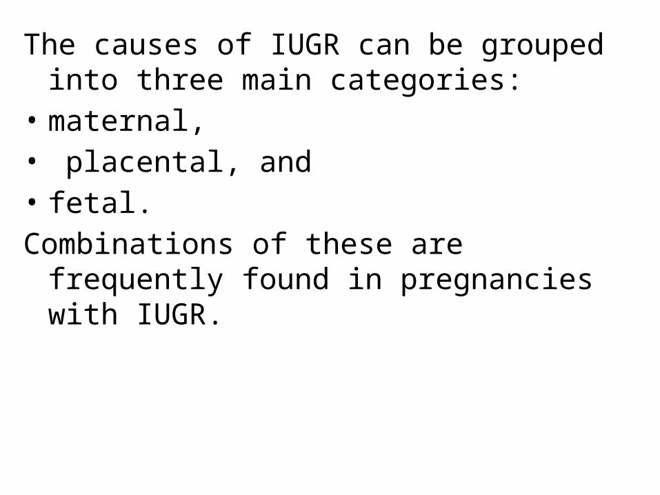
The causes of IUGR can be grouped into three main categories:
• maternal,• placental, and • fetal. Combinations of these are frequently found in
pregnancies with IUGR.
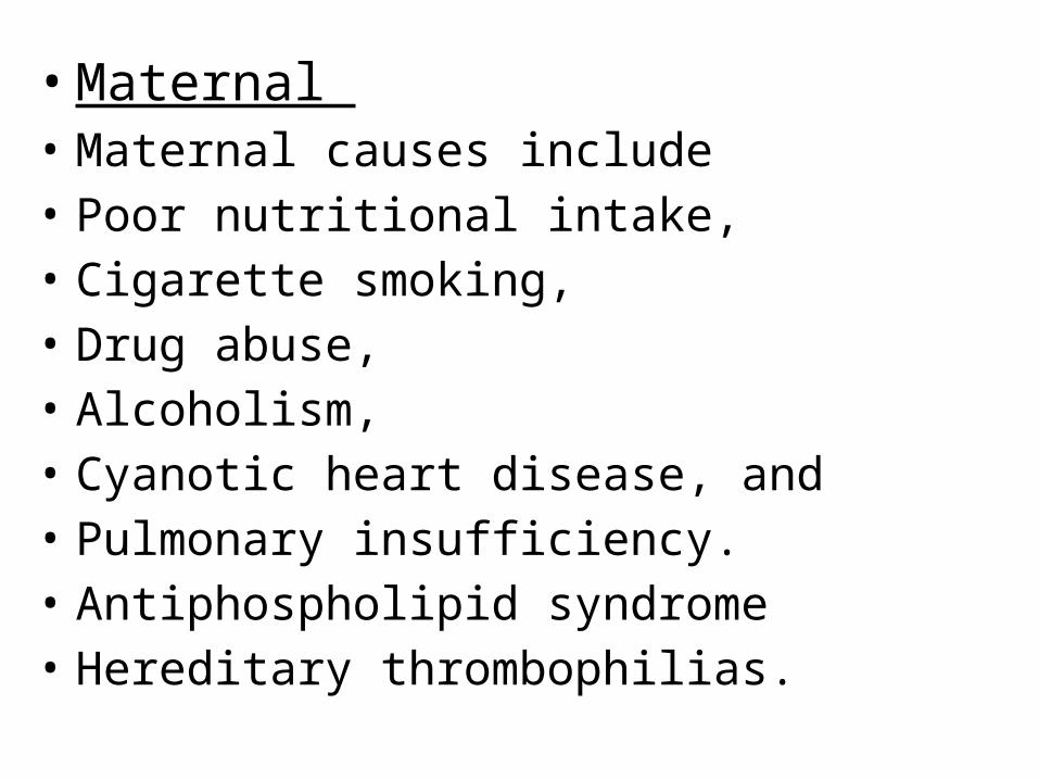
• Maternal • Maternal causes include• Poor nutritional intake, • Cigarette smoking, • Drug abuse, • Alcoholism, • Cyanotic heart disease, and • Pulmonary insufficiency. • Antiphospholipid syndrome • Hereditary thrombophilias.
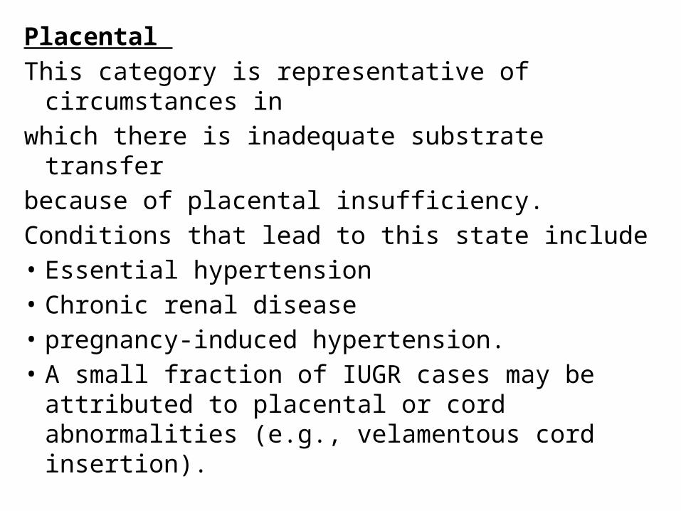
Placental This category is representative of circumstances inwhich there is inadequate substrate transferbecause of placental insufficiency.Conditions that lead to this state include• Essential hypertension• Chronic renal disease• pregnancy-induced hypertension. • A small fraction of IUGR cases may be attributed
to placental or cord abnormalities (e.g., velamentous cord insertion).
Fetal causes of IUGRExamples of fetal causes include:• Intrauterine infection (listeriosis and TORCH
[toxoplasmosis, other infections, rubella, cytomegalovirus infection, and herpes simplex] agents)
• Congenital anomalies. • Genetic diseases• Multiple pregnancy
• Two types of fetal growth restriction have been described: symmetric and asymmetric.
• In fetuses with symmetric growth restriction, growth of both the head and the body is inadequate. The head-to-abdominal circumference ratio may be normal, but the absolute growth rate is decreased. Symmetric growth restriction is most commonly seen in association with intrauterine infections or congenital fetal anomalies.
• When asymmetric growth restriction occurs, usually late in pregnancy, the brain is preferentially spared at the expense of "nonvital" abdominal viscera. As a result, the head size is proportionally larger than the abdominal size.
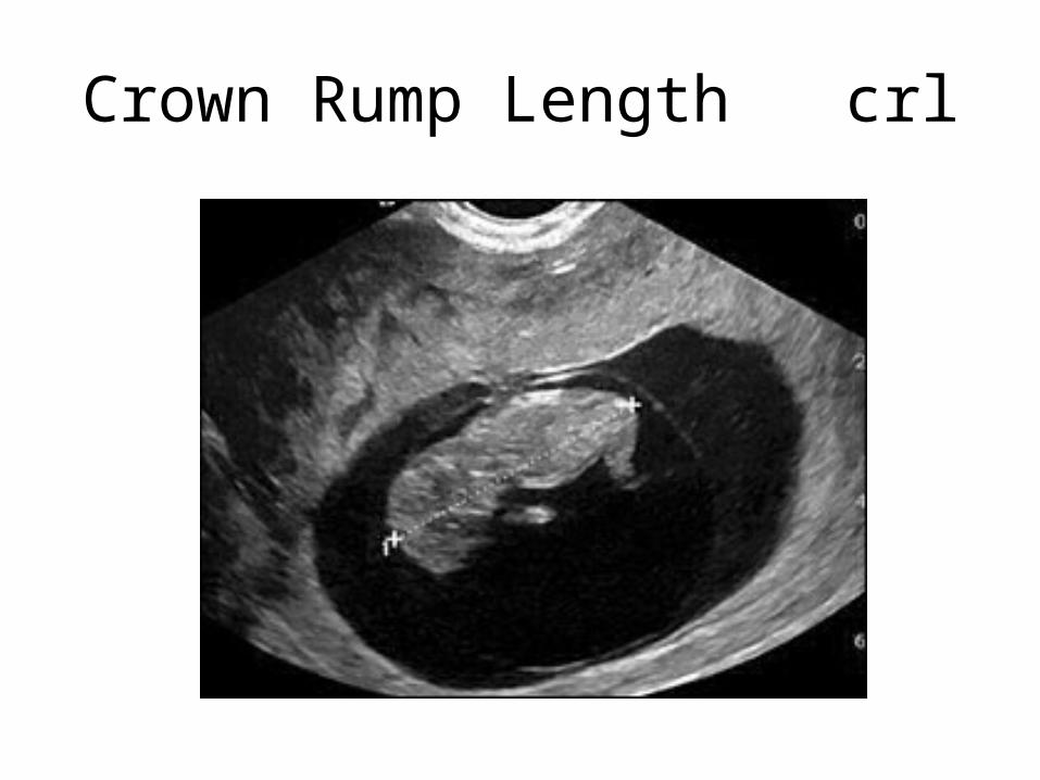
Crown Rump Length crl
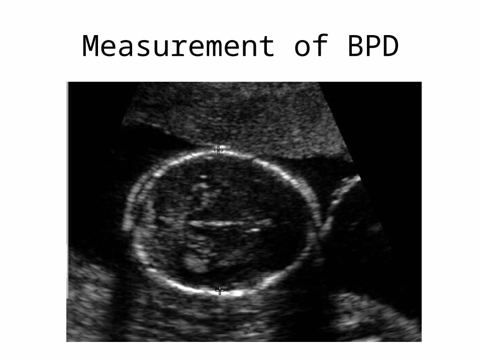
Measurement of BPD
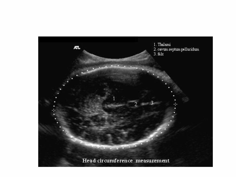
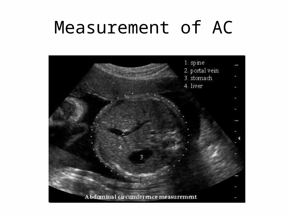
Measurement of AC
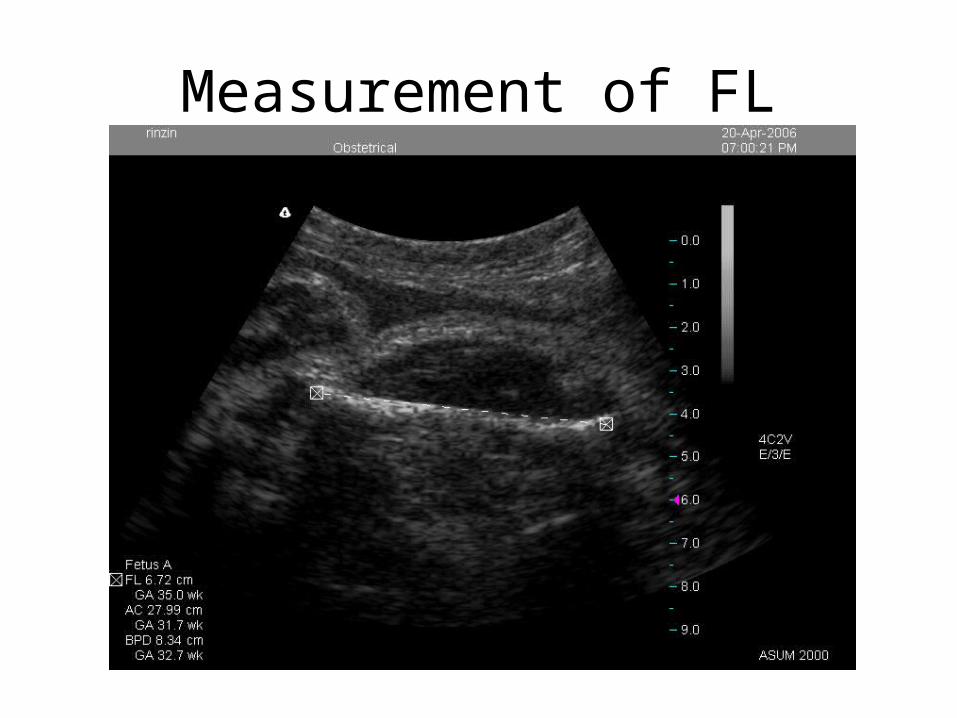
Measurement of FL
DIAGNOSIS Growth restriction may go undiagnosed unless
the obstetrician • establishes the correct gestational age of the
fetus.• identifies high-risk factors from the obstetric
data base• serially assesses fetal growth by fundal height
or ultrasonography
• MANAGEMENT • Prepregnancy
• An important part of preventive medicine is to anticipate risks that can be modified before a woman becomes pregnant.
• Improving nutrition and stopping smoking are two approaches that should improve fetal growth in women who are underweight, who smoke, or both.
• For women with antiphospholipid antibodies associated with the delivery of a prior IUGR infant, low-dose aspirin (81 mg/day) in early pregnancy may reduce the likelihood of recurrent IUGR.
• For patients with one of the hereditary thrombophilias, low-dose heparin (5000 U twice daily) with or without low-dose aspirin has also been shown to reduce the risk of recurrent IUGR.
• Antepartum • Once a fetus has been identified as having decreased
growth, the obstetrician should direct his or her efforts toward modifying any associated factors that can be changed.
• Because poor nutrition and smoking exert their main effects on birth weight in the latter half of pregnancy, cessation of smoking and improved nutrition can have a positive impact. T
• The working woman who becomes fatigued is more likely to have a low-birth-weight infant. Work leave or, in some cases of maternal disease, hospitalization will increase uterine blood flow and may improve the nutrition of the fetus at risk.
• The most important clinical decisions revolve around the timing and mode of delivery.
• The objective is to expedite delivery before the occurrence of fetal compromise but after fetal lung maturation.
• This requires regular fetal monitoring with a twice-weekly nonstress test (NST) and biophysical profile.
• Most institutions use a modified biophysical profile that includes an NST and AFI.
• A simple technique is available whereby a pregnant woman can help in the assessment of fetal well-being. She assesses fetal movement (kick count) each evening while resting comfortably on her left side.
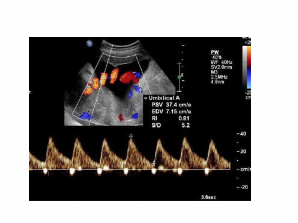
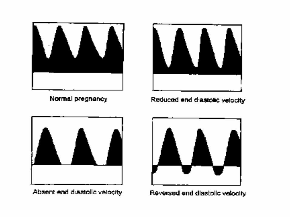
• Doppler-derived umbilical artery systolic-to-diastolic ratios are abnormal in IUGR fetuses. Fetuses with growth restriction tend to have increased resistance to flow and to demonstrate low, absent, or reversed diastolic flow.
Fetuses clinically suspected of IUGR could be approached as follows: • For cases in which results of fetal monitoring are normal and
ultrasonic findings strongly suggest normal growth, no clinical intervention is warranted.
• For cases in which ultrasonic findings strongly suggest IUGR, with or without abnormal fetal surveillance, delivery is indicated at gestational ages of 34 weeks or later, or at any reasonable gestational age if pulmonary maturity is documented.
• In the presence of severe oligohydramnios, delivery should be strongly considered without assessing lung maturity because these fetuses are at great risk of asphyxia, and the stress associated with IUGR usually accelerates fetal pulmonary maturity.
• For those cases in which ultrasonic findings are equivocal for IUGR, bed rest, fetal surveillance, and serial ultrasonic measurements at three weekly intervals are indicated.
LABOR AND DELIVERY • • IUGR per se is not a contraindication for induction of
labor, but there should be a low threshold to perform a cesarean section because of the poor capacity of the IUGR fetus to tolerate asphyxia.
• As a result, during labor, these high-risk patients must be electronically monitored to detect the earliest evidence of fetal distress.
• A combined obstetric-neonatal team approach to delivery is mandatory because of the likelihood of neonatal asphyxia.
After birth, the infant should be carefullyexamined to rule out the possibility of congenitalanomalies and infections. The monitoring of blood glucose levels is
important, because the fetuses do not have adequate hepatic glycogen stores, and hypoglycemia is a common finding.
Hypothermia is common in these infants. Respiratory distress syndrome is more common in
the presence of fetal distress, because fetal acidosis reduces surfactant synthesis and release.
• PROGNOSIS
The long-term prognosis for infants with IUGR must be assessed according to the varied etiologies of the growth restriction.
If infants with chromosomal abnormalities, autoimmune disease, congenital anomalies, and infection are excluded, the outlook for these newborns is generally good.