intraoperative echo: decision making in mitral valve...
TRANSCRIPT

Intraoperative Echo: Decision Making in Mitral Valve Disease
Vera H. Rigolin, MD, FASEVice-President, American Society of Echocardiography
Professor of MedicineNorthwestern University
Bluhm Cardiovascular InstituteMedical Director, Echocardiography Laboratory
Northwestern Memorial HospitalChicago, IL USA

Disclosures
• None

Important Information Needed from the Echo
• Pre-op– What is the mechanism of the valve disorder– How severe is the valve lesion– What are the other abnormalities in the heart
• Post-op (in the OR)– Is there residual regurgitation?– Is there unacceptable stenosis?– Is there SAM?– How is the LV? How is the RV?

Mitral Valve Anatomy
Otto, CM. N Engl J Med 2001;345:740
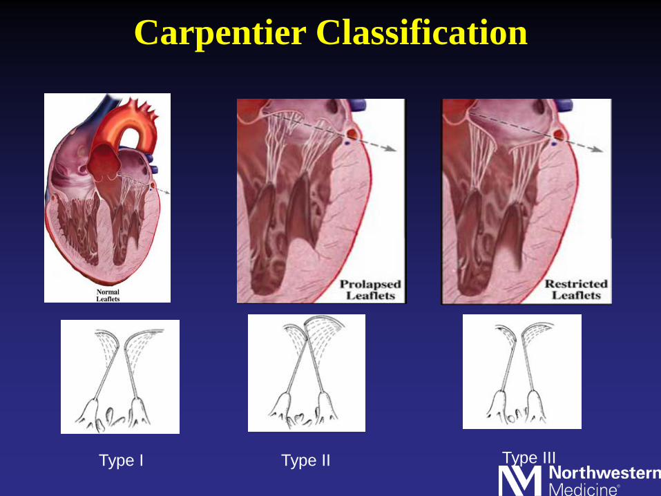
Carpentier Classification
Type I Type II Type III

Mitral Valve Anatomy

TEE of the Mitral Valve
•Identification of disease mechanism
•Identification of prolapsing/flail segments
•Extent/direction of MR jets
•Identification of additional pathology
Omran AS et al. J Soc Echocardiogr 2002;15:950-7

Illustration of traditional 2D and novel 3D echocardiography(a) energy beam formed from the conventional ultrasound crystal array(b) energy beam formed from the new crystal array(c) the resulting 3D pyramid of data can be examined in short- axis (plane C) or in long-axis (plane B)

Type I Mitral Regurgitation
Controls (unpublished)
DAIPm1 = 25.7 (16 - 39)DAP = 34.8 (24 - 51)C3D = 130.9 (89 - 168)C2D = 127.2 (87 - 164)A2D = 1231.3 (555 - 2003) Courtesy of Nausheen Akhter, MD
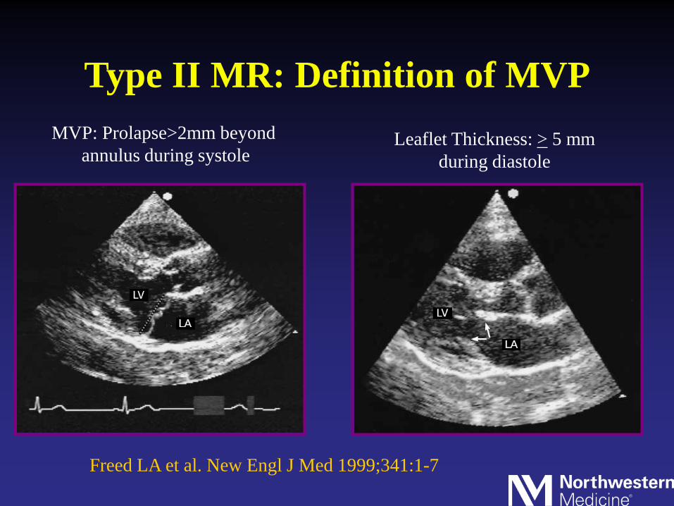
Type II MR: Definition of MVPMVP: Prolapse>2mm beyond
annulus during systoleLeaflet Thickness: > 5 mm
during diastole
Freed LA et al. New Engl J Med 1999;341:1-7

Barlow’s Valve
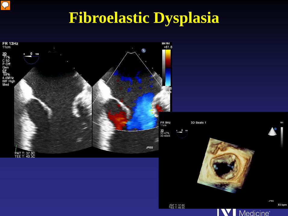
Fibroelastic Dysplasia

Type 3a Mitral Regurgitation/Mitral Stenosis

Type 3b Mitral Regurgitation


Feasibility of Real Time 3D TEE
Percent “Excellent” visualization
»MV: 85-91%» IAS: 84%» LAA: 86%» LV: 77%» AV: 18%» TV: 11%
Sugeng L et al. J Am Coll Cardiol 2008;52:446-9

Identification of MV Scallops Using 3D TEE
P1 P2P3
A1A2 A3

New Findings Using 3D TEE for Valve Surgery
Abraham TP et al. Am J Cardiol 1997;80:1577-1582
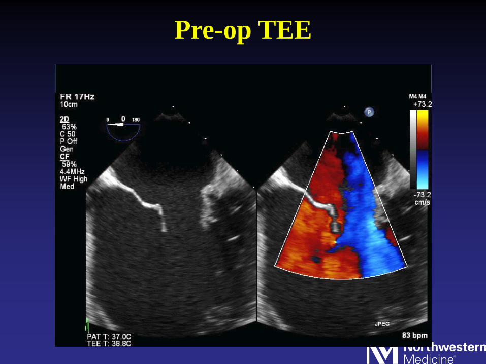
Pre-op TEE

Pre-op TEE

3D TEE

3D Color

Patient #2


3D TEE


Coming of Bypass
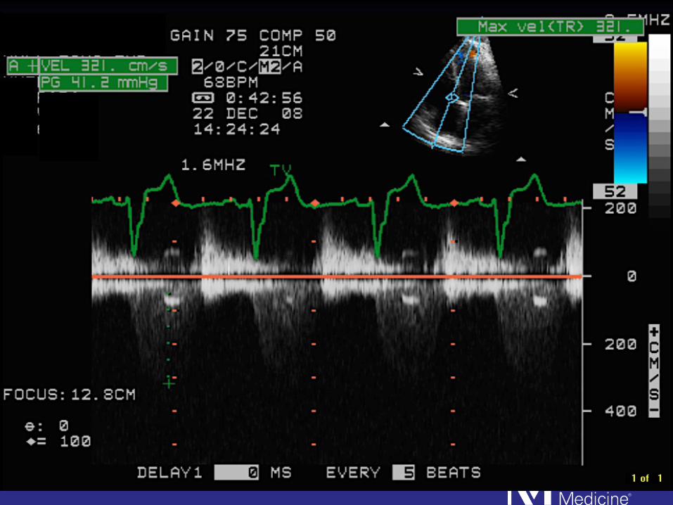
• RV and LV function• SAM• MR• MS

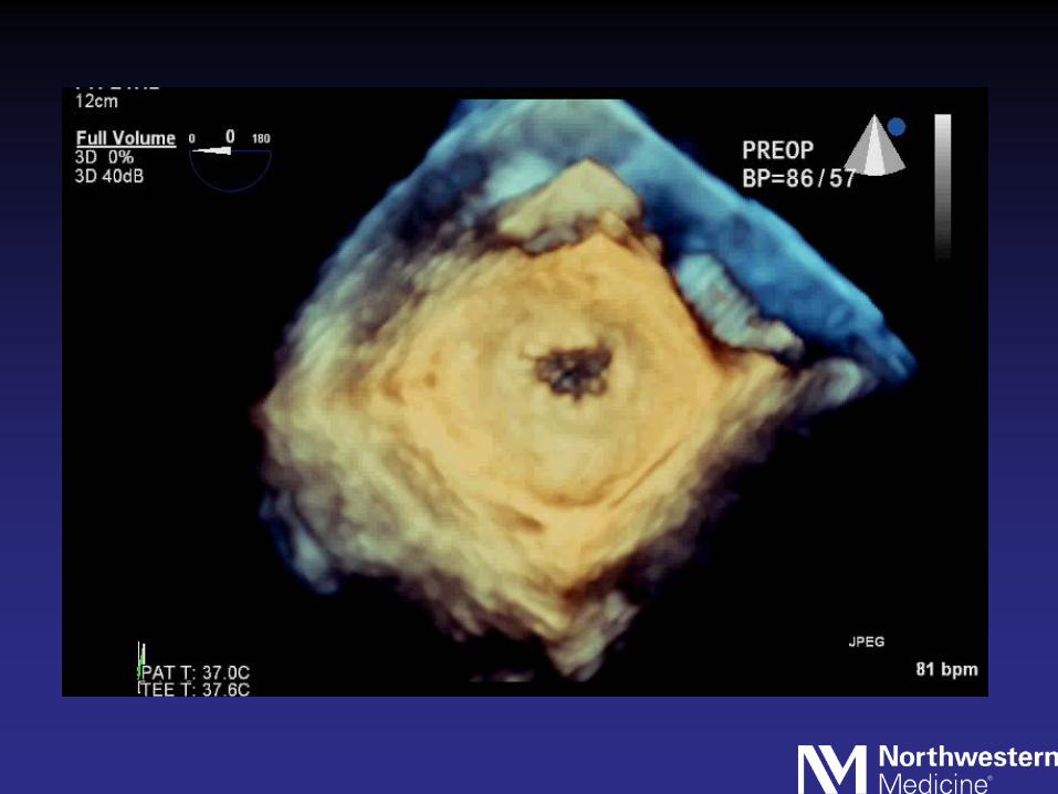
Preop TEE


MV Measurements
• Annulus – 4.0 cm• PL=2.3 cm• AL=2.3 cm• Csept=2.1 cm
Maslow AD et al. J Am Coll Cardiol1999;34(7):2096-104
AL/PL<1.3C-sept<2.5 cm


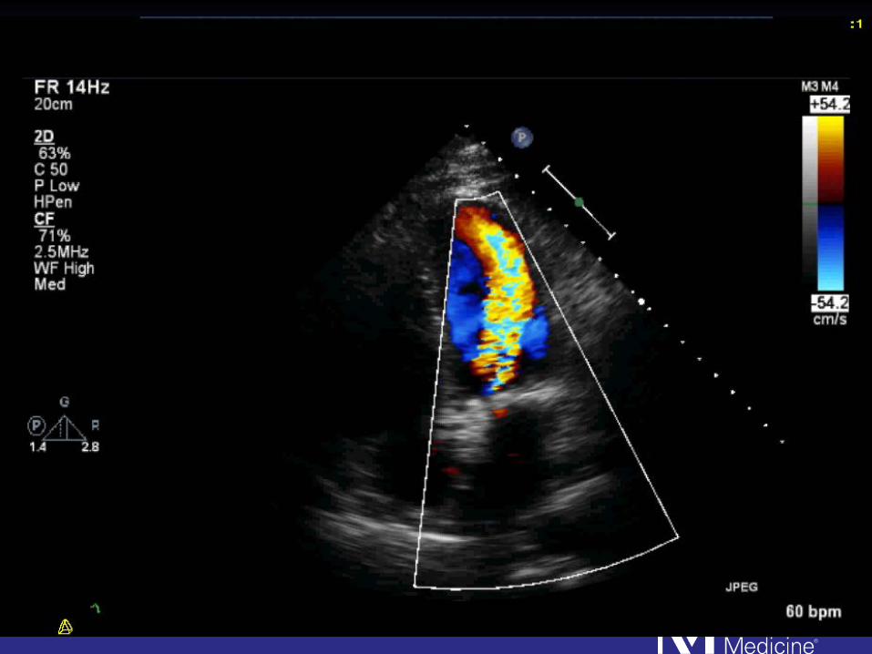
After Fluids and Reducing Pressers

Preop


VC=0.5 cm

EROA=0.76 cm2

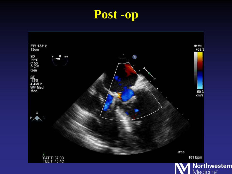
Post -op

Post-op

Late Complications

History
• 78yo s/p MV repair 3 mo prior (at OSH) using Annuloplasty band
• Post-op: Acute renal failure• Now presents with VRE bacteremia likely 2/2
catheter-related blood stream infection with infected vas cath


History
• 56 yr old male s/p MV repair 6 yrs prior • h/o MV prolapse• He presents with SOB


Summary
• Echo is an invaluable tool to assist in the management of pts with mitral valve disease
• TEE provides comprehensive assessment of anatomy, mechanism of disease and hemodynamic information
• 2D and 3D TEE combined provide the most comprehensive information

Thank You