intraoperative biventricular pacing applications, techniques, early results vincent a. gaudiani, md...
TRANSCRIPT

Intraoperative Biventricular PacingIntraoperative Biventricular PacingApplications, Techniques, Early ResultsApplications, Techniques, Early Results
Vincent A. Gaudiani, MDVincent A. Gaudiani, MDLuis J. Castro, MDLuis J. Castro, MD
Audrey L. Fisher, MPHAudrey L. Fisher, MPH
Published in The Heart Surgery Forum, Volume 6, Issue 6, 2003.

Traditional Pacing - What Is It? Traditional Pacing - What Is It?
Traditional PacingTraditional Pacing
• Unipolar Pacing – requires a Unipolar Pacing – requires a singlesingle dipole dipole from the active electrode on the heart to from the active electrode on the heart to the ground on the skinthe ground on the skin
• Bipolar Pacing – requires a Bipolar Pacing – requires a singlesingle dipole dipole from the active electrode on the heart to a from the active electrode on the heart to a ground on the heartground on the heart

Biventricular Pacing - What Is It? Biventricular Pacing - What Is It?
Biventricular PacingBiventricular Pacing
• Requires Requires two two dipoles about 180 degrees dipoles about 180 degrees apart on heart. A dipole from an active apart on heart. A dipole from an active electrode on the anterior RV to a ground electrode on the anterior RV to a ground and a second dipole from an active and a second dipole from an active electrode on the posterolateral LV to a electrode on the posterolateral LV to a ground.ground.

Why Biventricular Pacing?Why Biventricular Pacing?
• With two dipoles far apart activating the With two dipoles far apart activating the ventricles simultaneously, ventricles simultaneously, electricalelectrical activation is quickeractivation is quicker (90 – 110 ms) than (90 – 110 ms) than traditional pacing and therefore traditional pacing and therefore mechanical mechanical activation is more synchronousactivation is more synchronous..

Biventricular PacingBiventricular Pacing
• Another term for biventricular pacing is Another term for biventricular pacing is cardiac resynchronization therapy cardiac resynchronization therapy (CRT).(CRT).

Biventricular PacingBiventricular Pacing
What are the common clinical examples of What are the common clinical examples of suboptimal ventricular synchronization?suboptimal ventricular synchronization?
1.1. LBBBLBBB
2.2. Pacemaker SyndromePacemaker Syndrome
3.3. Some VT’sSome VT’s
4.4. Any QRS > 130 ms (IVCD’s)Any QRS > 130 ms (IVCD’s)

Biventricular PacingBiventricular Pacing
Which patient groups are most likely to Which patient groups are most likely to suffer reduced cardiac output when poorly suffer reduced cardiac output when poorly synchronized?synchronized?
Patients with a combination of:Patients with a combination of:
• Large LVIDd’sLarge LVIDd’s
• QRS > 130 msQRS > 130 ms
• Low EF’sLow EF’s

Biventricular PacingBiventricular Pacing
What are the clinical consequences of What are the clinical consequences of ventricular dyssynchrony?ventricular dyssynchrony?
• Abnormal septal wall motionAbnormal septal wall motion
• Reduced dP/dtReduced dP/dt
• Reduced diastolic filling timeReduced diastolic filling time
• Prolonged MR durationProlonged MR duration

Cardiac Resynchronization TherapyCardiac Resynchronization Therapy
• Cardiac resynchronization Cardiac resynchronization in association with an in association with an optimized AV delay optimized AV delay improves hemodynamic improves hemodynamic performance by forcing performance by forcing the left ventricle to the left ventricle to complete contraction and complete contraction and begin relaxation earlier, begin relaxation earlier, allowing an increase in allowing an increase in ventricular filling time.ventricular filling time.
• Coordinated activation of Coordinated activation of the ventricles and septum.the ventricles and septum.
ECG depicting cardiac resynchronizationECG depicting cardiac resynchronization
ECG depicting IVCDECG depicting IVCD

• Transvenous ApproachTransvenous Approach– Standard pacing leads in RA and RVStandard pacing leads in RA and RV– Specially designed left heart lead placed in a left ventricular cardiac Specially designed left heart lead placed in a left ventricular cardiac
vein via the coronary sinusvein via the coronary sinus
Achieving Cardiac ResynchronizationAchieving Cardiac ResynchronizationMechanical Goal: Pace Right and Left VentriclesMechanical Goal: Pace Right and Left Ventricles
Cardiac Resynchronization SystemCardiac Resynchronization System

Proposed Mechanisms of Proposed Mechanisms of Cardiac ResynchronizationCardiac Resynchronization
• More synchronous left More synchronous left ventricular contraction ventricular contraction (towards its own center (towards its own center of mass)of mass)
• Improved AV interval Improved AV interval optimizationoptimization
• Mitral valve closure Mitral valve closure earlier in systoleearlier in systole

0.14
0.16
0.18
0.20
0.22
0.24
500 600 700 800 900 1000
dP/dtmax (mmHg/s)
MVO
2/H
R (R
elat
ive
Uni
ts)
LV Pacing
Dobutamine
Nelson et al. Nelson et al. CirculationCirculation 2000;102:3053-3059. 2000;102:3053-3059.
CRT Improves Cardiac Function at CRT Improves Cardiac Function at Diminished Energy CostDiminished Energy Cost
p< 0.05

Is Cardiac Resynchronization Is Cardiac Resynchronization Pro-arrhythmic?Pro-arrhythmic?
0.010.0187 ± 14287 ± 14217 ± 2017 ± 20Ventricular Arrhythmia Ventricular Arrhythmia Duration (min)Duration (min)
0.020.0276 ± 14776 ± 1471 ± 31 ± 3PVC RunsPVC Runs
NSNS3,394 3,394 ± 2,970± 2,9701,255 1,255 ± 1,535± 1,535PVC CountPVC Count
PPWithout CRWithout CRWith CRWith CRSinus Rhythm Group; N=12Sinus Rhythm Group; N=12
Walker, et al. Walker, et al. Am J CardiolAm J Cardiol 2000;86:231-3. 2000;86:231-3.

Randomized Clinical TrialsRandomized Clinical Trials
• MIRACLEMIRACLE– Multicenter InSyncMulticenter InSync®® Randomized Clinical Evaluation Randomized Clinical Evaluation
• MUSTICMUSTIC– Multisite Stimulation in CardiomyopathyMultisite Stimulation in Cardiomyopathy
Data from these trials document symptomatic improvement and Data from these trials document symptomatic improvement and increased exercise capacity in patients who have moderate to increased exercise capacity in patients who have moderate to severe heart failure and ventricular dysynchrony when treated with severe heart failure and ventricular dysynchrony when treated with cardiac resynchronization therapy.cardiac resynchronization therapy.

Measurable OutcomesMeasurable Outcomes
• NYHA functional classification NYHA functional classification
• Quality of LifeQuality of Life
• 6-Minute Hall Walk Distance6-Minute Hall Walk Distance
• Peak VOPeak VO22

Comparison of Clinical ResultsComparison of Clinical Results
8% improvement8% improvementPeak VOPeak VO22
23% improvement23% improvement6-Minute Hall Walk6-Minute Hall Walk
32% improvement32% improvementQuality of LifeQuality of Life
ImprovementImprovementNot assessedNot assessedNYHA Functional NYHA Functional ClassClass
MIRACLEMIRACLE**** Trial TrialMUSTICMUSTIC** Trial Trial
* Cazeau S, Leclercq C, Lavergne T, et al. * Cazeau S, Leclercq C, Lavergne T, et al. N Engl J MedN Engl J Med. 2001; 344:873-880.. 2001; 344:873-880.** Abraham WT, et al. ACC/NASPE 2001 Scientific Sessions. Results not yet published.** Abraham WT, et al. ACC/NASPE 2001 Scientific Sessions. Results not yet published.
+ + Results consistent with MUSTIC trial resultsResults consistent with MUSTIC trial results
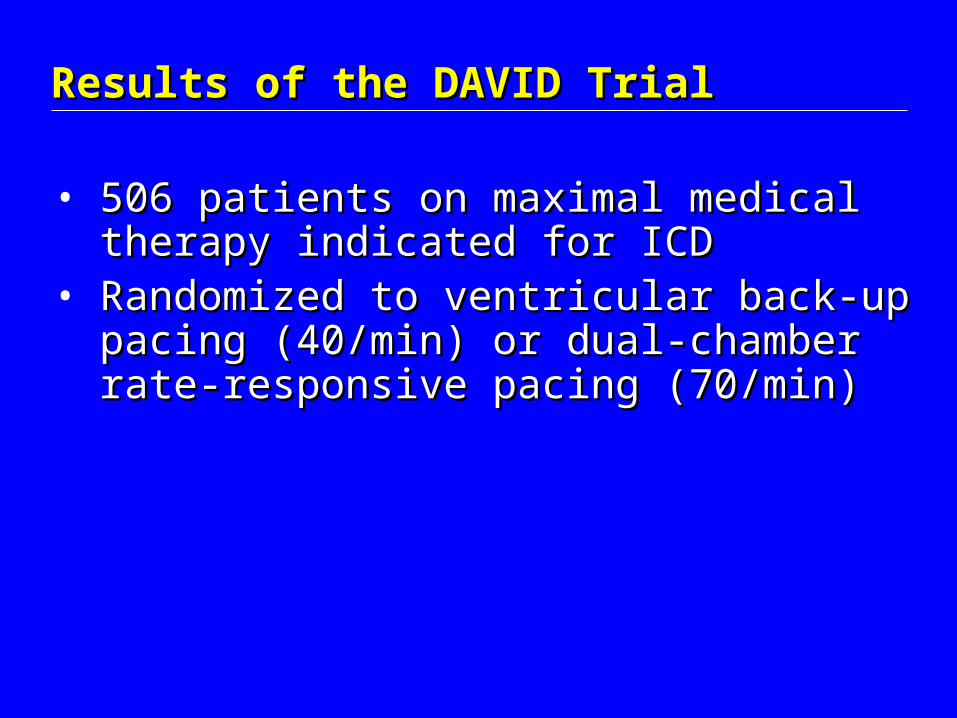
Results of the DAVID TrialResults of the DAVID Trial
• 506 patients on maximal medical therapy 506 patients on maximal medical therapy indicated for ICDindicated for ICD
• Randomized to ventricular back-up pacing Randomized to ventricular back-up pacing (40/min) or dual-chamber rate-responsive (40/min) or dual-chamber rate-responsive pacing (70/min)pacing (70/min)

Results of the DAVID TrialResults of the DAVID Trial
Ventricular Backup Pacing40/min
Dual-chamber Rate-responsive70/min
Endpoint:Endpoint:
One-year survival free composite time to death or first One-year survival free composite time to death or first hospitalization for CHFhospitalization for CHF
83.9%1-yr free of death or CHF hospitalization
Results:*p < 0.03
Conclusion: “Dual-chamber pacing offers no clinical advantage over ventricular backup pacing and may be detrimental by increasing… death or hospitalization for CHF,” for patients with standard indications for ICD therapy, EF<40%, and no indication for bradycardic pacing.
73.3%1-yr free of death or CHF hospitalization

Results of the COMPANION TrialResults of the COMPANION Trial
1600 patients with active CHF and QRS > 1600 patients with active CHF and QRS > 120 ms with maximal medical therapy120 ms with maximal medical therapy
Endpoint: Endpoint:
Combined All-Cause Mortality and All-Cause HospitalizationCombined All-Cause Mortality and All-Cause Hospitalization
Biventricular Pacer + ICD
Biventricular Pacer Only
Medical Therapy Only0
-20%
-40%
% Reduction in Mortality +/-Hospitalization

Biventricular PacingBiventricular Pacing
How can biventricular pacing help cardiac How can biventricular pacing help cardiac surgery patients?surgery patients?
• Use temporary DDD biventricular pacing Use temporary DDD biventricular pacing in all patients with large LV’s, low EF’s in all patients with large LV’s, low EF’s ++ wide QRS’swide QRS’s
• Implant permanent LV epicardial Implant permanent LV epicardial electrode in those likely to benefitelectrode in those likely to benefit

How To Do Temporary BiV PacingHow To Do Temporary BiV Pacing
1.1. Sew temporary electrodes to anterior Sew temporary electrodes to anterior RV and posterolateral LV RV and posterolateral LV
2.2. Attach BOTH to the Attach BOTH to the negativenegative pole of the pole of the gray cablegray cable
3.3. Place a skin ground in the positive polePlace a skin ground in the positive pole
You have now created TWO unipolar You have now created TWO unipolar pacing dipoles that will activate pacing dipoles that will activate
the RV + LV simultaneouslythe RV + LV simultaneously

Temporary BiV Lead PlacementTemporary BiV Lead Placement

Biventricular PacingBiventricular Pacing
• Virtually all cardiac surgery patients with Virtually all cardiac surgery patients with poor LV function who require temporary poor LV function who require temporary pacing postoperatively should have pacing postoperatively should have temporary biventricular leads as well as temporary biventricular leads as well as atrial leads.atrial leads.
• Who should have a permanent LV lateral Who should have a permanent LV lateral electrode placed at the time of operation?electrode placed at the time of operation?

Biventricular PacingBiventricular Pacing
The following groups may benefit from a The following groups may benefit from a posterolateral LV epicardial electrode placed posterolateral LV epicardial electrode placed at the time of cardiac operation:at the time of cardiac operation:
• Those with pacers already in placeThose with pacers already in place• Those with large, hypocontractile LV’s Those with large, hypocontractile LV’s
who are likely to need pacingwho are likely to need pacing• Some Maze patientsSome Maze patients• Those who may need ICD’sThose who may need ICD’s

How To Do Permanent BiV PacingHow To Do Permanent BiV Pacing
Sew a steroid eluting epicardial pacing wire Sew a steroid eluting epicardial pacing wire posterolaterally on all those with large LVIDd posterolaterally on all those with large LVIDd and low EF:and low EF:
• Who already have pacers in placeWho already have pacers in place
• Who are likely to need permanent pacingWho are likely to need permanent pacing
• Leave it buried under clavicleLeave it buried under clavicle

Permanent BiV Lead PlacementPermanent BiV Lead Placement

How To Do Permanent BiV PacingHow To Do Permanent BiV Pacing
Where is the optimal location for the LV Where is the optimal location for the LV wire?wire?

OutcomesOutcomes

Biventricular Pacing:Biventricular Pacing:Preoperative Characteristics (n=25)Preoperative Characteristics (n=25)
Mean Age (yrs)Mean Age (yrs) 75 75
NYHA 3+NYHA 3+ 80%80%
Previous MIPrevious MI 40%40%
Previous Cardiac SurgeryPrevious Cardiac Surgery 32%32%
DiabetesDiabetes 32%32%
Renal FailureRenal Failure 20%20%
Cerebrovascular DiseaseCerebrovascular Disease 20%20%
Peripheral Vascular DiseasePeripheral Vascular Disease 20%20%
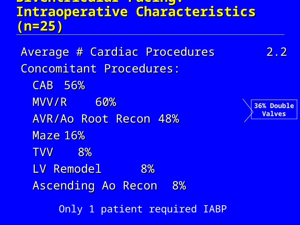
Biventricular Pacing:Biventricular Pacing:Intraoperative Characteristics (n=25)Intraoperative Characteristics (n=25)
Average # Cardiac Procedures Average # Cardiac Procedures 2.2 2.2
Concomitant Procedures:Concomitant Procedures:
CABCAB 56%56%
MVV/RMVV/R 60%60%
AVR/Ao Root ReconAVR/Ao Root Recon 48%48%
MazeMaze 16%16%
TVVTVV 8% 8%
LV RemodelLV Remodel 8% 8%
Ascending Ao ReconAscending Ao Recon 8% 8%
36% DoubleValves
Only 1 patient required IABP

Biventricular Pacing: Biventricular Pacing: Intraoperative BiV Pacing - # of ProceduresIntraoperative BiV Pacing - # of Procedures
• Excluding pacing procedures,Excluding pacing procedures,– (8) pts had one procedure(8) pts had one procedure– (9) pts had two procedures(9) pts had two procedures– (7) pts had three procedures(7) pts had three procedures– (1) pt had five procedures(1) pt had five procedures
PtsPts BiV Procedure List
4 CAB
1 CAB, LVA
2 AVR
1 Redo AoRR
1 Redo AoRR, ASC Ao
1 Redo AVR, MVR
1 ARE, MVR
1 AoRR, MVV
3 AVR, MVV, CAB
1 AVR, MVR, CAB, ASC Ao, LVA
1 Redo AVR, CAB, ASC Ao
3 MVV, CAB, MAZE
1 MVV,MAZE
1 MVV, CAB
21
MVV, TVVMVV

Biventricular Pacing:Biventricular Pacing:Distribution of LVIDd (n=25)Distribution of LVIDd (n=25)
0
1
2
3
4
5
6
7
8
# P
ati
en
ts
<5 5.0-5.5 5.6-6.0 6.1-7.0 > 7.0
LVIDd (cm)

Biventricular Pacing: Biventricular Pacing: Procedural Categories (n=25)Procedural Categories (n=25)
Lead Only (5)20%
Upgrade (7)28%New Pacer (13)
52%

Biventricular Pacing: Biventricular Pacing: Preoperative CharacteristicsPreoperative Characteristics
CategoryCategory Average Average LVEFLVEF
Average Average
LVIDdLVIDd
Lead Only (5)Lead Only (5) 43%43% 5.95.9
New Pacer (13)New Pacer (13) 29%29% 6.86.8
Upgrade (7)Upgrade (7) 30%30% 5.55.5
Total (25)Total (25) 32%32% 6.36.3

Biventricular Pacing: Biventricular Pacing: Patient Characteristics: EF vs. LV SizePatient Characteristics: EF vs. LV Size
Figure 1. Ejection Fraction vs. LV Size
0
10
20
30
40
50
60
70
0 1 2 3 4 5 6 7 8 9 10
Left Ventricular Internal Dimension (cm)
% E
ject
ion
Fra
ctio
n (
EF
)
NORMAL(EF>50% & LVID<5.7 cm)

Figure 2. QRS Interval vs. LV Size
0
50
100
150
200
0 1 2 3 4 5 6 7 8 9 10
Left Ventricular Internal Dimension (cm)
QR
S In
terv
al (
ms
)
NORMAL(QRS<120 ms & LVIDd<5.7 cm)
Paced Preop
Not Paced Preop
Biventricular Pacing: Biventricular Pacing: Patient Characteristics: QRS vs. LV SizePatient Characteristics: QRS vs. LV Size

Biventricular Pacing: Biventricular Pacing: ResultsResults
• 1 operative mortality1 operative mortality
• 3 late deaths3 late deaths
• 1 patient had two postop strokes1 patient had two postop strokes
• 1 patient required subsequent VT ablation1 patient required subsequent VT ablation

Biventricular Pacing: Biventricular Pacing: Postoperative Survival (Days)Postoperative Survival (Days)
Kaplan-Meier Survival
Days from Surgery
4003002001000
Cu
mu
lativ
e S
urv
iva
l
1.0
.9
.8
.7
.6
.5
.4
.3
.2
.1
0.0
Survival Function
Censored

Surgical Implications of CRT - OverallSurgical Implications of CRT - Overall
If we are to improve our knowledge of who If we are to improve our knowledge of who will benefit from permanent LV will benefit from permanent LV electrodes, we mustelectrodes, we must
1.1. Renew our interest in preoperative EKG, Renew our interest in preoperative EKG, for instance RBBB v. LBBBfor instance RBBB v. LBBB
2.2. Improve our knowledge about Improve our knowledge about intraoperative echo diagnosis of intraoperative echo diagnosis of dyssynchronydyssynchrony
3.3. Learn optimal LV electrode placementLearn optimal LV electrode placement

Surgical Implications of CRT – Ischemic MRSurgical Implications of CRT – Ischemic MR
• Because dyssynchrony contributes to Because dyssynchrony contributes to “ischemic” MR, we must consider it a “ischemic” MR, we must consider it a correctable part of the syndrome that correctable part of the syndrome that neither ring nor prosthetic valve placement neither ring nor prosthetic valve placement addresses.addresses.
• Dyssynchrony tethers the posterior leaflet.Dyssynchrony tethers the posterior leaflet.

Surgical Implications of CRT - LV RemodelingSurgical Implications of CRT - LV Remodeling
• LV aneurysmectomy to physically remodel LV aneurysmectomy to physically remodel the heart can no longer be complete the heart can no longer be complete unless we “electrically” remodel the heart unless we “electrically” remodel the heart as well.as well.

Surgical Implications of CRT - MazeSurgical Implications of CRT - Maze
• The maze operation in some patients will The maze operation in some patients will no longer be complete unless we restore no longer be complete unless we restore AV synchrony and LV synchrony as wellAV synchrony and LV synchrony as well

Surgical Implications of CRT - PacedSurgical Implications of CRT - Paced
• Chronically paced patients with large, Chronically paced patients with large, hypocontractile hearts who require cardiac hypocontractile hearts who require cardiac operations are easy to upgrade to operations are easy to upgrade to biventricular pacing and likely to benefitbiventricular pacing and likely to benefit

Biventricular Pacing ConclusionsBiventricular Pacing Conclusions
• We prefer temporary biventricular DDD We prefer temporary biventricular DDD pacing for postop pacing in all patients with pacing for postop pacing in all patients with large, low EF heartslarge, low EF hearts
• We consider placing a permanent We consider placing a permanent epicardial lead in patients with poor LV epicardial lead in patients with poor LV function and prolonged QRS who are likely function and prolonged QRS who are likely to need permanent pacing or who currently to need permanent pacing or who currently have permanent pacershave permanent pacers