interstitial lung diseases sevda Özdoğan md, prof. chest diseases
TRANSCRIPT

Interstitial Lung Diseases
Sevda Özdoğan MD, Prof.
Chest Diseases

Definition Interstitial lung diseases covers
a group of diffuse lung disorders that involve infiltration of alveolar air spaces or thickening of pulmonary interstitial structures
This heterogenious group of diseases have similar clinical, radiological and physiological characteristics
Alveolus
Interstitium
Type I pneumocyt
Type II pneumocyt
Capillair
Endotheel cel
AlveolusAlveolus
Interstitium
Type I pneumocyt
Type II pneumocyt
Capillair
Endotheel cel
Alveolus

Classification
Alveolar proteinosis,microlithiasis
Berilliosis, hypersensitivity pn, histiocytosisOccupational
radiation
40-50%


pneumoconiosis

Hypersensitivity pneumonitis

Collagen tissue diseases associated with ILD
SLE RA PSS Sjögren sydrome Polymyositis-dermatomyositis Mixed connective tissue disease Ankylosing spondylitis
Other Systemic Diseases
Sarcoidosis Vasculites (Wegener, Churg Strauss) Hemorragic Syndromes (Goodpasture,
Idiopathic pulmonary hemosiderosis) Hystiositosis X Chronic Gastric aspiration Lymphangitis Carcinomatosa Chronic pulmonary edema Chronic uremia Alveolary proteinosis

Epidemiology
The known insidance and prevalance rate of ILD depens on some reports which may not represent the population
Estimated prevalance in man is 80.9/100 000 population
Estimated prevalance in woman is 67.2/100 000 population

SYMPTOMS Dyspnea: Progressive, prominent in
exercise. Sudden worsening of dyspnea, particularly
if associated with pleural pain, may indicate a spontaneous pneumothorax.
Cough: A dry cough

Hemoptysis: Diffuse alveolar hemorrhage syndromes, Vasculitides
Churg strauss, Wegener Granulomatosis Behçet’s disease
Can be due to other complications Lung cancer Pulmonary tuberculosis
Symptoms of Right Hearth Failure
HISTORY
The cause of the illness is often recognized from the patient's history.
Entire lifelong employment Prior medication use Symptoms of a systemic disease
Musculoskeletal pain, weakness, fatigue, fever, joint pains or swelling, photosensitivity, dry mouth or eye

Gender:
Female
Lymphangioleiomyomatosis (Premenopausal)
pulmonary involvement in tuberous sclerosis
Connective tissue diseases
Male
ILD associated with rheumatoid arthritis
occupational exposures, pneumoconiosis.

Smoking history
Current or former smokers Hystiocytosis X DIP, IPF, RBILD
Never or exsmokers Sarcoidosis Hypersensitivity
pneumonitis.
Active smoking can lead to complications in Goodpasture's syndrome, in which pulmonary hemorrhage is far more frequent in current smokers.

Family history
Tuberous sclerosis, Neurofibromatosis
Idiopathic pulmonary fibrosis, Sarcoidosis

PHYSICAL EXAMINATION
Physical examination is usually nonspecific
Crackles or "velcro rales" are common in most forms of ILD, although they are less likely to be heard in the granulomatous lung diseases, especially sarcoidosis.
Scattered late inspiratory high-pitched rhonchi (inspiratory squeaks) are frequently heard on chest examination in patients with bronchiolitis.
Cyanosis Tachypnea Tachicardia Cor pulmonale (edema, hepatomegali) Extrapulmonary manifestations of
systemic diseases Erythema nodosum (sarcoidosis, tb) Telenjiectasis (scleroderma) Joint deformities (RA)

Clubbing: Common in IPF, Asbestosis

Diagnosis History
Chest radiography Chest x-ray (can be normal in 10%) HRCT
The routine laboratory evaluation is not much helpfull but should be done (biochemical and hematologic tests)

Chest x-ray
All available previous chest radiograms should be reviewed to ascertain the onset, progression, chronicity and stability of the disease Diffuse alveolar (aciner) pattern, lineer, nodular or reticulonodular infiltrations Cystic changes (honeycombing) can be
present

5-10 mm nodules aciner pattern
Cystic changes (Honeycombing)

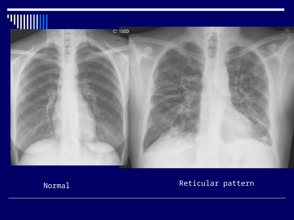
Reticular patternNormal

The distribution of the lesions (Upper or lover zone distribution) can be usefull in narroving the differential diagnosis
The presence and the pattern of adenopathy may provide additional clues

HRCT More sensitive and specific than chest
radiographs Graund grass attenuation Lung consolidation Reticular opacities Nodules Cysts Fibrotic changes
Honey combing

Pulmonary Function Testing
A restrictive defect with reductions in total lung capacity (TLC), functional residual capacity (FRC), and residual volume (RV)
Flow rates are decreased (FEV1 and FVC), but the changes are in proportion to the decreased lung volumes; thus, the FEV1/FVC ratio is usually normal or increased
A reduction in the DLCO is a commonly found

Arterial Blood Gas analysis
The resting arterial blood gases may be normal or may reveal hypoxemia
A normal resting PaO2 does not rule out significant hypoxemia during exercise or sleep, which is common in ILD.
Carbon dioxide retention is rare and usually a manifestation of end-stage disease.

Invasive Diagnostic techniques FOB
Bronchoalveolar lavage (BAL) Transbronchial lung biopsy
VATS
Treatment
Avoid the exposed etiologic factor if there is any
Supportive care (Oxygen, pulmonary rehabilitation)
Treat the underlying disease if there is any
Corticosteroids Immunesuppressive treatment Lung transplantation

Sarcoidosis
Sarcoidosis is a multisystem granulomatous disorder of unknown cause(s). It commonly affects young and middle-aged adults and frequently presents with bilateral hilar lymph-adenopathy, pulmonary infiltration, ocular and skin lesions.
VII İnternational Sarcoidosis Meeting 1976

The histologic hallmark is noncaseating granulomas

Etiology
The cause of Sarcoidosis remains unknown
Despite the lack of specific infectious cause Tuberculosis Atipic mycobacteria Epstein Barr virus Fungal infections Mycoplasma are investigated

Organ involvement in sarcoidosis
OrganOrgan % % of patientsof patients
Mediastinal lymph nodesMediastinal lymph nodes 95-98%95-98%LungsLungs >90%>90%LiverLiver 50-80%50-80%SpleenSpleen 40-80%40-80%EyesEyes 20-50%20-50%Peripheral lymph nodesPeripheral lymph nodes 30%30%SkinSkin 25%25%Nervous systemNervous system 10%10%Heart (clinically)Heart (clinically) 5%5%

Chest x-ray
Bilateral hiler adenopathy
Bilateral hiler adenopathy+ paranchymal infiltrates

Chest radiographic stages of sarcoidosis
Stage Stage FrequencyFrequency
00 NormalNormal 5-10%5-10%
II BHLBHL 50%50%
IIIIBHL and parenchymal BHL and parenchymal infiltratesinfiltrates 25%25%
IIIIII Parenchymal infiltrates Parenchymal infiltrates without BHLwithout BHL 15%15%
IVIV Signs of fibrosisSigns of fibrosis 5-10%5-10%

HRCT
Hilar and mediastinal lymphadenopathy Beaded or irregular thickening of the bronchovascular
bundles Nodules along bronchi, vessels, and subpleural regions Ground glass opacification Parenchymal masses or consolidation Parenchymal bands Cysts Traction bronchiectasis Fibrosis with distortion of the lung architecture

Clinical Symptoms
2/3 of the patients are asymtomatic diagnosed in an incidental chest radiogram
Respiratory symptoms Dyspnea (marked on exertion)
Cough (nonproductive)
Chest discomfort (Chest pain often difficult to describe)
Chest tightness and Wheesing (In chronic fibrocystic sarcoidosis)

Systemic / constitutional symptoms
Fatigue (up to 70% of patients)
Fever (usually low-grade, but up to 40°C possible)
Weight loss(2-6 kg during 10-12 weeks)
Symptoms of involved system

Löfgren’s Syndrome
It is a well defined presentation of sarcoidosis that consists of Erythema nodosum Polyartritis Bilateral hiler lymphadenopathy
The unset is usually abrupt In most cases spontaneous remission
occurs

Heerfordt’s Syndrome
Also known as Uveoparotid fever Consists of Fever Anterior uveitis Parotid and lacrimal gland enlargement Bilateral hiler adenopathy Facial paralysis
Has an abrupt unset

Diagnostic Evaluation History (occupational and environmental exposure, symptoms) Physical examination Postero-anterior chest radiogaphy Pulmonary function tests: spirometry and CO diffusion capacity Serum chemistries: calcium, liver enzymes, creatinine, blood urea
nitrogen, ACE Urine analysis Electrocardiography Routine opthalmologic examination Tuberculin skin test Tissue biopsy

Bronchoscopic evaluation FOB can be usefull in tissue sampling by
Bronchial mucosal biopsy (there may be nodularity, mucosal edema, hypervascularity)
Transbronchial needle aspiration of the mediastinal lymph nodes
Transbronchial lung biopsy
BAL sampling Increased lymphocyts to 20-50% (<10% in normal
subjects)

The easiest accessible biopsy site should be preferred for tissue sampling A sklin nodule Superficial lymph node Nasal mucosa, salivary gland
Mediastinoscopy VATS or thoracotomy rarely needed
Treatment
>80% of the patients undergo spontaneous remission depending on the stage of the disease with the highest rate in the early stages
Which patients should be treated??

Treatment indications
Systemic symptoms (Fever, weight loss) Progressive symptomatic pulmonary
involvement (Dyspnea, cough, hypoxemia) Multipl vital organ involvement Myocardial involvement Eye involvement that is resistant to topical
treatment Santral Nervous system involvement Hypercalcemia

Corticosteroids minimum of 8-12 months
Supportive management of patients who have advanced disease and corpulmonale
Special topical treatments for extrapulmonary disease
