chronic obstructive pulmonary disease e. sevda Özdoğan md chest diseases
TRANSCRIPT
Chronic Obstructive Chronic Obstructive Pulmonary DiseasePulmonary Disease
E. Sevda Özdoğan MD
Chest diseases
COPDCOPD
COPD is a disease state characterized by airflow limitation that is not fully reversible. The airflow limitation is usually both progressive and associated
with an abnormal inflammatory response of the lungs to noxious particles or gases.
TerminologyTerminology
Chronic BronchitisDefined as the presence of cough and sputum production for at least 3 months in each of 2 consecutive years, is not necessarily associated with airflow limitation.
EmphsemaIrreversibl dilatation and destruction of the airways distal
to terminal bronchie (without fibrosis) – Centrlobular emphysema (respiratory bronchioles)– Panlobular emphsema (resp. Br+alv ductus and
alveolus)
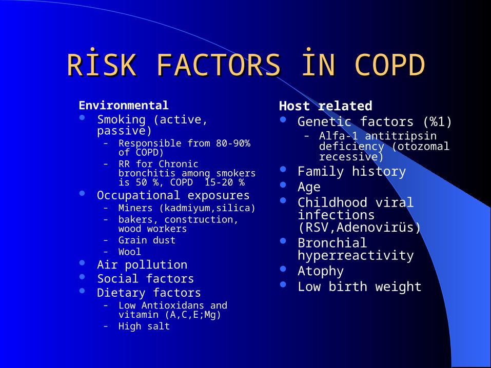
RİSK FACTORS İN COPDRİSK FACTORS İN COPDEnvironmental Smoking (active, passive)
– Responsible from 80-90% of COPD)– RR for Chronic bronchitis among
smokers is 50 %, COPD 15-20 % Occupational exposures
– Miners (kadmiyum,silica)– bakers, construction, wood workers– Grain dust– Wool
Air pollution Social factors Dietary factors
– Low Antioxidans and vitamin (A,C,E;Mg)
– High salt
Host related Genetic factors (%1)
– Alfa-1 antitripsin deficiency (otozomal recessive)
Family history Age Childhood viral infections
(RSV,Adenovirüs) Bronchial hyperreactivity Atophy Low birth weight
Pathophysiological Pathophysiological MechanismsMechanisms
InflammationProtease-antiprotease imbalanceOxydative stress
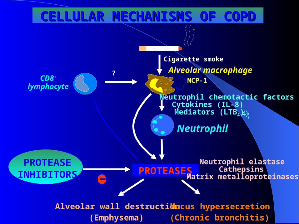
Cigarette smoke
Alveolar macrophage
Neutrophil
PROTEASES
Alveolar wall destruction(Emphysema)
Mucus hypersecretion(Chronic bronchitis)
PROTEASEINHIBITORS
Neutrophil chemotactic factors
CELLULAR MECHANISMS OF COPDCELLULAR MECHANISMS OF COPD
Neutrophil elastaseCathepsins
Matrix metalloproteinases
Cytokines (IL-8)Mediators (LTB4) 4 ))
?CD8+
lymphocyte
-
MCP-1
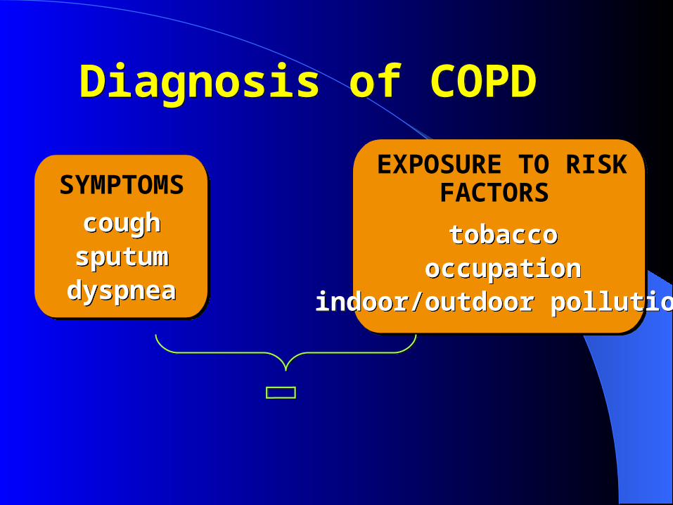
SYMPTOMS
coughcoughsputumsputumdyspneadyspnea
EXPOSURE TO RISKFACTORS
tobaccotobaccooccupationoccupation
indoor/outdoor pollutionindoor/outdoor pollution
Diagnosis of COPDDiagnosis of COPD
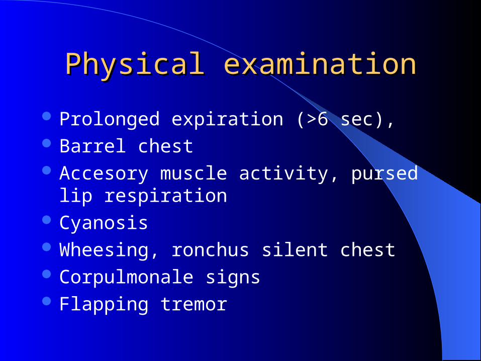
Physical examinationPhysical examination
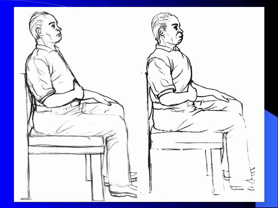
Prolonged expiration (>6 sec), Barrel chest Accesory muscle activity, pursed lip respiration Cyanosis Wheesing, ronchus silent chest Corpulmonale signs Flapping tremor
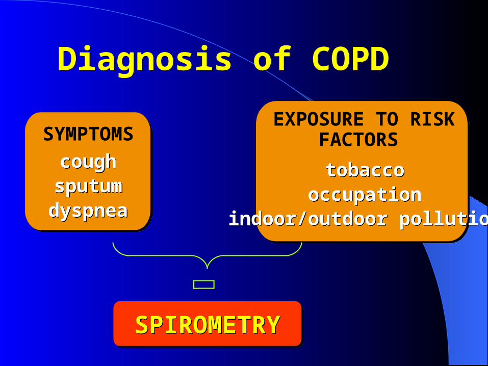
SYMPTOMS
coughcoughsputumsputumdyspneadyspnea
EXPOSURE TO RISKFACTORS
tobaccotobaccooccupationoccupation
indoor/outdoor pollutionindoor/outdoor pollution
SPIROMETRYSPIROMETRY
Diagnosis of COPDDiagnosis of COPD
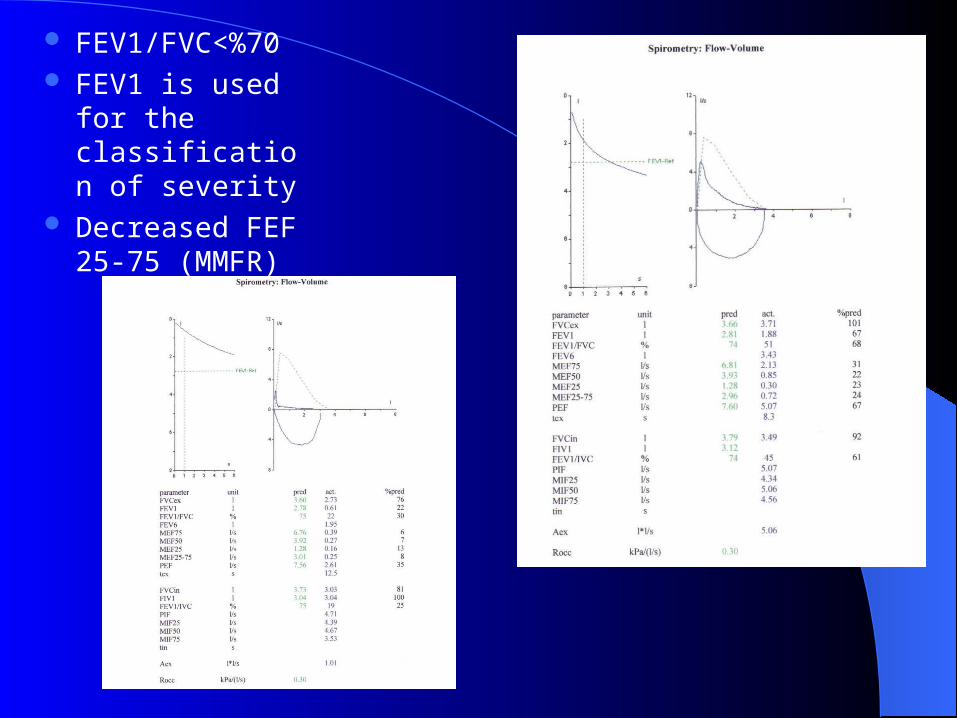
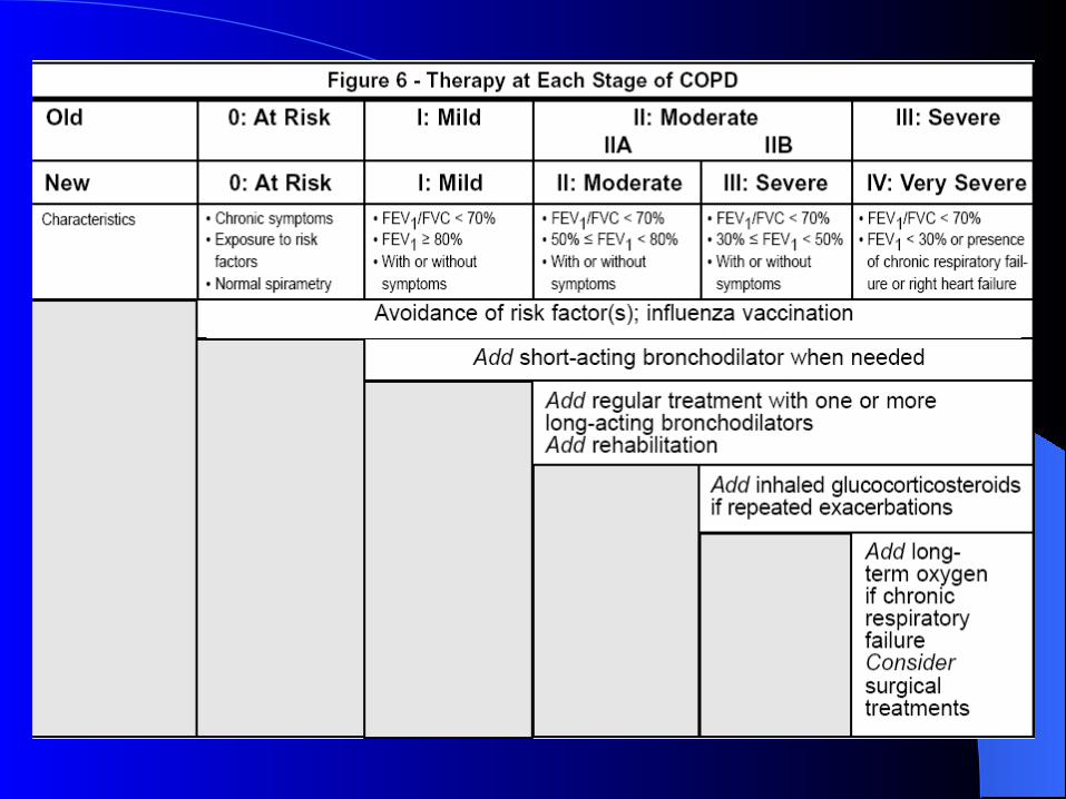
FEV1/FVC<%70 FEV1 is used for
the classification of severity
Decreased FEF 25-75 (MMFR)
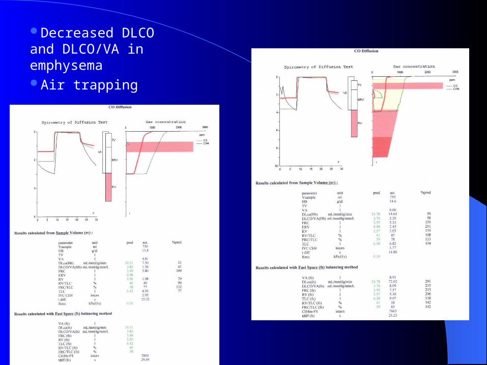
Decreased DLCO and DLCO/VA in emphysemaAir trapping
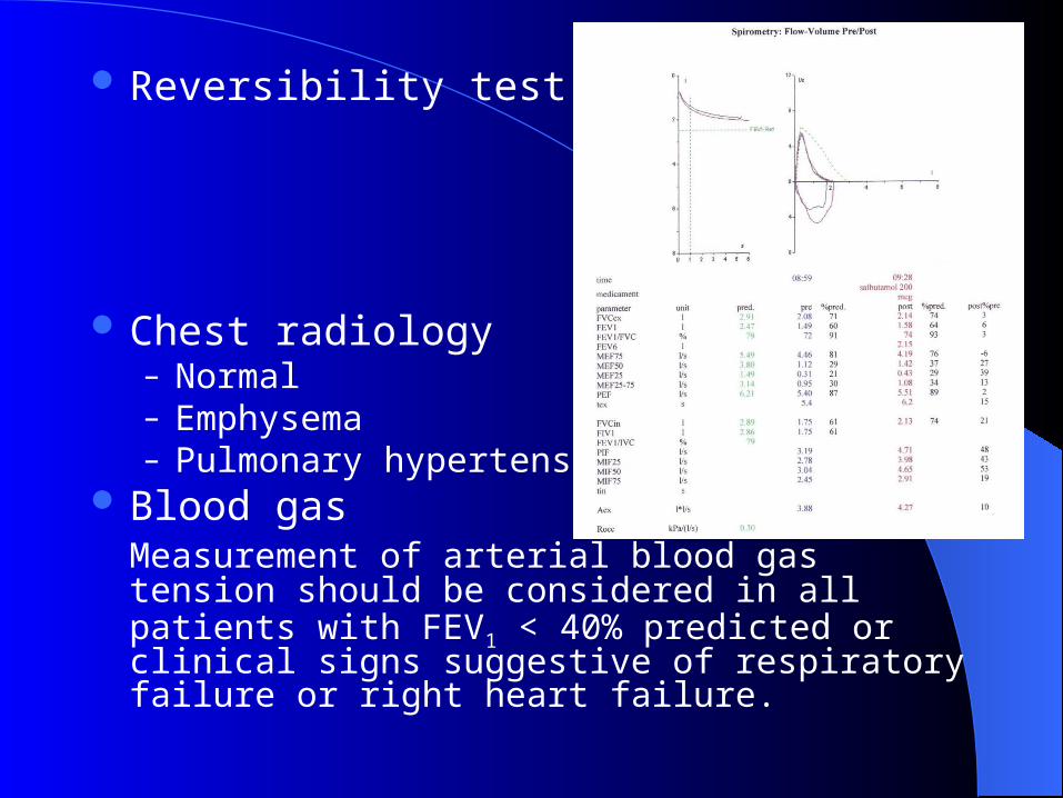
Reversibility test (-)
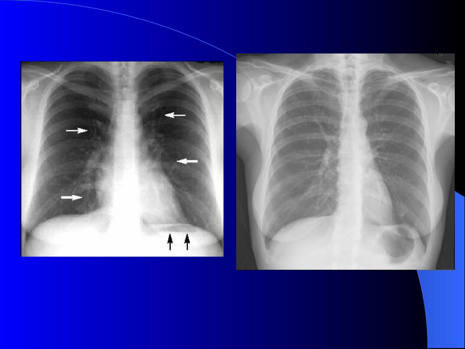
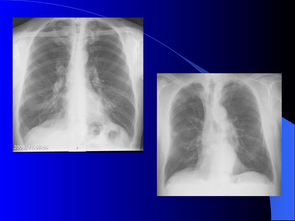
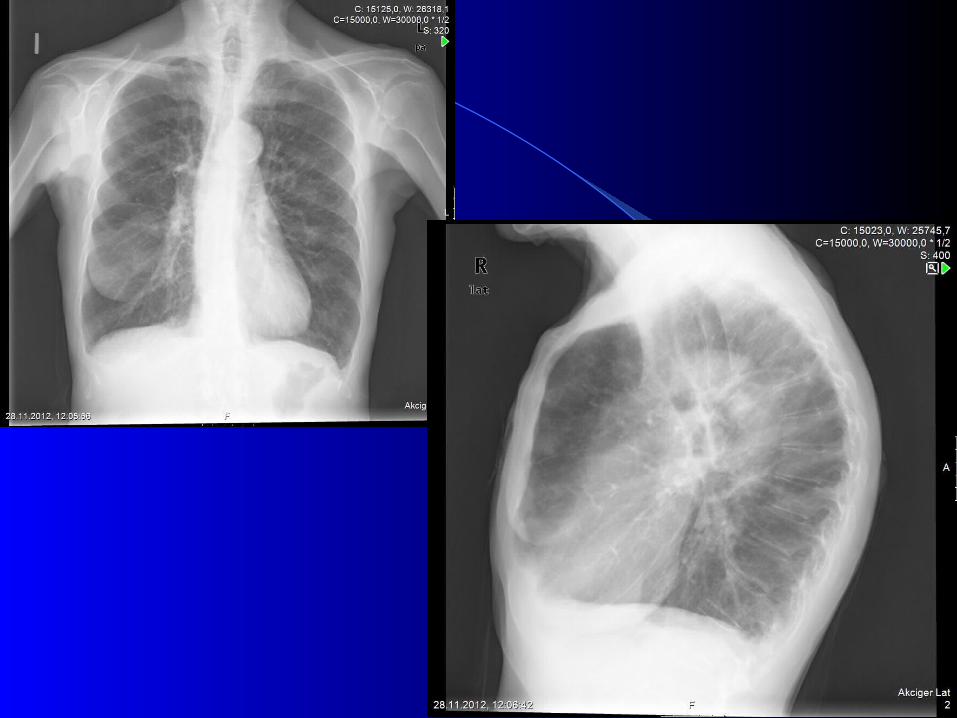
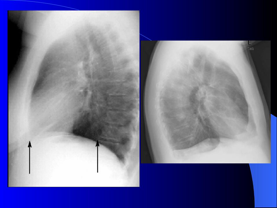
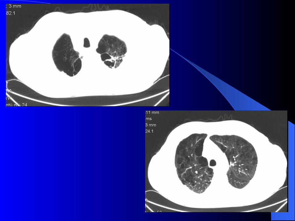
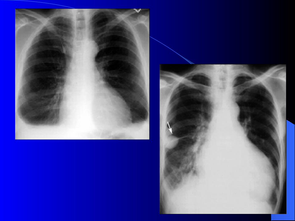
Chest radiology– Normal– Emphysema– Pulmonary hypertension
Blood gasMeasurement of arterial blood gas tension should be considered in all patients with FEV1 < 40% predicted or clinical signs suggestive of respiratory failure or right heart failure.
Routine blood tests:– Polistemia– Liver function abnormality– Renal function abnormalities
ECG: – Axis changes due to diafragmatic flattening or
right ventricular hypertrophy– P pulmonale– Right bundle block– İmpaired R progression
Alfa-1 antitripsin deficiency tests: – COPD before the age of 45– Family history– Nonsmokers– Panlobular emphysema in the lower lobes
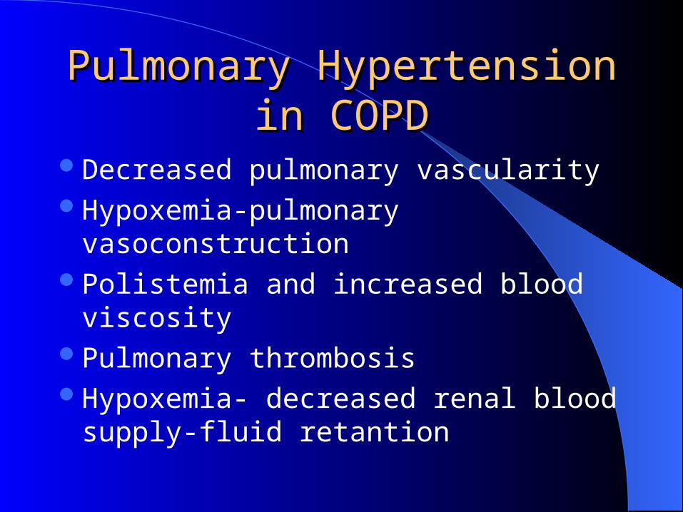
Pulmonary Hypertension in Pulmonary Hypertension in COPDCOPD
Decreased pulmonary vascularityHypoxemia-pulmonary vasoconstructionPolistemia and increased blood viscosityPulmonary thrombosisHypoxemia- decreased renal blood supply-
fluid retantion
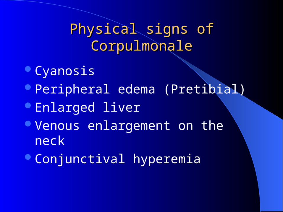
Physical signs of CorpulmonalePhysical signs of Corpulmonale
CyanosisPeripheral edema (Pretibial)Enlarged liverVenous enlargement on the neckConjunctival hyperemia
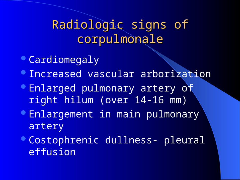
Radiologic signs of corpulmonaleRadiologic signs of corpulmonale
CardiomegalyIncreased vascular arborizationEnlarged pulmonary artery of right hilum
(over 14-16 mm)Enlargement in main pulmonary arteryCostophrenic dullness- pleural effusion
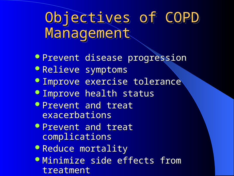
Objectives of COPD Objectives of COPD ManagementManagementObjectives of COPD Objectives of COPD ManagementManagement
Prevent disease progression Relieve symptomsImprove exercise toleranceImprove health statusPrevent and treat exacerbationsPrevent and treat complicationsReduce mortalityMinimize side effects from treatment
Prevent disease progression Relieve symptomsImprove exercise toleranceImprove health statusPrevent and treat exacerbationsPrevent and treat complicationsReduce mortalityMinimize side effects from treatment
Reduce Risk Factors
• Reduction of total personal exposure to tobacco smoke, occupational dusts and chemicals, and indoor and outdoor air pollutants are important goals to prevent the onset and progression of COPD.
• Smoking cessation is the single most effective-and cost-effective- intervention to reduce the risk of developing COPD and stop its progression
Vaccines: – Influenza vaccines reduce serious illness
and death in COPD patients by 50%. Give once (in Autumn) each year.
– Pneumococcal vaccine
Pharmacologic treatmentPharmacologic treatment
Can improve and prevent symptoms,Reduce the frequency and severity of
exacerbations,Improve health status,Improve exercise tolerance.

BronchodilatorsBronchodilators These medications are central to symptom
management in COPD.
Short acting beta agonists LABA Anticholinergics (long and short acting) Theophyline
Regular treatment with long-acting bronchodilators is more effective and convenient than treatment with short-acting bronchodilators, but more expensive.
Combining drugs with different mechanisms and durations of action may increase the degree of bronchodilation for equivalent or lesser side effects.
Theophylline is effective in COPD, but due to its potential toxicity inhaled bronchodilators are preferred when available.
Regular treatment with inhaled glucocorticosteroids is not always necessary only appropriate for patients with:
– symptomatic improvement and a documented spirometric response to inhaled glucocorticosteroids or
– If FEV1 < 50% predicted and repeated exacerbations (for example, 3 in the last three years).
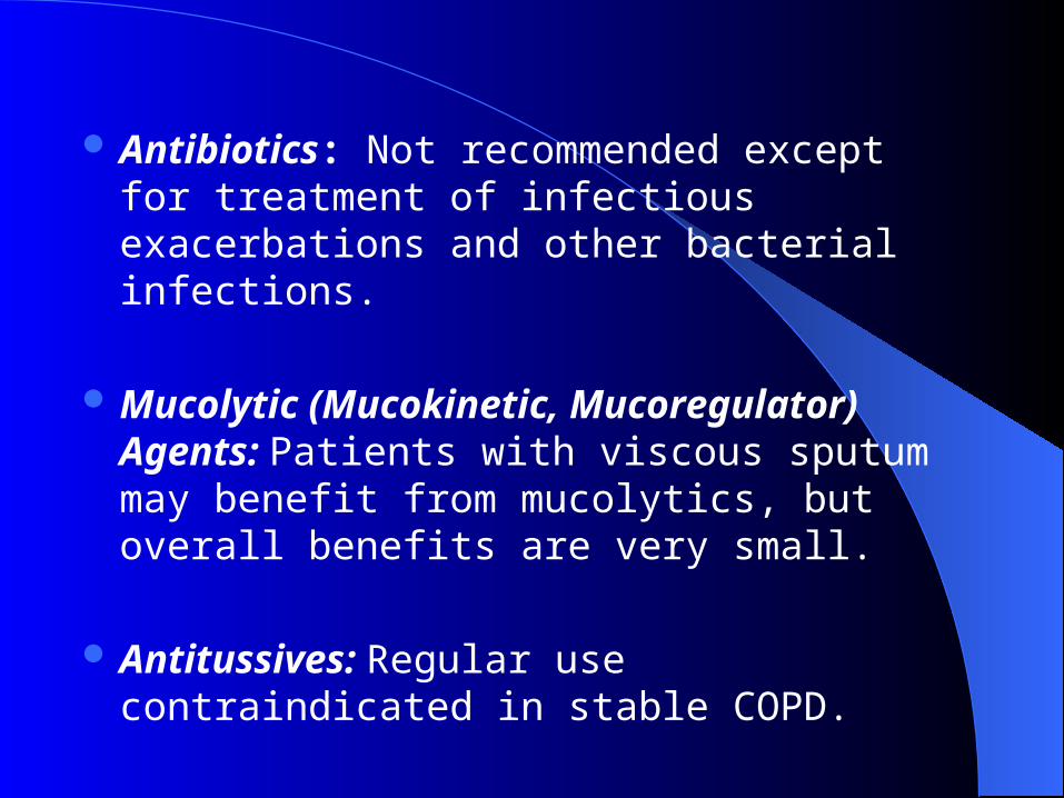
Antibiotics: Not recommended except for treatment of infectious exacerbations and other bacterial infections.
Mucolytic (Mucokinetic, Mucoregulator) Agents: Patients with viscous sputum may benefit from mucolytics, but overall benefits are very small.
Antitussives: Regular use contraindicated in stable COPD.
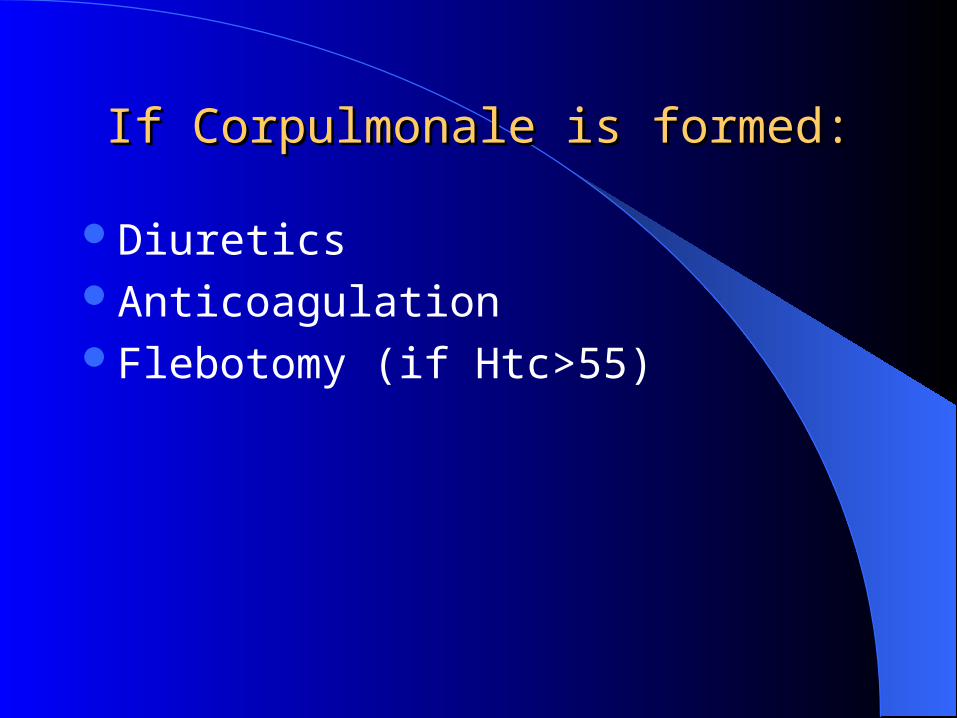
If Corpulmonale is formed:If Corpulmonale is formed:
DiureticsAnticoagulationFlebotomy (if Htc>55)
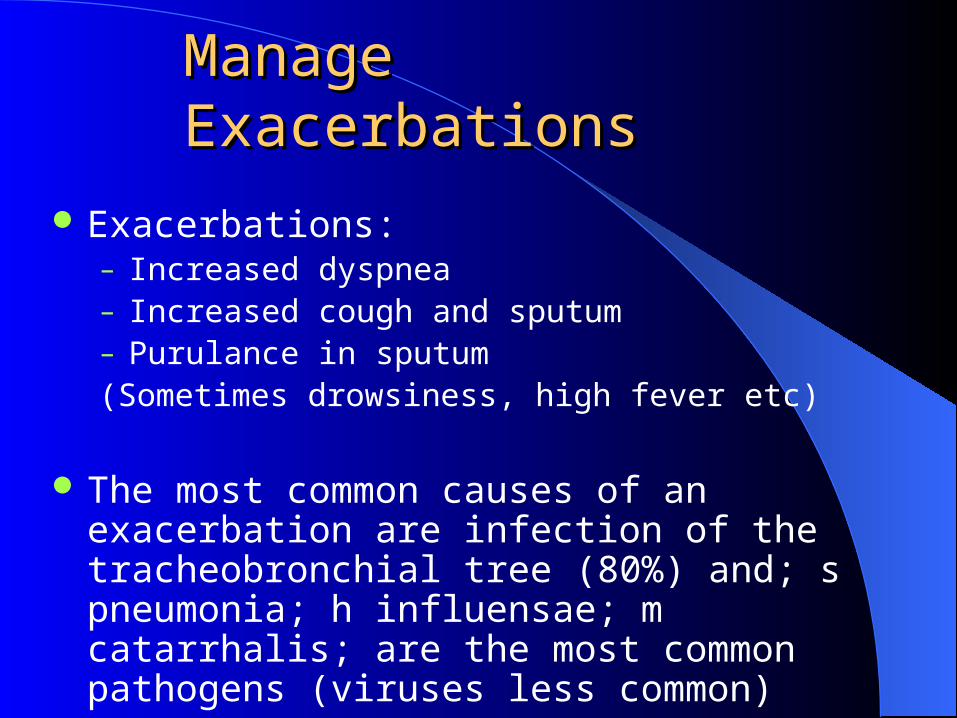
Manage ExacerbationsManage Exacerbations
Exacerbations:– Increased dyspnea– Increased cough and sputum– Purulance in sputum(Sometimes drowsiness, high fever etc)
The most common causes of an exacerbation are infection of the tracheobronchial tree (80%) and; s pneumonia; h influensae; m catarrhalis; are the most common pathogens (viruses less common)
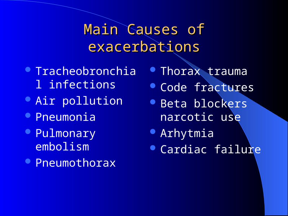
Main Causes of exacerbationsMain Causes of exacerbations
Tracheobronchial infections
Air pollution Pneumonia Pulmonary embolism Pneumothorax
Thorax trauma Code fractures Beta blockers narcotic
use Arhytmia Cardiac failure
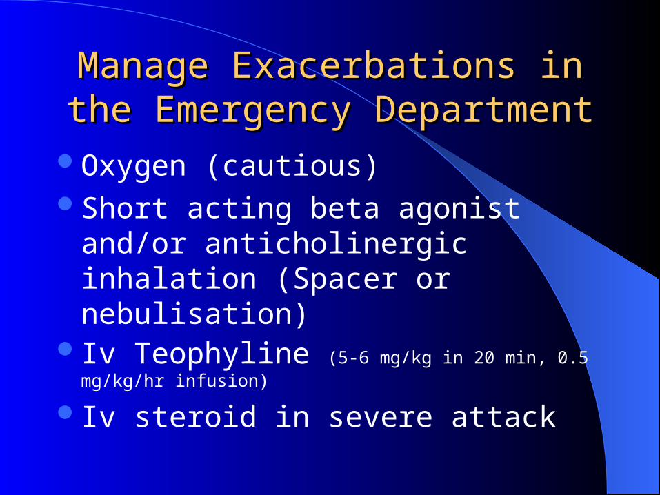
Manage ExacerbationsManage Exacerbations in the in the Emergency DepartmentEmergency Department
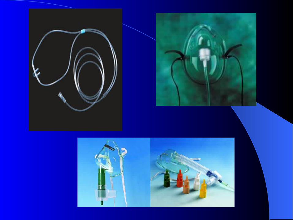
Oxygen (cautious)Short acting beta agonist and/or
anticholinergic inhalation (Spacer or nebulisation)
Iv Teophyline (5-6 mg/kg in 20 min, 0.5 mg/kg/hr infusion)
Iv steroid in severe attack
Antibiotics (iv, oral)Check fluid imbalance, nutritionTreat concomitant disease (Corpulmonale, px
etc)Prophylactic anticoagulationNoninvasive (BİPAP) and invasive mechanic
ventilation
Noninvasive intermittent positive pressure ventilation (NIIPPV) in acute exacerbations improves blood gases and pH, reduces in-hospital mortality, decreases the need for invasive mechanical ventilation and intubation, and decreases the length of hospital stay
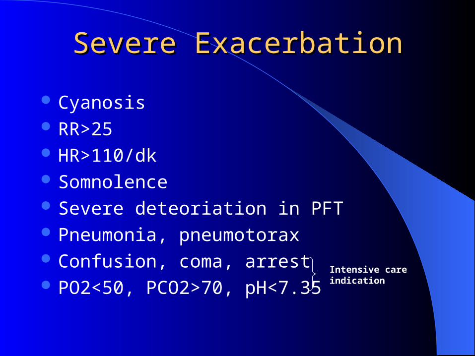
Severe ExacerbationSevere Exacerbation
Cyanosis RR>25 HR>110/dk Somnolence Severe deteoriation in PFT Pneumonia, pneumotorax Confusion, coma, arrest PO2<50, PCO2>70, pH<7.35
Intensive care indication
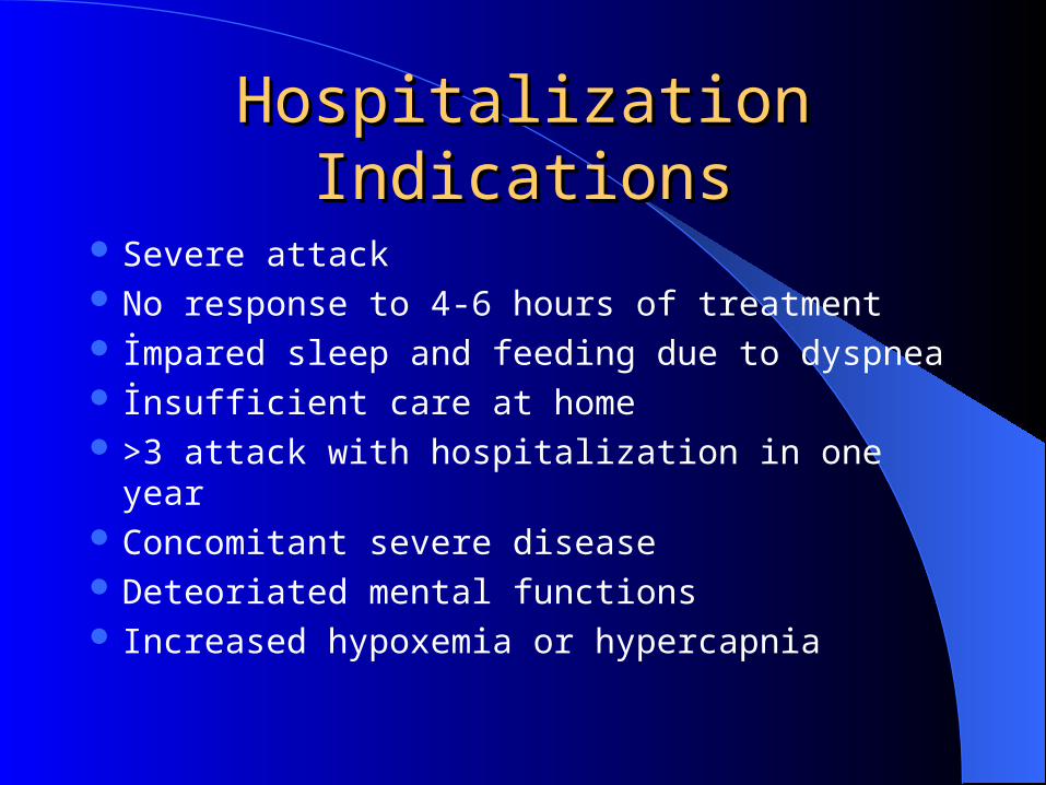
Hospitalization IndicationsHospitalization Indications
Severe attack No response to 4-6 hours of treatment İmpared sleep and feeding due to dyspnea İnsufficient care at home >3 attack with hospitalization in one year Concomitant severe disease Deteoriated mental functions Increased hypoxemia or hypercapnia
Non-Pharmacologic Treatment
RehabilitationOxygen therapySurgical interventions
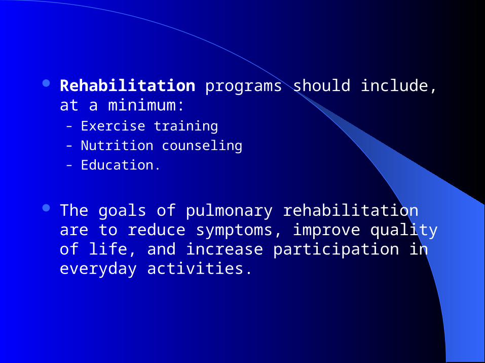
Rehabilitation programs should include, at a minimum:– Exercise training– Nutrition counseling– Education.
The goals of pulmonary rehabilitation are to reduce symptoms, improve quality of life, and increase participation in everyday activities.
Oxygen TherapyOxygen Therapy
The long-term administration of oxygen (>15 hours per day) to patients with chronic respiratory failure increases survival and has a beneficial impact on pulmonary arterial pressure, polycythemia (hematocrit > 55%), exercise capacity, lung mechanics, and mental state.
The goal of long-term oxygen therapy is to increase the baseline PaO2 at rest to at least 60 mm Hg at sea level, and/or produce SaO2 at least 90%, which will preserve vital organ function by ensuring an adequate delivery of oxygen.
Low flow rates 2-3lt/min should be given
Surgical TreatmentsSurgical Treatments
Bullectomy, lung volume reduction surgery (LVRS) and lung transplantation may be considered in carefully selected patients with Stage IV: Very Severe COPD.
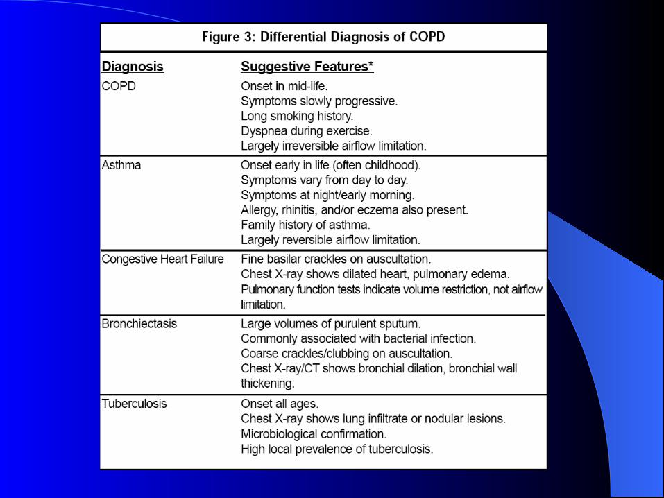
COPD IS NOT ASTHMA !COPD IS NOT ASTHMA !
• Different causes
• Different inflammatory cells
• Different mediators
• Different inflammatory consequences
• Different response to treatment

http://www.youtube.com/watch?v=aktIMBQSXMo