interpretation of immunohistochemical stains...
TRANSCRIPT

INTERPRETATION OF IMMUNOHISTOCHEMICAL STAINS - DIFFICULTIES AND PITFALLS
Gabor Fischer Diagnostic Services Manitoba
University of Manitoba

IHC INTERPRETATIONS –LOCAL DATA
Diagnostic Services Manitoba
Number of pathologists: 48 surgical pathologists, neuropathologists, autopsy pathologists, hematopathologists
AP residency training program: 11 residents
Number of surgical cases: ~99,000
Number non-gynecologic cytology cases: ~19,000

IHC INTERPRETATIONS –LOCAL DATA
How many immunostains do we order per year?
57,184 (2015)
What is the most commonly ordered IHC?
pancytokeratin (CK AE1/AE3): 3358 (5.9%)
Followed by
ki67 (2166)
CD3 (1877) p63 (1807) CD20 (1672)

INCREASED UTILIZATION OF IMMUNOSTAINS
Diagnostic Cytopathology 2015;43:688–695
Number of cytology cases in which immunostains were ordered increased more than 3x in a 4-year interval (2007-11)
American Journal of Clinical Pathology 2011;136:81-87
The introduction of targeted therapies led to a 600% increase in IHC utilization to diagnose lung squamous cell and adenocarcinomas (combined cytology and biopsy specimens, before and after 2005)

Immunohistochemistry
INTUITION VS. SCIENCE IN THE IHC WORLD
Technical requirements, protocols, preparations have to be based strictly on “science” Interpretation has its` rules, but it has an intuitive component (“art”)

INTUITION VS. SCIENCE IN THE IHC WORLD
Pathologists, residents, students responded
The “correct” answer is 7.5%
Wide range of responses
43 pathologists: 0-100% mean: 29.7%

INTUITION VS. SCIENCE IN THE IHC WORLD
Study has many limitations we don`t rank exact probabilities (%) based on morphology
The antibodies can determine the probability of a given diagnosis Antibody A is positive in X % of the cases, B is in Y% of the cases
We don`t calculate these probabilities in a routine practice
We synthesize all available information Intuitive estimation may be very far from mathematical calculation

DISCREPANCIES IN REPORTING IHC RESULTS
May be related to technical issues
fixation processing reagents instruments methodology
or interpretation
Technical and methodological aspects are well investigated
Interpretation/evaluation receives less attention

INCORRECT INTERPRETATIONS AND CONCLUSIONS
Two potential problems
Interpretation of the immunostain
Negative stain is interpreted as positive or a positive as negative
May or may not affect the final diagnostic interpretation
Final diagnostic interpretation The stain is interpreted correctly, but it leads to an incorrect conclusion

INCORRECT INTERPRETATIONS AND CONCLUSIONS
Silva, Da, Leonard et al. The American Journal of Surgical Pathology, 2008, Vol.32(5), p.773-783
Aberrant E-cadherin expression in lobular breast carcinomas

CAUSES OF DISCREPANT IHC INTERPRETATIONS Lack of definition of “positive”
Applying the cut-off numbers and scoring systems when appropriate
Staining intensity Weak, ambiguous – are they strong enough to call them positive?
Area to assess the stain Random vs. central vs. peripheral Solid vs. necrotic Invasive vs. in situ tumor cells Tumor cell vs. not
Staining patterns (cytoplasmic, nuclear, membranous, dot-like)
Misleading literature
Inter– and intraobserver variability

WHAT IS POSITIVITY?
Definition and threshold issue
Intensity – qualitative component
What staining intensity is considered to be positive?
Extent – quantitative component
What percentage of the cells have to be positive?
only 1 cell? or a certain% of the cells?
Do you have to combine the two and if so, based on what guidelines?

WHAT IS POSITIVITY?
Different for diagnostic and prognostic interpretations
Diagnostic – Class I Sometimes one positive cell can be enough CMV, Herpes
In many scenarios the threshold is not well-defined high interobserver variability vague, ambigous, non-contributory
Clear, strong positivity is not an issue
Prognostic biomarkers – Class II Should follow well defined guidelines

AREAS TO BE ASSESSED
Highest proliferative invasive area at the periphery of the tumor for mitotic index, proliferation markers
Invasive vs. in-situ Her2 – invasive
Necrosis nonspecific background staining
Poorly differentiated areas within the tumor for prognostic markers
Well-differentiated areas for diagnostic applications (typical patterns are better preserved)
Van Diest et al.J Clin Pathol 1997;50:801-804

Napsin A was introduced as a marker for lung adenocarcinoma
Some papers reported positivity in squamous cell carcinomas (up to 26%)
Really??? It would be a major problem…
WHAT CELLS ARE POSITIVE?
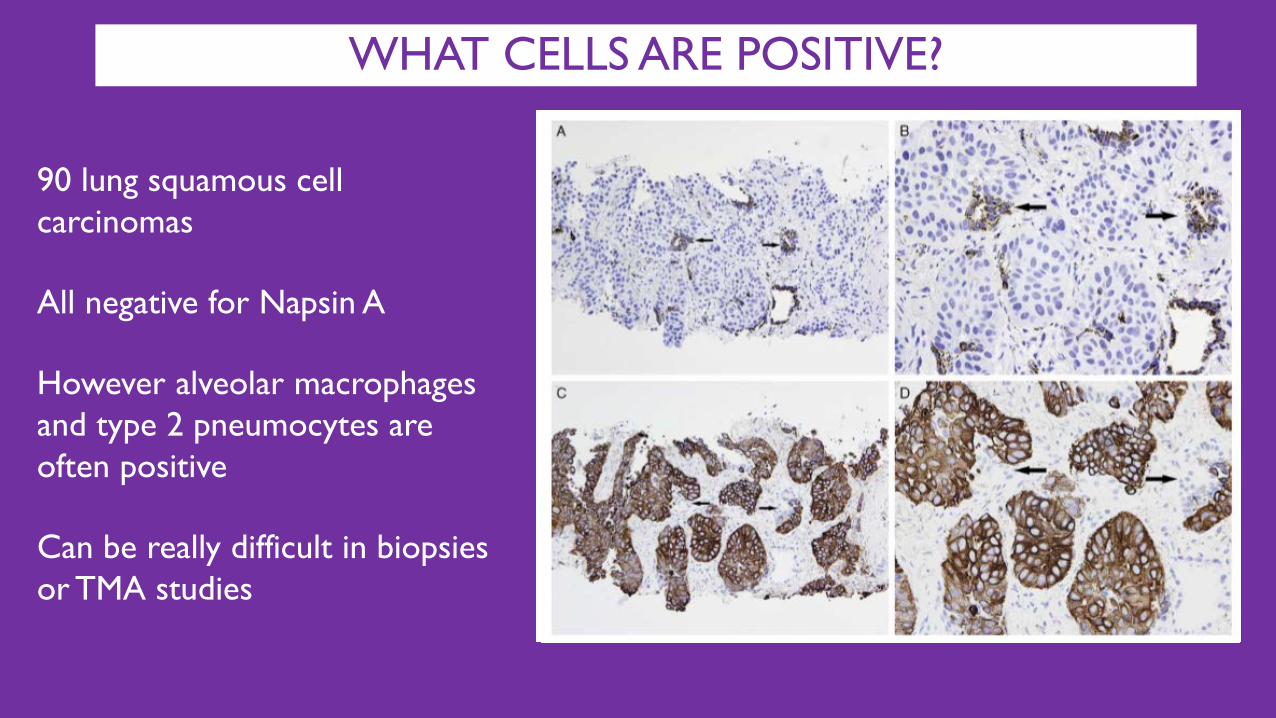
90 lung squamous cell carcinomas
All negative for Napsin A
However alveolar macrophages and type 2 pneumocytes are often positive
Can be really difficult in biopsies or TMA studies
WHAT CELLS ARE POSITIVE?

Ye JX et al. Histol Histopathol. 2015 May;30(5):581-8
Gastrointestinal spindle cell tumor
H&E CD117 DOG-1 Tryptase SMA
WHAT CELLS ARE POSITIVE?

Mast cells can be “passenger” cells in spindle cell lesions
Mast cells are positive for CD 117
Mast cells can be spindled or ovoid
DOG-1 stains ICC (Interstitial Cells of Cajal)
ICCs may be hyperplastic in leiomyomas
WHAT CELLS ARE POSITIVE?

LOCALIZATION OF IMMUNOSTAINS AT THE CELLULAR LEVEL
Membranous
Cell adhesion molecules: E-cadherin, CD56, Ber-EP4
Cell surface/transmembrane receptors/proteins: CD10, CEA, most leukocyte antigens (CD3 and CD20), EMA, CD117
Proteins linking surface molecules to cytoskeleton: β-catenin, dystrophin
Cytoplasmic –different patterns
Granular: chromogranin, HMB45
Fibrillary: intermediate filaments (desmin, cytokeratins, vimentin) may appear membranous, due to condensation beneath membrane
Diffuse: myoglobin, thyroglobulin

LOCALIZATION OF IMMUNOSTAINS AT THE CELLULAR LEVEL
Nuclear Cell-cycle associated proteins: ki67, p16
Transcription factors: TTF-1, CDX-2, myogenin, PAX-5
Tumor suppressor genes: p53, p63, WT-1, Rb
Nuclear enzymes and proteins: TdT, mismatch products
Steroid hormone receptors: ER/PR
Calcium-binding proteins: S-100, calretinin
also show cytoplasmic pattern
Some viral proteins: CMV, herpes
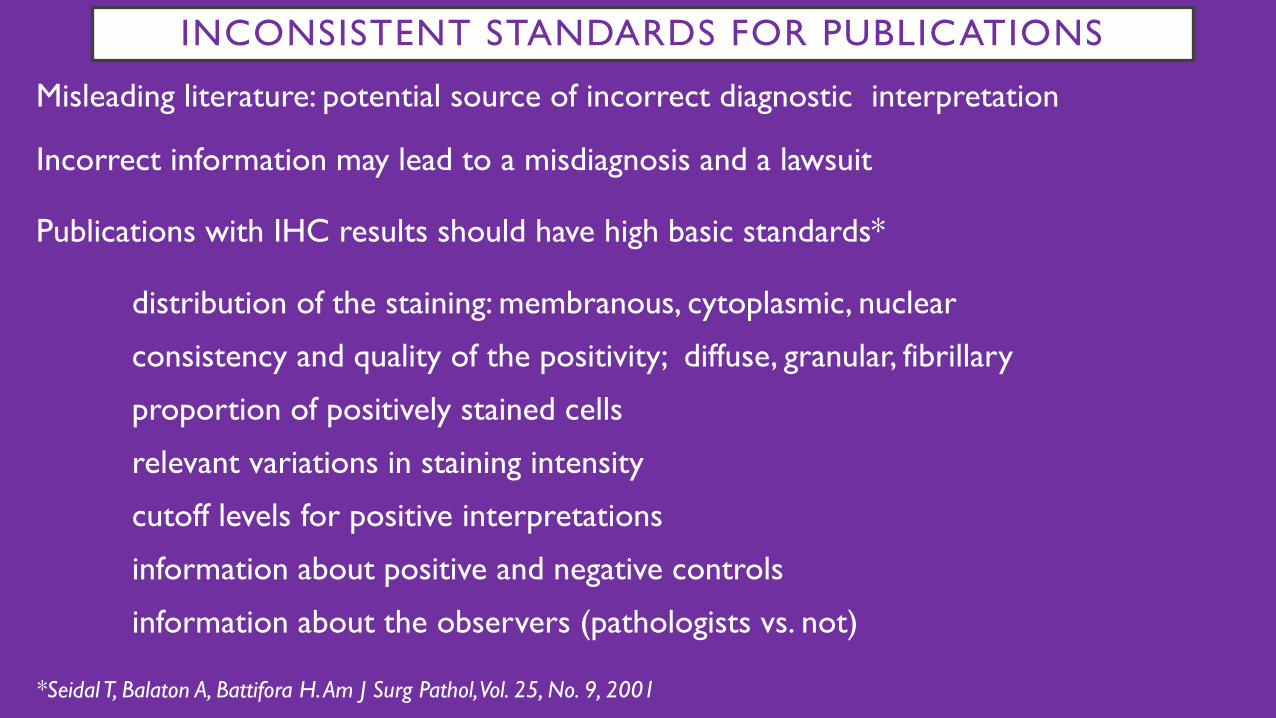
INCONSISTENT STANDARDS FOR PUBLICATIONS
Misleading literature: potential source of incorrect diagnostic interpretation
Incorrect information may lead to a misdiagnosis and a lawsuit
Publications with IHC results should have high basic standards*
distribution of the staining: membranous, cytoplasmic, nuclear
consistency and quality of the positivity; diffuse, granular, fibrillary
proportion of positively stained cells
relevant variations in staining intensity
cutoff levels for positive interpretations
information about positive and negative controls
information about the observers (pathologists vs. not)
*Seidal T, Balaton A, Battifora H. Am J Surg Pathol, Vol. 25, No. 9, 2001

THE CHANGING WORLD OF IHC MARKERS
New ICH stains are marketed to address a specific need
The final role of the markers in practice may be different
accumulation of evidence takes years (sensitivity and specificity)
some stains perform below expectations, others do well
some stains deliver unexpected advantages
Healthy scepticism helps when we read about a new marker
Continuous learning is essential

THE CHANGING WORLD OF IHC MARKERS
Journey of an antibody
Expanding or diminishing roles
New generation antibodies for organ specific scenarios
Advantages and pitfalls
Changing recommended panels for differential diagnostic scenarios

THE CHANGING ROLE OF IHC MARKERS
RCC – not really specific for renal cell carcinoma
TTF1 – less sensitive and specific for lung adenocarcinoma than we thought
CK5/6 – became very useful for intraductal proliferations in breast
CD117 – useful marker for seminoma, mast cells
CA19-9 – not specific for pancreatic origin
CA125 – not a specific Mullerian marker
CK19- not specific for pancreatobiliary origin

CA 125
NOT specific for Mullerian origin
Tumor site CA 125 positivity rate Ovary 98%
Endometrium 93% Pancreas 82%
Lung 66% Cervix 64% Thyroid 50% Breast 35%
Gremel G. et al, Histopathology; 64: 293-305

CK 19 NOT specific for pancreatic origin
Tumor site CK19 positivity rate Cholangiocarcinoma 100%
Pancreas 100% Gallbladder 100%
Lung 100% Colon 100% Ovary 100% Cervix 95%
Urothelial 95%

HMB-45 40-year-old woman with a rubbery uterine mass (4.5 cm)

HMB-45 Signed out as uterine angiomyolipoma Call from the clinician – Really? Are you sure? Can you send it out?

THE JOURNEY OF HMB 45
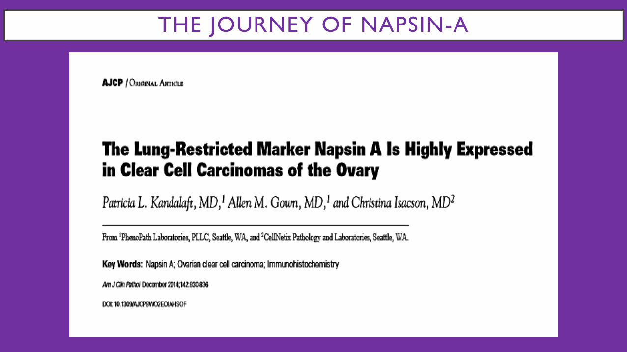
THE JOURNEY OF NAPSIN-A

SPECIFICITY– NAPSIN A

SPECIFICITY– NAPSIN A
Criteria for staining evaluation: Only coarse granular cytoplasmic staining was considered positive
Scoring: based on intensity and percentages of positive cells
All scores >2 were considered positive

SPECIFICITY– NAPSIN A
Tumor site Napsin-A positivity rate Lung adenocarcinoma 100%
Renal cell carcinoma, papillary 87%
Thyroid papillary carcinoma 48%
Renal cell carcinoma, clear 29%
Endometrioid adenocarcinoma 10%
Lung squamous cell carcinoma 0%
Breast ductal carcinoma 0%
Colon adenocarcinoma 0%

TTF-1 SPECIFICITY
62-year-old man, liver mass lesions, unknown primary

TTF-1 SPECIFICITY
Aberrant expression rate is clone dependent
8G7G3/1: around 2%, SPT24: 10% or higher (colon)

BIRTH OF AN ANTIBODY

AGONY OF AN ANTIBODY
CA 125 GCDFP- 15

DEATH OF AN ANTIBODY
NEURON SPECIFIC ENOLASE

BREAST MARKERS ER Mammoglobin GCDFP-15 50% of metastatic breast carcinomas are negative for all 3
GATA-3 positive in 96% of metastatic breast carcinomas
choriocarcinoma 100%
urothelial carcinoma 84 -100%
chromophobe RCC 51%
cholangiocarcinoma 9%
lung adenocarcinoma 9%

PITFALLS IN PROSTATE IHC INTERPRETATION
Scenarios related to certain IHC stains
Basal cell markers
Prostate specific markers

PITFALLS IN PROSTATE IHC INTERPRETATION
Racemase (AMACR): intense cytoplasmic granular positivity in carcinoma frequently overexpressed in HGPIN
certain carcinoma types may not overexpress AMACR atrophic, hormon treated, foamy gland
benign mimickers may overexpress it adenosis, atrophy, hyperplasia
never interpret AMACR in isolation
don`t use it as a prostate marker present in colon, lung, breast, kidney, ovary, bladder

PITFALLS IN PROSTATE IHC INTERPRETATION
HGPIN
Atrophy

PITFALLS IN PROSTATE IHC INTERPRETATION
Basal cell markers can be negative in benign mimickers adenosis
staining may be minimal, racemase may not help “atypical glandular proliferation, adenosis can not be excluded”
partial atrophy
go with morphology
staining can completely mimic carcinoma (-basal, +AMACR)
False positive staining of adenocarcinoma for basal cell markers
extremely rare, but can happen in Gleason 3 patterns
usually patchy, may be diffuse (for both 34BE12 and p63)

PITFALLS IN PROSTATE IHC INTERPRETATION
Gleason 3 with HMWCK positivity

PITFALLS IN PROSTATE IHC INTERPRETATION
High grade prostatic adenocarcinoma vs. invasive urothelial carcinoma Recommended panel: 2 prostatic and 2 urothelial markers
Prostate marker Features
PSA specific / cytoplasmic granular
PSAP specific / cytoplasmic granular
PSMA
sensitive, but stains 17% of urothelial carcinomas prostate: cytoplasmic + apical or membranous non-
prostate: cytoplasmic only Prostein (p501S) specific / perinuclear

PITFALLS IN PROSTATE IHC INTERPRETATION
High grade prostatic adenocarcinoma vs. invasive urothelial carcinoma Recommended panel: 2 prostatic and 2 urothelial markers
Urothelial marker Features
GATA-3 very specific, less sensitive
34BE12 sensitive, but may stain PD prostate carcinomas
p63 specific, less sensitive
Uroplakin, thrombomodulin specific / less sensitive

EFFECT OF THE LOCAL ENVIRONMENT – METASTATIC TUMORS
73-year-old woman with a 4 cm thyroid mass and compressive symptoms
Lesions in brain, lung, adrenal and lymph nodes
Thyroid FNA + immunostains: thyroglobulin (+), TTF-1 (+) and calcitonin (-)
Pap Thyr
TTF-1
Diagnosed as poorly differentiated (insular) thyroid carcinoma
Kanjanahattakij N et al. CytoJournal .2015;12:27

EFFECT OF THE LOCAL ENVIRONMENT – METASTATIC TUMORS
Thyr
Thyr
TTF-1
TTF-1
H&E
H&E
Total thyroidectomy + lymph node dissection to relieve symptoms + diagnosis Thyroid shows tumor foci with glandular and papillary features Thyroid and lymph nodes show different IHC patterns (thyroglobulin + vs -)
Thyroid
Lymph node

EFFECT OF THE LOCAL ENVIRONMENT – METASTATIC TUMORS
Diagnosis was changed to primary lung adenocarcinoma with widespread metastases (including thyroid)
Patient died 7 months later
Thyroglobulin was not expressed by the tumor cells
Nonspecific uptake and staining due to diffusion artefact from the surrounding follicles?
from the needle as it punctured through the follicles?

INTRA - AND INTEROBSERVER VARIABILITY
TMA (tissue microarray) selected from radical prostatectomies
All stained for PDX-1 (pancreatic duodenal homeobox-1)
Transcription factor overexpressed in prostatic adenocarcinoma
Expressed in several types of carcinomas (gastric, pancreatic, prostate)
Cytoplasmic stain

INTRA - AND INTEROBSERVER VARIABILITY
Stains evaluated by 4 independent observers 2 pathologists with interest in GU pathology 2 medical doctors with no formal training in pathology
Stains were scored twice by each participants 2-week interval between the reads
Intra – and interobserver reproducibility was recorded
Time spent with slides was also recorded

INTRA - AND INTEROBSERVER VARIABILITY
Scoring: Intensity: 0-3 (0: no staining, 3: most intense)
Extent: 1-3 (1: < 33%, 2: 34-66%, 3: >67%)
Two scores are multiplied (final score: 0-9)

INTRA - AND INTEROBSERVER VARIABILITY

INTRA - AND INTEROBSERVER VARIABILITY
Staining intensity – good results intraobserver: very high agreement interobserver: high (from substantial to very high)
Extent of staining – terrible numbers intraobserver: poor agreement interobserver: poor agreement
Non-pathologists spent more time on evaluation (2x) impoved in the second run

CROWDSOURCING IHC INTERPRETATION EXPERIMENT
Outsourcing of tasks typically performed by experts to a large crowd Kasparov vs. the World chess game (1999, internet)
Plurality vote decided World Team`s moves - 50,000 people from 75 countries
Kasparov won with whites
he had “never expended as much effort on any other game in his life”
Crowdsourcing experiments in medicine retinal fundus photography classification, malaria parasite quantification
IHC experiment from Italy*
13 breast images with MIB1 immunostain (positive-negative)
crowd: 28 respondents from 18 countries, non-pathologists
pathologists` count is gold standard
*Della Mea et al. Diagnostic Pathology 2014 9(Suppl 1):S6


CROWDSOURCING IHC INTERPRETATION EXPERIMENT
Crowdsourced median percentage is similar to gold standard
Counting is time-consuming by experts, may be difficult by software
Authors` conclusion: “method may be more aimed to research than routine”
When large number of images need ad hoc evaluation

THANK YOU!
QUESTIONS?