interphase. nucleus nucleoli chromatin. interphase,naberbiology.com/documents/ap lab 3...
TRANSCRIPT

I=igure 3.2: Whitefish Blastula
The whitefish blastula is often used for the study of cell division. As soon as the egg is fertilized it begins to divide, and nuclear division after nuclear division follows. You will be provided with slides of whitefish blastula which have been sectioned in various planes in relation to the mitotic spindle. You will be able to see side and polar views of the spindle apparatus.
Procedure
Examine prepared slides of either onion root tips or whitefish blastula. Locate the meristematic region of the onion, or locate the blastula, with the lOX objective and then use the 40X objective to study individual cells. For convenience in discussion, biologists have described certain stages, or phases, of the continuous mitotic cell cycle, as outlined on this page and the next. Identify one cell that clearly represents each phase. Sketch and label the cell in the boxes provided.
1. The nondividing cell is in a stage calied interphase. The nucleus may have one or more dark-stained nucleoli and is filled with a fine network of threads, the chromatin. During interphase, DNA replication occurs.
Interphase
4. At the beginning of anaphase, the cen tromere regions of each pair of chromatids separate and are moved by the spindle fibers toward opposite pl)les of the spindle,
. dragging the rest of the chromatid behind them. Once the two chromatids separate, cach is called a chromosome. These daughter chromosomes continue their poleward movement until they form two compact clumps. one at each spindle pole.
L
Anaphase
5. Telophase, the last stage of division, is marked by a pronounced condensation of the chromosomes, followed by the formation of a new nuclear cnvelope around each group of chromosomes. The chromosomes gradually uncoil to form the fine chromatin network seen in interphase. and the nucleoli and nuclear envelope reappear. Cytokinesis may occu r. This is the division of the cytoplasm into two cell s. In plants, a new cell wall is laid clown between the daughter cell s. In animal cells, the old cell will pinch off in the middle along a cleavage furrow to form two new daughter cells.
Telophase
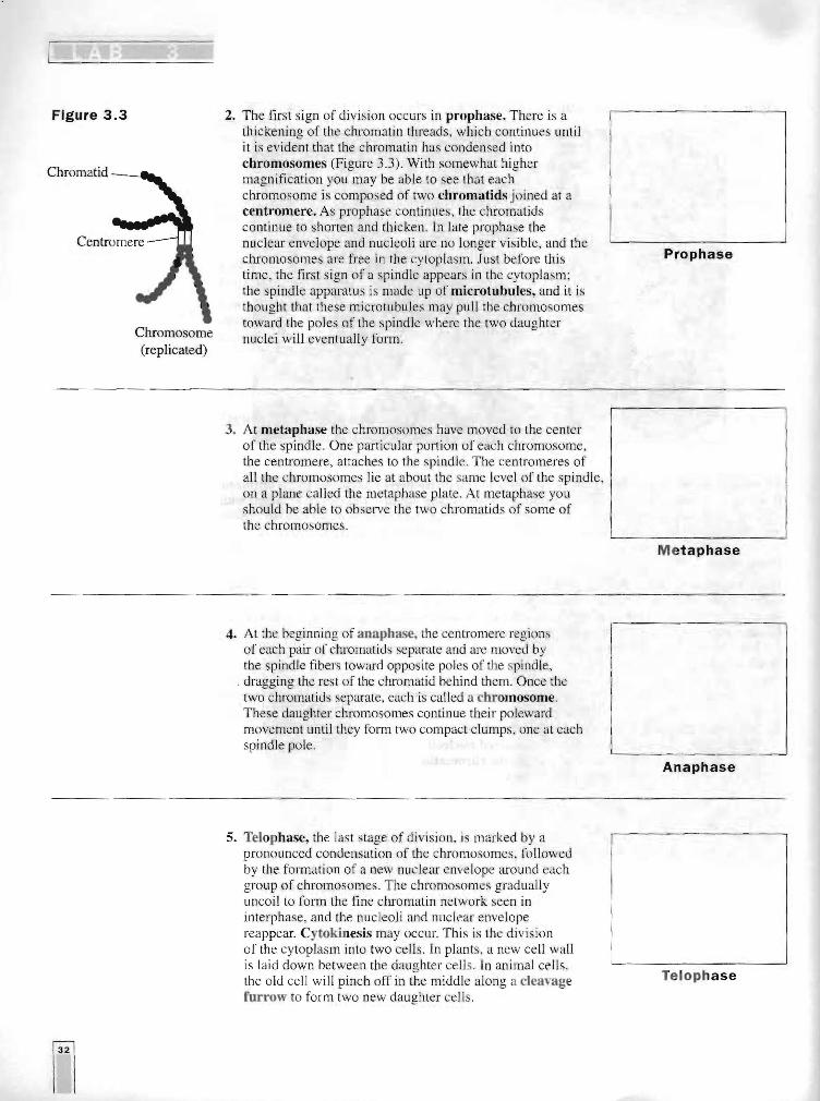
Figure 3.3
Chromatid _____
Centromere
Chromosome (replicated)
2. The first sign of division occurs in prophase. There is a thickening of the chromatin threads, which cOI<tinues until it is evident that the chromatin has condensed into chromosomes (Figure 3.3). With somewhat Iligher magnification you may be able to see Ihat each chromosome is composed of two chromatids joined at a centromere. As prophase continues, the chromatids continue to shorten and thicken. In late prophase the nuclear envelope and nucleoli are no longer visible, and the chromosomes are free in the cytoplasm. Just before this time, the first s ign of a spindle appears in the cytoplasm; the spindle apparatus is made up of microtubuies, and it is
. thought that these microtubules may pull the chromosomes toward the poles of the spindle where the two daughter nuclei will eventually form.
Prophase
3. At metaphase the chromosomes have moved to the center of the spindle. One particular portion of each chromosome, the centromere, attaches La the spindle. The centromeres of all the chromosomes lie at about the same level of the spindle. on a plane called the metaphase plate. At metaphase yo u should be able to observe the two chromatids of some of the chromosomes.
Metaphase

Analysis Questions
1. Explain how mitosis leads to two daughter cells , each of which is diploid and genetically identical to the original cell. What activities are going on in the cell during interphase?
2. How does mitosis differ in plant and animal ce lls? How does plant mitosis accommodate a rigid, inflexible cell wall?
3. What is the role of the centrosome (the area surrounding the cen trioles)? Is it necessary for mitosis? Defend your answer.
EXERCISE 3A.2: Time for Cell Replication
To estimate the relative length of time that a cell spends in the various stages of cell division, you will examine the meristematic region of a prepared slide of the onion root tip. The I nglh of the cell cycle is approximately 24 hours for cells in actively divid ing onion root tips.
Procedure
It is hard to imagine that you can estimate how much rime a cell spends in each phase of cell division from a slide of dead cells, yet this is precisely what you will do in this part of the lab. Since you are working with a prepared slide, you cannot get any information about how long it takes a cell to divide. What you can determine is how many cells re in each phase. From this, you can infer the percentage of time each cell spends in each phase.
1. Observe every cell in one high-power field of view and determine which phase of the cell cycle it i in. This is best done in pairs . The partner observing the slide calls out the phase of each cell while the other partner records. Then sw itch so th recorder becomes the observer and vice versa. Count at least two fu ll fields of view. If you have not counted at least 200 cells, then count a third field of view.
2. Record your data in Table 3.1.

c
3. Calculate the percentage of cells in each phase, and record in Table 3.1.
Consider that it takes, on average, 24 hours (or 1,440 minutes) for onion root tip cells to complete the cell cycle. You can calculate the amount of time spent in each phase of the cell cycle from the percentage of cells in that stage.
Percentage of cells in stage x 1,440 minutes = _ _ __ minutes of cell cycle spent in stage
Table 3.1
Field 1
Number of Cells
Field 2 Field 3 Total
Percent of Total
Cells Counted
Time In
Each Stage
Interphase
Prophase
Metaphase
Anaphase
I
I
I
I
I
I
Telophase
I
Total Cells Counted
Questions
1. If your observations had not been restricted to the area of the root tip that is actively dividing, how would your results have been different?
2. Based on the data in Table 3.1, what can you infer about the relative length of time an onion root tip cell spends in each stage of cell division?
3. Draw and label a pie chart of the onion root tip cell cycle using the data from Table 3.1.
Title: _______

Figure 3.12
Telophase U. Place the chromosomes at opposite sides of the dividing cell. At this time a nuclear envelope forms and, in our simulation, the cytoplasm divides .
..>Q~ 1 1 (al 1 1
(b)
Analysis and Investigation
1. List three major differences between the events of mitosis and meiosis.
2. Compare mitosis and meiosis with respect to each of the following in Table 3.2:
Table 3.2
Mitosis Meiosis-Chromosome Number of Parent Cells
Number of DNA Replications
Number of Divisions
Number of Daughter Cells Produced
Chromosome Number of Daughter Cells
Purpose/ Function

:
3. How are meiosis I and meiosis II different?
4. How do oogenesis and spermatogenesis differ?
5. Why is meiosis important for sexual reproduction?
EXERCISE 38. 2: Crossing Over during Meiosis in Sordaria
Sordaria ,jimicola is an ascomycete fungus that can be used to demonstrate the results of crossing over during meiosis. Son/aria is a haploid organism for most of its life cycle. It becomes diploid only when the fusion of the mycelia (filamentlike groups of cells) of two different strains results in the fusion of the two different types of haploid nuclei to form a diploid nucleus. The diploid nucleus must then undergo meiosis to resume its haploid state.
Meiosis, followed by one mitotic division, in Sordaria results in the formation of eight haploid ascospores contained within a sac called an ascus (plural. asd). Many asci are contained within a fruiting body called a perithecium (ascocarp). When ascospores are mature the ascus ruptures, releasing the ascospores. Each ascospore can develop into a new haploid fungus. The life cycle of Sordariafimicola is shown in Figure 3.13.
Procedure
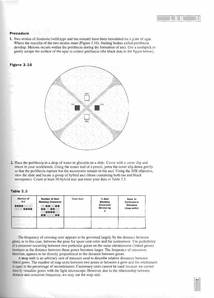
1. Two strains of Sordaria (wild type and tan mutant) have been inoculated on a plate of agar. Where the mycelia of the two strains meet (Figure 3.16), fruiting bodies called perithecia develop. Meiosis occurs within the perithecia during the formation of asci. Use a toothpick to gently scrape the surface of the agar to collect perithecia (the black dots in the figure below).
Figure 3.16
2. Place the perithecia in a drop of water or glycerin on a slide. Cover with a cover slip and return to your vmrkbench. Using the eraser end of a pencil, press the cover slip down gently so that the perithecia rupture but the ascospores rema,in in the asci. Using the lOX objective, view the slide and locate a group of hybrid asci (those containing both tan and black ascospores). Count at least SO hybrid asci and enter your data in Table 3.3.
Table 3.3
Number of Number of Asci Total Asci % Asci Gene to 4:4 Showing Crossover Showing Centromere
••••0000 0000••••
00••00•• ..00••00 00••••00
Crossover Divided by
2
Distance (map units)
• • 0000••
The frequency of crossing over appears to be governed largely by the distance between genes, or in this case, between the gene for spore coat color and the centromere. The probability of a crossover occurring between two particular genes on the same chromosome (linked genes) increases as the distance between those genes becomes larger. The frequency of crossover, therefore, appears to be directly proportional to the distance between genes.
A map unit is an arbitrary unit of measure used to describe relative distances between linked genes. The number of map units between two genes or between a gene and the centromere is equal to thc percentage of recombinants. Customary units cannot be used because we cannot directly visualize genes with the light microscope. However, due to the relationship between distance and crossover frequency, we may use the map unit.

Analysis of Results 1. Using your data in Table 3.3, determine the djstance between the gene for spore color and
the centromere. Calculate the percentage of crossovers by dividing the number of crossover asci (2:2:2:2 or 2:4:2) by the total number of asci X 100. To calculate the map distance, divide the percentage of crossover asci by 2. The percentage of crossover asci is divided by 2 because only half of the spores in each ascus are the result of a crossover event (Figure 3.15). Record your results in Table 3.3.
2. Draw a pair of chromosomes in MI and MIl and show how you would get a 2:4:2 arrangement of ascospores by crossing over. (Hint: refer to Figure 3.15).