intermittent hypoxia-induced cardiovascular remodeling is ... · with increased cardiovascular...
TRANSCRIPT

Intermittent Hypoxia-InducedCardiovascular Remodeling Is Reversed byNormoxia in a Mouse Model of Sleep ApneaAnabel L. Castro-Grattoni, MS; Roger Alvarez-Buvé, MS; Marta Torres, PhD; Ramon Farré, PhD;Josep M. Montserrat, MD, PhD; Mireia Dalmases, MD; Isaac Almendros, PhD; Ferran Barbé, MD, PhD;and Manuel Sánchez-de-la-Torre, PhD
BACKGROUND: Intermittent hypoxia (IH) is the principal injurious factor involved in thecardiovascular morbidity and mortality associated with OSA. The gold standard for treat-ment is CPAP, which eliminates IH and appears to reduce cardiovascular risk. There is noexperimental evidence on the reversibility of cardiovascular remodeling after IH withdrawal.The objective of the present study is to assess the reversibility of early cardiovascularstructural remodeling induced by IH after resumption of normoxic breathing in a novelrecovery animal model mimicking OSA treatment.
METHODS: Weinvestigated cardiovascular remodeling inC57BL/6mice exposed to IH for 6weeksvs the normoxia group and its spontaneous recovery after 6 subsequent weeks under normoxia.
RESULTS: Aortic expansive remodeling was induced by IH, with intima-media thickening andwithout lumen perimeter changes. Elastic fiber network disorganization, fragmentation, andestrangement between the end points of disrupted fibers were increased by IH. Extracellularmatrix turnover was altered, as visualized by collagen and mucoid interlaminar accumula-tion. Furthermore, left ventricular perivascular fibrosis was increased by IH, whereas car-diomyocytes size was unaffected. These cardiovascular remodeling events induced by IH werenormalized after recovery in normoxia, mimicking CPAP treatment.
CONCLUSIONS: The early structural cardiovascular remodeling induced by IH was normalizedafter IH removal, revealing a novel recovery model for studying the effects of OSA treatment.Our findings suggest the clinical relevance of early detection and effective treatment of OSAin patients to prevent the natural course of cardiovascular diseases. CHEST 2016; -(-):---
KEY WORDS: atherosclerosis recovery; cardiovascular disease; continuous positive airwaypressure; intermittent hypoxia; obstructive sleep apnea
ABBREVIATIONS: ECM = extracellular matrix; H&E = hematoxylinand eosin; IH = intermittent hypoxia; IMT = intima-media thicknessAFFILIATIONS: From the Respiratory Department (Drs Dalmases,Barbé, and Sánchez-de-la-Torre; Ms Castro-Grattoni; and Mr Alvarez-Buvé), Hospital Universiti Arnau de Vilanova and Santa Maria, IRBLleida, University of Lleida, Lleida, Catalonia, Spain; the Sleep Labo-ratory (Drs Torres and Montserrat), Pneumology Department,Hospital Clinic of Barcelona, Barcelona, Spain; the Unitat de Biofísicai Bioenginyeria (Drs Farré, Montserrat, and Almendros), Facultat deMedicina, Universitat de Barcelona, and the Institut InvestigacionsBiomèdiques August Pi Sunyer (IDIBAPS), Barcelona, Spain; andthe Centro de Investigación Biomédica en Red de EnfermedadesRespiratorias (CIBERES) (Drs Torres, Farré, Montserrat, Dalmases,Almendros, Barbé, and Sánchez-de-la-Torre), Madrid, Spain.
FUNDING/SUPPORT: This work was supported by the Spanish Respi-ratory Society (SEPAR), the Associació Lleidatana de Respiratori(ALLER), and the Spanish Fondo de Investigaciones Sanitarias PI14/00004, Instituto de Salud Carlos III (ISCIII), European RegionalDevelopment Fund (ERDF) “Una manera de hacer Europa”.CORRESPONDENCE TO: Manuel Sánchez-de-la-Torre, PhD, HospitalArnau de Vilanova-Santa María, IRB Lleida, CIBERES, Avda RoviraRoure 80, 25198, Lleida, Spain; e-mail: [email protected] ! 2016 American College of Chest Physicians. Published byElsevier Inc. All rights reserved.DOI: http://dx.doi.org/10.1016/j.chest.2015.11.010
[ Original Research ]
journal.publications.chestnet.org 1
FLA 5.4.0 DTD ! CHEST172_proof ! 12 March 2016 ! 5:26 pm ! EO: CHEST-15-2057

OSA is a highly prevalent disorder that affects 6% to15% of the general population and is caused by repetitiveupper airway occlusion during sleep.1,2 OSA is animportant public health problem because of its associationwith increased cardiovascular morbidity and mortality,including hypertension, coronary artery disease,congestive heart failure, heart attack, and stroke.3,4 Themajor OSA components associated with cardiovascularconsequences are large swings in intrathoracic pressure,postapneic arousals, and intermittent hypoxia (IH). IH isthe main detrimental event leading to cardiovascularmorbidity and mortality.5,6
Sympathetic overactivation, oxidative stress, andsystemic inflammation are the main intermediarymechanisms associated with IH.4,7 These abnormalitiesall contribute to the development of early and latecardiovascular remodeling, including increased bloodpressure, endothelial dysfunction, carotid intima-mediathickness (IMT), arterial stiffness, and acceleratedprogression of atherosclerosis, and induce cardiacrhythm and structural disturbances.4,8
Murine models have been used to study the adaptive anddegenerative hemodynamic and structural alterations ofthe cardiovascular system induced by IH.9 IH inducesblood pressure elevation, endothelial dysfunction,enlargement of aortic IMT, cardiac hypertrophy, and
extracellular matrix (ECM) alterations; increased systemicinflammation and activation of proinflammatorypathways in cardiovascular tissue; and increased riskof developing atherosclerotic plaques.10
CPAP, the gold standard therapy for patients withOSA, effectively improves daytime symptoms andquality of life, and might be an effective treatment forcardiovascular risk reduction.11,12 Randomized controlledtrials have demonstrated that CPAP therapy reducesblood pressure, sympathetic overactivity, and coagulationabnormalities and improves left ventricular ejectionfraction.13-16 CPAP has also been shown to improveendothelial function, IMT, and arterial stiffness in smallstudies.5,17 However, there is no experimental evidencethat elimination of IH reverses the cardiovascularremodeling induced by injurious hypoxic challenge.
To address this important issue, we established a murinemodel of recovery in which normal room air breathingis resumed after chronic IH challenge. We hypothesizedthat the resumption of normoxic conditions, whichmimics CPAP treatment, could reverse the earlycardiovascular morphological remodeling induced byIH. This recovery model will enable the study of themechanisms involved in the therapeutic effects of OSAtreatments in reversing injuries induced by IH indifferent organs.
Materials and MethodsStudy DesignThe study was approved by the Ethical Committee for AnimalResearch of the University of Barcelona and was performed on6-week-old pathogen-free C57BL/6 male mice (Charles RiverLaboratories). The animals were housed in standard cages in atemperature- and light-controlled room (22!C-24!C; 14 hours oflight, 10 hours of dark). A total of 40 mice were randomly assignedto IH exposure (n ¼ 20 mice) or normoxia (n ¼ 20 mice) for6 weeks. After this IH phase, 10 mice from each group wereanesthetized (urethane 20%, 1 g/kg) and euthanized byexsanguination, and aortas and hearts were excised. The remainingIH mice were subsequently subjected to a 6-week normoxic recoveryphase to mimic CPAP treatment of patients with OSA andsacrificed, and tissue samples were excised as described below. Theexperimental design of the protocol is shown in Figure 1A. Thegroups were labeled N, normoxia; IH, intermittent hypoxia; NþR,normoxia with recovery phase; and IHþR, intermittent hypoxia withrecovery phase.
Intermittent HypoxiaChronic IH was applied as previously described.18 For 6 weeks, mice inthe IH group received 60 hypoxic events/h (20 s at 5% O2 per min),during 6 h/d, corresponding to severe OSA. Control mice withnormoxic breathing were placed in an identical system, but the hypoxicgas from the reservoir was replaced by room air. In the normoxicrecovery phase, all mice were subjected to identical normoxic conditions.
Histomorphological AnalysesThe mid thoracic aorta and left ventricle of the heart samples wereperfused with phosphate-buffered saline, fixed with4% paraformaldehyde, and embedded in paraffin for furtherhistological analysis by an investigator blinded to the experimentalgroup. The samples were stained with hematoxylin and eosin (H&E,Master Diagnostica), Gomori trichrome stain (Artisan Link SpecialStaining System; DAKO), or Alcian blue (Alcian blue 2.5; Bio-Optica).For measurements, images from four consecutive sections wereprocessed using Image J (National Institutes of Health) and AdobePhotoshop CS6 (Adobe Systems Inc) software. All stained sectionswere captured with a digital microimaging network instrument (Leica-DMD-108; Leica Microsystems), and aortic autofluorescence wasvisualized using a fluorescence microscope (Olympus-BX51; Olympus).
Intima-media thickness: The cross-sectional IMT was quantified bymorphometric analysis of the H&E stained sections (300measurements for each animal).
Alcian blue staining: The integrated density of the blue staining wasquantified and adjusted to the corresponding aortic wall area to detectmucoid deposition.
Cardiac hypertrophy: The cross-sectional area of the cardiacmyofibers with a circular running pattern was analyzed quantitativelyusing H&E stained sections (300 cardiomyocytes for each animal).
Cardiovascular fibrosis: Gomori trichrome stain was used to detectfibrosis in aortic and cardiac tissue. The fibrotic tissue was determinedby measuring the positive collagen area adjusted to the total tissue area.
2 Original Research [ -#- CHE ST - 2 0 1 6 ]
FLA 5.4.0 DTD ! CHEST172_proof ! 12 March 2016 ! 5:26 pm ! EO: CHEST-15-2057

Elastic-network analysis: The aortic autofluorescence was used toperform elastic fiber analysis. The elastin disruption (ie, the completefragmentation of one elastic fiber) and the distance between bothends of a fragmented fiber were quantified (adjusted by total aorticarea and shown as percent space without fiber). In addition, wequantified the area with elastic fiber disorganization based on theinability to count the amount of organized elastic fiber.19
Data AnalysisResults were expressed as the mean $ SEM. Depending onnormality and variance homogeneity, analysis of variance andStudent t test or Mann-Whitney U test were performed. Statisticalsignificance was set at a probability value of less than .05.Structural parameters were adjusted for body weight using a linearregression model.
Results
Body Weight
The body weight at baseline was similar in both groups.However, 6 weeks of IH decreased animal body weight(P ¼ .005). After the normoxic recovery phase, thebody weights of mice in the IHþR group were similarto those in the NþR group, suggesting a normalizationof body weight after IH withdrawal (Fig 1B).
Morphological Vascular Remodeling
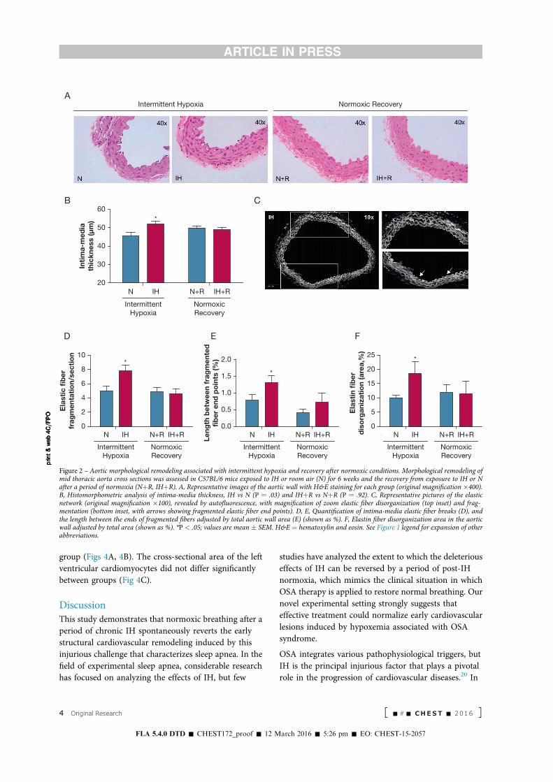
Intima-media thickness: The aortic IMT was increasedby IH exposure vs that of the N group (P ¼ .03). Afternormoxia, the IMT of mice in the IHþR group wasnormalized compared with its control, suggesting arecovery of aortic remodeling (Figs 2A, 2B). The aorticlumen perimeter did not exhibit significant changes,indicating expansive remodeling of the aortic wallinduced by IH. Moreover, mice in the NþR and IHþRgroups did not show statistically significant differencesin lumen perimeter.
Elastin fiber disorganization and disruption: Sixweeks of IH exposure induced elastin fiber disruption andincreased the distance between both ends of thefragmented fibers (Figs 2C-E). These alterations werereduced compared with those of the NþR group,suggesting that the aortas of the IHþR group were
subjected to a recovery remodeling process (Figs 2D, 2E).Furthermore, mice exposed to IH displayed an increase inzones of elastin fiber disorganization in the aortic wall,which was not observed in mice in the IHþR groupcompared with those in the NþR group (Fig 2F).
Aortic Mucoid deposition: Alcian blue stainingrevealed greater mucoid deposition in the vascularwall of the IH group between subintimal elastic fibers,specifically in regions neighboring the aortic lumen.Mucoid deposition in the aortic wall in the NþR andIHþR groups was similar to that in the normoxia group,suggesting normalization after normoxic recovery(Figs 3A, 3B).
Aortic fibrosis: The collagen fiber content in the aorticwall was higher in mice exposed to IH for 6 weeks,suggesting the induction of collagen synthesis during IHexposure. Recovery under normoxic conditions of theIHþR group resulted in a decrease in aortic fibrosis,similar to the NþR group (Figs 3A, 3C).
Morphological Cardiac Remodeling
Mice exposed to IH for 6 weeks exhibited increasedcardiac perivascular fibrosis compared with thenormoxia group (Figs 4A, 4B). After the normoxicrecovery phase, the extracellular collagen content of theIHþR group was no different from that of the NþR
NormoxiaA B
N
IH
Normoxia
Normoxia
Normoxia
Intermittent hypoxia
Intermittent hypoxia
Intermittent HypoxiaPhase (6-wk)
Normoxic RecoveryPhase (6-wk)
N+R
IH+R 20N
Intermittent Hypoxia Normoxic Recovery
IH N+R IH+R
25
Bod
y w
eigh
t (g
)
30
35
**
print&
web4C=F
PO
print&
web4C=F
PO
print&
web4C=F
PO
print&
web4C=F
PO
Figure 1 – Mouse growth is altered by intermittent hypoxia and normalized after normoxic recovery. A, Experimental design of the study (n ¼ 10, pergroup): male C57BL/6 mice exposed to room air (N) or to IH for 6 weeks, and mice exposed to N or IH and subsequently subjected to a period ofnormoxia (6 more weeks; NþR and IHþR). B, Box-plot representation of body weight in N and IH groups at 6 weeks (P ¼ .005) and in NþR andIHþR groups at 12 weeks (P ¼ .136). **P < .01 for intergroup comparisons. IH ¼ intermittent hypoxia; IHþR ¼ intermittent hypoxia plus normoxicrecovery; N ¼ normoxia; NþR ¼ normoxia plus normoxic recovery.
journal.publications.chestnet.org 3
FLA 5.4.0 DTD ! CHEST172_proof ! 12 March 2016 ! 5:26 pm ! EO: CHEST-15-2057
group (Figs 4A, 4B). The cross-sectional area of the leftventricular cardiomyocytes did not differ significantlybetween groups (Fig 4C).
DiscussionThis study demonstrates that normoxic breathing after aperiod of chronic IH spontaneously reverts the earlystructural cardiovascular remodeling induced by thisinjurious challenge that characterizes sleep apnea. In thefield of experimental sleep apnea, considerable researchhas focused on analyzing the effects of IH, but few
studies have analyzed the extent to which the deleteriouseffects of IH can be reversed by a period of post-IHnormoxia, which mimics the clinical situation in whichOSA therapy is applied to restore normal breathing. Ournovel experimental setting strongly suggests thateffective treatment could normalize early cardiovascularlesions induced by hypoxemia associated with OSAsyndrome.
OSA integrates various pathophysiological triggers, butIH is the principal injurious factor that plays a pivotalrole in the progression of cardiovascular diseases.20 In
0N IH
IntermittentHypoxia
NormoxicRecovery
N+R IH+R
2
4
6
8
Ela
stic
fibe
rfr
agm
enta
tion/
sect
ion 10
*
D
20N IH
IntermittentHypoxia
NormoxicRecovery
N+R IH+R
30
40
50
Intim
a-m
edia
thic
knes
s (μ
m)
60
Intermittent Hypoxia Normoxic Recovery
*
0.0N IH
IntermittentHypoxia
NormoxicRecovery
N+R IH+R
0.5
1.0
1.5
Leng
th b
etw
een
frag
men
ted
fiber
end
poi
nts
(%) 2.0
*
E
0N IH
IntermittentHypoxia
NormoxicRecovery
N+R IH+R
5
10
15
20
Ela
stin
fibe
rdi
sorg
aniz
atio
n (a
rea,
%) 25 *
F
A
B C
print&web4C=F
PO
print&web4C=F
PO
print&web4C=F
PO
print&web4C=F
PO
Figure 2 – Aortic morphological remodeling associated with intermittent hypoxia and recovery after normoxic conditions. Morphological remodeling ofmid thoracic aorta cross sections was assessed in C57BL/6 mice exposed to IH or room air (N) for 6 weeks and the recovery from exposure to IH or Nafter a period of normoxia (NþR, IHþR). A, Representative images of the aortic wall with H&E staining for each group (original magnification %400).B, Histomorphometric analysis of intima-media thickness, IH vs N (P ¼ .03) and IHþR vs NþR (P ¼ .92). C, Representative pictures of the elasticnetwork (original magnification %100), revealed by autofluorescence, with magnification of zoom elastic fiber disorganization (top inset) and frag-mentation (bottom inset, with arrows showing fragmented elastic fiber end points). D, E, Quantification of intima-media elastic fiber breaks (D), andthe length between the ends of fragmented fibers adjusted by total aortic wall area (E) (shown as %). F, Elastin fiber disorganization area in the aorticwall adjusted by total area (shown as %). *P < .05; values are mean $ SEM. H&E ¼ hematoxylin and eosin. See Figure 1 legend for expansion of otherabbreviations.
4 Original Research [ -#- CHE ST - 2 0 1 6 ]
FLA 5.4.0 DTD ! CHEST172_proof ! 12 March 2016 ! 5:26 pm ! EO: CHEST-15-2057

addition to the evidence of adverse events caused byIH,9 other studies have demonstrated beneficial effectsof IH in both animal models and patients with OSA.21
The opposing effects induced by IH depend mainlyon the experimental time; long-term exposure(4-8 weeks) is required to cause detrimental effects.22
In the present study, we assessed several morphologicalcardiovascular changes resulting from the direct effectof IH for 6 weeks, a common experimental paradigmto mimic severe OSA in patients. Our results confirmedthe hypothesis that restoring normoxia by removingIH stress facilitates homeostatic cardiovascularrestoration.
Vascular remodeling is dependent on dynamicinteractions between local growth factors, vasoactivesubstances, and hemodynamic stimuli and is a responseto long-standing changes in hemodynamic conditions.23
IH24 and sleep fragmentation19 are independent factorsthat promote vascular remodeling in the aorta. IMTremodeling is an early predisposing event inatherosclerosis and plaque formation and is associatedwith increased cardiovascular risk.25 Patients with OSAexhibit increased IMT in association with inflammatorymarkers and nocturnal oxygen desaturation.26 Ourfindings confirm previous observations of expansiveaortic remodeling with increased IMT without vascular
0N
IntermittentHypoxia
NormoxicRecovery
IH N+R IH+R
5
10
15
20
Blu
e st
ain/
µm
2
N IH N+R IH+R
Intermittent Hypoxia Normoxic Recovery
B
A
C
*
0N
IntermittentHypoxia
NormoxicRecovery
IH N+R IH+R
10
20
Aor
tic c
olla
gen
posi
tive
area
(%)
30
*
print&
web4C=F
PO
print&
web4C=F
PO
print&
web4C=F
PO
print&
web4C=F
PO
Figure 3 – Aorta extracellular matrix remodeling induced by intermittent hypoxia and progression after recovery in normoxic conditions. Remodelingof extracellular components of the aortic wall was assessed in C57BL/6 mice exposed to IH or room air (N) at 6 weeks and in mice exposed to N or IHand subsequently subjected to normoxia (NþR, IHþR). A, top, Representative images of Alcian blue staining from the mid thoracic aorta to detectaortic wall mucoid deposition (original magnification %200; mucins in blue); A, bottom, collagen-positive area of the intima-media (%). B, Repre-sentative images of Gomori trichrome stain to measure aortic wall fibrosis (original magnification %200; collagen in green). C, Intima-media mucoiddeposition shown as the ratio of the total blue density to the total aortic wall area. (%).*P < .05; values are mean $ SEM. See Figure 1 legend forexpansion of abbreviations.
journal.publications.chestnet.org 5
FLA 5.4.0 DTD ! CHEST172_proof ! 12 March 2016 ! 5:26 pm ! EO: CHEST-15-2057

dilatation as a result of IH exposure in mice.27
Importantly, our novel experimental data on IMTnormalization after normoxic recovery are in agreementwith clinical data on patients with OSA who weretreated with CPAP.17
We also observed that IH increased elastic fiberdisorganization and disruption. The increase in theestrangement of the two end points of the disruptedlamina, reported in the present study, suggests a highertensile stress in the aortic wall exposed to IH, leading toa stronger fiber break. Perturbations in the continuity ofthe elastic lamina have been implicated in early phasesof atherosclerosis28 and in vascular remodeling inducedby sleep fragmentation.19 Changes in elastin structureand distribution have been reported in a rat model ofIH, but quantitative morphometric analysis was notperformed.29 However, we have quantitatively assessedelastic fiber organization and fragmentation of the aorticwall. Strikingly, our results demonstrate that thenormoxic recovery in mice that had been previouslyexposed to IH enabled a normalization of the vascularelastic fiber network alterations.
Changes in the ECM have been implicated in thepathogenesis of atherosclerosis and play an important
role in intercellular networking. These changes can leadto a fibroproliferative response, promoting lipid bindingto the vascular wall and inducing foam cell formation.30
We observed abnormal ECM turnover in the aortic wallin mice exposed to IH, which suggests that IH promotescollagen and mucopolysaccharide (proteoglycans andglycosaminoglycans) synthesis and deposition ininterlaminar spaces. Importantly, we observed that thisECM remodeling could be normalized after a recoveryperiod in normoxic conditions, which indicates thepossible activation of inhibitory and degradationpathways of collagen and mucopolysaccharide synthesis.
The ECM response to IH stress also includesmorphological myocardial remodeling. We observedthat IH induced perivascular fibrosis in the left ventricle,whereas interstitial fibrosis was not increased, inagreement with previous studies.31 Perivascular fibrosisis substantially associated with the impairment ofcoronary blood flow and is involved in the progressionof heart failure.32 Because of significant independentassociations between OSA and heart failure, manystudies have evaluated CPAP as a treatment for patientswith OSA who have heart failure.33,34 In the presentstudy, we observed a normalization of coronary
0.0N
IntermittentHypoxia
NormoxicRecovery
IH
IHN IH+RN+R
N+R IH+R
0.1
0.2
0.3
0.4
Per
ivas
cula
r co
llage
npo
sitiv
e ar
ea (%
)
0.5
Intermittent Hypoxia Normoxic Recovery
B
A
*
300N
IntermittentHypoxia
NormoxicRecovery
IH N+R IH+R
400
500
600
700
Car
diom
yocy
tes
area
(µm
2 )
800C
print&web4C=F
PO
print&web4C=F
PO
print&web4C=F
PO
print&web4C=F
PO
Figure 4 – Cardiac morphological remodeling associated with intermittent hypoxia and the effect of recovery in normoxic conditions. Morphologicalremodeling of cardiac tissue was assessed in mice exposed to IH or room air (N) at 6 weeks and in mice exposed to IH or N that were subsequentlysubjected to normoxia (NþR, IHþR). A, Representative images of the left ventricle with Gomori trichrome stain to detect perivascular fibrosis (originalmagnification %200; collagen in green). B, Analysis of perivascular fibrosis measured as collagen-positive area (%). C, Histomorphometric analysis ofthe left ventricular cardiomyocyte area of fibers with a circular pattern did not reveal statistically significant differences. *P < .05; values are mean $SEM. See Figure 1 legend for expansion of abbreviations.
6 Original Research [ -#- CHE ST - 2 0 1 6 ]
FLA 5.4.0 DTD ! CHEST172_proof ! 12 March 2016 ! 5:26 pm ! EO: CHEST-15-2057

perivascular fibrosis after recovery under normoxicconditions. Normoxia restoration was sufficient toreduce perivascular fibrosis, most likely because of thereduction of the fibroinflammatory response andoxidative stress production in myocardial tissue. Thisfinding has clinical relevance and suggests that patientswith OSA who have heart disease would benefit fromeffective breathing normalization, most likely because ofthe resulting improved coronary blood flow.
Cardiac remodeling includes hypertrophy that canexist in a state of compensation or progress to adecompensated state with time. We did not observeleft ventricular hypertrophy, consistent with previousstudies.35 However, other studies have observedcardiac hypertrophy induced by IH.31,36 The largedisparity in results for left ventricular hypertrophymay reflect differences in species or strain or even theside of the heart,22 which could explain our negativeresult for left ventricular hypertrophy.
Aortic wall and left ventricular remodeling inducedby IH is the result of multiple interactions betweenintermediary mechanisms, including oxidative stress,systemic and tissue inflammation, metabolicderegulation, endothelial dysfunction, sympatheticoveractivation, and blood pressure overload.24,37 Ourstudy did not focus on assessing changes in bloodpressure; however, two similar studies found thatC57BL/6 mice exhibit increments in blood pressure after14 and 90 days of IH exposure.22,38 Arterial blood pressureincreases (10 to 20 mm Hg) in rodent models of IH arecomparable with those of other experimental animalmodels of hypertension.39 Thus, in mice that are exposedto IH, increases in blood pressure may induce functional,mechanical, and structural changes in the aortic wall inresponse to hemodynamic and biomechanical stress.Moreover, IMT, elastin fiber disruption, and interlaminarcollagen accumulation induce arterial stiffness,40 therebycontributing to systemic vascular resistance and arterialblood pressure elevation.
Reversibility of structural cardiovascular damagehas been demonstrated in several animal models ofhypertension through spontaneous reversion orthrough the use of several forms of antihypertensivetreatment.41-47 Celiprolol reduced cardiovascularalterations induced by hypoxic stress in mice exposedto IH.48 The reversal of structural changes induced byelevated blood pressure suggests that several of ourresults could be explained by a reduction in bloodpressure after the recovery phase in normoxic conditions.
The current study has several limitations. Recurrentapnea in patients results in IH, hypercapnia, sleeparousal, sleep fragmentation, and changes inintrathoracic pressure that may contribute tocardiovascular remodeling. However, our study focusedexclusively on IH stress, which is a limitation becausethe mice model of hypoxemia associated with sleepapnea does not represent the totality of the complexdisorder. However, IH is the most importantpathophysiological component of sleep apnea thatunderlies cardiovascular complications, which was theprincipal outcome of our study. The most commonindex of cardiac hypertrophy is the measure of heart orventricular weights related to body weight. We did notassess this parameter, but relating heart to body weightis not valid when the investigated groups do not exhibitsimilar body growth patterns, as we observed in thisstudy.49 The main strength of this work is that the useof a conventional mouse strain allowed us to assess thecardiovascular impact induced by IH per se and thesubsequent recovery process under normoxicconditions, avoiding other confounding factors.
ConclusionsThe current study demonstrates that IH inducespreatherosclerotic remodeling characterized by IMT,elastin disruption and disorganization, accumulationof collagen fibers, and mucoid elements on the aorticwall. We also observed initial myocardial remodelinginduced by IH exposure, specifically perivascularfibrosis. These cardiovascular remodeling events arevirtually reversed when the IH stress was removed andmice were returned to normoxic conditions, mimickingthe effective treatment of the hypoxic component ofOSA. The clinical relevance of our findings suggests thatearly detection of patients with OSA and the subsequenttherapeutic intervention to normalize breathing mayalter the natural course of cardiovascular diseases thatare promoted by cyclic hypoxia and reoxygenation.Furthermore, we propose for the first time a murinemodel of IH followed by normoxia to study the potentialbenefits of IH resolution with CPAP treatment inpatients with OSA, including restoring normal structureand function of the different organs challenged bythis sleep breathing disorder. This recovery model maybe a useful tool for future studies aimed at identifyingpossible cellular and molecular mechanisms andsignaling pathways involved in the homeostatic andadaptive response to IH. Additionally, this model maybe used in future studies to assess OSA treatments.
journal.publications.chestnet.org 7
FLA 5.4.0 DTD ! CHEST172_proof ! 12 March 2016 ! 5:26 pm ! EO: CHEST-15-2057
AcknowledgmentsAuthor contributions: M. S. contributed tostudy concept and design, data acquisition,data analysis and interpretation, drafting ofthe manuscript, and critical revision of themanuscript for important intellectual contentand approved the final version. He is theguarantor of the paper. A. L. C. contributedto data acquisition, data analysis andinterpretation, drafting of the manuscript,and critical revision of the manuscript forimportant intellectual content and approvedthe final version. R. F. and F. B. contributedto study concept and design, data acquisition,data analysis and interpretation, drafting ofthe manuscript, and critical revision of themanuscript for important intellectual contentand approved the final version. J. M. M.contributed to study concept and design, dataacquisition, data analysis and interpretation,and critical revision of the manuscript forimportant intellectual content and approvedthe final version. R. A. contributed to dataacquisition, data analysis and interpretation,and critical revision of the manuscript forimportant intellectual content and approvedthe final version. M. T., M. D., and I. A.contributed to data acquisition and criticalrevision of the manuscript for importantintellectual content and approved the finalversion.
Financial/nonfinancial disclosure: Nonedeclared.
Role of sponsors: The sponsor had no role inthe design of the study, the collection andanalysis of the data, or the preparation of themanuscript.
Other contribution: The authors thankAnna Casanovas, MD, PhD, Josep Esquerda,MD, PhD, and Serafí Cambray, PhD, forintellectual support, Gerard Castellá, MS, andJoan Valls, PhD, for statistical contributions,and Maricel Abornés, MMM, Ana Martinez,MLT, Olga Minguez, MLT, Lydia Pascual,CNS, and Alicia Martín, RMA, for technicalsupport.
References1. Durán J, Esnaola S, Rubio R, Iztueta A.
Obstructive sleep apnea-hypopnea andrelated clinical features in a population-based sample of subjects aged 30 to 70 yr.Am J Respir Crit Care Med.2001;163(3 Pt 1):685-689.
2. Peppard PE, Young T, Barnet JH,Palta M, Hagen EW, Hla KM. Increasedprevalence of sleep-disordered breathingin adults. Am J Epidemiol. 2013;177(9):1006-1014.
3. Marin JM, Carrizo SJ, Vicente E,Agusti AG. Long-term cardiovascularoutcomes in men with obstructive sleepapnoea-hypopnoea with or withouttreatment with continuous positive airwaypressure: an observational study. Lancet.2005;365(9464):1046-1053.
4. Sánchez-de-la-Torre M, Campos-Rodriguez F, Barbé F. Obstructive sleepapnoea and cardiovascular disease. LancetRespir Med. 2013;1(1):61-72.
5. Kohler M, Stradling JR. Mechanisms ofvascular damage in obstructive sleepapnea. Nat Rev Cardiol. 2010;7(12):677-685.
6. Baguet J-P, Barone-Rochette G,Tamisier R, Levy P, Pépin J-L.Mechanisms of cardiac dysfunction inobstructive sleep apnea. Nat Rev Cardiol.2012;9(12):679-688.
7. Barceló A, Miralles C, Barbé F, Vila M,Pons S, Agustí AG. Abnormal lipidperoxidation in patients with sleepapnoea. Eur Respir J. 2000;16(4):644-647.
8. Torres G, Sánchez-de-la-Torre M,Barbé F. Relationship between OSA andhypertension. Chest. 2015;148(3):824-832.
9. Farré R, Montserrat JM, Navajas D.Morbidity due to obstructive sleep apnea:insights from animal models. Curr OpinPulm Med. 2008;14(6):530-536.
10. Dematteis M, Godin-Ribuot D, Arnaud C,et al. Cardiovascular consequences ofsleep-disordered breathing: contributionof animal models to understanding thehuman disease. ILAR J. 2009;50(3):262-281.
11. Jenkinson C, Davies RJ, Mullins R,Stradling JR. Comparison of therapeuticand subtherapeutic nasal continuouspositive airway pressure for obstructivesleep apnoea: a randomised prospectiveparallel trial. Lancet. 1999;353(9170):2100-2105.
12. Hirshkowitz M, Sharafkhaneh A. Positiveairway pressure therapy of OSA. SeminRespir Crit Care Med. 2005;26(1):68-79.
13. Barceló A, Piérola J, de la Peña M, et al.Impaired circadian variation of plateletactivity in patients with sleep apnea. SleepBreath. 2012;16(2):355-360.
14. Durán-Cantolla J, Aizpuru F,Montserrat JM, et al; Spanish Sleep andBreathing Group. Continuous positiveairway pressure as treatment for systemichypertension in people with obstructivesleep apnoea: randomised controlled trial.BMJ. 2010;341:c5991.
15. Barbé F, Durán-Cantolla J, Sánchez-de-la-Torre M, et al; Spanish Sleep andBreathing Network. Effect of continuouspositive airway pressure on the incidenceof hypertension and cardiovascular eventsin nonsleepy patients with obstructivesleep apnea: a randomized controlled trial.JAMA. 2012;307(20):2161-2168.
16. Wons AM, Kohler M. Established vasculareffects of continuous positive airwaypressure therapy in patients withobstructive sleep apnoea-an update.J Thorac Dis. 2015;7(5):912-919.
17. Drager LF, Bortolotto LA, Figueiredo AC,Krieger EM, Lorenzi GF. Effects ofcontinuous positive airway pressure onearly signs of atherosclerosis in obstructivesleep apnea. Am J Respir Crit Care Med.2007;176(7):706-712.
18. Torres T, Laguna-Barraza R, Dalmases M,et al. Male fertility is reduced by chronicintermittent hypoxia mimicking sleepapnea in mice. Sleep. 2014;37(11):1757-1765.
19. Carreras A, Zhang SX, Peris E, et al.Chronic sleep fragmentation inducesendothelial dysfunction and structuralvascular changes in mice. Sleep.2014;37(11):1817-1824.
20. Fletcher EC. Invited review: physiologicalconsequences of intermittent hypoxia:systemic blood pressure. J Appl Physiol.2001;90(4):1600-1605.
21. Almendros I, Wang Y, Gozal D. Thepolymorphic and contradictory aspectsof intermittent hypoxia. Am J PhysiolLung Cell Mol Physiol. 2014;307(2):L129-L140.
22. Campen MJ, Shimoda LA,O’Donnell CP. Acute and chroniccardiovascular effects of intermittenthypoxia in C57BL/6J mice. J ApplPhysiol. 2005;99(5):2028-2035.
23. Renna NF, Las Heras N de, Miatello RM.Pathophysiology of vascular remodelingin hypertension. Int J Hypertens. 2013;2013:808353. http://dx.doi.org/10.1155/2013/808353.
24. Gileles-Hillel A, Almendros I, Khalyfa A,Zhang SX, Wang Y, Gozal D. Earlyintermittent hypoxia inducesproatherogenic changes in aortic wallmacrophages in a murine model ofobstructive sleep apnea. Am J Respir CritCare Med. 2014;190(8):958-961.
25. Hodis HN, Mack WJ, LaBree L, et al. Therole of carotid arterial intima-mediathickness in predicting clinical coronaryevents. Ann Intern Med. 1998;128(4):262-269.
26. Minoguchi K, Yokoe T, Tazaki T, et al.Increased carotid intima-media thicknessand serum inflammatory markers inobstructive sleep apnea. Am J Respir CritCare Med. 2005;172(5):625-630.
27. Arnaud C, Beguin PC, Lantuejoul S, et al.The inflammatory preatheroscleroticremodeling induced by intermittenthypoxia is attenuated by RANTES/CCL5inhibition. Am J Respir Crit Care Med.2011;184(6):724-731.
28. Jones GT, Jiang F, McCormick SP,Dusting GJ. Elastic lamina defects are anearly feature of aortic lesions in theapolipoprotein E knockout mouse. J VascRes. 2005;42(3):237-246.
29. Xu XM, Yao D, Cai XD, et al. Effect ofchronic continual- and intermittenthypoxia-induced systemic inflammationon the cardiovascular system in rats. SleepBreath. 2015;19(2):677-684.
30. Lan TH, Huang XQ, Tan HM. Vascularfibrosis in atherosclerosis. CardiovascPathol. 2013;22(5):401-407.
31. Ramirez TA, Jourdan-Le Saux C, Joy A,et al. Chronic and intermittent hypoxiadifferentially regulate left ventricularinflammatory and extracellular matrixresponses. Hypertens Res. 2012;35(8):811-818.
32. Dai Z, Aoki T, Fukumoto Y,Shimokawa H. Coronary perivascularfibrosis is associated with impairment ofcoronary blood flow in patients with non-ischemic heart failure. J Cardiol.2012;60(5):416-421.
8 Original Research [ -#- CHE ST - 2 0 1 6 ]
FLA 5.4.0 DTD ! CHEST172_proof ! 12 March 2016 ! 5:26 pm ! EO: CHEST-15-2057

33. Kaneko Y, Floras JS, Usui K, et al.Cardiovascular effects of continuouspositive airway pressure in patients withheart failure and obstructive sleepapnea. N Engl J Med. 2003;348(13):1233-1241.
34. Egea CJ, Aizpuru F, Pinto JA, et al;Spanish Group of Sleep BreathingDisorders. Cardiac function afterCPAP therapy in patients with chronicheart failure and sleep apnea: amulticenter study. Sleep Med. 2008;9(6):660-666.
35. Fagan KA. Selected contribution:pulmonary hypertension in micefollowing intermittent hypoxia. J ApplPhysiol. 2001;90(6):2502-2507.
36. Chen L, Zhang J, Gan TX, et al. Leftventricular dysfunction and associatedcellular injury in rats exposed to chronicintermittent hypoxia. J Appl Physiol.2008;104(1):218-223.
37. Dewan NA, Nieto FJ, Somers VK.Intermittent hypoxemia and OSA:implications for comorbidities. Chest.2015;147(1):266-274.
38. Dematteis M, Julien C, Guillermet C, et al.Intermittent hypoxia induces earlyfunctional cardiovascular remodeling inmice. Am J Respir Crit Care Med.2008;177(2):227-235.
39. Kanagy NL. Vascular effects ofintermittent hypoxia. ILAR J. 2009;50(3):282-288.
40. Wagenseil JE, Mecham RP. Elastin inlarge artery stiffness and hypertension.J Cardiovasc Transl Res. 2012;5(3):264-273.
41. Weiss L, Lundgren Y, Folkow B. Effectsof prolonged treatment with adrenergicb-receptor antagonists on bloodpressure, cardiovascular design andreactivity in spontaneously hypertensiverats (SHR). Acta Physiol Scand.1974;91(4):447-457.
42. Freslon JL, Giudicelli JF. Comparedmyocardial and vascular effects ofcaptopril and dihydralazine duringhypertension development inspontaneously hypertensive rats. Br JPharmacol. 1983;80(3):533-543.
43. Sihm I, Schroeder AP, Aalkjaer C, et al.Normalization of structural cardiovascularchanges during antihypertensivetreatment with a regimen based on theACE-inhibitor perindopril. Blood Press.1995;4(4):241-248.
44. Richard V, Joannides R, Henry JP,et al. Fixed-dose combination ofperindopril with indapamide inspontaneously hypertensive rats:haemodynamic, biological and
structural effects. J Hypertens.1996;14(12):1447-1454.
45. Palmieri V, Devereux RB. Angiotensinconverting enzyme inhibition anddihydropyridine calcium channelblockade in the treatment of leftventricular hypertrophy in arterialhypertension. Minerva Cardioangiol.2002;50(3):169-174.
46. Bernátová I, Pechánová O, Pelouch V,Simko F. Regression of chronic L-NAME-treatment-induced leftventricular hypertrophy: effect ofcaptopril. J Mol Cell Cardiol. 2000;32(2):177-185.
47. Paulis L, Matuskova J, Adamcova M, et al.Regression of left ventricular hypertrophyand aortic remodelling in NO-deficienthypertensive rats: effect of l-arginine andspironolactone. Acta Physiol (Oxf).2008;194(1):45-55.
48. Nishioka S, Yoshioka T, Nomura A,et al. Celiprolol reduces oxidativestress and attenuates left ventricularremodeling induced by hypoxic stressin mice. Hypertens Res. 2013;36(11):934-939.
49. Wang Y, Wisloff U, Kemi OJ. Animalmodels in the study of exercise-inducedcardiac hypertrophy. Physiol Res.2010;59(5):633-644.
journal.publications.chestnet.org 9
FLA 5.4.0 DTD ! CHEST172_proof ! 12 March 2016 ! 5:26 pm ! EO: CHEST-15-2057