intermediary metabolism of fructose3€¦ · reaching consequences to carbohydrate and lipid...
TRANSCRIPT

754S Am J C/in Nutr 1993;58(suppl):754S-65S. Printed in USA. © 1993 American Society for Clinical Nutrition
Intermediary metabolism of fructose�3
Peter A Mayes
ABSTRACT Most of the metabolic effects of fructose arc
due to its rapid utilization by the liver and it by-passing the phos-
phofructokinase regulatory step in glycolysis, leading to far
reaching consequences to carbohydrate and lipid metabolism.
These consequences include immediate hepatic increases in py-
ruvate and lactate production, activation of pyruvate dehydro-
genase, and a shift in balance from oxidation to esterification of
nonesterified fatty acids, resulting in increased secretion of very-
low-density-lipoprotein (VLDL). These effects are augmented by
long-term absorption of fructose, which causes enzyme adapta-
tions that increase lipogenesis and VLDL secretion, leading to
triglyceridemia, decreased glucose tolerance, and hyperinsuli-
nemia. Acute loading of the liver with fructose causes sequestra-
tion of inorganic phosphate in fructose-l-phosphate and dimin-
ished ATP synthesis. Consequently, the inhibition by Al? of the
enzymes of adenine nucleotide degradation is removed and uric
acid formation accelerates with consequent hyperuricemia. These
effects are of particular significance to potentially hypertrigly-
ceridemic or hyperuricemic individuals. Am J Clin Nutr1993;58(suppl):754S-765S.
KEY WORDS Fructose, intermediary metabolism, sucrose,
immediate effects, long-term effects, liver, carbohydrate metab-
olism, lipid metabolism, enzyme adaptation, lipogenesis, very-
low-density lipoproteins, insulin, nonesterified fatty acids, hy-
pertriglyceridemia, hyperuricemia
Introduction
Virtually all the unique metabolic properties of fructose are
due to two primary factors: its rapid uptake by the liver, and its
entry to the pathway of glycolysis or gluconeogenesis at the tn-
ose phosphate level after bypassing the phosphofructokinase reg-
ulatory step. The metabolic consequence is the provision of in-
creased substrate in all the metabolic pathways leading from tn-
ose phosphate (Fig 1).Current interest in fructose metabolism has arisen because of
its increased use as a sweetener in the food industry in the form
of high-fructose corn syrup (HFS). Also, whether based on sound
science or not, fructose has been promoted as an appetite de-
pressant, as a food for non-insulin-dependent diabetics, for par-
enteral feeding, and as a food supplement for endurance athletes.
Representing half of the sucrose molecule, fructose has been red-
ognized for many years as being largely responsible for the met-
abolic effects of high-sucrose diets. Concern has arisen because
of the realization that fructose, at elevated concentrations, can
promote metabolic changes that are actually or potentially dele-
terious, eg, hyperlipidemia, hyperuricemia, nonenzymic fructo-
sylation of proteins, lactacidemia, and disturbances in copper me-
tabolism. The paper concentrates on the general metabolism of
fructose, particularly concerning its impact on carbohydrate, lipid
and punine metabolism. Other aspects referred to are the subjects
of other reviews in this volume.
Of further consideration is whether augmented insulin secretion
is responsible for some of the metabolic effects of fructose intake.
Compared with glucose, fructose feeding does not directly cause
an increase in plasma insulin (1) whereas sucrose feeding does (2).
Thus whenever intake of fructose is accompanied by glucose, the
added effect of insulin secretion must be taken into account. Not
only is this the case with sucrose itself but some HFSs are equiv-
alent to hydrolyzed sucrose because they are - 50% glucose and50% fructose (3). Similar considerations apply to supplements for
athletes that contain both fructose and glucose.
General metabolism
Utilization of blood fructose and uptake into tissues
The consequence of the digestion of sucrose and other fructose-
containing foods such as honey, fruits, and some vegetables, is
absorption and transport of fructose by the intestinal epithelium
into the hepatic portal vein. Therefore, all fructose absorbed flows
through the liver initially. Because of the presence of an active
hepatic enzyme system for metabolizing fructose, fructose readily
passes into the liver, accounting for a fractional uptake of 55%
and 71%, respectively, of the fructose presented to the liver after
tube feeding fed or starved rats (4). In humans it was shown that
the liver metabolized at least half of the fructose injected intrave-
nously (5). In the perfused rat liver we found a value of 40% for
the fractional extraction of fructose (6). As a consequence of the
high rate of extraction of fructose by the liver, correspondingly
low concentrations of fructose are found in the systemic blood
vessels after meals containing fructose or sucrose are consumed
(4, 7, 8). Some 20% of fructose administered intravenously is taken
up by the kidney (9). Thus, somewhat less than this amount would
be expected to be taken up by this organ after oral feeding where
the liver takes up some 50% of the initial influx. A considerably
smaller fraction would be available for adipose tissue (10) and
I From the Division of Biochemistry, Department of Veterinary BasicSciences, Royal Veterinary College, University of London.
2 Supported by the British Heart Foundation.
3 Address reprint requests to PA Mayes, Division of Biochemistry,Department of Veterinary Basic Sciences, Royal Veterinary College,
University of London, London NW1 0Th, UK.
by guest on July 14, 2014ajcn.nutrition.org
Dow
nloaded from

Co2
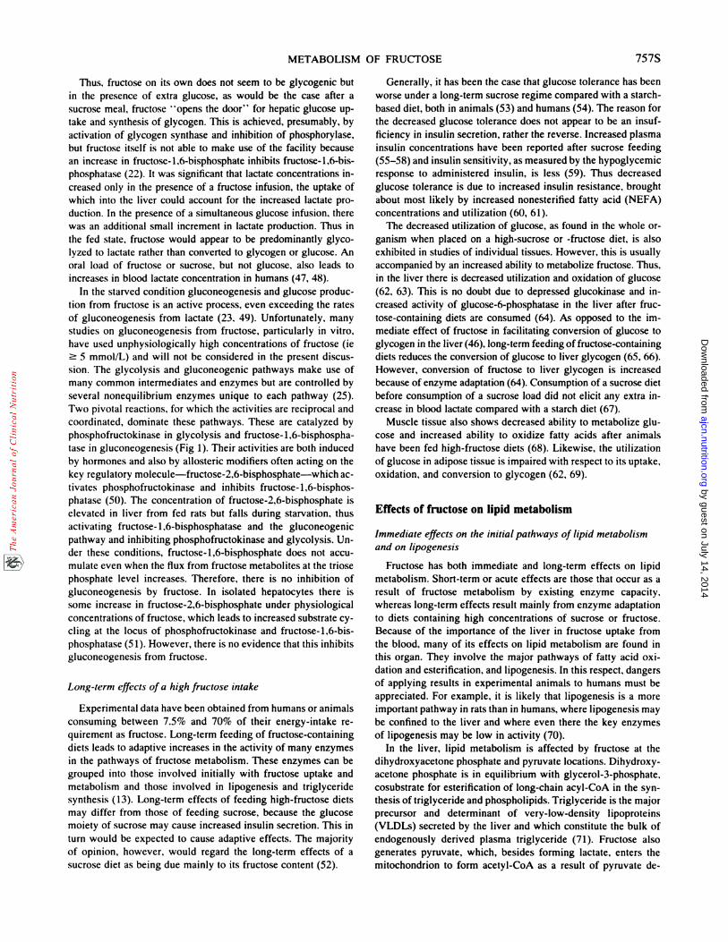
METABOLISM OF FRUCTOSE 755S
Hexokinase will catalyse the phosphorylation of most hexose
sugars, including fructose. However, when fructose is present
FRUCT�SE
FRUCTOSE 1-P
Glyceraldehyde
I ciucose 6.pase_�jcr3I G1uc::::L,.4��
GLUCOSE6-P’��..�..�.#{216}. Glycogen
FRUCTOSE 6-P
[ FRUCTOSE 1.6- bis.P�e �( ) I �4O6PHOFRUCTOKIP4ASE
FRUCTOSE 1 ,6-bis-P
FIG 1. Fructose utilization in the liver showing its interrelationship with glucose and fatty acid metabolism. Pase,
phosphatase; P, phosphate.
skeletal muscle (11). A recent study (12) showed that when fruc-
tose is infused into exercising subjects to maintain a concentration
of 5.5 mmol/L, which is above physiological concentrations and
above the glucose concentration, there is considerably more fruc-
tose oxidation by exercising and resting muscles.
In a previous review (13) we pointed out that many investiga-
tions both in vivo and in vitro had been carried out using unphy-
siological concentrations of fructose and that deductions made
from such observations could be misleading, particularly when
applied to humans consuming normal quantities of sucrose and
fructose. Of particular relevance to hepatic metabolism arc the con-
centrations of fructose likely to be attained in the hepatic portal
vein. In humans (14) and baboons (8) maximal concentrations of
2.2 mmol/L have been recorded after a fructose or sucrose meal.
We found values within the range 1.1-2.2 mmolfL, when fed or
starved rats were given a large fructose meal by gastric intubation
(4). In humans a maximum concentration of 1.0 mmol fructoscfL
was recorded in peripheral blood (14). Because the normal blood
fructose concentration is zero when no fructose is being absorbed,
fructose concentrations in blood will vary from zero up to the
maximum recorded above, according to the quantity in the diet.
Specific metabolic pathways offructose utilization in individual
tissues
with glucose at physiological concentrations its phosphorylation
is largely inhibited by glucose (10). Nevertheless, it is probably
via this route that fructose, after uptake from the blood, enters
the metabolic pathways in adipose tissue and muscle. Of far
greater significance is the pathway discovered by Hers (15),
which involves three particular enzymes (fructokinase, aldolase
B, and triokinase), two of which arc specific for fructose metab-olism (Fig 1). These enzymes arc present in liver and kidney and
probably in the small intestine of some species-such as the
golden hamster (16), guinea pig (17), and dog (18)-that are able
to convert some absorbed fructose into glucose. However, this is
not possible in the intestine of either humans (19) or rats (17),
which do not contain glucose-6-phosphatasc, the enzyme nec-
essary for releasing glucose. Thus in these two species there is
no conversion of fructose to glucose during absorption. In this
respect the rat is a good model of human fructose metabolism.
Fructose is rapidly phosphorylated by AlP in the liver to form
fructose 1-phosphate, catalyzed by the first enzyme of the fructose
pathway-fructokinasc (20). This enzyme is virtually specific for
fructose because it is a kctohexokinase, and fructose is the only
ketohexose of physiological significance in the diet. The high ac-
tivity of fructokinase underlies the ability of the liver to extract so
much of the fructose passing through it. Fructose-i-phosphate is
split by liver aldolase (aldolase B) into glyceraldehyde and dihy-
droxyacetonc phosphate, a member of the glycolysis sequence of
intermediates. Aldolase B also functions in the liver in glycolysis
by guest on July 14, 2014ajcn.nutrition.org
Dow
nloaded from

756S MAYES
to split fructose-1,6-bisphosphate to glyceraldehyde-3-phosphate
and dihydroxyacetone phosphate. The third enzyme in the fructose
pathway is tniokinase (21), which catalyzes the phosphorylation of
glyceraldehyde by Al? to form glyceraldehyde-3-phosphate, an-
other intermediate of the glycolytic pathway.
Thus, the pathways of glucose and fructose metabolism in the
liver converge at the triose phosphate stage of metabolism and
from this point on, their metabolism is qualitatively similar. Fruc-
tose has arrived at this stage in metabolism, without passing
through the main rate-controlling step in glycolysis catalyzed by
phosphofructokinase (22). In this way fructose, on presentation
to the liver, is rapidly phosphorylated, providing increased sub-
strate to the metabolic pathways leading from triose phosphate
(ie, glycolysis, glycogenesis, gluconeogenesis, lipogenesis, and
possibly fatty acid estenification). Thus, major products of fruc-
tose metabolism in the liver are glucose, glycogen, and lactate
(23). Smaller quantities arc oxidized to carbon dioxide, ketonc
bodies or converted to tniacylglyccrol (13). The general dispo-
sition of fructose carbon between its major end products is altered
by changes in nutritional and endocrine status, eg, gluconeogen-
esis from fructose is increased during starvation, diabetes, or ad-
ministration of ethanol or glucagon (24).
Biosynthesis offructose in mammalian tissues
Although the primary purpose of this paper is to review the
effects of exogenously derived fructose, discussion of the metab-
olism of fructose is not complete without a very brief reference
to its synthesis in a few specialized tissues.
Free fructose is found in the lens, seminal fluid, and the fetal
circulation of ungulates and whales. It is formed by the sorbitol
(polyol) pathway (25), which is responsible for fructose forma-
tion from glucose. This pathway is present in the lens, seminal
vesicles, and placenta of the groups mentioned above. It increases
in activity as glucose concentrations rise in diabetics in those
tissues that are not insulin sensitive, such as the lens. Glucose
undergoes reduction by NADPH to sorbitol, catalyzed by aldose
reductase, followed by oxidation of sorbitol to fructose by son-
bitol (polyol) dehydrogenase in the presence of NAD. Both son-
bitol and fructose accumulate in the human lens in diabetics,
causing osmotic damage, which is probably involved in the path-
ogenesis of diabetic cataract. Sorbitol dehydnogenase, but not
aldose reductase, is found in liver and is responsible for the con-
version of any exogenously derived sorbitol to fructose.
Effects of fructose on carbohydrate metabolism
Effects on the concentrations of important intermediates
Experiments with both the perfused liven (23, 26, 27) and cath-
etenized human subjects (9) indicate that in the starved state,
� 66% of a fructose dose is converted to glucose and up to 25%
is released as lactate. Up to a further 8% may form glycogen
(23). After administration of fructose either orally or by intra-
venous loading there is a rapid and marked increase in glucose
1-phosphate in those tissues containing the fructose pathway, ie,
liver (28-31), kidney (30, 32), and small intestine (33, 34). Re-
cent investigations using �‘ P magnetic-resonance spectroscopy
have confirmed the accumulation of fructose 1-phosphate in the
human liver after intravenous administration of fructose (35). Al-
though most of these effects were obtained by using unphysio-
logical concentrations of fructose, experiments with isolated he-
patocytes using graded concentrations of fructose (31) demon-
strated that significant elevations of fructose 1-phosphate
occurred at concentrations of 0.5 and 1 .0 mmol fructosc/L, which
are available in the portal vein in vivo after a fructose meal.
Other important intermediates whose concentrations may alter
as a result of fructose administration include glycerol 3-phos-
phate, fructose 2,6-bisphosphate, pyruvate, lactate, ATP, and in-
organic phosphate (Pi). These will be discussed in following 5cc-
tions.
Effects on glycolysis, glycogenesis, and gluconeogenesis
As a result of the loading of the initial pathways of fructose
metabolism there is a tendency for intermediates of glycolysis to
increase in concentration, resulting in an increased flux through
the pathway, evidenced by increased lactate formation and raised
blood lactate concentrations (36, 37). In the span of glycolytic
reactions from glyceraldehyde-3-phosphate to pyruvate and lac-
tate, the rate-controlling step is catalyzed by pyruvate kinase.
This enzyme is normally under feed-forward control because of
allostenic activation by fructosc-1,6-bisphosphatc. Although this
metabolite may double in concentration when fructose is added
to hepatocytes (31), of more significance are the large increases
in fructose-1-phosphate concentrations, which extend a similar
but more enhanced activation of pyruvate kinase (38), thus fa-
cilitating passage of fructose carbon to pyruvate and lactate.
Glycogen synthesis and breakdown is controlled by a complex
series of reactions involving covalent modification by protein
phosphorylation and dephosphorylation. Briefly, regulation cen-
ters around two rate-controlling enzymes-glycogcn synthase
and glycogen phosphorylase. (See ref 25 for a general review.)
The active form of glycogen synthase (synthase a) is the de-
phosphoenzyme, whereas the inactive synthase b is phosphoryl-
ated. On the other hand active glycogen phosphorylase a is the
phosphoenzymc, whereas the inactive b form is dephosphory-
lated. Protein kinases carry out phosphorylations and protein
phosphatases carry out dephosphorylations of these enzymes.
Both processes are controlled by hormonal and allostenic modi-
fiers. Control of glycogen metabolism in liver differs in some
detail from that of glycogen metabolism in muscle and has been
reviewed by Hers (39).
A study of the literature reveals a disparity in results on whether
fructose promotes liven glycogen deposition, with the balance of
studies in vivo in the fed condition indicating that fructose is a
better promoter of glycogenesis than is glucose (31). The net dep-
osition of glycogen appears to result from both activation of gly-
cogen synthase (40, 41) and inhibition of glycogen phosphorylase
(40, 42). This appears to be brought about by several mechanisms.
Phosphorylase a is inhibited by fructosc-1-phosphate (42-44),
which accumulates after administration of fructose. Also, glucose-
6-phosphate increases in concentration and activates glycogen syn-
thase and inhibits phosphorylase (45). We have carried out per-
fusions with the liver of fed rats, using whole blood containing
physiological concentrations of glucose, amino acids, and free fatty
acids into which was infused glucose, fructose, or both sugars at
physiological rates (46). When either sugar was infused alone.
there was a net output of glucose from the liver, with no change
in glycogen concentration. However, when glucose and fructose
were infused together, there was a marked uptake of glucose and
an increase in glycogen. Fructose uptake was the same with or
without a concomitant glucose infusion.
by guest on July 14, 2014ajcn.nutrition.org
Dow
nloaded from
METABOLISM OF FRUCTOSE 757S
Thus, fructose on its own does not seem to be glycogenic but
in the presence of extra glucose, as would be the case after a
sucrose meal, fructose ‘ ‘opens the door’ ‘ for hepatic glucose up-
take and synthesis of glycogen. This is achieved, presumably, by
activation of glycogen synthase and inhibition of phosphorylase,
but fructose itself is not able to make use of the facility because
an increase in fructose-1,6-bisphosphate inhibits fructose-i,6-bis-
phosphatase (22). It was significant that lactate concentrations in-
creased only in the presence of a fructose infusion, the uptake of
which into the liver could account for the increased lactate pro-
duction. In the presence of a simultaneous glucose infusion, there
was an additional small increment in lactate production. Thus in
the fed state, fructose would appear to be predominantly glyco-
lyzed to lactate rather than converted to glycogen or glucose. An
oral load of fructose or sucrose, but not glucose, also leads to
increases in blood lactate concentration in humans (47, 48).
In the starved condition gluconcogenesis and glucose produc-
tion from fructose is an active process. even exceeding the rates
of gluconeogenesis from lactate (23, 49). Unfortunately, many
studies on gluconcogenesis from fructose, particularly in vitro,
have used unphysiologically high concentrations of fructose (ic
� 5 mmol/L) and will not be considered in the present discus-
sion. The glycolysis and gluconeogenic pathways make use of
many common intermediates and enzymes but arc controlled by
several nonequilibrium enzymes unique to each pathway (25).
Two pivotal reactions, for which the activities arc reciprocal and
coordinated, dominate these pathways. These arc catalyzed by
phosphofructokinase in glycolysis and fructose-i,6-bisphospha-
tase in gluconeogenesis (Fig 1). Their activities arc both induced
by hormones and also by allostenic modifiers often acting on the
key regulatory molecule-fructosc-2,6-bisphosphatc-which ac-
tivates phosphofructokinase and inhibits fructosc-1,6-bisphos-
phatase (50). The concentration of fructose-2,6-bisphosphatc is
elevated in liver from fed rats but falls during starvation, thus
activating fructose-i ,6-bisphosphatase and the gluconeogenic
pathway and inhibiting phosphofructokinasc and glycolysis. Un-
der these conditions, fructose- 1,6-bisphosphate does not accu-
mulate even when the flux from fructose metabolites at the triose
phosphate level increases. Therefore, there is no inhibition of
gluconcogenesis by fructose. In isolated hepatocytes there is
some increase in fructose-2,6-bisphosphate under physiological
concentrations of fructose, which leads to increased substrate cy-
cling at the locus of phosphofructokinasc and fructosc-i,6-bis-
phosphatase (51). However, there is no evidence that this inhibits
gluconeogenesis from fructose.
Long-term effects of a high fructose intake
Experimental data have been obtained from humans or animals
consuming between 7.5% and 70% of their energy-intake re-
quirement as fructose. Long-term feeding of fructose-containing
diets leads to adaptive increases in the activity of many enzymes
in the pathways of fructose metabolism. These enzymes can be
grouped into those involved initially with fructose uptake and
metabolism and those involved in lipogenesis and triglyceride
synthesis (13). Long-term effects of feeding high-fructose diets
may differ from those of feeding sucrose, because the glucose
moiety of sucrose may cause increased insulin secretion. This in
turn would be expected to cause adaptive effects. The majority
of opinion, however, would regard the long-term effects of a
sucrose diet as being due mainly to its fructose content (52).
Generally, it has been the case that glucose tolerance has been
worse under a long-term sucrose regime compared with a starch-
based diet, both in animals (53) and humans (54). The reason for
the decreased glucose tolerance does not appear to be an insuf-
ficiency in insulin secretion, rather the reverse. Increased plasma
insulin concentrations have been reported after sucrose feeding
(55-58) and insulin sensitivity, as measured by the hypoglycemic
response to administered insulin, is less (59). Thus decreased
glucose tolerance is due to increased insulin resistance, brought
about most likely by increased nonestenified fatty acid (NEFA)
concentrations and utilization (60, 61).
The decreased utilization of glucose, as found in the whole or-
ganism when placed on a high-sucrose or -fructose diet, is also
exhibited in studies of individual tissues. However, this is usually
accompanied by an increased ability to metabolize fructose. Thus,
in the liver there is decreased utilization and oxidation of glucose
(62, 63). This is no doubt due to depressed glucokinase and in-
creased activity of glucose-6-phosphatasc in the liver after fruc-
tose-containing diets are consumed (64). As opposed to the im-
mediate effect of fructose in facilitating conversion of glucose to
glycogen in the liver (46), long-term feeding of fructosc-containing
diets reduces the conversion of glucose to liver glycogen (65, 66).
However, conversion of fructose to liver glycogen is increased
because of enzyme adaptation (64). Consumption of a sucrose diet
before consumption of a sucrose load did not elicit any extra in-
crease in blood lactate compared with a starch diet (67).
Muscle tissue also shows decreased ability to metabolize glu-
cose and increased ability to oxidize fatty acids after animals
have been fed high-fructose diets (68). Likewise, the utilization
of glucose in adipose tissue is impaired with respect to its uptake,
oxidation, and conversion to glycogen (62, 69).
Effects of fructose on lipid metabolism
Immediate effects on the initial pathways of lipid metabolism
and on lipogenesis
Fructose has both immediate and long-term effects on lipid
metabolism. Short-term or acute effects arc those that occur as a
result of fructose metabolism by existing enzyme capacity,
whereas long-term effects result mainly from enzyme adaptation
to diets containing high concentrations of sucrose or fructose.
Because of the importance of the liver in fructose uptake from
the blood, many of its effects on lipid metabolism arc found in
this organ. They involve the major pathways of fatty acid oxi-
dation and estcnification, and lipogenesis. In this respect, dangers
of applying results in experimental animals to humans must be
appreciated. For example, it is likely that lipogenesis is a more
important pathway in rats than in humans, where lipogenesis may
be confined to the liver and where even there the key enzymes
of lipogenesis may be low in activity (70).
In the liver, lipid metabolism is affected by fructose at the
dihydroxyacetone phosphate and pyruvate locations. Dihydroxy-
acetone phosphate is in equilibrium with glycerol-3-phosphatc,
cosubstrate for esterification of long-chain acyl-CoA in the syn-
thesis of triglyceride and phospholipids. Triglyceride is the major
precursor and determinant of very-low-density lipoprotcins
(VLDLs) secreted by the liver and which constitute the bulk of
endogenously derived plasma triglyceride (71). Fructose also
generates pyruvate, which, besides forming lactate, enters the
mitochondnion to form acetyl-CoA as a result of pyruvate de-
by guest on July 14, 2014ajcn.nutrition.org
Dow
nloaded from

758S MAYES
hydrogenase (PDH) activity. Here it can act as a carbon source
for three products: carbon dioxide, after oxidation in the citric
acid cycle; long-chain fatty acids, after entering the lipogenesis
sequence of reactions; and, ketone bodies (72, 73). Acetyl-CoA
is the major carbon source for lipogenesis. However, as lipogen-
esis occurs in the cytosol, acetyl-CoA must be transported
through the mitochondnial membrane as citrate, reforming acetyl-
CoA in the cytosol by the action of the lipogenic enzyme, AlT-citrate lyase. Acctyl-CoA is converted to long-chain fatty acid
via the important cytosolic intermediate malonyl-CoA. By these
pathways, fructose provides carbon atoms for both the glycerol
and the acyl portions of the acylglycerol molecule.
The activity of PDH is a key factor in determining the fate of
pyruvate. It is activated by a decreased ratio of [AlT] to [ADP]
and by an increase in pyruvate (74); it is inhibited by increased
concentrations of NEFA (74, 75). As discussed in reference 76,
fructose administration, particularly at high amounts, causes de-
pletion not only of AlT but of all adenine nucleotides; therefore,
the ratio of [ATP] to [ADP] may not change appreciably. Oral
administration of large amounts of fructose to humans produces
hypcruriccmia, which is known to follow depletion of Al? and
other adcninc nucleotides (77, 78). However, no decrease in he-
patic [ATP] was found in fructose-fed rats (79). Adenine nude-
otide concentrations and PDH activity did not change when fruc-
tose was added to the perfused rat liver at physiological concen-
trations of 1 .3 mmol/L. However, at 1 1 .0 mmol/L both AlP and
total adenine nucleotides were decreased and PDH was activated
(80). At a fructose concentration of 1 .5 mmol/L there was a per-
ceptible increase in PDH activity, which was not accompanied
by any change in adenine nucleotide concentrations (13). Of
more significance was the fact that at this physiological concen-
tration of fructose, the inhibitory effect of increased oleate (a
NEFA) on PDH activity was reversed. Generally, these results
support the view that fructose causes increases in PDH activity
by increasing pyruvate concentrations, rather than by decreasing
the ratio of [Al?] to [ADP] (81). In summary, it would appear
that some activation of PDH occurs with high physiological con-
centrations of fructose, especially in antagonism to the depressant
effect of increased concentrations of NEFAs.
When fructose was injected intravenously into rats there was
an immediate though transient increase in glycerol-3-phosphate
and pyruvate concentrations (82). In contrast, after glucose ad-
ministration there was no immediate increase. When fructose was
administered intrapenitoneally there was an increase in hepatic
dihydroxyacetone phosphate, glycerol-3-phosphate, pyruvate,
and lactate (83). Similar results have been obtained in the per-
fused liver (27, 84, 85). Acetyl-CoA concentrations also increase
(72). It is also likely that malonyl-CoA concentrations rise when
fructose is administered because an increase has been recorded
in hepatocytes in the presence of lactate and pyruvate (86), both
of which increase in the presence of fructose. Thus, fructose
causes increased concentrations of its metabolites in both the
glycerol-3-phosphatc and glycolytic and acetyl-CoA branches of
its metabolism. Although many lipogenic intermediates increase
in concentration after administration of fructose, there is little
evidence that fructose on its own has an immediate effect on
lipogenesis. Thus, fructose stimulated lipogenesis from acetate
in chick liver slices, but not when fructose was added alone (87).
Using the tnitiated water technique we were unable to demon-
strate any increase in lipogenesis in perfused livers from fed rats
in the presence of physiological concentrations of fructose (13).
It is likely that if fructose were administered with an equal
amount of glucose, there might well be increased lipogenesis,
particularly if this was accompanied by a rise in insulin concen-
tration.
Immediate effects on fatty acid oxidation and esterification and
on lipoprotein formation and utilization
Although fructose does not seem to cause any marked imme-
diate increase in lipogenesis by itself, it does have marked and
immediate effects on the fate of NEFAs. These fatty acids arise
as a result of lipid mobilization in adipose tissue or from hydro-
lysis of triglyceride-rich lipoprotcins by lipoprotcin lipase. We
established in the perfused rat liver that NEFAs, which are taken
up by the liver to the extent of 30-40% per pass, are either oxi-
dized or esterified (88, 89). An inverse relationship was dem-
onstrated between the quantity of fatty acids oxidized to carbon
dioxide and kctonc bodies on the one hand, and those esterified
in liver acylglyccrols and in VLDLS on the other. Regulation of
this balance controlled both ketogenesis and VLDL secretion in
a reciprocal manner. For a given load of fatty acids taken up by
the liver, livers from fed animals oxidized less fatty acids but
esterified more than did livers from starved animals, demonstrat-
ing that a mechanism exists, which is affected by the nutritional
state, for regulating the partition of fatty acids between these two
major pathways. We showed (89) that livers from fasted animals
maintained a constant fractional rate of cstcrification irrespective
of the NEFA load, demonstrating that glycerol-3-phosphate
availability could not have been rate limiting in fatty acid ester-
ification. It has now been firmly established that the regulatory
site for controlling the balance between oxidation and esterifi-
cation lies in the oxidative pathway, where long-chain acyl
groups enter the mitochondrion (90). The rate-limiting step in
mitochondnial oxidation of long-chain fatty acids is catalyzed by
palmitoyltransferasc I. Fatty acids failing to enter the mitochon-
dna for oxidation do not accumulate but are esterified immedi-
ately. Palmitoyltransfcrasc I is inhibited by malonyl-CoA (90),
whose concentration increases in the fed condition when there is
active lipogcnesis. This can explain the change in balance be-
twecn esterification and oxidation of fatty acids between livers
of fed and starved animals.
To test whether fructose and insulin had direct and immediateeffects on VLDL secretion by the liver, we perfused livers from
fed rats with whole blood into which [‘4C]oleate NEFA was in-
fused to maintain a constant physiological concentration (46, 91).
Increased concentrations of insulin or an infusion of a physio-
logical quantity of fructose decreased oxidation and increased
estenification of fatty acids and secretion of VLDL triglyceride
from the liver. When insulin was added together with fructose,
the separate effects were additive with a further decrease in ox-
idation and enhancement of estenification and VLDL secretion.
Malonyl-CoA is the product of acctyl-CoA carboxylase activityand it is known that this enzyme is activated by covalent modi-
fication by insulin and inhibited by glucagon (92). Therefore,
insulin and fructose can independently increase the concentration
of malonyl-CoA. Fructose may also enhance estenification byraising the concentration of glyccrol-3-phosphate (93, 94) al-
though this seems unlikely if physiological concentrations of
fructose do not in fact influence the concentration of this inter-
mediate (82). Also, we have shown that mitochondnial glycerol
phosphate acyltransferase activity is enhanced by insulin (95),
by guest on July 14, 2014ajcn.nutrition.org
Dow
nloaded from

METABOLISM OF FRUCTOSE 759S
and this could also account for a direct effect of insulin in pro-
moting esterification and VLDL secretion.
These effects of fructose on NEFA metabolism are unique to
this sugar. In a direct comparison with glucose, the effect of an
infusion providing physiological concentrations of fructose in the
perfusate of the perfused liver were compared with a comparable
infusion of glucose. Only the fructose infusion boosted
[‘4C]NEFA estenification and [‘4C]VLDL secretion (46). When
similar amounts of the two sugars were then infused simultane-
ously to simulate sucrose digestion and absorption, there were
no further increases in NEFA esterification or secretion as VLDL.
Perfusions were also carried out (73) to test the effect of a fruc-
tose concentration above the physiological range (8.9 mmollL)
against a concentration within the physiological range (1.5 mmol/
L). At the higher concentration, VLDL production was cut toone-third of the amount found under the physiological concen-
tration. Because lipoprotein production is a complex energy-re-
quining process, the lower ATP availability associated with
high-fructose concentrations (76) was probably the reason for the
reduced VLDL output under these conditions.
When a 20% fructose solution was given intravenously, serum
triglycerides decreased in female baboons but increased in males,
whereas a comparable solution of glucose caused a decrease in
both sexes (96). Although the increase in triglycerides is most
likely the result of a direct effect on the liver, as described above,
the fall in triglyceride concentration is probably due to a decrease
in NEFA concentration, which in turn provides less NEFA sub-
strate for the liver and therefore less VLDL production. In hu-
mans, fructose causes a decrease in NEFAs with little change in
insulin concentrations (97), indicating that any reduced secretion
of VLDL is due to decreased NEFA availability and any in-
creased secretion is most likely due to the direct hepatic action
of fructose. The net output of VLDL by the liver is therefore a
balance between these two opposing effects of fructose.
Fructose is both antiketogenic and ketogenic depending on the
circumstances. In vivo, in the starved state, a physiological intake
of fructose is antiketogenic. This is nearly always due to its in-
hibition of NEFA mobilization from adipose tissue (97), which
results in reduced uptake of the main ketogenic substrate by the
liver. It appears that there is also a direct antiketogenic effect of
fructose on the liver, as demonstrated in vivo in the presence of
a constant plasma NEFA concentration (98). Similar experiments
have been carried out in the perfused liver and isolated hepato-
cytes in the presence of added NEFA (72, 84, 91, 99, 100). Most
of these experiments were carried out in the starved state. It is
unclear whether or not malonyl-CoA inhibition of carnitine pal-
mitoyltransferase I can explain these results. It would require the
immediate activation of acctyl-CoA carboxylase, which has not
been reported under these conditions. Although glyccrol-3-phos-
phate availability does not appear to be rate-limiting on esterifi-
cation of NEFAS, it is possible that boosting its concentration
above the normal concentration as a result of fructose metabolism
could cause a greater fractional estenification of NEFAs and less
oxidation to ketonc bodies (98).
That fructose can also be ketogenic was first shown in perfused
livers from starved rats infused with the sugar at 20 mmollL (72).
The phenomenon was also demonstrated with livers from fed
animals by using lower but still unphysiological concentrations
of fructose (73, 101). To test whether this effect could be ob-
tamed with fructose within physiological concentrations we stud-
ied ketogenesis in perfused livers from fed animals and showed
that ketogenesis progressively increased as the concentration of
fructose was raised (13). At 1.5 mmol fructose/L, there was a
significant increase compared with control perfusions in the ab-
sence of fructose. Some of these studies also used [‘4C]fructosc.
At 20 mmol/L, all of the carbon atoms found in ketone bodiescame from fructose (72) whereas at 8.9 mmol/L, half came from
fructose and at the physiological concentration of i .5 mmol fruc-
tose/L, 15% of the carbon atoms originated from fructose (73).
Radiolabeled fructose has also been used to follow the course
taken by the sugar in the various metabolic pathways. Initially,
there is no dilution of fructose label because there is no endog-
enous pool of free fructose, but as fructose is converted into
intermediates common with glucose and fatty acid metabolism,
the label is extensively diluted. Therefore, although it is not pos-
sible to quantify the fate of fructose in a whole animal or tissue
without knowledge of the specific radioactivity of intermediary
pools, some idea may be obtained of the fate of administered
fructose. Thus, when the metabolism of starved rats given either
[ U-’4C]glucosc or [U-’4C]fructose by gastric intubation was com-
pared, it was shown that although � 2.5 times as much newly
synthesized triglyceride came from total body glucose as from
administered fructose, twice as much of the administered fructose
formed triglyceride compared with a similar quantity of admin-
istered glucose (102). These data reflect the fact that fructose is
taken up predominantly by the liver whereas glucose is utilized
mainly by the extrahepatic tissues. In liver slices, radiolabeled
fructose is converted to lactate, pyruvate, carbon dioxide, and
triglyceride more rapidly than is glucose (103). Similar results
have been reported for incorporation into plasma lipids in
guinea pigs (104) and baboons (105). After administration of
[‘4C}fructosc, more radioactivity is found in glyceride glycerol
than in the fatty acid portions of hepatic triglyceride (102, 103),
due in part to greater dilution and exchange of label in the path-
way from fructose to long-chain fatty acids than in that to glyc-
erol-3-phosphatc (Fig 1). When [U-’4Clfructose was infused to
maintain a concentration of 1.5 mmol/L in the perfusate of livers
prepared from fed rats, 12% was oxidized to carbon dioxide and
2.4% was converted to ketone bodies; 4.1% was in liver lipids,
1.6% was incorporated into VLDLS, and only 0.4% was in total
cholesterol (13). As the fructose load was increased, proportion-
ally less of the labeled fructose entered lipid products except for
ketone bodies, which remained the same. Thus, ketonc bodies
and lactate act as overflows of excess carbon as fructose saturates
the metabolic pathways. Hence, ketone bodies fulfill a similar
role in the liver with respect to carbohydrate as they do when
fatty acids are present in excess.
Long-term effects of a high fructose intake
Enzyme adaptation is also responsible for many of the long-
term effects of a high-fructose diet on lipid metabolism. as well
as for the long-term effects on carbohydrate metabolism. In the
liver, fructokinase activity increased in rats on a fructose-rich diet
(106). Fructose or sucrose feeding also increased the activity of
glycerol-3-phosphate dehydrogenase, which is necessary for the
conversion of fructose to glycerol-3-phosphate via the glycolytic
intermediate dihydroxyacetone phosphate (107, 108). After 50 d
on a fructose diet, but not on a sucrose diet, pyruvate kinase in
rats increased substantially (109). Similarly fructose diets in-
creased liver PDH activity in rats (1 10). In the pathway of lipo-
genesis and triglyceride formation, ATP-citratc lyase (108, 111,
112), acetyl-CoA carboxylase (111-113), fatty acid synthase
by guest on July 14, 2014ajcn.nutrition.org
Dow
nloaded from

760S MAYES
(1 1 i-i 14), and phosphatidate phosphohydrolase (1 15) are re-
ported to be increased in activity in animals on fructose-contain-
ing diets. Also, the enzymes responsible for generation of reduc-
ing power for lipogencsis-glucose-6-phosphatc dehydrogenase
(107-109, iii, 112, 116), 6-phosphogluconate dehydrogcnase
(107, 108, 1 1 1, 1 12), and NADP malate dehydrogenase (‘malic
enzyme’) (107, 1 1 1, 1 12, 1 16)-are all increased in activity.
Many of these adaptive changes in liver enzyme activity arc not
unique for fructose because glucose feeding will also increase
their activities (107), illustrating that this is a more general effect
of soluble carbohydrates. Of considerable interest is that sucrose
or fructose feeding reduces the activities of hexokinase, pyruvate
kinase, PDH, ATP-citrate lyase, acetyl-CoA carboxylase, fatty
acid synthase, glucose-6-phosphatc dehydrogenase, and NADP
malate dehydrogenase in adipose tissue, whereas feeding glucose
or starch enhances their activities (52). It would appear from a
review of these enzyme activities that lipogenesis is elevated in
the livers but depressed in adipose tissue of sucrose- or fructose-
fed animals. In keeping with this conclusion the concentrations
of the following intermediates in the liver are elevated: pyruvate,
malate, acctyl-CoA (1 17, 1 18), acetoacetyl-CoA, and long-chain
acyl-CoA (1 17). However, glycerol-3-phosphate concentrations
apparently do not increase (119).
Biosynthesis of long-chain fatty acids (lipogenesis) has been
shown to increase in liver slices from rats fed either fructose
(120) or sucrose (121, 122), as measured directly by using la-
beled acetate or tritiated water. Increased incorporation of labeled
fructose has also been shown (118, i23). In the whole animal,
incorporation of tritiated water into liver fatty acids was elevated
in rats fed fructose compared with those fed glucose (124). Also,
F‘4C]fructosc was incorporated more rapidly into fatty acids andacylglycerol glycerol in plasma and liver in rats on a fructose
diet than in rats on a glucose diet (125).
In isolated livers perfused with whole blood, we studied li-
pogenesis in rats that had been fed the standard laboratory diet
or a sucrose-supplemented diet (13, 126). Half the perfusions
were infused with fructose to maintain a physiological perfusate
concentration of 1 .4 mmolfL. Incorporation of 3H2O into liver
acylglycerol fatty acids increased significantly only in the group
of perfusions in which fructose was infused into sucrose-fed rats.
Thus, lipogenesis is increased on sucrose diets but only when
fructose specifically acts as the substrate. This finding is similar
to those found in liver slices from rats on a glucose diet, which
enhanced selectively the conversion of [‘4C]glucose to fatty ac-
ids, but not � ‘4Cjfructose, and where slices from rats on a fructose
diet selectively incorporated [‘4Cjfructose into fatty acids but not
[‘4C]glucose. They demonstrate the highly specific and selective
nature of the lipogenic adaptations to different sugars in the diet,
clearly based on the enzyme adaptations previously discussed.
Plasma triglycerides increase in both humans and rats when
diets are enriched with carbohydrate, especially sucrose and fruc-
tose (127). This has been, and still is, of considerable interest in
view of the fact that raised concentrations of plasma triglyceride
may be an independent factor associated with coronary heart dis-
case (128). The adaptive changes in enzyme activity, evidence
of increase in lipogenic potential, plus the direct actions of fruc-
tose previously described, all lead to increased output of VLDL
triglyceride from the liver, increasing the amount in the circula-
tion. The resultant concentration of triglyceride also depends on
the rate of hydrolysis by lipoprotein lipase and any impairment
of this enzyme would also lead to increased concentrations of
plasma triglyceride (129). However, long-term fructose or su-
crose feeding results in hyperinsulinemia (55-58) and increases
post hepanin lipolytic activity, indicating an enhancement of li-
poprotcin lipase activity (52). Therefore, raised plasma concen-
trations of triglyceride in fructose- or sucrose-fed animals, in-
cluding humans, reflects an increased rate of VLDL secretion by
the liver together with an increased rate of VLDL catabolism.
Fructose feeding enhances the release of NEFAs from adipose
tissue (60), increasing its plasma concentration (60, 61), because
of a decreased rate of re-esterification of NEFAs within adipose
tissue (69). Esterification of NEFAs in adipose tissue depends on
glucose utilization, which is depressed under conditions of fruc-
tose feeding. NEFAs are the major precursors of VLDL triglyc-
eride in the liver (71). Therefore, they will augment VLDL for-
mation in animals on high-fructose or -sucrose diets. Mindful of
the fact that NEFAs arc always present in plasma, we studied the
effects on VLDL secretion of a physiological infusion of fruc-
tose, and of sucrose supplementation of the diet, in isolated per-
fused livers infused with [1-’4C]oleate NEFA (126). VLDL tn-
glyceride production increased in both the livers infused with
fructose alone and in those derived from sucrose-fed rats. When
livers from sucrose-fed animals were infused with fructose, the
rate of secretion more than doubled. Because shifts in balance
between oxidation and estenification of NEFAs in the direction
of estenification have been shown to increase secretion of VLDL
triglyceride (89), we also measured these indexes. Oxidation of
[ 4C]oleatc to 4C0 was highest and estenification was lowest in
control perfusions when livers from normal animals were used.
Fructose infusion or sucrose feeding shifted the balance in favor
of estenification and increased output of [‘4C]VLDL. The corn-
bination of fructose infusion plus sucrose feeding showed the
lowest rate of oxidation and the highest rate of estenification and
output of [‘4C]VLDL. The marked extra increase in VLDL output
in the combined group was probably due to increased lipogenesis
from fructose in this group (13, 126).
The potential adverse effects of fructose on lipid metabolism
in humans has been reviewed (130, 131). It was concluded from
a review of a large number of studies that normal individuals
consuming diets containing average quantities of fructose have
normal fasting triglyceride concentrations. However, this is only
a measure of triglyceride clearance and does not give information
on prandial and postprandial concentrations of triglycerides,
which might be raised on these diets. The condition was exac-
erbated in individuals having defects in carbohydrate metabolism
leading to hypertniglycenidemia, even with low intakes of fruc-
tose. Similar conclusions apply to plasma cholesterol, which may
increase if higher than normal amounts of VLDLS arc present. In
addition, the combination of saturated fat and fructose in the diet
would appear to favor elevated cholesterol concentrations.
Effects of fructose on purine metabolism
The hyperuricemic effect of fructose
It was first reported (132) that when fructose was administered
intravenously to both normal children and those with hereditary
fructose intolerance, there was an increase in serum and urinary
uric acid. The hyperunicemic effect appears to be dose dependent
and the fructose infusion needs to be > 0.5 gkg body wt ‘
to cause detectible hyperunicemia. Also, the effect is fructose
specific because comparable infusions of either glucose or galac-
by guest on July 14, 2014ajcn.nutrition.org
Dow
nloaded from

METABOLISM OF FRUCTOSE
FRUCTOSEI�CT��1
FRUCTOSE 1�Ptose do not raise the plasma uric acid concentration (133). Pa-
tients suffering from gout arc more sensitive to fructose admin-
istration than are normal subjects, as are diabetics (130). These
results apply to fructose administered parenterally, but is it pos-
sible to increase blood uric acid concentrations when fructose is
consumed orally? The effect of feeding fructose at a dose of 1 g!
kg body wt was studied in healthy subjects, in patients with gout,
and in children of parents with gout (78). Hyperunicemia oc-
curred in all groups but it was more marked and prolonged in
those subjects, or offspring of parents, with gout. Subjects con-
suming higher than � 18% of their energy intake as sucrose (ic,
9% as fructose) showed significant increases in serum uric acid
(130). These results suggest that the average intake of fructose
or sucrose in a mixed diet is on the borderline for provoking
hyperunicemia in normal individuals. Note that gouty patients do
have lower serum uric acid conccntrations when fed lower-fruc-
tose diets; even healthy individuals may be at risk of hyperuni-
cemia when consuming a diet high in fructose.
Pathways of adenine nucleotide catabolism and biosynthesis
To understand the mechanism of the hyperunicemic effect of
fructose a knowledge of puninc nucleotide metabolism and its
regulation is necessary. Adenylate kinase maintains equilibrium
between the adenine nucleotides ATP, adenosinc diphosphate
(ADP), and adenosine monophosphate (AMP) (Fig 2). Thus, all
three adenine nuclcotides are able to be degraded and formed via
AMP. However, as with many other common metabolites, the
pathway of degradation of adenine nucleotides is not the simple
reverse of the pathway of biosynthesis. In the degradative path-
way, inosine monophosphate (IMP) is formed from AMP by the
action of AMP deaminase. Phosphate is then removed by 5’-
nucleotidase to form inosine. Alternatively, inosine may be
formed from AMP via adenosinc but experiments in isolated he-
patocytes (31) have indicated that this is not as important a route
for AMP degradation as that via IMP. In addition, the reaction
catalyzed by AMP deaminase appears to be the rate-limiting step
in the breakdown of hepatic adeninc nucleotides (134-136), and
Pi at normal intracellular concentrations is an important inhibitor
of this enzyme. Inosinc is broken down to uric acid via hypo-
xanthine and xanthinc. In humans and other primates this is the
end product of purinc metabolism and is excreted as such, but
other mammals possess the enzyme unicase, which allows further
oxidation to the much more soluble allantoin. A consequence of
the absence of unicase in humans is the occurrence of gout, which
is associated with hypcruniccmia.
IMP is also the first formed punine nucleotide in the pathway
of de novo synthesis from nibose 5-phosphate. This pathway in-
volves no less than 1 1 separate steps (137). Of these, only the
first two need to be mentioned because they catalyze the rate-
controlling reactions, both of which are held in check by a feed-
back regulation because of high concentrations of puninc nude-
otides. In addition, 5-phosphonibosyl-1-pyrophosphatc (PRPP),
the product of the first reaction in the pathway is an allostenic
activator of the second reaction in the pathway catalyzed by
PRPP glutamyl amidotransferase (Fig 2).
Mechanism underlying the hyperuricemic action of fructose
The essential finding that led to the explanation as to why
fructose administration caused increased uric acid formation was
the observation that it was accompanied by a sharp fall not only
76lS
e 3.f�
AMP+ATP 1 � 2ADP
Adenosine Adenylo- � IMP4� � 9 steps
Inossne � 5-Phospho-p ribosylamine
ATPMJdIEOS1O#{128} AMP. GMP
� I4
PRPP GL.UTAMYI.
Hypoxanthine M4IDOTR4j�G:ER*.SE
A
XANTHINE �
(O)�DASE) PRPP
AMP. ADP. AlP
Xanthine S
_____________ 4XANTHINE I PRPP
o&{�DmGB� �SYN�1ETASE
(OXIDASE)
Uric acid Ribose 5-P
FIG 2. The relationship between fructose metabolism and uric acidformation in the liver. P. phosphate; AMP, adenosine monophosphate:
ADP, adenosine diphosphate; ATP, adenosine triphosphate; Pi, organic
phosphate; GMP, guanosine monophosphate; PRPP, 5-phosphonibosyl-
1-pyrophosphate; IMP, inosine monophosphate.
in hepatic AlT concentration but also in total adeninc nucleotides
(76). The reason for this became apparent when it was shown
that fructosc-1-phosphatc accumulated and Pi concentrations fell
(27). Thus, the sequence of events after fructose administration
involves rapid phosphorylation of the sugar to fructose-1-phos-
phate because of the high activity of fructokinase. This leads to
depletion of ATP due to inhibition of oxidative phosphorylation
of ADP because of a shortage of Pi sequestered in fructose-i-
phosphate. The lowering of AlP concentration is also assisted
by the activity of tniokinase in utilizing Al? in the phosphory-
lation of glyceraldehyde to glyccraldehyde-3-phosphatc. The de-
pletion of Pi and ATP leads to the removal of the allostenic in-
hibition on the enzymes that degrade AMP, respectively, AMP
deaminase and 5’-nuclcotidase. There is a rise in inosinc con-
centration that leads to increased formation of uric acid with de-
pletion of the total adenine nucleotide pool.
The depletion of adenine nucleotides also leads to stimulation
of the pathway of punine nucleotide synthesis as a feedback re-
action (Fig 2). The lowering of the concentration of adenine nu-
cleotides removes the allostenic inhibition of the first two steps
in the pathway catalyzed by PRPP synthetase and PRPP glutamyl
amidotransferase. The production of PRPP acts as a further ac-
tivator of PRPP glutamyl amidotransferase, leading to IMP syn-
thesis. If 5’-nucleotidase is still active, IMP will be degraded to
uric acid rather than converted to AMP. In this way the initial
production of uric acid from the adenine nuclcotidc pool is aug-
mented by dc novo synthesis.
by guest on July 14, 2014ajcn.nutrition.org
Dow
nloaded from

762S MAYES
quantities of fructose may be at increased risk.
The reduction in concentration of ATP and other high energy
phosphates has other profound effects on metabolism and other
functions dependent on a continual supply of these vital sources
of free energy, eg, inhibition of protein (76) and nucleic acid
synthesis (138).
Summary and conclusions
Many investigations both in vivo and in vitro have been carried
out by using unphysiological concentrations of fructose. Deduc-
tions made from such observations can be misleading, particu-
larly when applied to humans consuming normal quantities of
sucrose and fructose. This aspect has been taken into account in
compiling this review.
Specific enzymes in the liver-fructokinase, aldolase B, and
tniokinase-allow fructose ready access to the tniose phosphate
pool and all pathways leading from it, after bypassing the phos-
phofructokinase regulatory step in glycolysis. In the fed state,
this allows a greater saturation of the glycolysis pathway with
consequent lactate production, activation of PDH, and domi-
nance of the oxidative pathways leading to carbon dioxide for-
mation and ketone body production when under fructose load.
Glucose production, glycogenesis, and lipogenesis are not en-
hanced but there is an immediate shift in the balance between
oxidation and estenification of NEFAs in favor of estenification.
This leads to augmented output of VLDLS from the liver. In the
liver of starved animals, the enzymes of gluconeogenesis are ac-
tive and fructose will form much more glucose under these con-
ditions.
As a result of enzyme adaptation to the long-term feeding of
fructose or sucrose diets, the pattern of fructose metabolism is
changed. Enhanced activity of fructose-1,6-bisphosphatase, gly-
cogen synthase, and glucose-6-phosphatase allow more fructose
to form glycogen and glucose. Enhanced activity of lipogenic
enzymes in the liver, but not in adipose tissue, stimulates long-
chain fatty acid synthesis, which augments the immediate hepatic
actions of fructose in promoting VLDL triglyceride output. This
leads to hypertniglycenidemia. Fructose metabolism in adipose
tissue also causes impaired glucose utilization and impaired es-
tenification of fatty acids, which promotes release of NEFAs with
consequent raised plasma NEFA concentrations and increased
VLDL production. The increased triglyceride and NEFA con-
centrations impair utilization of glucose in muscle, decreasing
glucose tolerance and increasing insulin resistance with conse-
quent hypeninsulinemia. This, in turn, will stimulate the already
increased VLDL production by the liver. Because VLDLS con-
tam � 20% cholesterol, there is a corresponding rise in plasma
cholesterol, with little change in LDL cholesterol.
Acute loading of the liver with fructose is also a cause of
hyperunicemia. This is due to utilization of Al? in the phospho-
rylation of fructose and sequestration of Pi in fructose-1-phos-
phate, preventing oxidative regeneration of AlT from ADP. Be-
cause the enzymes of adeninc nucleotide degradation are inhib-
ited by AlT and Pi, removal of the inhibition leads to destruction
of the total adeninc nucleotide pool and generation of uric acid.
Both the hyperlipidemic and hyperuricemic effects of fructose
can be demonstrated in humans. Although these effects seem
minimal in healthy individuals on normal diets, individuals on
very-high-fructose diets and individuals who are actually or p0-
tentially hyperlipidemic or hyperunicemic who take in average
It will be apparent that although there have been noteworthy
contributions on fructose metabolism in humans, most investi-
gations have been carried out in laboratory animals, particularly
rats. This is inevitable when precise information about intracel-
lular processes is required, involving invasive and critical pro-
cedures. It is clear that systematic investigations in humans are
needed to ascertain the precise amounts, both of fructose con-
sumption and of its concentration in the blood, at which dde-
tenious effects such as hyperlipidemia and hyperunicemia occur.
Nuclear magnetic resonance spectroscopy is clearly of value as
a noninvasive technique in monitoring intracellular changes in
the concentration of key metabolites. U
References
1. Bruckdorfer KR, Kang 55, Yudkin J. Plasma concentrations of
insulin, corticosterone, lipids and sugars in rats fed on meals withglucose and fructose. Proc Nutr Soc 1973;32: 12A-B.
2. Reiser 5, Michaelis 0, Putney J, Hallfrisch J. Effect of sucrosefeeding on the intestinal transport of sugars in two strains of rats.J Nutr 1975;105:894-905.
3. Silliman K, Coulston AM. Sugars in the diet. In: Kretchmer N,Hollenbeck CB, eds. Sugars and sweetners. London: CRC Press,
1991: 17-35.4. Topping DL, Mayes PA. The concentrations of fructose, glucose
and lactate in the splanchnic blood vessels of rats absorbing fruc-
tose. Nutr Metab 1971;13:331-8.5. Mendeloff Al, Weichselbaum TE. Role of the human liver in the
assimilation of intravenously administered fructose. Metabolism1953;2:450-8.
6. Topping DL, Mayes PA. Comparative effects of fructose and glu-cose on the lipid and carbohydrate metabolism of perfused rat liver.
Br J Nutr 1976;36:113-26.7. Macdonald I, Turner U. Serum fructose levels after sucrose or its
constituent monosaccharides. Lancet 1968; 1:841-3.8. Crossley JN, Macdonald I. The influence in male baboons of a high
sucrose diet on the portal and arterial levels of glucose and fructose
following a sucrose meal. Nutr Metab 1970;12:171-8.9. BjOrkman 0, Felig P. Role of the kidney in the metabolism of
fructose in 60-hour fasted humans. Diabetes 1982:31:516-20.10. Froesch ER, Ginsberg JL. Fructose metabolism of adipose tissue.
I. Comparison of fructose and glucose metabolism in epididymaladipose tissue of normal rats. J Biol Chem 1962;237:3317-24.
1 1 . Bergstrom J, Hultman E. Synthesis of muscle glycogen in man afterglucose and fructose infusion. Acta Med Scand 1967;182:93-107.
12. Ahlborg G, BjOrkman 0. Splanchnic and muscle fructose metab-olism during and after exercise. J Appl Physiol 1990;69:1244-51.
13. Mayes PA, Laker ME. Effects of acute and long-term fructose ad-ministration on liver lipid metabolism. In: Macdonald I, Vrana A,
eds. Metabolic effects of dietary carbohydrates. Prog BiochemPharmacol 1986;21 :33-58.
14. Holdsworth CD, Dawson AM. Absorption of fructose in man. Proc
Soc Exp Biol Med 1965;118:142-5.
15. Hers HG. Fructose metabolism. (La m#{233}tabolisme du fructose.)Brussels: Arscia, 1957 (in French).
16. Wilson TH, Vincent TN. Absorption of sugars in vitro by the in-
testine of the golden hamster. J Biol Chem 1955:216:851-66.17. Ginsburg V, Hers HG. On the conversion of fructose to glucose by
guinea pig intestine. Biochim Biophys Acta 1960:38:427-34.18. Shoemaker WC, Yanof HM, Turk LN, Wilson TH. Glucose and
fructose absorption in the unanesthetized dog. Gastroenterology
l963;44:654-63.19. Cook GC, Jacobson J. Individual variation in fructose metabolism
in man. Br J Nutr 1971;26:187-95.
20. Hers HG. Fructokinase of the liver. (La fructokinase du foie.)Biochim Biophys Acta 1952;8:416-23.
by guest on July 14, 2014ajcn.nutrition.org
Dow
nloaded from

METABOLISM OF FRUCTOSE 763S
21. Hers HG. Tniokinase. In: Colowick SP, Kaplan NO, eds. Methods
in enzymology. Academic Press, New York: l962;5:362-4.
22. Underwood AH, Newsholme EA. Properties of phosphofructoki-nase from rat liver and their relation to the control of glycolysis
and gluconeogenesis. Biochem J 1965;95:868-75.
23. Exton JH, Park CR. Control of gluconeogenesis in liver. I. General
features of gluconeogenesis in the perfused livers of rats. J Biol
Chem 1967;242:2622-36.24. Van den Berghe G. Metabolic effects of fructose in the liver. Curr
Top Cell Regul 1978;13:97-135.
25. Murray RK, Granner DK, Mayes PA, Rodwell VW. Harper’s
biochemistry. 23rd ed. Norwalk, CT: Appleton and Lange,1993:206-11.
26. Sestoff L, Fleron P. Determination of the kinetic constants of fruc-tose transport and phosphorylation in the perfused rat liver.
Biochim Biophys Acta 1974:345:27-38.
27. Woods HF, Eggleston LV, Krebs HA. The cause of hepatic accu-mulation of fructose-i-phosphate on fructose loading. Biochem J
l970;1 19:501-10.
28. Kjerulf-Jensen K. The phosphate esters formed in the liver tissueof rats and rabbits during assimilation of hexoses and glycerol. Acta
Physiol Scand 1942;4:249-58.
29. Gunther MA, Sillero A, Sols A. Fructokinase assay with a specificspectrophotometnic method using l-phosphofructokinase. Enzymol
Biol Clin (Basel) 1967:8:341-52.30. Bunch HB, Lowry OH, Meinhardt L, Max P, Chyu KJ. Effect of
fructose, dihydroxyacetone, glycerol and glucose on metabolites
and related compounds in liver and kidney. J Biol Chem1970;245:2092-102.
3 1 . Van den Berghe G. Fructose: metabolism and short-term effects oncarbohydrate and purine metabolic pathways. In: Macdonald I,Vrana A, eds. Metabolic effects of dietary carbohydrates. Prog
Biochem Pharmacol 1986;21:i-32.32. Burch HB, Choi 5, Dence CN, Alvey TR, Cole BR, Lowry OH.
Metabolic effects of large fructose loads in different parts of the
rat nephron. J Biol Chem 1980;255:8239-44.
33. Kjerulf-Jensen K. The hexosemonophosphonic acids formed withinthe intestinal mucosa during absorption of fructose, glucose and
galactose. Acta Physiol Scand 1942;4:225-48.
34. Lamers JMJ, HUlsmann WC. The effect of fructose on the stores
of energy-rich phosphate in rat jejunum in vivo. Biochim BiophysActa 1973;3l3:l-8.
35. Segebarth C, Gnivegn#{233}eAR, Longo R, Luyten PR, den HollanderJA. In vivo monitoring of fructose metabolism in the human liverby means of �‘P magnetic resonance spectroscopy. Biochimie
1991 ;73: 105-8.
36. Kayc R, Williams ML, Barbero G. Comparative study of glucose
and fructose metabolism in infants with reference to utilization and
to the accumulation of glycolytic intermediates. J Clin Invest1958:37:752-62.
37. Sahebjami H, Scalettar R. Effects of fructose infusion on lactate
and uric acid metabolism. Lancet l971;1:366-9.
38. Eggleston LV, Woods HF. Activation of liver pyruvate kinase by
fructose-i-phosphate. FEBS Lett 1970;6:43-5.
39. Hers HG. The control of glycogen metabolism in the liver. Annu
Rev Biochem 1976;45:167-89.40. Hue L, Stalmans W, Van den Berghe G, Hers HG. Effect of fruc-
tose injection on the activity of glycogen phosphorylase and syn-
thetase. Stockholm: Royal Academy of Sciences, 1973.
41. Whitton PD, Hems DA. Glycogen synthesis in the perfused liver
of streptozotocin-diabetic rats. Biochem J 1975;150:153-65.
42. Thurston JH, Jones EM, Hauhart RE. Decrease and inhibition of
liver glycogen phosphorylase after fructose. An experimental
model for the study of hereditary fructose intolerance. Diabetes1974;23:597-604.
43. Kaufmann U, Froesch ER. Inhibition of phosphorylase-a by fruc-
tose-1-phosphate, a-glycerophosphate and fructose- 1 ,6-diphos-
phate. Explanation for fructose-induced hypoglycemia in heredi-
tary fructose intolerance and fructose- 1,6-diphosphatase defi-
ciency. Eur J Clin Invest 1973;3:407-13.44. Van den Berghe G, Hue L, Hers HG. Effect of the administration
of fructose, on the glycogenolytic action of glucagon. An investi-gation of the pathogeny of hereditary fructose intolerance. Biochem
J 1973;l34:637-45.45. Hers HG, Stalmans W, de Wuif H, Laloux M, Hue L. The control
of glycogen metabolism in the liver. In: Lundquist F, Tygstrup N,eds. Regulation of hepatic metabolism. Copenhagen, Munksgaard:
1974:237-49.
46. Topping DL, Mayes PA. Comparative effects of fructose and glu-cose on the lipid and carbohydrate metabolism of perfused rat liver.
BrJ Nutr i976;36:113.-26.47. Cook GC. Absorption and metabolism of D(-) fructose in man.
Am J Clin Nutr 1971;24:i302-7.48. Macdonald I, Keyser A, Pacy D. Some effects in man of varying
the load of glucose, sucrose, fructose, or sorbitol on various me-
tabolites in blood. Am J Clin Nutr 1978;31:1305-1 1.
49. Ross BD, Hems R, Krebs HA. The rate of gluconeogenesis fromvarious precursors in the perfused rat liver. Biochem J 1967;
102:942-51.
50. Hers HG, Van Schaftingen E. Fructose 2,6-bisphosphate 2 yearsafter its discovery. Biochem J 1982;206:l-i2.
51. Clark DG, Filsell OH, Topping DL. Effects of fructose concentra-tion on carbohydrate metabolism, heat production and substrate
cycling in isolated rat hepatocytes. Biochem J 1979:184:501-7.
52. Vr#{225}naA, Fabry P. Metabolic effects of high sucrose or fructoseintake. World Res Nutr Diet 1983;42:56-lOi.
53. Uram J, Friedman L, Kline 0. Influence of diet on glucose toler-
ance. Am J Physiol 1958:192:521-4.54. Cohen A, Teitelbaum A, Balogh M, Green J. Effect of interchang-
ing bread and sucrose as main source of carbohydrate in a low fat
diet on the glucose tolerance curve of healthy volunteer subjects.
Am J Clin Nutr 1966:19:59-62.55. Shafrir E, Teitelbaum A, Cohen AM. Hyperlipidemia and impaired
glucose tolerance in Acomys cahirinus maintained on synthetic car-
bohydrate diets. Isr J Med Sci l972;8:990-2.56. Bruckdorfer KR, Yudkin J. A comparison of dietary starch and
sucrose in the pig. Nutr Metab 1975;i9:225-32.
57. Reaven GM, Risser TR, Chen YDI, Reaven EP. Characterizationof a model of dietary-induced hypertriglycenidemia in young, non-
obese rats. J Lipid Res i979;20:371-8.58. Sleder J, Chen YDI, Cully MD, Reaven GM. Hypeninsulinemia in
fructose induced hypertniglycenidemia in the rat. Metabolism
1980;29:303-5.
59. Vr#{225}naA, Slabochov#{225} Z, Fabry P, Kazdov#{225} L. Influence of diet with
a high starch or sucrose content on glucose tolerance, serum insulin
level and insulin sensitivity in rats. Physiol Bohemoslov 1974;
23:305-10.
60. Vr#{225}naA, Fabry P. Effect of dietary fructose on free fatty acidrelease from adipose tissue and serum free fatty acid concentrationin the rat. Nutr Metab 1974;17:74-83.
61. Merkens IS, Tepperman HM, Tepperman J. Effects of short-termdietary glucose and fructose on rat serum triglyceride concentra-
tion. J Nutr 1980;110:982-8.
62. Bender AE, Thadini PV. Some metabolic effects of dietary sucrose.Nutr Metab i970;i2:22-39.
63. Tuovinen CGR, Bender AE. Some metabolic effects of prolongedfeeding of starch, sucrose, fructose and carbohydrate-free diet in
the rat. Nutr Metab 1975;19:i6i-72.
64. Freedland RA, Harper AE. Metabolic adaptations in higher ani-mals. I. Dietary effects on liver glucose-6-phosphatase. J BiolChem 1957;228:743-5 1.
65. Vr#{225}naA, Fabry P. Kazdov#{225}L. Liver glycogen synthesis and glu-
cose tolerance in rats adapted to diets with a high proportion of
fructose or glucose. Nutr Metab 1978;22:262-8.
by guest on July 14, 2014ajcn.nutrition.org
Dow
nloaded from

764S MAYES
66. Vr#{225}naA, Fabry P. Kazdov#{225} L, Zvolankov#{225} K. Effect of the type
and proportion of dietary carbohydrate on serum glucose levels andliver and muscle glycogen synthesis in the rat. Nutr Metab
1978;22:313-20.
67. Kelsay JL, Behall KM, Moser PB, Prather ES. The effect of kind
of carbohydrate in the diet and use of oral contraceptives on me-
tabolism of young women. I. Blood and urinary lactate, uric acid,
and phosphorus. Am J Clin Nutr i977;30:2016-22.
68. Vr#{225}naA, Poledne R, Fabry P. Kazdov#{225} L. Palmitate and glucose
oxidation by diaphragm of rats with fructose induced hypertrigly-
cenidemia. Metabolism i978;27:885-8.
69. Vr#{225}naA, Fabry P. Slabochov#{225} Z, Kazdov#{225}L. Effect of dietaryfructose on free fatty acid release from adipose tissue and serum
free fatty acid concentration in the rat. Nutr Metab l974;i7:74-
83.
70. Bjorntorp P, Sj#{246}strOm L. Carbohydrate storage in man: specu-
lations and some quantitative considerations. Metabolism 1978;
27:1853-65.
7i. Havel Ri, Felts JM, Van Duyne CM. Formation and fate of endog-
cnous triglycerides in blood plasma of rabbits. J Lipid Res 1962;
3:297-308.
72. Soling HD, Willms B, Janson G. Ketogenic action of fructose in
the isolated perfused rat and guinea pig liver. FEBS Lett 1970;
11:324-7.
73. Laker ME, Mayes PA. [U-’4C] Fructose and lipid metabolism
in the perfused rat liver. Biochem Soc Trans 1979:7:1279-
81.
74. Wieland OH, Siess EA, Weiss L, et al. Regulation of the mam-
malian pyruvate dehydrogenase complex by covalent modification.
In: Davies DD, ed. Rate control of biological processes. Cam-
bridge: Cambridge University Press, 1973:371-400.
75. Laker ME, Mayes PA. Investigations into the direct effects of in-
sulin on hepatic ketogenesis, lipoprotein secretion and pyruvate
dehydrogenase activity. Biochim Biophys Acta i984;795:427-30.
76. Maenpaa PH, Raivio KO, Kekom#{228}ki MP. Liver adenine nucleo-
tides: fructose-induced depletion and its effect on protein synthesis.
Science i968;i6i: 1253-4.
77. Perheentupa J, Raivio K. Fructose-induced hyperuricaemia. Lancet
1967;2:528-31.
78. Stirpe F, Corte ED, Bonetti E, Abbondanza A, Abbati A,
Dc Stefano F. Fructose-induced hyperunicaemia. Lancet 1970;2:1310-i.
79. Romsos DR, Leveille GA. Effect of dietary fructose on in vitro and
in vivo fatty acid synthesis in the rat. Biochim Biophys Acta
1974;360: i-i 1.
80. Topping DL, Mayes PA. Effects of fructose concentration on ad-
enine nucleotide concentrations and pyruvate dehydrogenase activ-
ity of perfused rat liver. Biochem Soc Trans 1977;5:1001-2.81. Patzelt C, Loffler G, Wieland OH. Interconversion of pyruvate de-
hydrogenase in the isolated perfused rat liver. Eur J Biochem
i973;33: 117-22.
82. Zakim D, Herman RH. The effect of intravenous fructose and glu-
cose on the hepatic a-glycerophosphate concentration in the rat.
Biochim Biophys Acta 1968;165:374-9.
83. Bunch HB, Max P, Chyu K, Lowry OH. Metabolic intermediates
in liver of rats given large amounts of fructose or dihydroxyace-
tone. Biochem Biophys Res Commun i969;34:6i9-26.
84. Wieland 0, Matschinsky F. On the antiketogenic effect of glycerol
and fructose. (Zu den antiketogenen Wirkungen von glycerin und
fructose.) Life Sci 1962;2:49-54.
85. Exton JH, Park CR. Control of gluconeogenesis in liver III. Effects
of L-lactate, pyruvate, fructose, glucagon, epinephnine and adeno-
sine-3’5’-monophosphate on gluconeogenic intermediates in per-
fused rat liver. J Biol Chem 1969;244:1424-33.
86. McGarry JD, Takabayashi Y, Foster DW. The role of malonyl-CoA
in the coordination of fatty acid synthesis and oxidation in isolated
rat hepatocytes. J Biol Chem i978;253:8294-300.
87. Goodnidge AG. Regulation oflipogenesis. Stimulation of fatty acid
synthesis in vivo and in vitro in the liver of the newly hatched
chick. Biochem J 1970;i18:259-63.
88. Mayes PA, Felts JM. Liven function studied by liver perfusion. Proc
Eur Soc Study Drug Tox 1966;7:16-29.89. Mayes PA, Felts JM. Regulation of fat metabolism in the liver.
Nature 1967;215:716-8.
90. McGarny JD, Foster DW. Regulation of hepatic fatty acid oxidation
and ketone body production. Annu Rev Biochem 1980:49:395-420.
91. Topping DL, Mayes PA. The immediate effects of insulin and fruc-tose on the metabolism of the perfused liver. Changes in lipoprotein
secretion, fatty acid oxidation and estenification, lipogenesis and
carbohydrate metabolism. Biochem J 1972:126:295-311.
92. Witters LA, Monianity D, Martin DB. Regulation of hepatic acetyl
coenzyme A carboxylase by insulin and glucagon. J Biol Chem
1979;254:6644-9.
93. Borrebach R, Chnistiansen R, Chnistopherson BO, Bremer J. The
role ofacyltransferases in fatty acid utilization. Cinc Res i976;38:I-
16 to 21.
94. Declerecq PE, Debeen Ii, Mannaerts GP. Role of glycerol-3-phos-
phate and glycerophosphate acyltransferase in the nutritional con-
trol of hepatic tniacylglycerol synthesis. Biochem J 1982:204:247-
56.
95. Bates EJ, Topping DL, Sooranna SP, Saggerson D, Mayes PA.
Acute effects of insulin on glycerol phosphate acyltransferase ac-
tivity, ketogenesis and serum free fatty acid concentration in per-
fused liver. FEBS Lett 1977;84:225-8.
96. Jourdan MH. A difference between male and female baboons in
the serum glyceride response to rapid intravenous injections of
fructose. J Physiol 1969;20l:27P-8P.
97. Rozen P, Shafnir E. Comparison of changes in plasma free fatty
acids, glycerol and insulin following glucose and fructose loads.
Isr J Med Sci 1972:8:838-40.98. Rawat AK, Menahan LA. Antiketogenic action of fructose, glyc-
eraldehyde, and sorbitol in the rat in vivo. Diabetes 1975:24:926-
32.
99. Exton JH, Edson NL. The antiketogenic action of sorbitol. Biochem
J 1964;91:478-83.100. Ontko JA. Metabolism of free fatty acids in isolated liver cells. J
Biol Chem 1972:247:1788-800.
101. Sestoft L. Regulation of fructose metabolism in the perfused rat
liver. Interrelation with onthophosphate, glucose, ketone body and
ethanol metabolism. Biochim Biophys Acta 1974:343:1-16.
102. Mahurama Y, Macdonald I. Incorporation of orally administered
glucose-U’4C and fructose-U’4C into the triglyceride of the liver,
plasma and adipose tissue of rats. Metabolism 1973;22: 1205-15.103. Pereira JN, Jangaard NO. Different rates of glucose and fructose
metabolism in rat liver tissue in vitro. Metabolism i97i;20:392-400.
104. Gale MM, Crawford MA. Different rate of incorporation of glucose
and fructose into plasma and liver lipids in guinea pig. Metabolism
1969;i8: 1021-5.
105. Jourdan MH. The incorporation of fructose and glucose, given in-
travcnously, into the serum lipids of baboons. Nutr Metab 1972:
14:339-48.
106. Heinz F. The enzymes of carbohydrate degradation. Prog BiochemPharmacol 1973;8:1-56.
107. Fitch WH, Chaikoff IL. Extent and patterns of adaptation of en-
zyme activities in livers of normal rats fed diets high in glucose
and fructose. J Biol Chem i960;235:554-7.
108. Moser PB, Berdamier CD. Effect of early sucrose feeding on themetabolic patterns of mature rats. J Nutr i974;104:687-94.
109. Naismith DJ. Differences in the metabolism of dietary carbohy-
drates studied in the rat. Proc Nutn Soc 1971;30:259-65.
1 10. Vr#{225}naA, Roulin J, Loriette C, Kazdov#{225}L. Basal pyruvate de-
hydrogenase activity in the liver, adipose tissue and brain of rats
by guest on July 14, 2014ajcn.nutrition.org
Dow
nloaded from

METABOLISM OF FRUCTOSE 765S
with fructose-induced hypentniglycenidemia. Nutn Rep Int 1983;
28: i437-44.
1 1 1 . Shafnir E, Benchimol A, Orevi M. Hyperlipidemia and hepatic hy-
perlipogenesis in spiny mice �Acomys cahirinus) on carbohydratediets; relationship to peripheral unresponsiveness and low insulin
release. Isr J Med Sci 1975; 1 i :738-52.
1 12. Winder WW, Booth FW, Fitts RH, Holloszy JO. Effect of exercise
on response of liver lipogenic enzymes to a high fructose diet. Proc
Soc Exp Biol Med 1975:148:1150-i 154.I 13. Waterman RA, Romsos DR. Tsai AC, Miller ER, Leveille GA.
Effects of dietary carbohydrate source on growth, plasma metab-
olites and lipogenesis in rats, pigs and chicks. Proc Soc Exp Biol
Med 1975:150:220-5.1 i4. Bruckdorfer KR, Khan IH, Yudkin J. Fatty acid synthetase activity
in the liver and adipose tissue of rats fed with various carbohy-
drates. Biochem J 1972; 129:439-46.
1 15. Sturton G, Pritchard PH, Han LY, Bnindley DN. The involvement
of phosphatidate phosphohydrolase and phospholipase A activities
in the control of hepatic glycerolipid synthesis. Biochem J
1978; 174:667-70.
1 16. Sagawa-Katayama Y, Monita N. Effects of a high fructose diet on
lipogenic enzyme activities in some organs of rats fed ad libitum.
J Nutr 1975;105:1377-83.
117. Griffaton G, Rozen R, Ardouin B, Lowy R. Effects of lipogenic
precursors in rat liver, according to the nature of the dietary car-
bohydrate. Enzyme 1974;i7:319-32.
1 i8. Zakim D. The effect of fructose on hepatic synthesis of fatty acids.
Acta Med Scand 1972:542:205-14.
i 19. Zakim D, Pardini RS, Herman RH, Sauberlich HE. Mechanism for
the differential effects of high carbohydrate diets on lipogenesis in
rat liver. Biochim Biophys Acta 1967;i44:242-51.
120. Chnistophe J, Mayer J. Influence of diet on utilization of glucose
and incorporation of acetate-i-C’4 into liven fatty acid and choles-
terol in rats. Am J Physiol 1959;197:55-9.
121. Fabry P, Poledne R, Kazdov#{225} L, Braun T. The effect of feeding
frequency and type of dietary carbohydrate on hepatic lipogenesis
in the albino rat. Nutr Dicta 1968;i0:81-90.
122. Teppenman J, Tepperman HM. Gluconeogenesis, lipogenesis and
the Sherningtonian metaphor. Fed Proc 1970;29:1284-93.
123. Chevalier MM, Wiley JH, Leveille GA. Effect of dietary fructose
on fatty acid synthesis in adipose tissue and liven of the rat. J Nutr1972; 102:337-42.
124. Romsos DR, Leveille GA. Effect of dietary fructose on in vitro and
in vivo fatty acid synthesis in the rat. Biochim Biophys Acta
1974;360: 1-1 i.i25. Maruhama Y, Macdonald I. Some changes in the triglyceride me-
tabolism of rats on high fructose or glucose diets. Metabolism
1972:21:835-42.
126. Gardner RS, Mayes PA. The singular and combined effects of acute
fructose loading and adaptation to a high sucrose diet on hepatic
very-low-density lipoprotein secretion. Biochem Soc Trans
i974;2:1 127-8.
127. Macdonald I. Effects of dietary carbohydrate on serum lipids. Prog
Biochem Pharmacol 1973;8:216-41.
128. Carlson LA, Bottiger LE. Ischaemic heart disease in relation to
fasting values of plasma triglycerides and cholesterol. Lancet
i972;i :865-8.
129. Cnyer A, Riley SE, Williams ER, Robinson DS. Effects of fructose,
sucrose and glucose feeding on plasma insulin concentrations and
on adipose-tissue cleaning factor lipase activity in the rat. Biochem
J i974;i40:56i-3.130. Reiser 5, Hallfnisch J. Metabolic effects of dietary fructose. Boca
Raton, FL: CRC Press, 1987.131. Henry RR, Crapo PA. Current issues in fructose metabolism. Annu
Rev Nutr 199i;1i:21-39.132. Perheentupa J, Raivio K. Fructose-induced hyperunicaemia. Lancet
i967;2:528-31.
i33. Narins RG, Weisberg JS, Meyers AR. Effects of carbohydrates on
uric acid metabolism. Metabolism 1974:23:455-65.
134. Van den Berghe G, Bnonfman M, Vanneste R, Hers HG. The mech-
anism of adenosine tniphosphate depletion in the liver after a load
of fructose. A kinetic study of liver adenylate deaminase. Biochem
J 1977;162:601-9.
135. Van den Berghe G, Bontemps F, Hers HG. Punine catabolism in
isolated rat hepatocytes. Influence of coformycin. Biochem J
1980; 188:913-20.
136. Vincent MF, Van den Berghe G, Hers HG. The pathway of adenine
nucleotide catabolism and its control in isolated rat hepatocytes
subjected to anoxia. Biochem J 1982:202:117-23.
137. Murray RK, Granner DK, Mayes PA, Rodwell VW. Harper’s bio-
chemistry. 23rd ed. Norwalk, CT: Appleton and Lange 1993:363-77.138. Bode JC, Zelder 0, Rumpelt HJ, Wittkamp U. Depletion of liver
adenosine phosphates and metabolic effects of intravenous infusion
of fructose or sorbitol in man and in the rat. Eur J Clin Investi973;3:436-41.
by guest on July 14, 2014ajcn.nutrition.org
Dow
nloaded from