introduction to intermediary metabolism · routed through the body for use as needed by its cells....
TRANSCRIPT
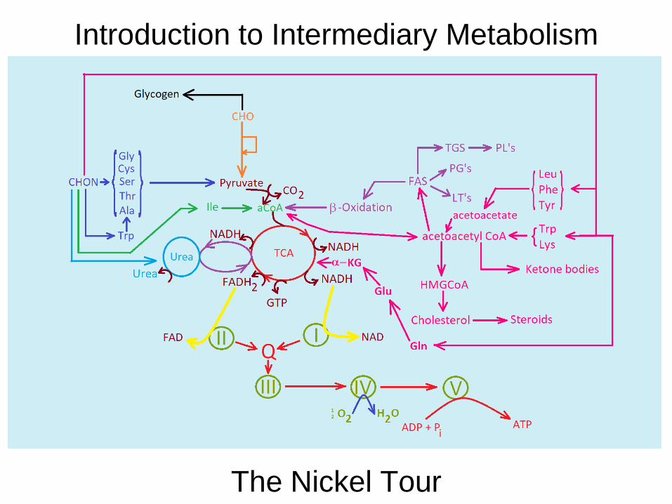
Introduction to Intermediary Metabolism
The Nickel Tour
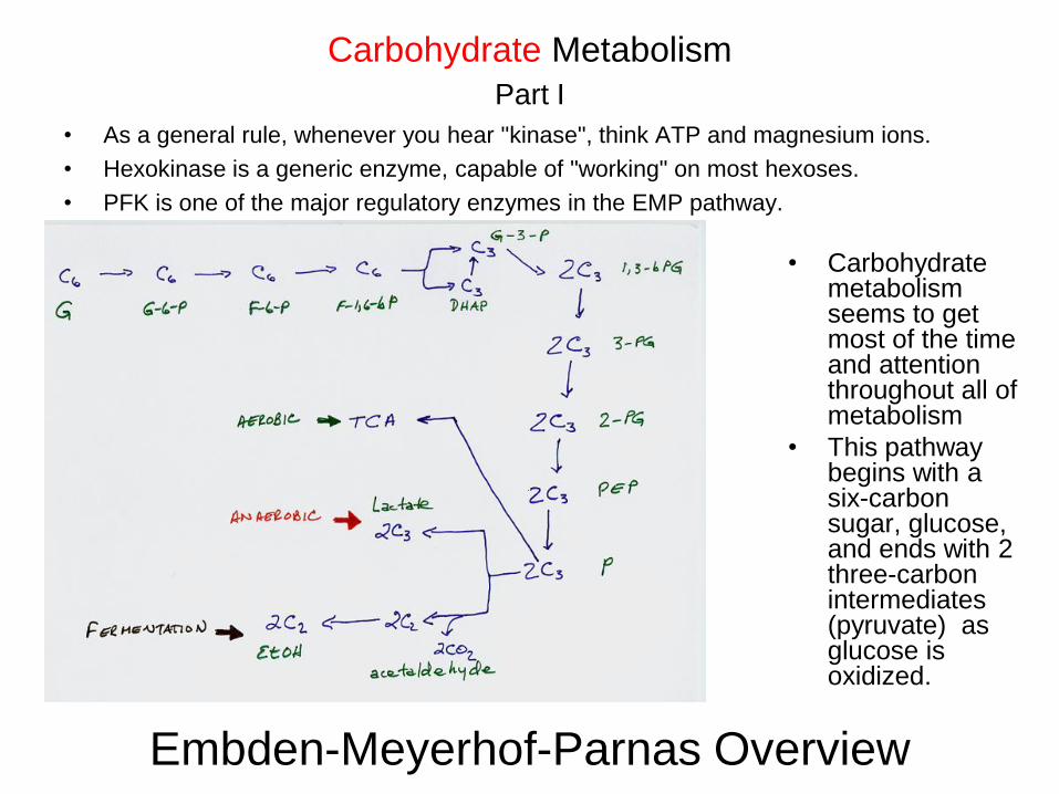
• As a general rule, whenever you hear "kinase", think ATP and magnesium ions.
• Hexokinase is a generic enzyme, capable of "working" on most hexoses.
• PFK is one of the major regulatory enzymes in the EMP pathway.
Carbohydrate Metabolism
Part I
• Carbohydrate metabolism seems to get most of the time and attention throughout all of metabolism
• This pathway begins with a six-carbon sugar, glucose, and ends with 2 three-carbon intermediates (pyruvate) as glucose is oxidized.
Embden-Meyerhof-Parnas Overview
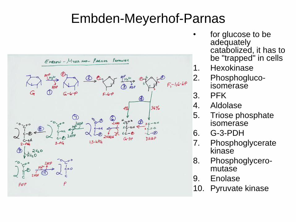
Embden-Meyerhof-Parnas • for glucose to be
adequately catabolized, it has to be "trapped" in cells
1. Hexokinase
2. Phosphogluco-isomerase
3. PFK
4. Aldolase
5. Triose phosphate isomerase
6. G-3-PDH
7. Phosphoglycerate kinase
8. Phosphoglycero-mutase
9. Enolase
10. Pyruvate kinase

2,3-bPG • 2,3-bPG reduces the affinity of Hb for O2 – is a primary
compensatory factor in going to higher altitudes -- Is a side-rxn of EMP
• E.g., if live in San Francisco, to adapt (short term) to life at Lake Tahoe, body increases [2,3-bPG] so more oxygen is released to the cells in the body

Anaerobic/Fermentative Metabolism
• The stoichiometry – aerobic AND anaerobic -- (review your Chem 121) is that per molecule of glucose (1 six-carbon sugar), TWO molecules of three-carbon sugars are formed, i.e., one times six is six, as are two times three.
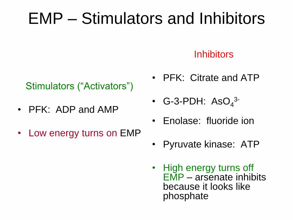
EMP – Stimulators and Inhibitors
Stimulators (“Activators”)
• PFK: ADP and AMP
• Low energy turns on EMP
Inhibitors
• PFK: Citrate and ATP
• G-3-PDH: AsO43-
• Enolase: fluoride ion
• Pyruvate kinase: ATP
• High energy turns off EMP – arsenate inhibits because it looks like phosphate

ATP Summary – Used and Gained --
AEROBIC
ATP Used
• Hexokinase: -1
• PFK: -1
• Total USED = 2 ATP
ATP Gained
• G-3-PDH: +6 ()
• Phosphoglycerate
kinase: +2
• Pyruvate kinase: +2
• Total GAINED: 10
ATP
Overall: 8 ATP produced
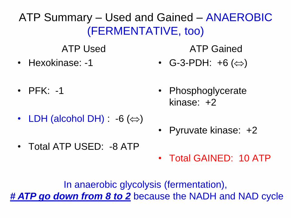
ATP Summary – Used and Gained – ANAEROBIC
(FERMENTATIVE, too)
ATP Used
• Hexokinase: -1
• PFK: -1
• LDH (alcohol DH) : -6 ()
• Total ATP USED: -8 ATP
ATP Gained
• G-3-PDH: +6 ()
• Phosphoglycerate
kinase: +2
• Pyruvate kinase: +2
• Total GAINED: 10 ATP
In anaerobic glycolysis (fermentation),
# ATP go down from 8 to 2 because the NADH and NAD cycle

Fructose Metabolism • Every now and again, a diabetic patient's parent, sibling or other relative reads about how diabetes is a disease of
glucose metabolism.
• On occasion, they read a little more and discover that fructose is a carbohydrate, but not glucose.
• They then come in to see you as the health care person who knows something about diabetes and ask you, "Since fructose isn't glucose, can I substitute all of my relative's carbohydrate needs with fructose?"
• Your answer is, of course, no.
• So, why is it that your answer is no?
• The catabolism of fructose and how it intertwines with triglyceride (TGS) synthesis follows.
1. Hexokinase
(adipose tissue)
2. Fructokinase (liver)
3. F-1-P aldolase
4. Triose kinase
5. Triose phosphate isomerase (TPI)
6. DHAP DH
7. Glycerol phosphate DH
8. Phosphatase
9. Glycerol kinase

Galactose Metabolism • Galactose is an important constituent of lactose,
• Galactose may be used in the synthesis of glycoproteins
• Galactose may be used in the synthesis of glycogen
1. Galactokinase
2. Galactose-1-phosphate uridyl transferase
3. Lactose synthetase (BOTH catalytic unit and specificity protein)
4. Lactose synthetase (catalytic unit only)
5. UDP-galactose-4-epimerase
6. UDP-glucose pyrophosphorylase
7. Glycogen synthetase

Glycogen Metabolism Regulated by
cAMP • Phosphodiesterase (PDE)
inhibited by:
– Theophylline
– Caffeine
– Theobromine
• These compounds are called xanthines.
• When PDE is inactivated, cAMP levels build up, making it easier for patients to breathe (controversy).
• Elevated levels of cAMP drive protein synthesis, enzyme
cascades and changes in membrane permeability
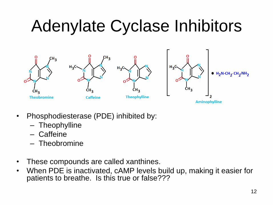
Adenylate Cyclase Inhibitors
• Phosphodiesterase (PDE) inhibited by:
– Theophylline
– Caffeine
– Theobromine
• These compounds are called xanthines.
• When PDE is inactivated, cAMP levels build up, making it easier for patients to breathe. Is this true or false???
12

In General: Glycogen – olysis and genesis
• Glycogenolysis (destruction of glycogen)
• Glycogenesis (production of glycogen)
• As the body doesn't like to be confused during times of
stress, cAMP inhibits glycogenesis and activates
glycogenolysis.
• One example of this occurs when epinephrine binds with
the appropriate receptor on the cell membrane of a
target cell.
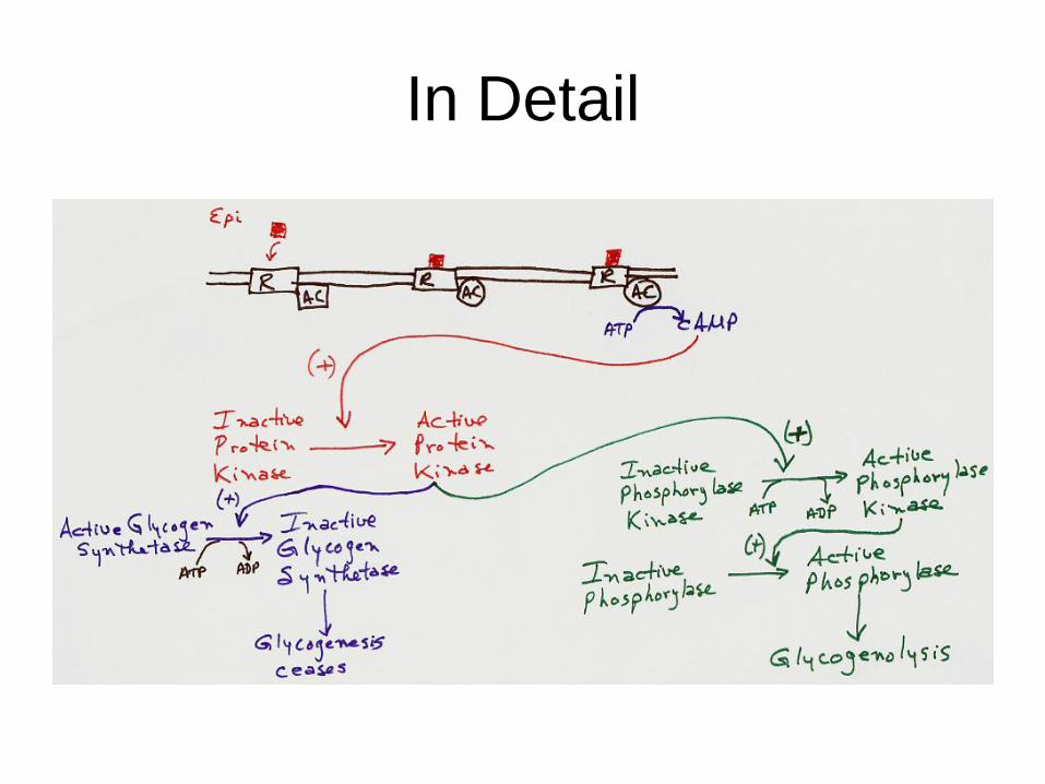
In Detail

G Proteins
15

IP3 drives changes in:
Ca2+ concentration
Ca2+ mobilization
GABA, AVP, ANG,
TSH utilize IP3
Second Messenger: IP3
16
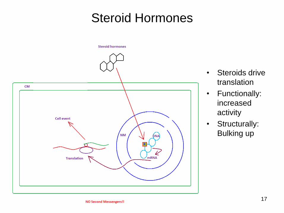
Steroid Hormones
• Steroids drive
translation
• Functionally:
increased
activity
• Structurally:
Bulking up
17
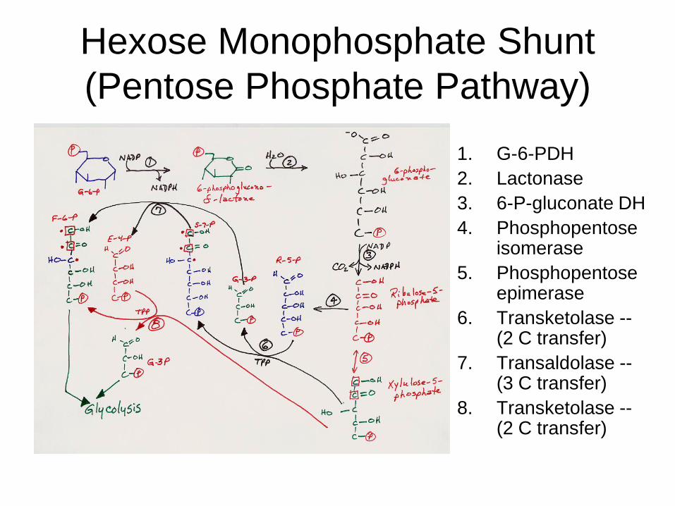
Hexose Monophosphate Shunt
(Pentose Phosphate Pathway)
1. G-6-PDH
2. Lactonase
3. 6-P-gluconate DH
4. Phosphopentose isomerase
5. Phosphopentose epimerase
6. Transketolase -- (2 C transfer)
7. Transaldolase -- (3 C transfer)
8. Transketolase -- (2 C transfer)

• 2 NADPH – reductive power
• Ribose – nucleoside/nucleotide synthesis
• People with G-6-PDH deficiency don’t make enough NADPH to reduce
G-S-S-G and causes health problems
Hexose Monophosphate Shunt (Pentose
Phosphate Pathway): Clinical/Significance
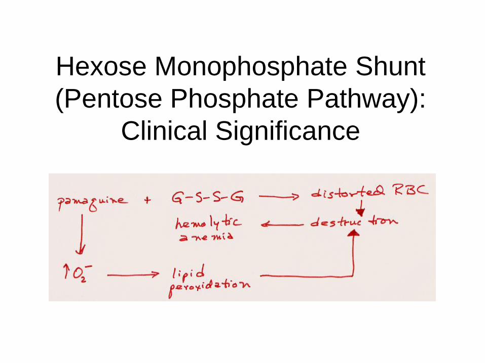
Hexose Monophosphate Shunt
(Pentose Phosphate Pathway):
Clinical Significance

Gluconeogenesis
• Gluconeogenesis is NOT the absolute reverse of glycolysis.
• Some enzymes are the same -- 4 are NOT
• When the body produces new glucose, it utilizes various substrates as necessary.
• These include the carbon skeletons of amino acids and anaerobic end-products of catabolism.
• The carbon skeletons of Gly, Ala, Thr, Ser and Cys feed into gluconeogenesis via pyruvate, as does lactate.
• The carbon skeletons of Asn and Asp feed in to OAA.
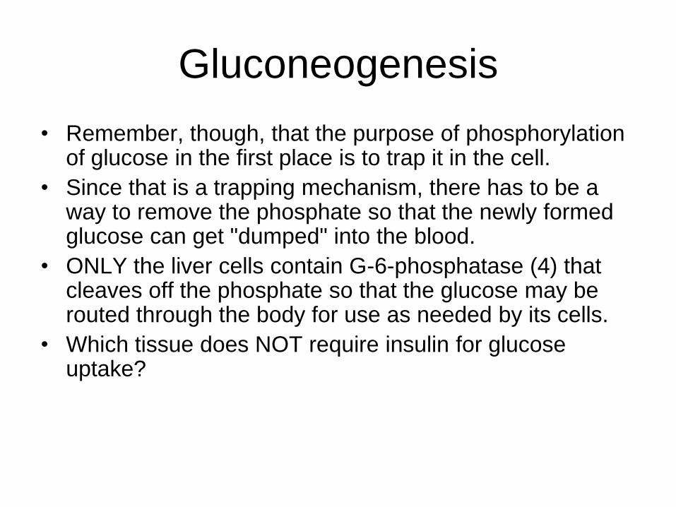
• Remember, though, that the purpose of phosphorylation of glucose in the first place is to trap it in the cell.
• Since that is a trapping mechanism, there has to be a way to remove the phosphate so that the newly formed glucose can get "dumped" into the blood.
• ONLY the liver cells contain G-6-phosphatase (4) that cleaves off the phosphate so that the glucose may be routed through the body for use as needed by its cells.
• Which tissue does NOT require insulin for glucose uptake?
Gluconeogenesis
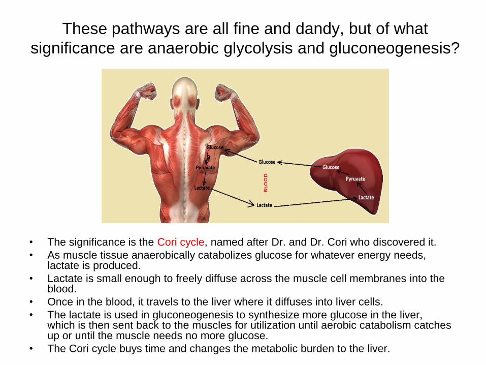
These pathways are all fine and dandy, but of what
significance are anaerobic glycolysis and gluconeogenesis?
• The significance is the Cori cycle, named after Dr. and Dr. Cori who discovered it.
• As muscle tissue anaerobically catabolizes glucose for whatever energy needs, lactate is produced.
• Lactate is small enough to freely diffuse across the muscle cell membranes into the blood.
• Once in the blood, it travels to the liver where it diffuses into liver cells.
• The lactate is used in gluconeogenesis to synthesize more glucose in the liver, which is then sent back to the muscles for utilization until aerobic catabolism catches up or until the muscle needs no more glucose.
• The Cori cycle buys time and changes the metabolic burden to the liver.
Aerobic Energy Sources from Intermediary Metabolism
• There are three systems that provide energy to cells:
– System 1: Phosphagen System
– System 2: Creatine Phosphate System
– System 3: The Krebs’ Cycle (TCA; Citric Acid Cycle)
• The first system is the phosphagen system. In this system, the source of the energy is ATP. During muscular contraction, ATP is hydrolyzed to ADP, Pi and energy. When this happens, there is only enough energy for 5-6 seconds.
• So how do our cells get additional energy?
• Our cells get it via a compound called phosphocreatine (PCr or CrP; System 2). The concentration of PCr is about 2-3 times greater than the concentration of ATP. When PCr is available, it is hydrolyzed to Cr and Pi and energy. The Pi is used to re-phosphorylate ADP to make more ATP.
• This gives us about 15 seconds of maximal contractions and is used for short bursts.
• As long as the system (cell and/or tissues and/or body) remains in an aerobic state and fuel is present, the TCA (System 3) will continue to provide energy to the cells. 24
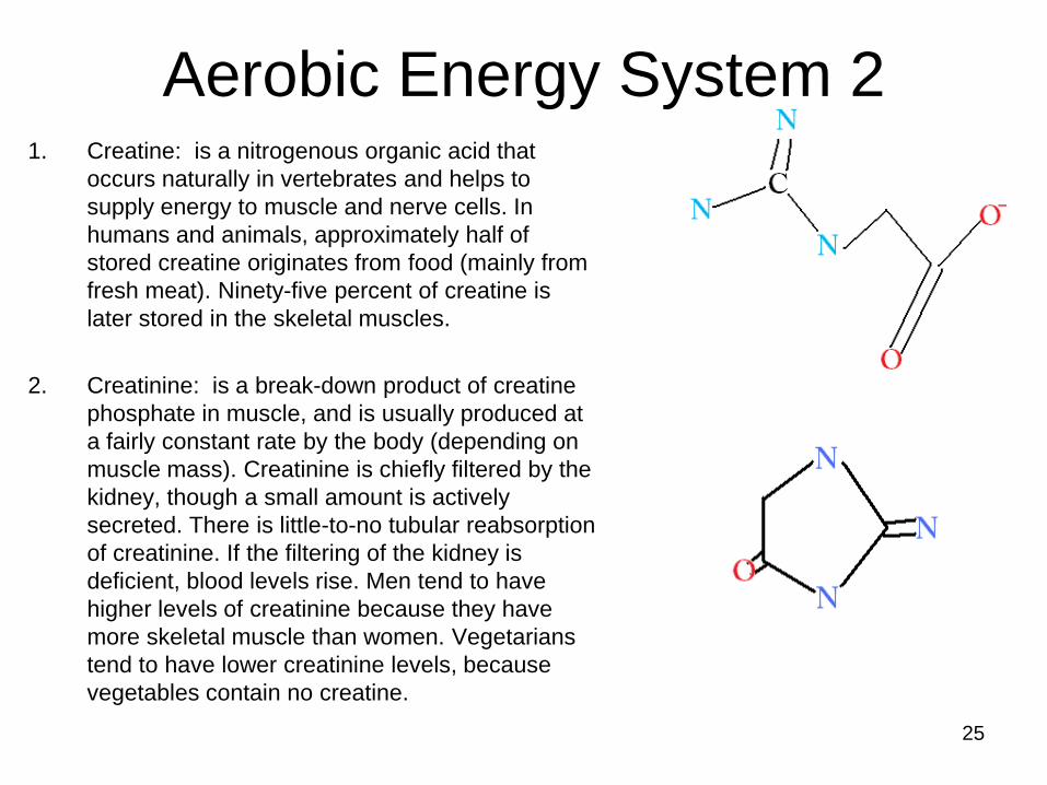
1. Creatine: is a nitrogenous organic acid that
occurs naturally in vertebrates and helps to
supply energy to muscle and nerve cells. In
humans and animals, approximately half of
stored creatine originates from food (mainly from
fresh meat). Ninety-five percent of creatine is
later stored in the skeletal muscles.
2. Creatinine: is a break-down product of creatine
phosphate in muscle, and is usually produced at
a fairly constant rate by the body (depending on
muscle mass). Creatinine is chiefly filtered by the
kidney, though a small amount is actively
secreted. There is little-to-no tubular reabsorption
of creatinine. If the filtering of the kidney is
deficient, blood levels rise. Men tend to have
higher levels of creatinine because they have
more skeletal muscle than women. Vegetarians
tend to have lower creatinine levels, because
vegetables contain no creatine.
25
Aerobic Energy System 2
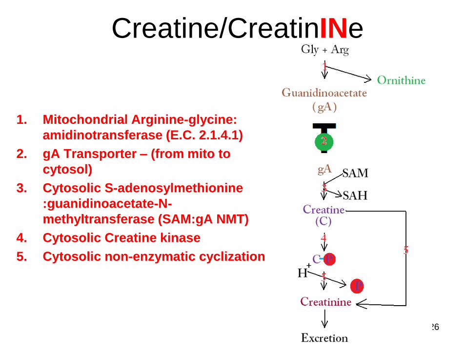
Creatine/CreatinINe
26
1. Mitochondrial Arginine-glycine:
amidinotransferase (E.C. 2.1.4.1)
2. gA Transporter – (from mito to
cytosol)
3. Cytosolic S-adenosylmethionine
:guanidinoacetate-N-
methyltransferase (SAM:gA NMT)
4. Cytosolic Creatine kinase
5. Cytosolic non-enzymatic cyclization
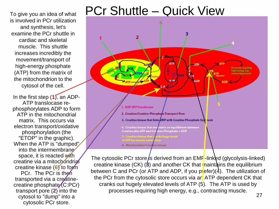
PCr Shuttle – Quick View To give you an idea of what
is involved in PCr utilization
and synthesis, let's
examine the PCr shuttle in
cardiac and skeletal
muscle. This shuttle
increases incredibly the
movement/transport of
high-energy phosphate
(ATP) from the matrix of
the mitochondrion to the
cytosol of the cell.
27
In the first step (1), an ADP-ATP translocase re-
phosphorylates ADP to form ATP in the mitochondrial matrix. This occurs via
electron transport/oxidative phosphorylation (the
"ETOP" in the graphic). When the ATP is "dumped"
into the intermembrane space, it is reacted with
creatine via a mitochondrial creatine kinase (6) to form
PCr. The PCr is then transported via a creatine-creatine phosphate (C:PCr) transport pore (2) into the cytosol to "dump" into a
cytosolic PCr store.
The cytosolic PCr store is derived from an EMP-linked (glycolysis-linked)
creatine kinase (CK) (3) and another CK that maintains the equilibrium
between C and PCr (or ATP and ADP, if you prefer)(4). The utilization of
the PCr from the cytosolic store occurs via an ATP dependent CK that
cranks out hugely elevated levels of ATP (5). The ATP is used by
processes requiring high energy, e.g., contracting muscle.
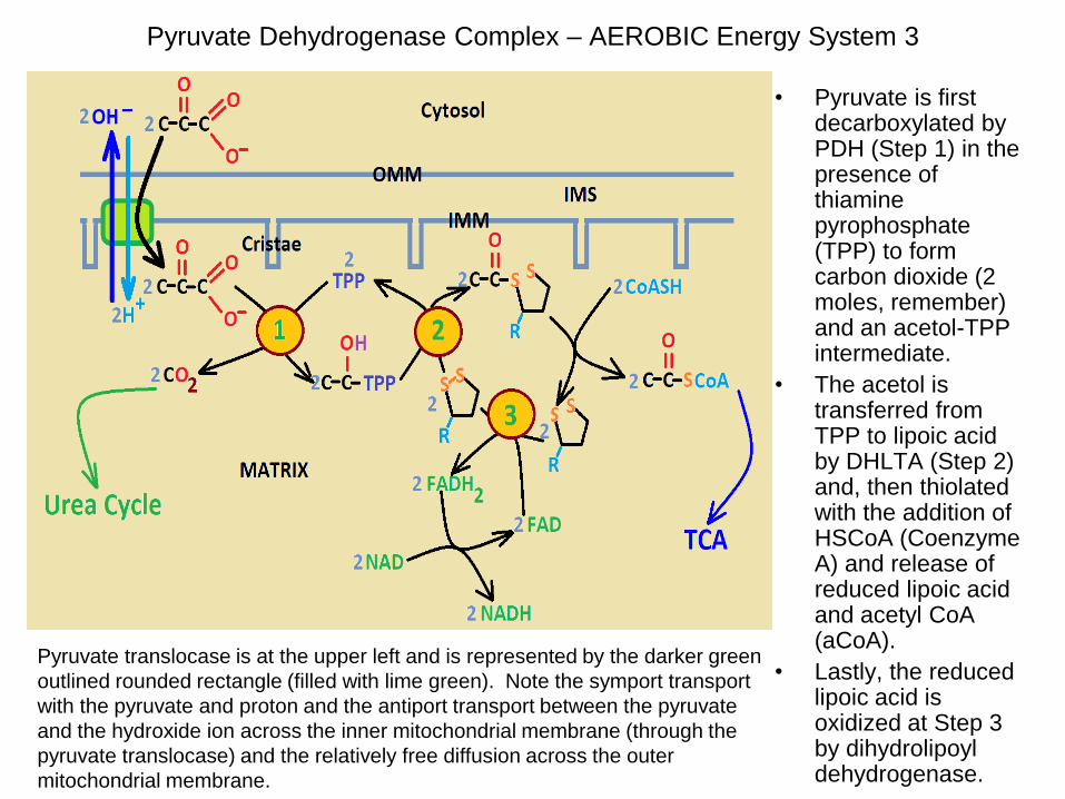
Pyruvate Dehydrogenase Complex – AEROBIC Energy System 3
• Pyruvate is first decarboxylated by PDH (Step 1) in the presence of thiamine pyrophosphate (TPP) to form carbon dioxide (2 moles, remember) and an acetol-TPP intermediate.
• The acetol is transferred from TPP to lipoic acid by DHLTA (Step 2) and, then thiolated with the addition of HSCoA (Coenzyme A) and release of reduced lipoic acid and acetyl CoA (aCoA).
• Lastly, the reduced lipoic acid is oxidized at Step 3 by dihydrolipoyl dehydrogenase.
Pyruvate translocase is at the upper left and is represented by the darker green
outlined rounded rectangle (filled with lime green). Note the symport transport
with the pyruvate and proton and the antiport transport between the pyruvate
and the hydroxide ion across the inner mitochondrial membrane (through the
pyruvate translocase) and the relatively free diffusion across the outer
mitochondrial membrane.
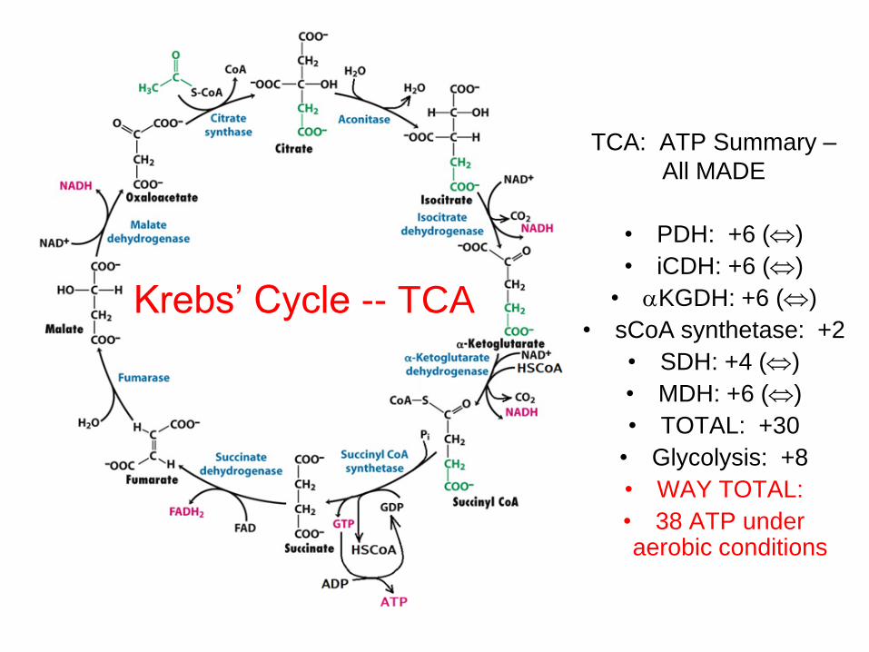
Krebs’ Cycle -- TCA
• PDH: +6 ()
• iCDH: +6 ()
• KGDH: +6 ()
• sCoA synthetase: +2
• SDH: +4 ()
• MDH: +6 ()
• TOTAL: +30
• Glycolysis: +8
• WAY TOTAL:
• 38 ATP under aerobic conditions
TCA: ATP Summary –
All MADE
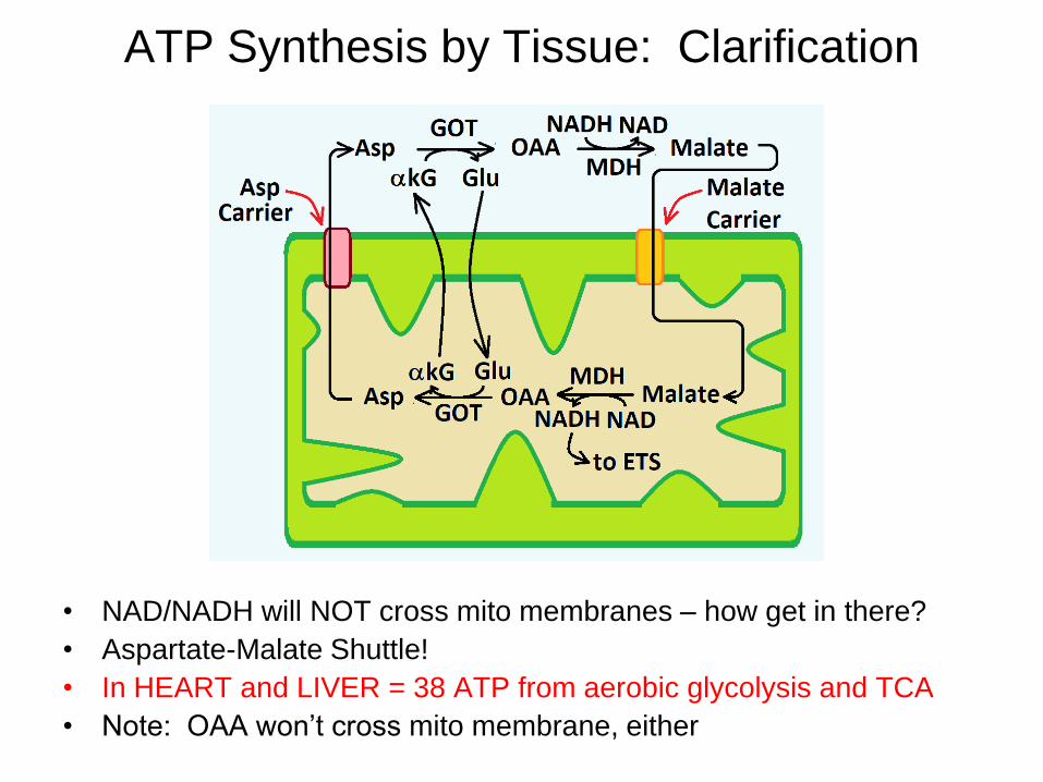
ATP Synthesis by Tissue: Clarification
• NAD/NADH will NOT cross mito membranes – how get in there?
• Aspartate-Malate Shuttle!
• In HEART and LIVER = 38 ATP from aerobic glycolysis and TCA
• Note: OAA won’t cross mito membrane, either
ATP Synthesis by Tissue: Clarification
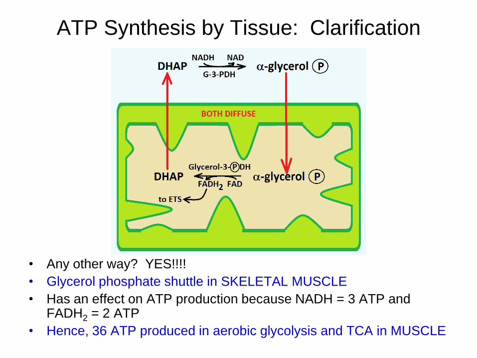
• Any other way? YES!!!!
• Glycerol phosphate shuttle in SKELETAL MUSCLE
• Has an effect on ATP production because NADH = 3 ATP and FADH2 = 2 ATP
• Hence, 36 ATP produced in aerobic glycolysis and TCA in MUSCLE
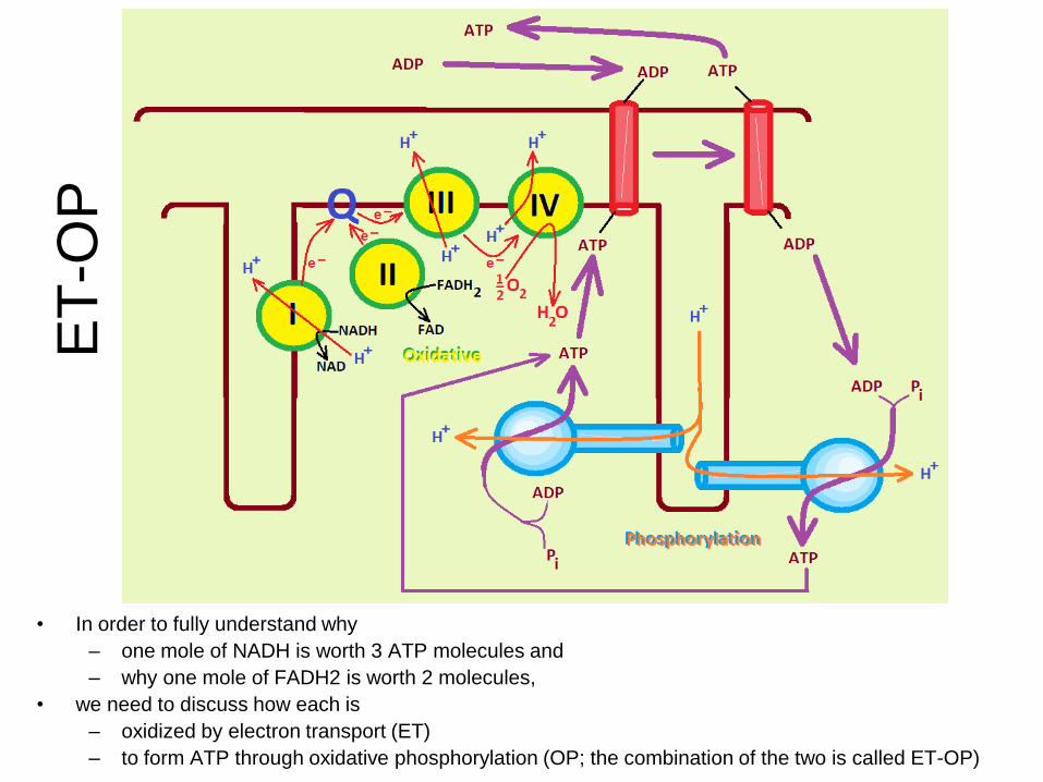
• In order to fully understand why
– one mole of NADH is worth 3 ATP molecules and
– why one mole of FADH2 is worth 2 molecules,
• we need to discuss how each is
– oxidized by electron transport (ET)
– to form ATP through oxidative phosphorylation (OP; the combination of the two is called ET-OP)
ET
-OP

Proton Pump Functioning 1. H+ moving out
of matrix at I, III, IV
2. pH gradient established – Fo opens and H+ run through to drive ATP’ase
3. [H+] in IMS decreasing to point of maximal flow into matrix
4. H+ being “recycled” and re-starting cycle – Fo fully closed
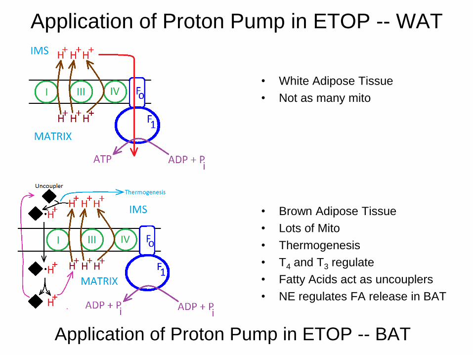
Application of Proton Pump in ETOP -- WAT
• White Adipose Tissue
• Not as many mito
Application of Proton Pump in ETOP -- BAT
• Brown Adipose Tissue
• Lots of Mito
• Thermogenesis
• T4 and T3 regulate
• Fatty Acids act as uncouplers
• NE regulates FA release in BAT

ET-OP Inhibition
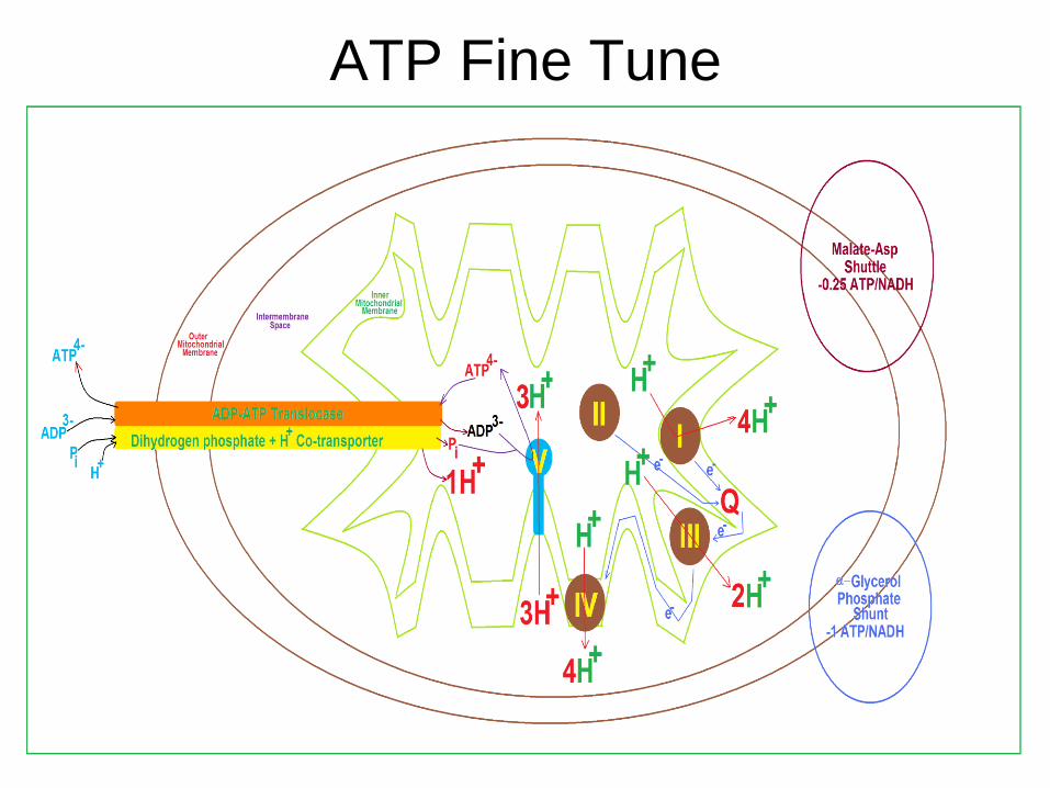
ATP Fine Tune
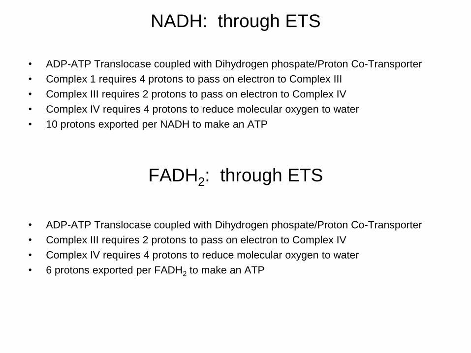
NADH: through ETS
• ADP-ATP Translocase coupled with Dihydrogen phospate/Proton Co-Transporter
• Complex 1 requires 4 protons to pass on electron to Complex III
• Complex III requires 2 protons to pass on electron to Complex IV
• Complex IV requires 4 protons to reduce molecular oxygen to water
• 10 protons exported per NADH to make an ATP
FADH2: through ETS
• ADP-ATP Translocase coupled with Dihydrogen phospate/Proton Co-Transporter
• Complex III requires 2 protons to pass on electron to Complex IV
• Complex IV requires 4 protons to reduce molecular oxygen to water
• 6 protons exported per FADH2 to make an ATP

Complex V and Co-Transporter
• 3 protons required to turn on Fo
• 1 proton co-transported with Pi
• 4 protons imported to synthesize 1 ATP
molecule
NADH
ATP
ATP
protons
NADH
protons
NADH
ATP5.2
4
10
1
#
2
2
2
5.14
6
1
#
FADH
ATP
ATP
protons
FADH
protons
FADH
ATP
Currently Accepted Stoichiometry
Suggested Source: http://www.life.uiuc.edu/crofts/bioph354/ATP_2e_ratios.html
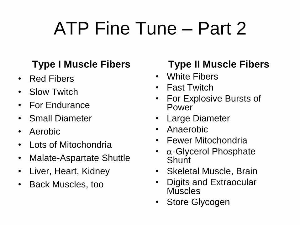
ATP Fine Tune – Part 2
Type I Muscle Fibers
• Red Fibers
• Slow Twitch
• For Endurance
• Small Diameter
• Aerobic
• Lots of Mitochondria
• Malate-Aspartate Shuttle
• Liver, Heart, Kidney
• Back Muscles, too
Type II Muscle Fibers
• White Fibers
• Fast Twitch
• For Explosive Bursts of Power
• Large Diameter
• Anaerobic
• Fewer Mitochondria
• -Glycerol Phosphate Shunt
• Skeletal Muscle, Brain
• Digits and Extraocular Muscles
• Store Glycogen
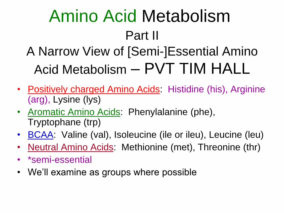
A Narrow View of [Semi-]Essential Amino
Acid Metabolism – PVT TIM HALL
• Positively charged Amino Acids: Histidine (his), Arginine (arg), Lysine (lys)
• Aromatic Amino Acids: Phenylalanine (phe), Tryptophane (trp)
• BCAA: Valine (val), Isoleucine (ile or ileu), Leucine (leu)
• Neutral Amino Acids: Methionine (met), Threonine (thr)
• *semi-essential
• We’ll examine as groups where possible
Amino Acid Metabolism Part II
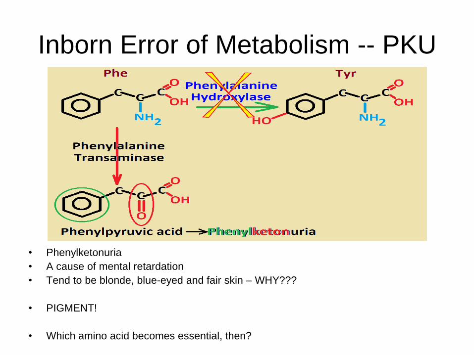
Inborn Error of Metabolism -- PKU
• Phenylketonuria
• A cause of mental retardation
• Tend to be blonde, blue-eyed and fair skin – WHY???
• PIGMENT!
• Which amino acid becomes essential, then?
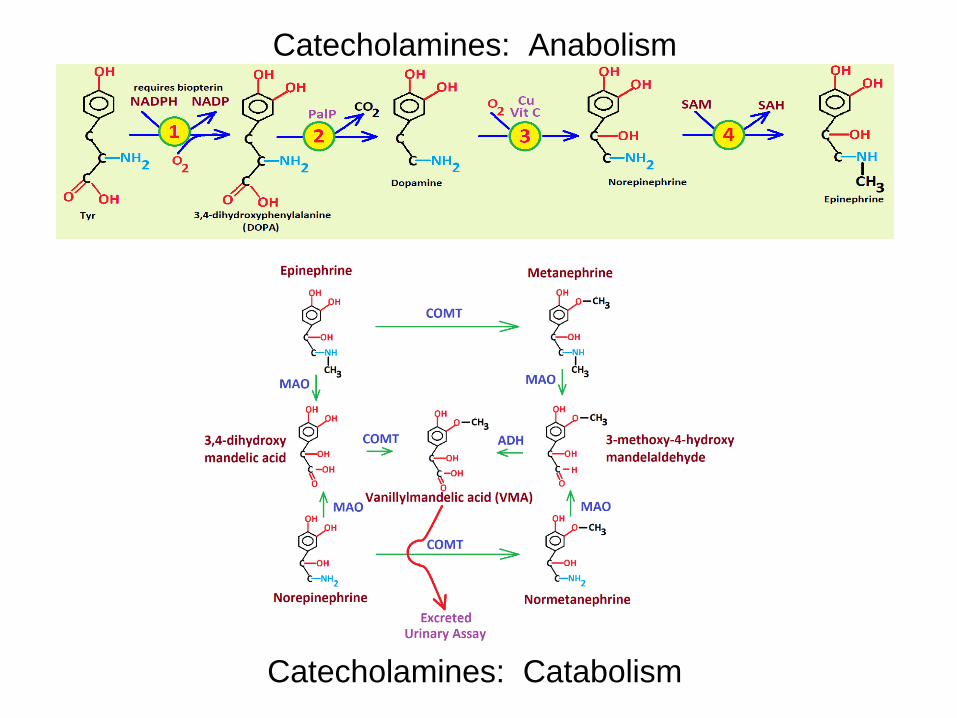
Catecholamines: Anabolism
Catecholamines: Catabolism
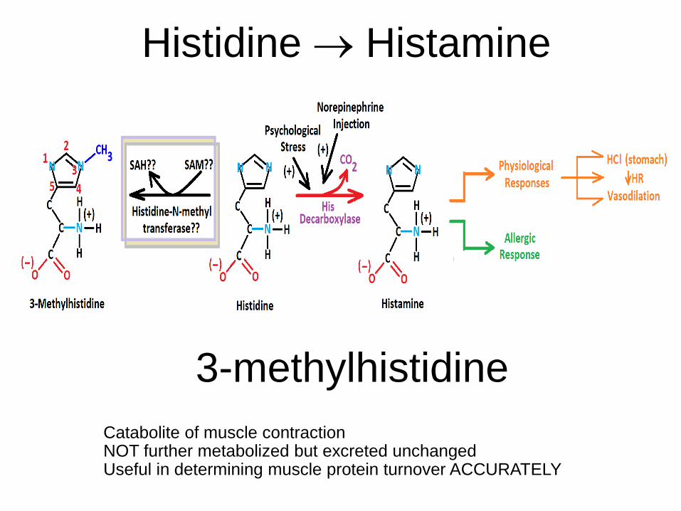
Histidine Histamine
3-methylhistidine
Catabolite of muscle contraction NOT further metabolized but excreted unchanged Useful in determining muscle protein turnover ACCURATELY
Significance of Lysine
• Provides crosslinking in collagen
• Provides crosslinking in elastin
• When crosslinking is inhibited – in severe Cu deficiency,
– after ingesting sweet pea toxin [-aminopropionitrile]
• This causes lathyrism
• There is increased solubility of the collagen
• Increased rigidity of elastin
• Both cause death – generally due to aortic rupture from a lack of elasticity at the aortic root – an “aortic blow out” or aneurism

Arginine and The Urea Cycle

46
N-acetylglutamate is an obligatory “on switch” for CPS-I.
Note relationships between Urea cycle, TCA, Creatine Synthesis
and Asp-Malate Shuttle.
Inside black lines is mito matrix
Outside black lines is cytosol
Creatine, Urea, TCA and Malate-Aspartate Shuttle Interconnections
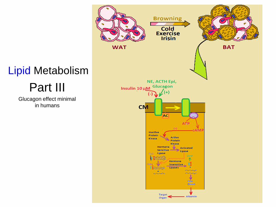
Lipid Metabolism
Part III Glucagon effect minimal
in humans

48
-O
xid
atio
n o
f
Fatt
y A
cid
s
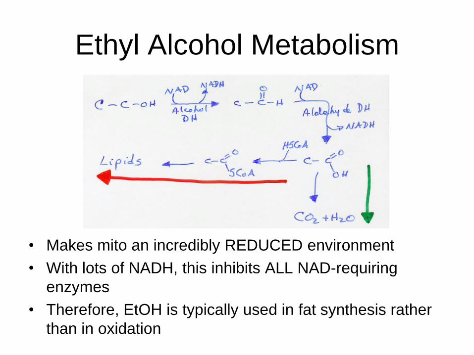
Ethyl Alcohol Metabolism
• Makes mito an incredibly REDUCED environment
• With lots of NADH, this inhibits ALL NAD-requiring
enzymes
• Therefore, EtOH is typically used in fat synthesis rather
than in oxidation
Fa
tty A
cid
Syn
the
sis

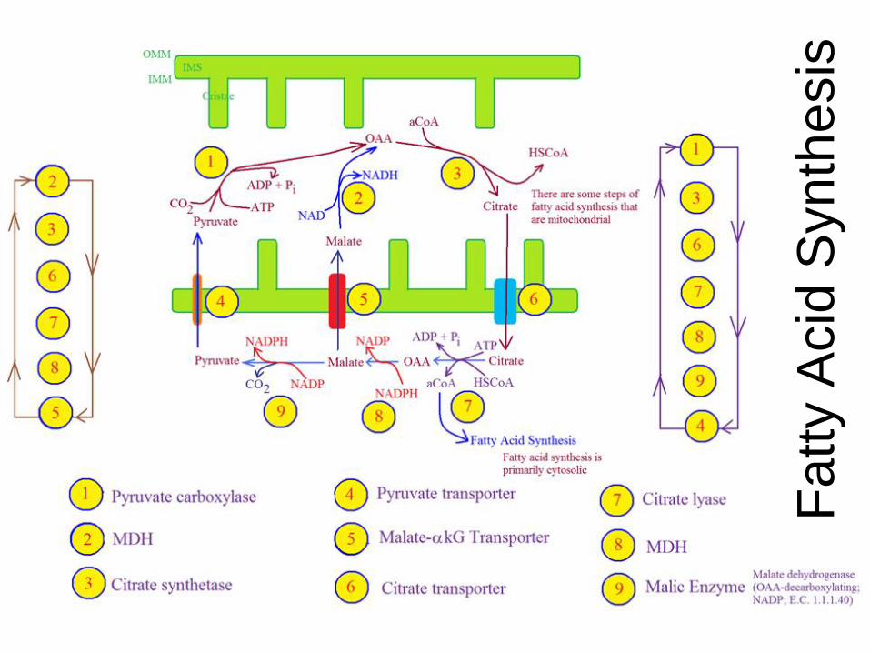
51

52
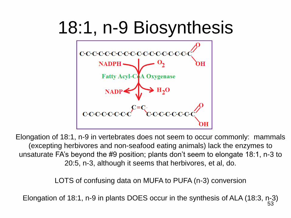
18:1, n-9 Biosynthesis
53
Elongation of 18:1, n-9 in vertebrates does not seem to occur commonly: mammals
(excepting herbivores and non-seafood eating animals) lack the enzymes to
unsaturate FA’s beyond the #9 position; plants don’t seem to elongate 18:1, n-3 to
20:5, n-3, although it seems that herbivores, et al, do.
LOTS of confusing data on MUFA to PUFA (n-3) conversion
Elongation of 18:1, n-9 in plants DOES occur in the synthesis of ALA (18:3, n-3)

Acetyl CoA Cholesterol Biosynthesis

HMGCoA Reductase and Inhibitors
(-hydroxy--methyl glutaryl coenzyme A)
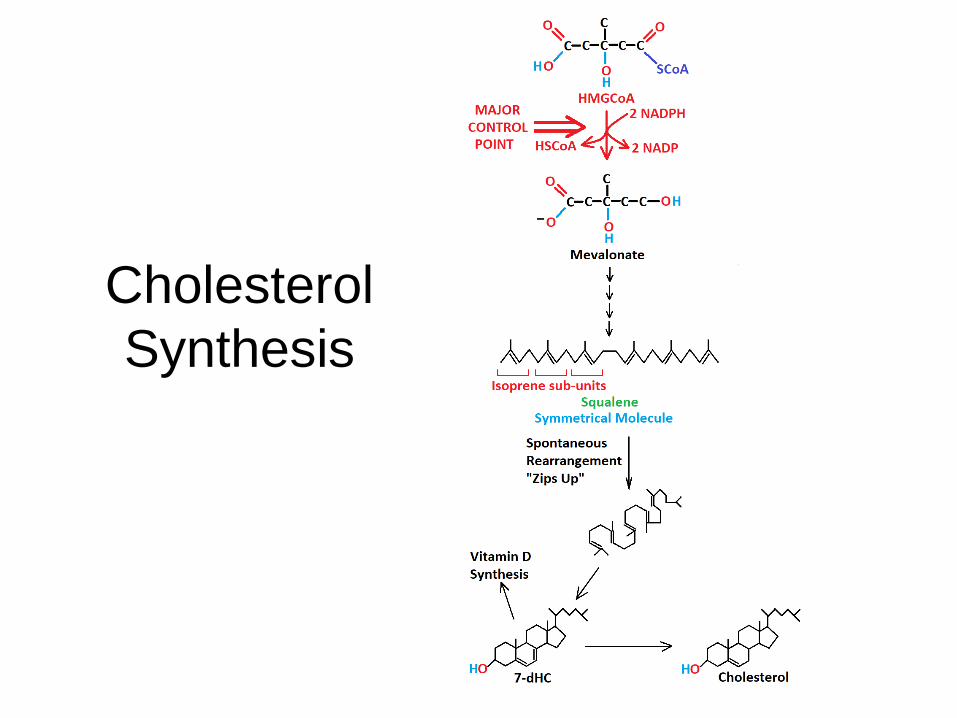
Cholesterol
Synthesis

57
Lecithin-Cholesterol Acyl Transferase: “LCAT”
• Used in lipid TRANSPORT
cholesteryl ester biosynthesis; esters then transported into the core of a developing lipoprotein;
makes HDL spherical; bound to HDL’s and LDL’s in the blood.
58
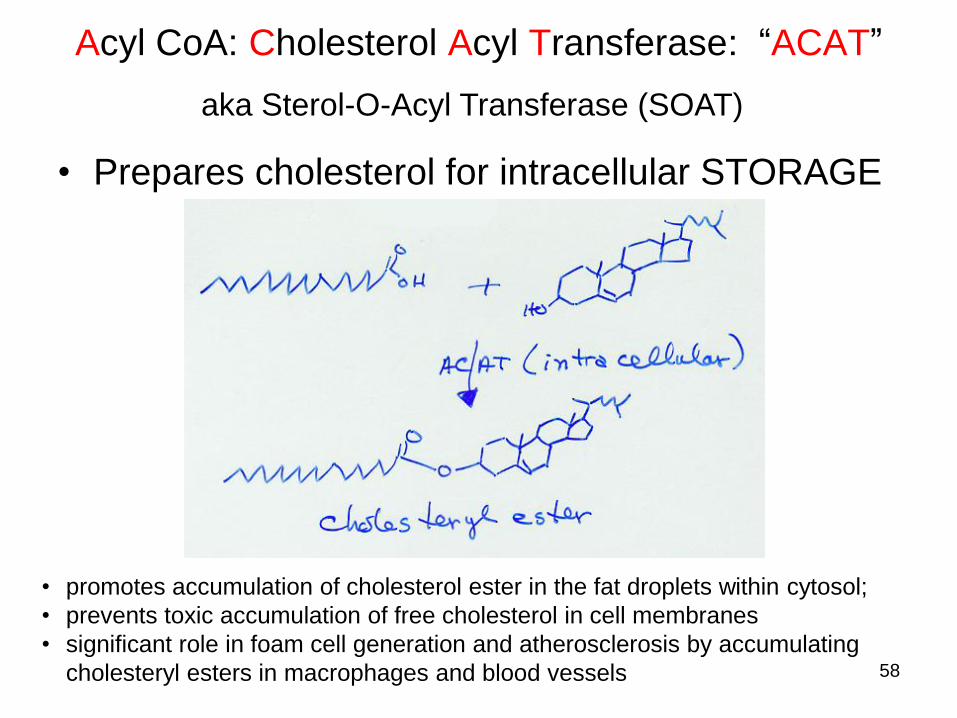
Acyl CoA: Cholesterol Acyl Transferase: “ACAT”
• Prepares cholesterol for intracellular STORAGE
aka Sterol-O-Acyl Transferase (SOAT)
• promotes accumulation of cholesterol ester in the fat droplets within cytosol;
• prevents toxic accumulation of free cholesterol in cell membranes
• significant role in foam cell generation and atherosclerosis by accumulating
cholesteryl esters in macrophages and blood vessels

Prostaglandins
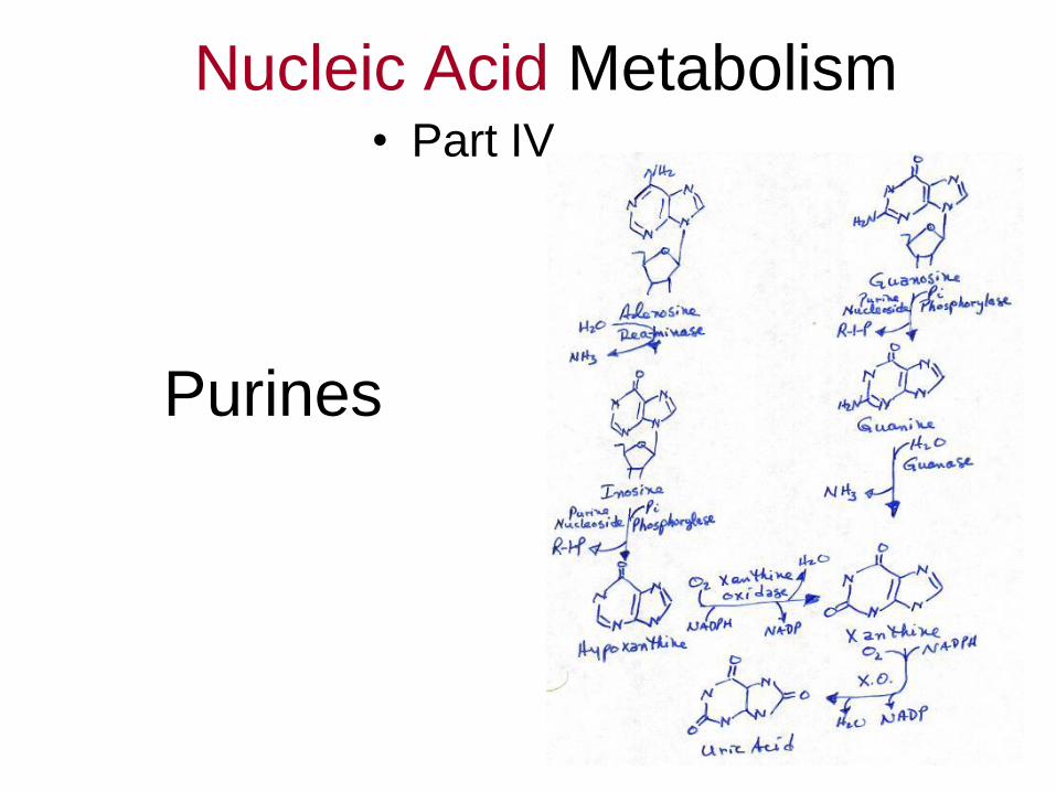
Purines
Nucleic Acid Metabolism • Part IV
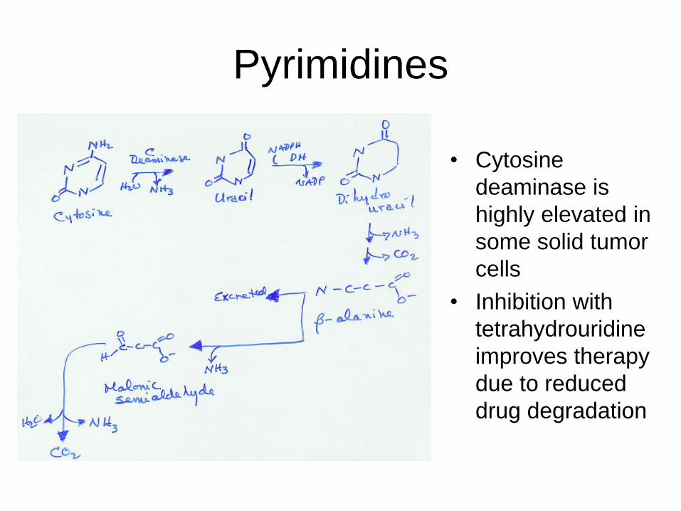
Pyrimidines
• Cytosine
deaminase is
highly elevated in
some solid tumor
cells
• Inhibition with
tetrahydrouridine
improves therapy
due to reduced
drug degradation

More
Pyrimidines

Integration of Metabolism SSDD
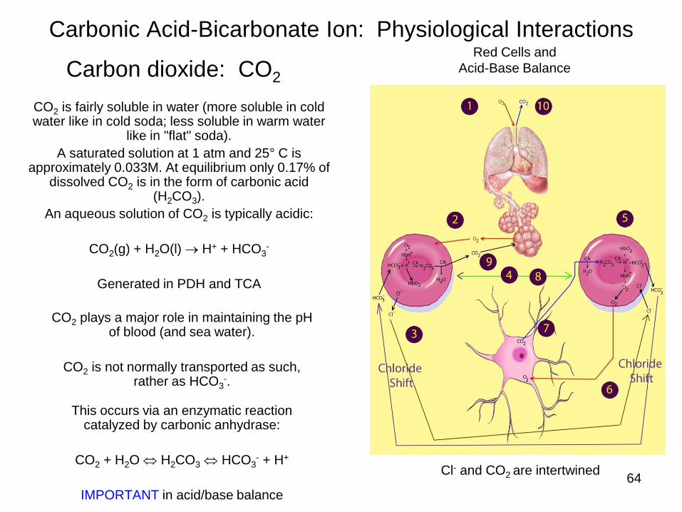
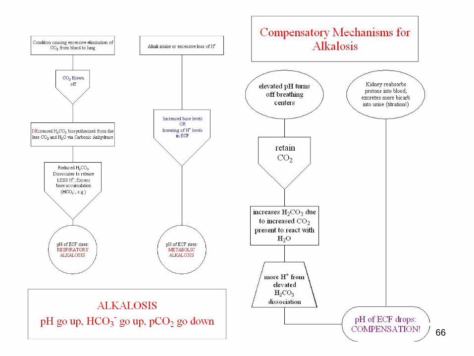
Carbonic Acid-Bicarbonate Ion: Physiological Interactions
64
Carbon dioxide: CO2
CO2 is fairly soluble in water (more soluble in cold water like in cold soda; less soluble in warm water
like in "flat" soda).
A saturated solution at 1 atm and 25° C is approximately 0.033M. At equilibrium only 0.17% of
dissolved CO2 is in the form of carbonic acid (H2CO3).
An aqueous solution of CO2 is typically acidic:
CO2(g) + H2O(l) H+ + HCO3-
Generated in PDH and TCA
CO2 plays a major role in maintaining the pH of blood (and sea water).
CO2 is not normally transported as such, rather as HCO3
-.
This occurs via an enzymatic reaction catalyzed by carbonic anhydrase:
CO2 + H2O H2CO3 HCO3- + H+
IMPORTANT in acid/base balance
Red Cells and
Acid-Base Balance
Cl- and CO2 are intertwined
65
66