interlocking horizontal mattress suture versus … article interlocking horizontal mattress suture...
TRANSCRIPT
ORIGINAL ARTICLE
Interlocking horizontal mattress suture versus Kakiuchi techniquein repair of Achilles tendon rupture: a biomechanical study
Matteo Guzzini1 • Riccardo Maria Lanzetti1 • Lorenzo Proietti1 • Daniele Mazza1 •
Mattia Fabbri1 • Edoardo Monaco1 • Germano Ferri1 • Andrea Ferretti1
Received: 26 June 2016 / Accepted: 1 March 2017 / Published online: 15 March 2017
� The Author(s) 2017. This article is published with open access at Springerlink.com
Abstract
Background In recent years, the type of surgical treatment
for Achilles tendon rupture has been the subject of con-
troversial debate. This biomechanical study evaluates for
the first time in literature the ultimate failure load (UFL) of
interlocking horizontal mattress (IHM) suture as compared
with Kakiuchi suture in Achilles tendon rupture. The
hypothesis is that IHM suture can be performed also for
Achilles tendon rupture and ensures higher resistance
compared with the traditional Kakiuchi suture.
Materials and methods Twenty fresh bovine Achilles
tendons were obtained. Ten preparations were randomly
assigned to each of two different groups: group A (10
specimens) sutured by IHM technique, and group B (10
specimens) sutured by Kakiuchi technique. Each construct
was mounted and fixed on a tensile testing machine. Static
preconditioning of 50 N was applied for 5 min as initial
tensioning to stabilize the mechanical properties of the
graft, then a load to failure test was performed at crosshead
speed of 500 mm/min.
Results Ten specimens were tested for each group. The
mean UFL was 228.6 ± 98.6 N in the IHM suture group
and 96.57 ± 80.1 N in the Kakiuchi suture group. Statis-
tical analysis showed a significant difference (p\ 0.05)
with better UFL in the IHM group. In both groups, the
failure mode registered in each specimen was suture
breakage (rupture of suture thread).
Conclusions IHM suture achieved better UFL compared
with Kakiuchi suture in an animal model of Achilles
tendon repair. These results seem to support IHM as a valid
option in Achilles tendon rupture.
Keywords Achilles tendon repair � Interlocking horizontal
mattress � Kakiuchi � Biomechanical study
Introduction
Achilles tendon injury is a common musculoskeletal dis-
order that is increasing in frequency, affecting between 5.5
and 9.9 per 100,000 individuals each year in North
America [14, 21]. To date, the type of surgical treatment
for Achilles tendon rupture has been the subject of con-
troversial debate, and the ideal form of treatment for
Achilles tendon rupture remains controversial [23]. For
many years, nonoperative treatment was the gold standard,
but recent studies have shown that surgical treatment offers
advantages over nonsurgical treatment in terms of clinical
outcomes and recurrence rate [15]. One of the most
important factors in modern surgery for Achilles tendon
rupture is the strength of the suture, which is vital to reduce
the number of re-ruptures and achieve early rehabilitation.
The re-rupture rate is currently about 5 % among those
treated with open surgical suture [3, 12, 24]. Early weight-
bearing and strengthening exercises are increasingly being
used to accelerate recovery [10, 13]. Therefore, strong
repair should resist distraction, allowing early rehabilita-
tion and promoting tendon healing [8, 17].
Early mobilization following Achilles tendon repair has
been reported to be beneficial in terms of postoperative
recovery and improved tendon vascularity [2, 11, 20, 25].
Clinically, patients with early mobilization after Achilles
tendon repair have been shown to have shorter rehabilita-
tion time and return-to-sport time [10, 28].
& Lorenzo Proietti
1 Azienda Ospedaliera Sant’Andrea Via di Grottarossa,
1035/1039, 00189 Rome, Italy
123
J Orthop Traumatol (2017) 18:251–257
DOI 10.1007/s10195-017-0455-x
Determining the best suture in terms of strength is one of
the most important challenges in this surgery. In this study,
it was decided to compare interlocking horizontal mattress
(IHM) versus Kakiuchi suture instead of standard tech-
niques such as Kessler, Bunnell or Krackow suture
because, nowadays, according to literature [9, 19, 22],
Kakiuchi suture is one of the sutures offering greater
guarantees in terms of stiffness and tightness in Achilles
tendon rupture (Fig. 1).
Interlocking horizontal mattress suture is the suture
giving the best clinical results in literature for hand flexor
repair [4]. The IHM suture technique is based on the
standard surgical horizontal mattress suture design. With a
regular forehand needle insertion, this technique
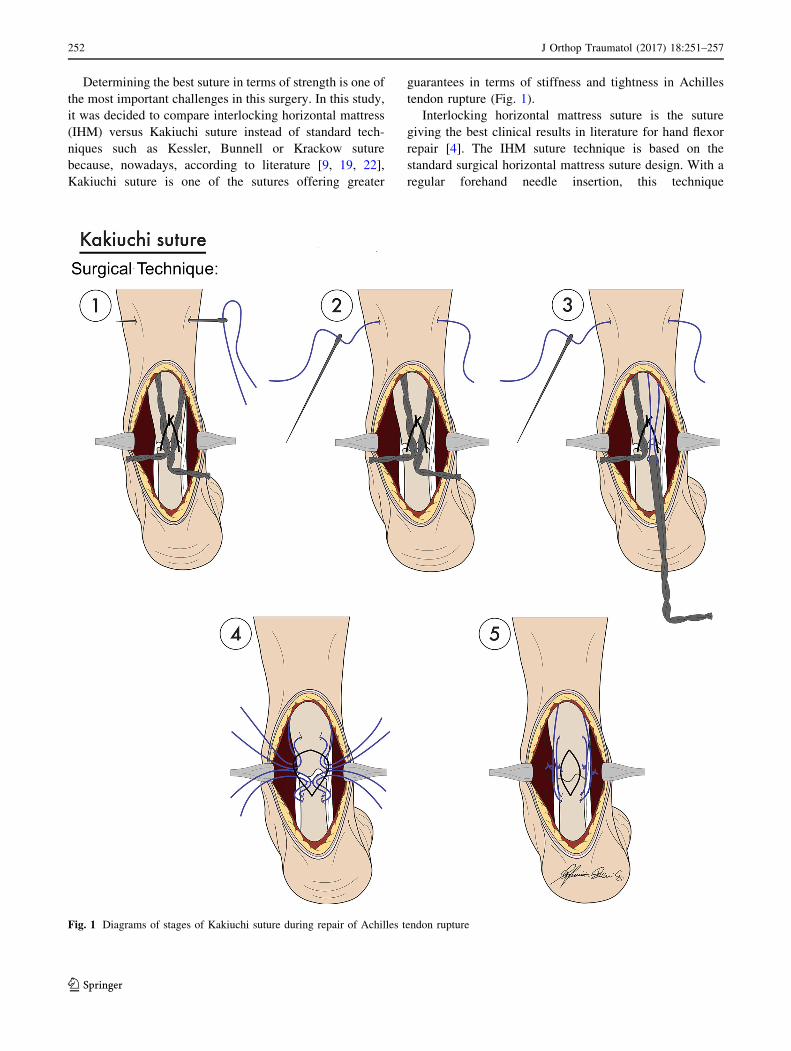
Fig. 1 Diagrams of stages of Kakiuchi suture during repair of Achilles tendon rupture
252 J Orthop Traumatol (2017) 18:251–257
123

commences on the far side of the repair and proceeds
toward the surgeon. This method results in a suture pattern
that has its strands running more longitudinally than the
oblique patterns of both the cross-stitch and interlocking
cross-stitch [5] (Fig. 2).
The aim of the authors is to perform IHM suture in
Achilles tendon rupture, a technique never described before
in literature.
The aim of this study is to evaluate the biomechanical
properties in terms of the ultimate failure load (UFL) of
the IHM suture versus the Kakiuchi suture for repair of
Achilles tendon rupture. Among the techniques described
over the years, there is no literature concerning IHM
suture in Achilles tendon rupture. The presented biome-
chanical study evaluates for the first time in literature the
UFL of IHM suture compared with Kakiuchi suture in
Achilles tendon rupture. The hypothesis is that IHM could
be performed also for Achilles tendon rupture and ensure
higher resistance with respect to traditional Kakiuchi
suture.
Materials and methods
Twenty fresh bovine Achilles tendons were obtained from
the forefeet of 6-month-old animals. They were kept moist
until testing by being wrapped in tissue paper soaked with
Ringer’s solution and stored in sealed polyethylene bags.
For this study, all applicable international, national, and/or
institutional guidelines for the care and use of animals were
followed. The same orthopedic surgeon performed tendon
harvesting and preparation for testing of each specimen.
All tendons were prepared with length of 120 mm, and a
lesion was produced in each tendon by using a scalpel to
create a clean division in the middle (60 mm). The width
and thickness of all tendons were measured and are
Fig. 2 Diagrams of stages of IHM suture during repair of Achilles tendon rupture
J Orthop Traumatol (2017) 18:251–257 253
123
presented in Table 1. The specimens were not statistically
different in terms of width or thickness (p\ 0.05). Two
different suture configurations were tested, using absorb-
able Ethicon PDS II� (polydioxanone) #2 suture (Fig. 2).
Ten preparations were randomly assigned to each of two
different groups: group A (10 specimens) sutured with
IHM technique, and group B (10 specimens) sutured with
Kakiuchi technique. Kakiuchi technique is performed by
passing transversely through the intact tendon a long
straight needle threaded with an absorbable suture, with the
same procedure being performed at two different levels on
both the proximal and distal stumps of the tendon. Proxi-
mally, the suture wire is passed at 0.5 and 1 cm from the
cut tendon edge; distally, the suture wire is passed at 0.5
and 1 cm from the rupture site (Fig. 1). In vivo the ankle is
held at 30� plantar flexion and the knee flexed to 90� whilethe sutures are tied. Direct observation confirms that the
space between the two stumps is eliminated [9]. The IHM
repair technique is performed by running an interlocking
horizontal mattress suture, starting at the far end. The
suture needle passes underneath the prior crossing suture to
lock each throw. When the suture is finished, it is tied at the
near end as shown in Fig. 2. In this technique, suture is
performed at 0.5 cm from the tendon edge. For IHM, there
are six loops for each suture.
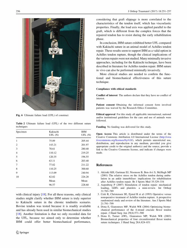
Each construct was mounted and fixed on a tensile
testing machine (model Z010, Zwick-Ruell, Ulm, Ger-
many) using two metal clamps connected to the load cell.
The load was applied parallel to the longitudinal axis of the
specimen to obtain the worst load scenario. The specifically
designed clamps were frozen to avoid graft slippage from
the clamp. Static preconditioning of 50 N was applied for
5 min as initial tensioning to stabilize the mechanical
properties of the graft, then a load to failure test was per-
formed at crosshead speed of 500 mm/min. Data regarding
the ultimate failure load (UFL) of each specimen were
recorded using Textexpert 8.1 software (Zwick-Ruell) and
evaluated using a load–displacement curve. We also
recorded the failure mode for each construct (Figs. 3, 4, 5,
6).
A total sample size of 18 was considered adequate for
overall comparison of the two techniques with respect to
construct measurements, assuming an effect size of 0.25,
alpha-value of 0.05, and beta-value of 0.20 (study power
80 %).
For all variables, normality of data was ascertained by
Kolmogorov–Smirnov test. Differences in construct tensile
properties (UFL) between the two groups were compared
by Mann–Whitney test. All data were analyzed by a single
blinded researcher. The Statistical Package for the Social
Sciences (SPSS) version 22 was used for calculations.
Results
Ten specimens were tested in each group, resulting in a
total of 20 specimens. The results are summarized in
Table 2. The mean UFL was 228.6 ± 98.6 N in the IHM
suture group and 96.5 ± 80.1 N in the Kakiuchi suture
group. Statistical analysis showed a significant difference
(p\ 0.05) with better UFL for the IHM group. In both
groups, the failure mode registered in each specimen was
suture breakage (rupture of suture thread).
Discussion
The most important finding of this study is that IHM suture
exhibited better UFL compared with Kakiuchi suture.
During the early rehabilitation phase, when passive
range-of-movement exercises are started, the forces in the
Achilles tendon of a healthy limb with the ankle at neutral
dorsiflexion range from 70.6 N with the knee in full
extension to 17.8 N with the knee flexed to 50� [17] .Achilles tendon forces at 10� and 20� of ankle dorsi-
flexion range from 183.2 to 83.4 N and from 401.8 to
215.5 N, respectively, with the knee in full extension and
with the knee flexed to 50� [22]. Forces of 190 N are
produced when walking in a cam walker with a 1-inch heel
raise [1]. When walking, these forces are 2.1 times higher
than those exerted by the body weight [6]. The forces on
the Achilles tendon during walking and running exceed the
strength of all repairs; thus, careful postoperative treatment
is imperative [27].
The results presented herein indicate that the IHM
technique applied in our study resulted in sufficient pri-
mary UFL (228.60 N) to withstand the forces experienced
during the early rehabilitation phase, except for passive
Table 1 Width and thickness of constructs
Specimen Kakiuchi IHM
Width, thickness (mm) Width, thickness (mm)
1 1.1, 0.7 1.5, 0.6
2 1.2, 0.6 1.2, 0.8
3 1.6, 0.8 1.4, 0.6
4 1.5, 0.8 1.2, 0.7
5 1.1, 0.9 1.4, 0.7
6 1.2, 1.1 1.2, 0.9
7 1.4, 0.7 1.1, 0.9
8 1.2, 0.9 1.4, 0.8
9 1.3, 0.7 1.3, 0.7
10 1.3, 0.8 1.5, 0.7
Mean Mean
1.29, 0.79 1.32, 0.74
254 J Orthop Traumatol (2017) 18:251–257
123
exercises with 20� of ankle dorsiflexion and knee in full
extension (401.8 N).
Another important finding is that we registered a
homogeneous mode of failure consisting in suture break-
age. No suture slippage across tendon was found, thus we
can speculate that both sutures distribute tension across the
tendon, avoiding excessive stress that could damage the
tendon itself, which has typically undergone degenerative
changes in Achilles tendon rupture.
Surgical repair of Achilles tendon rupture is commonly
performed, mostly in young active patients, and multiple
techniques for treatment of Achilles tendon rupture have
been described [7, 26, 28].
The aim of surgical treatment of acute Achilles tendon
rupture is to obtain the maximum primary mechanical
stability of the sutured tendon, which is crucial to ensure a
low rate of postoperative re-rupture and to allow early
rehabilitation. For this reason, the type of suture used can
affect the clinical outcome and change the postoperative
protocol. We tried to determine the load to failure by
simulating clinical failure, in which tendon re-rupture
occurs in the first 3 months after surgery as a result of
increased load. However, this study has several limitations.
First, this was an ex vivo animal study, so we could not
reproduce the tendon properties in vivo in humans [16, 18].
We used an apparently healthy tendon, which is different
from that usually found in the surgical scenario, where
rupture typically appears as a result of degenerative chan-
ges involving tissue of the tendon. Moreover, the lesion in
the tendon was produced by using a scalpel to create a
clean division, in contrast to the frayed ends associated
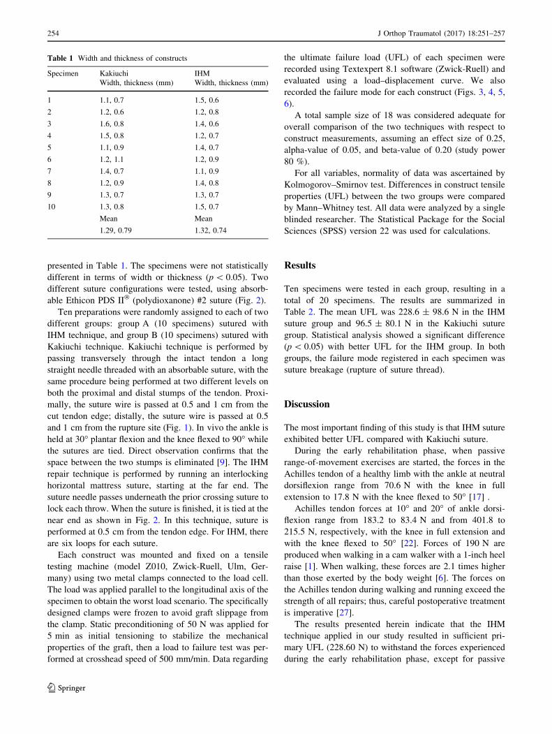
Fig. 3 Comparison between
IHM suture (a) and Kakiuchi
suture (b) performed on fresh
bovine Achilles tendon
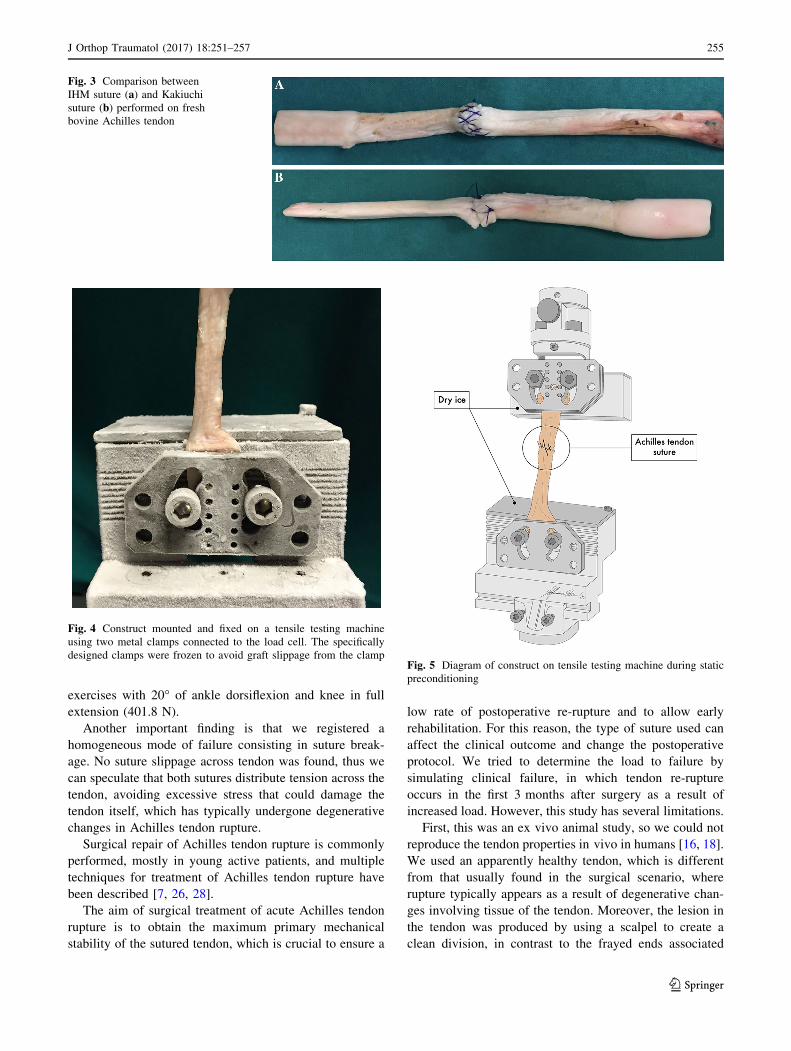
Fig. 4 Construct mounted and fixed on a tensile testing machine
using two metal clamps connected to the load cell. The specifically
designed clamps were frozen to avoid graft slippage from the clampFig. 5 Diagram of construct on tensile testing machine during static
preconditioning
J Orthop Traumatol (2017) 18:251–257 255
123
with clinical injury [18]. For all these reasons, only clinical
studies might clarify whether IHM suture is truly superior
to Kakiuchi suture in the chronic tendinitis scenario.
Bovine tendon was tested because it is readily available
and has already been used in similar biomechanical studies
[18]. Another limitation is that we only recorded data for
the UFL, because we aimed only to determine whether
IHM could offer better biomechanical performance,
considering that graft slippage is more correlated to the
characteristics of the tendon itself, which has viscoelastic
properties. Finally, the load axis was applied parallel to the
graft, which is different from the complex forces that the
repaired tendon has to resist during the early rehabilitation
phase.
In conclusion, IHM suture exhibited better UFL compared
with Kakiuchi suture in an animal model of Achilles tendon
repair. These results seem to support IHM as a valid option in
Achilles tendon rupture, though the clinical implications of
the various repairswere not studied.Manyminimally invasive
approaches, including for the Kakiuchi technique, have been
described in literature for Achilles tendon repair. IHM suture
in vivo can also be performed minimally invasively.
More clinical studies are needed to confirm the func-
tional and biomechanical effectiveness of this suture
technique.
Compliance with ethical standards
Conflict of interest The authors declare that they have no conflict of
interest.
Patient consent Obtaining the informed consent form involved
patients was waived by the Research Ethics Committee.
Ethical approval For this study all applicable international, national
and/or institutional guidelines for the care and use of animals were
followed.
Funding No funding was delivered for this study.
Open Access This article is distributed under the terms of the
Creative Commons Attribution 4.0 International License (http://crea
tivecommons.org/licenses/by/4.0/), which permits unrestricted use,
distribution, and reproduction in any medium, provided you give
appropriate credit to the original author(s) and the source, provide a
link to the Creative Commons license, and indicate if changes were
made.
References
1. Akizuki KH, Gartman EJ, Nisonson B, Ben-Avi S, McHugh MP
(2001) The relative stress on the Achilles tendon during ambu-
lation in an ankle immobiliser: implications for rehabilitation
after Achilles tendon repair. Br J Sports Med 35:329–334
2. Aspenberg P (2007) Stimulation of tendon repair: mechanical
loading, GDFs and platelets: a mini-review. Int Orthop
31:783–789
3. Cetti R, Christensen SE, Ejsted R et al (1993) Operative versus
nonoperative treatment of Achilles tendon rupture. A prospective
randomized study and review of the literature. Am J Sports Med
21:791–799
4. Dona E, Gianoutsos MP, Walsh WR (2004) Optimizing biome-
chanical performance of the 4-strand cruciate flexor tendon
repair. J Hand Surg Am 29(4):571–580
5. Dona E, Turner AWL, Gianoutsos MP, Walsh WR (2003)
Biomechanical properties of four circumferential flexor tendon
suture techniques. J Hand Surg 28A:824–831
Fig. 6 Ultimate failure load (UFL) of construct
Table 2 Ultimate failure load (UFL) of the two different suture
techniques
Specimen Kakiuchi IHM
UFL (N) UFL (N)
1 75.89 205.44
2 143.21 201.97
3 70.61 280.40
4 110.12 219.25
5 120.35 196.55
6 63.11 203.40
7 77.02 211.22
8 110.25 300.60
9 113.09 240.94
10 82.02 226.28
Mean Mean
96.57 228.60
256 J Orthop Traumatol (2017) 18:251–257
123
6. Froberg A, Komi P, Ishikawa M, Movin T, Arndt A (2009) Force
in the Achilles tendon during walking with ankle foot orthosis.
Am J Sports Med 37:1200–1207
7. Ismail M, Karim A, Shulman R, Amis A, Calder J (2008) The
Achillon achilles tendon repair: is it strong enough? Foot Ankle
Int 29:808–813
8. Schneppendahl J, Thelen S, Schek A et al (2012) Initial stability
of two different adhesives compared to suture repair for acute
Achilles tendon rupture—a biomechanical evaluation. Int Orthop
(SICOT) 36:627–632. doi:10.1007/s00264-011-1357-9
9. Kakiuchi M (1995) A combined open and percutaneous technique
for repair of tendo Achillis. Comparison with open repair. J Bone
Jt Surg Br 77(1):60–63
10. Kearney RS, Costa ML (2012) Current concepts in the rehabili-
tation of an acute rupture of the tendo Achillis. J Bone Jt Surg
[Br] 94-B:28–31
11. Kjaer M, Langberg H, Miller BF (2005) Metabolic activity and
collagen turnover in human tendon in response to physical
activity. J Musculoskelet Neuronal Interact 5:41–52
12. Lo IK, Kirkley A, Nonweiler B (1997) Operative versus nonop-
erative treatment of acute Achilles tendon ruptures: a quantitative
review. Clin Sports Med 7:207–211
13. Lovric V, Kanazawa T, Nakamura Y et al (2011) Effects of gaps
induced into theACL tendon graft on tendon-bone healing in a rodent
ACL reconstruction model. Muscle Ligament Tendon J 3:91–99
14. Maffulli N, Waterston SW, Squair J, Reaper J, Douglas AS
(1999) Changing incidence of Achilles tendon rupture in Scot-
land: a 15-year study. Clin J Sport Med 9:157–160
15. Moller M, Movin T, Granhed H, Lind K, Faxen E, Karlsson J
(2001) Acute rupture of tendo Achilles. A prospective ran-
domised study of comparison between surgical and non-surgical
treatment. J Bone Jt Surg (Br) 83(6):843–848
16. Mortensen HM, Skov O, Jensen PE (1999) Early motion of the
ankle after operative treatment of a rupture of the Achilles ten-
don: a prospective, randomized clinical and radiographic study.
J Bone Jt Surg [Am] 81-A:983–990
17. Orishimo KF, Burstein G, Mullaney MJ et al (2008) Effect of
knee flexion angle on Achilles tendon force and ankle joint
plantarflexion moment during passive dorsiflexion. J Foot Ankle
Surg 47:34–39
18. Ortiz C, Wagner E, Mococain P, Labarca G, Keller A, Del Buono
A, Maffulli N (2012) Biomechanical comparison of four methods
of repair of the Achilles tendon: a laboratory study with bovine
tendons. J Bone Jt Surg Br 94(5):663–667. doi:10.1302/0301-
620X.94B5.27642
19. Rebeccato A, Santini S, Salmaso G, Nogarin L (2001) Repair of
the achilles tendon rupture: a functional comparison of three
surgical techniques. J Foot Ankle Surg 40(4):188–194
20. Schepull T, Aspenberg P (2013) Early controlled tension improves
the material properties of healing human Achilles tendons after
ruptures: a randomized trial. Am J Sports Med 41:2550–2557
21. Suchak AA, Bostick G, Reid D, Blitz S, Jomha N (2005) The
incidence of Achilles tendon ruptures in Edmonton, Canada. Foot
Ankle Int 26:932–936
22. Vadala A, De Carli A, Vulpiani MC, Iorio R, Vetrano M,
Scapellato S, Suarez T, Di Salvo F, Ferretti A (2012) Clinical,
functional and radiological results of Achilles tenorraphy surgi-
cally treated with mini-open technique. J Sports Med Phys Fit-
ness 52(6):616–621
23. Wallace RG, Heyes GJ, Michael AL (2011) The non-operative
functional management of patients with a rupture of the tendo
Achillis leads to low rates of re-rupture. J Bone Jt Surg [Br]
93-B:1362–1366
24. Winter E, Ambacher T, Maurer F et al (1995) Surgical therapy of
Achilles tendon rupture. Unfallchirurg 98:468–473
25. Wong J, Barrass V, Maffulli N (2002) Quantitative review of
operative and nonoperative management of Achilles tendon
ruptures. Am J Sport Med 30:565–575
26. Worth N, Ghosh S, Maffulli N (2007) Management of acute
Achilles tendon ruptures in the United Kingdom. J Orthop Surg
(Hong Kong) 15:311–314
27. Zandbergen RA, de Boer SF, Swierstra BA, Dayi J, Kleinrensink
GJ, Beumer A (2005) Surgical treatment of Achilles tendon
rupture: examination of strength of 3 types of suture techniques in
a cadaver model. Acta Orthop 76(3):408–411
28. Wu Z, Hua Y, Li H, Chen S, Li Y (2015) Biomechanical com-
parison of three methods for distal Achilles tendon reconstruc-
tion. Knee Surg Sports Traumatol Arthrosc 23(12):3756–3760
J Orthop Traumatol (2017) 18:251–257 257
123