interfacial interaction between transmembrane...
TRANSCRIPT

RESEARCH PAPER
Interfacial Interaction between Transmembrane Ocular Mucinsand Adhesive Polymers and Dendrimers Analyzed by SurfacePlasmon Resonance
I. Bravo-Osuna & M. Noiray & E. Briand & A. M. Woodward & P. Argüeso & I. T. Molina Martínez & R. Herrero-Vanrell & G. Ponchel
Received: 17 December 2011 /Accepted: 17 April 2012 /Published online: 8 May 2012# Springer Science+Business Media, LLC 2012
ABSTRACTPurpose Development of the first in vitro method based on bio-sensor chip technology designed for probing the interfacial interactionphenomena between transmembrane ocular mucins and adhesivepolymers and dendrimers intended for ophthalmic administration.Methods The surface plasmon resonance (SPR) technique wasused. A transmembrane ocular mucin surface was prepared on thechip surface and characterized by QCM-D (Quartz Crystal Microbal-ance with Dissipation) and XPS (X-ray photoelectron spectroscopy).The mucoadhesive molecules tested were: hyaluronic acid (HA),carboxymethyl cellulose (CMC), hydroxypropylmethyl cellulose(HPMC), chitosan (Ch) and polyamidoamine dendrimers (PAMAM).Results While Ch originated interfacial interaction with ocular trans-membrane mucins, for HA, CMC and HPMC, chain interdiffusionseemed to be mandatory for bioadherence at the concentrationsused in ophthalmic clinical practise. Interestingly, PAMAM dendrimersdeveloped permanent interfacial interactions with transmembraneocular mucins whatever their surface chemical groups, showing arelevant importance of co-operative effect of these multivalent sys-tems. Polymers developed interfacial interactions with ocularmembrane-associated mucins in the following order: Ch(1 %) >G4PAMAM-NH2(2 %) 0 G4PAMAM-OH(2 %) > G3.5PAMAM-COOH(2 %)>> CMC(0.5 %) 0 HA(0.2 %) 0 HPMC(0.3 %).Conclusions The method proposed is useful to discern be-tween the mucin-polymer chemical interactions at molecularscale. Results reinforce the usefulness of chitosan and den-drimers as polymers able to increase the retention time ofdrugs on the ocular surface and hence their bioavailability.
KEY WORDS chitosan . mucoadhesion . oculartransmembrane mucins . polyamidoamine (PAMAM)dendrimers . surface plasmon resonance (SPR)
ABBREVIATIONSCh chitosanCMC carboxymethyl celluloseHA hyaluronic acidHCLE telomerase-immortalized human
corneal-limbal epithelialHPMC hydroxypropylmethyl celluloseIEP isoelectric pointPAMAM polyamidoamine dendrimersQCM-D quartz crystal microbalance with dissipationRU resonance unitsSPR surface plasmon resonanceXPS x-ray photoelectron spectroscopy
INTRODUCTION
Ocular delivery requires the implementation of effectivestrategies to increase the drug residence time on the ocularsurface with the aim of minimizing systemic drug side effectsand reducing dosing frequency, which might improve pa-tient compliance, one of the main lack-points of chronic
I. Bravo-Osuna (*) : I. T. M. Martínez : R. Herrero-VanrellDept. of Pharmacy & Pharmaceutical Technology, School of PharmacyUniversity Complutense of Madrid (UCM)Madrid, Spaine-mail: [email protected]
M. Noiray : E. Briand :G. PonchelCNRS UMR 8612, Université de Paris SudLaboratoire de Physicochimie, Pharmacotechnie & BiopharmacieChatenay-Malabry, France
A. M. Woodward : P. ArgüesoDepartment of Ophthalmology, Harvard Medical SchoolSchepens Eye Research InstituteBoston, Massachusetts 02114, USA
Pharm Res (2012) 29:2329–2340DOI 10.1007/s11095-012-0761-1

therapies at topic ophthalmic level. In this context, the use ofbioadhesive formulations able to interact with the trans-membrane mucins covering the ocular surface, has gainedattention in the last decades (1,2). Furthermore, some pa-thologies of the ocular surface, such as “dry eye syndrome”,can be also beneficiated by the inclusion of bioadhesivepolymers in the eye drops because of their lubricant andhumectant nature (3). Recent evidences showed that trans-membrane ocular mucins and their O-glycans on the cell-surface glycocalyx have important biological roles in theprotection of corneal and conjunctival epithelia, such as,promoting boundary lubrication, and maintaining epithelialbarrier function (4–6).
Bioadhesion of dosage forms is a complex phenomenon,involving simultaneously interfacial interactions with theliving tissues and the development of adequate mechanicalproperties of the bulk formulation. However, whatever thenature of the materials constituting the dosage forms, rem-anence at the corneal surface primarily depends on thecharacteristics of the adhesive interface that can be createdwith the transmembrane ocular mucins. Thus, the develop-ment of a dedicated in vitro technique specifically designed toevaluate the interaction of polymers with transmembraneocular mucins would be a valuable tool for a rational selec-tion of excipients of ocular drug formulations. Consideringthe low polymer concentration used in topic ocular admin-istration, the classic techniques based on tensiometric orrheological measurements are not very suitable for evaluat-ing interfacial adhesive properties but rather bulk propertiesof the formulations, such as hydrogels. Furthermore, somerheological studies consisting in mixing mucins to the poly-mers to be studied for evaluating possible interactions typ-ically require the use of relatively high amounts of mucins. Itis a severe limitation in the case of ocular mucins, which areproduced in very low extent, are not commercially availableand must be substituted by gastrointestinal porcine mucinsin most of the cases (7,8), considerably limitating the perti-nence of results. Besides, these “bulk-like” in vitro mucoad-hesion tests only give a macroscopic and indirectmeasurement of polymers-mucin interactions thus it is notpossible to evaluate the interaction at a molecular level.
Apart of these methods, powerful techniques such as iso-thermal titration calorimetry, quartz crystal microbalance orsurface plasmon resonance (SPR) are currently available forthe evaluation of interactions at the molecular scale. Amongthem, SPR is a very attractive chip-based biosensor technique,allowing the direct observation of binding events and kineticsbetween molecular partners. Very interestingly, it has such ahigh sensitivity that only small amounts of samples are re-quired (9). Although the SPR technique was developed toevaluate specific interactions (ligand-receptor or antibody-antigen), some attempts of mucoadhesion evaluation by SPRtechniques have already been carried out (10,11).
The objective of the present work was to develop an invitro methodology, based on biosensor chip technology, ableto evaluate the interactions of ocular transmembranemucins with ophthalmic preparations. In a first step, themucin layer formed on the sensor chip was deeply charac-terized by SPR, quartz crystal microbalance with dissipation(QCM-D) and X-ray photoelectron spectroscopy (XPS). Ina second step, the interaction of transmembrane mucinseither with conventionaly used polymers, or with den-drimers, as new drug delivery systems currently exploredin the topical ophthalmic route (8), was assessed. It might bethe first in vitro method specifically designed to evaluate theinteraction of ocular transmembrane mucins with polymersor other adhesive molecules. It is expected that it wouldallow the evaluation of these interactions at molecular level.This technique could help on the comprehension of thecomplex mechanisms involved in mucoadhesion at theocular surface.
MATERIALS AND METHODS
Materials
Hyaluronic acid ophthalmic grade (Mw 800,000–1,200,000 g/mol), carboxymethyl cellulose medium viscosity(CMC; 400–800 cps, 2 % solution at 20 °C) and hydroxypro-pylmethyl cellulose (HPMC; 1390 cps, 2 % solution at 20 °C),were purchased from Abaran Materias Primas S.L. (Madrid,Spain). Chitosan (400,000 g/mol) was supplied by Fluka(Saint-Quentin Fallavier, France) and ethylenediamine 2-carbon core PAMAM dendrimers (G4 PAMAM-NH2, G4PAMAM-OH, G3.5 PAMAM-COOH) manufactured byDendritech Inc., were obtained from Sigma-Aldrich (Saint-Quentin Fallavier, France). Au-coated crystals for QCM-Danalysis were provided by Q-sense A B (Gothenburg, Sweden).Reactives and materials used for the SPR experiments wereobtained from Biacore-GE Healthcare (Orsay, France). Allchemicals were reagent grade and used as received.
METHODS
Ocular Mucin Isolation
Telomerase-immortalized human corneal-limbal epithelial(HCLE) cells were plated on T150 flasks (Costar Corning,Inc., Corning, NY) and grown in a medium optimized forproliferation of keratinocytes (keratinocyte serum-free me-dium; Gibco-Invitrogen Corp.; Carlesbad, CA) to achieveconfluence. After reaching confluence, cells were switchedto stratification medium containing DMEM/F12 (Gibco-Invitrogen Corp.) with 1 mM CaCl2, 10 ng/mL EGF
2330 Bravo-Osuna et al.

(Hyclone, Logan, UT) and 10 % calf serum (Gibco-Invitro-gen Corp.) for 7 days to promote stratification and optimalbiosynthesis of cell surface-associated mucins. Derivatizationand mucin profile of HCLE cell cultures have been previ-ously reported (12). Mucin was purified from stratified cul-tures of HCLE cells as previously described (13,14). Briefly,protein from cell cultures was extracted using RIPA buffer(150 μM NaCl, 50 μM Tris, pH 8.0, 1 % NP 40, 0.5 %deoxycholate, 0.1 % SDS) plus complete Protease InhibitorCocktail (Roche Biochemical; Indianapolis, IN). After ho-mogenization with a pellet pestle, the protein extract wascentrifuged at 13,500 rpm for 45 min and the proteinconcentration of the supernatant determined using thePierce BCA Protein Assay Kit (Thermo Scientific; Rock-ford, IL). High molecular weight mucins were separated bygel chromatography on a Sepharose CL-4B size exclusioncolumn. The void volume (Vo) containing the mucin frac-tion was digested with RNase A and DNase I (1 mg nucle-ase/100 mg protein) for 3 h. at room temperature, andfurther purified by isopycnic density gradient centrifugationin cesium chloride. The presence of the cell surface-associated mucin MUC16 in individual fractions wasassayed by agarose gel electrophoresis and western blotusing the M11 antibody (Neo Markers; Fremont, CA).
Mucin Immobilization on Surface Plasmon ResonanceChip (Biacore®)
Purified ocular mucin was immobilized on the Sia Kit Auchips (Biacore®) by incubation of the surface with 50 μl ofmucin aqueous solution (80 μg/ml) over night at roomtemperature. Afterwards, chips were thoroughly rinsed inMilliQ® water. Once the chip was prepared and inserted onthe Biacore® T100 apparatus, it was again washed withMilliQ® water at 30 μl/min (12 h). Resonance units (RU)were measured before and after the immobilization step tomonitor the final amount of mucin attached to the Au chip.Calculations of immobilized mucin amounts were per-formed on the basis of the Biacore® standard relation: 1RU 0 1 pg·mm−2. It is relevant to mention that this relationis valid for proteins in general, so in the case of highlyglycosylated proteins, such as mucins, this value might notbe completely accurate. The optical mucin layer was calcu-lated considering that 1 RU correspond to a resonanceangle shift of 10−4° (9).
Mucin Layer Characterization
Quartz Crystal Microbalance with Dissipation Measurement(QCM-D)
Experiments were performed at 35.0±0.1 °C using aQCM-D E1, Q-sense AB (Gothenburg, Sweden). The
device has been described in detail elsewhere (15). Brief-ly, AT cut quartz crystals oscillate at the resonant fre-quency (5 MHz) or at one of its overtones (3rd, 5th, 7th,9th, 11th and 13th). The drive circuit is then open-circuited and the exponential decay of the oscillationamplitude is monitored. The dissipation, D, is definedas the fraction of energy of the oscillation that is dissi-pated during one period of oscillation. The resonantfrequency decreases when materials are adsorbed onthe crystal surface. The frequency shift (Δf ) is relatedto the amount while the dissipation shift (ΔD) reflects theviscoelastic properties of the adlayer. An estimation ofthe adsorbed mass can be made according to the Sauer-brey equation (16) when the adlayer is homogeneous, notslipping and rigid, or according to the Voigt model whenthe adlayers present viscoelastic behavior (17). Ocularmucin immobilization on the gold sensor was achievedby circulating a 80 μg/ml aqueous solution of ocularmucin at 60 μl/min over the sensor’s surface until stabi-lization, using a peristaltic pump (Ismatec IPC-N4). Afterrising with milliQ® water at the same flux, the runningbuffer was changed to PBS pH 7.4 until stabilization.Both oscillation frequency shifts and energy dissipationchanges were monitored all over the assay. Furthermore,all frequency shifts were normalized with the overtonenumber.
X-Ray Photoelectron Spectroscopy (XPS)
The mucin layer formed on the Biacore® Au chip wasevaluated by XPS analysis. High-resolution XPS datawere acquired with a Thermo VG Scientific ESCALAB250 spectrometer with a hemispherical electron analyserusing a monochromatic Al Kα X-ray source operatingin an ultra high vacuum chamber with a base pressurein the 5×10⁻¹⁰ mbar range. The spectra were recordedat normal emission take-off angle, using an energy stepof 1 and 0.1 eV and a pass-energy of 100 and 40 eVfor survey spectra and core levels respectively. Compo-sitional analysis by XPS were performed by assuming ahomogeneous distribution of atoms present on the goldsurface covered by the mucin layer and using tabulatedatomic sensitivity factors.
From the attenuation intensity of the gold Au4f peak, onecan estimate the thickness (d) using the exponential attenuation:
Is ¼ I 0S � e �dl cos θð Þ
Where Isº is Au4f intensity before coating, Is the Au4fintensity after coating, λ is the mean free path of Au4felectrons in the mucin layer, and θ is the photoelectronemission angle (relative to the normal surface).
Ocular Mucoadhesion of Polymers and Dendrimers by SPR 2331

Screening of Adhesion Behaviour of Polymersand Dendrimers with Ocular Transmembrane Mucinsby Surface Plasmon Resonance
Polymers and Dendrimers Preparation
Three polymers typically included in ocular topical formu-lations, hyaluronic acid (HA), carboxymethyl cellulose(CMC), and hydroxypropylmethyl cellulose (HPMC) werechosen at the concentrations generally used for ocular sur-face administration in clinical practise (0.2 % HA, 0.5 %CMC, 0.3 % HPMC) (18). A chitosan solution (Ch) was alsoevaluated. Although there is no commercial ocular formu-lation including chitosan yet, it has been extensively studiedas a mucoadhesive agent for the improvement of drugocular bioavailability (19). Polymer solutions, except chito-san, were prepared by stirring at room temperature in PBS,pH 7.4 overnight and filtering (0.45 μm). In the case of Ch,the commercial polymer was first depolymerised in order toreduce its molecular weight to 20,000 g/mol (20). Onceprepared, depolymerised chitosan was dissolved in aceticacid solution (pH 6) with a final polymer concentration of1 % and filtered. In addition, PAMAM dendrimers, newdrug delivery systems currently intended for ophthalmicadministration, were also evaluated. The G4 dendrimerswith amino and hydroxyl surface groups and the G3.5PAMAM dendrimer with carboxylic groups at the surfacewere prepared at a concentration of 2 % in PBS, pH 7.4,according to other authors (8). Briefly, dendrimers in themethanolic commercial PAMAM solution were evaporatedunder vacuum (Rotavapor R-124 Büchi. Switzerland) andsubsequently dissolved in PBS pH 7.4.
Surface Plasmon Resonance Binding Experiments
Solutions of linear polymers and dendrimers were flowedover the mucin coated sensor chip surface within the Bia-core® T100 apparatus for 300 s at 10 μl/min. PBS, pH 7.4,was used as running medium except for Ch experiments,where acetic acid solution (pH 6) was used. Afterwards, thesensorgram (RU versus time) was collected until equilibriumwas reached. All studies were performed in triplicate at theocular surface physiological temperature: 35 °C.
Ocular Mucin-PAMAM Dendrimers Surface Plasmon ResonanceIsotherms
Five concentrations of PAMAM-NH2 and PAMAM-OHdendrimer in PBS (pH 7.4) or in acetate buffer (pH 5.5)were used in SPR assays. All analysis were performed at35 °C, with a 600 s period of ligand-analyte association and600 s period of dissociation, at 10 μl/min. Dendrimer con-centrations (10–80 nM) and contact time were optimized in
order to approach the steady state at all concentrations. Thecorresponding isotherms were prepared by plotting theamount of dendrimers attached to the ocular transmem-brane mucin layer versus dendrimer concentration.
Chip Regeneration
After SPR assays, the Au chips were carefully separatedfrom their plastic holders by addition of ethanol and thegold surfaces were cleaned in a 5:1:1 mixture of MilliQ®water, NH3 (25 %) and H2O2 (30 %), respectively, beforeheating for 15 min at 70 °C. Afterwards, they were rinsedwith water and dried with N2. Subsequently, chips wereexposed three times, 15 min each, in a UV-O3 chamber(UV/ozone procleaner™ Bioforce nanosciences. Paris,France) (21). Samples were rinsed with ethanol and driedwith N2 before each exposition. XPS evaluation of theregenerated gold surface was performed to determine theabsence of mucins on the chip surface. The same procedurewas followed to prepare the gold-coated crystals prior toQCM-D studies.
Statistical Analysis
Results were statistically analysed by one-way analysis of vari-ance (ANOVA) using the SPSS® 16.0 software. Post-ANOVAanalysis was carried out according to the Bonferroni’s multiplecomparisons test. Results were determined to be significantwhen p<0.05.
RESULTS
Ocular Mucin Isolation
Ocular surface mucins were purified from stratified culturesof human corneal limbal epithelial (HCLE) cells using size-exclusion chromatography followed by isopycnic densitygradient centrifugation (Fig. 1). HCLE cells produce cellsurface-associated mucins which can be distinguished onthe basis of their molecular size as they are eluted as a singlepeak in the void volume (Vo, Fig. 1a). As determined bywestern blot using the M11 antibody against MUC16, gra-dient centrifugation produced aliquots of purified mucinwith a buoyant density range of 1.26 to 1.44 g/ml(Fig. 1b). Soluble mucins in fractions within this densityrange were combined and used for further study.
Mucin Layer Preparation and Characterization
The first step in the development of a new mucoadhesion invitro test based on SPR measurements is characterization of
2332 Bravo-Osuna et al.

the ligand (mucin) layer on the gold chip. In this purpose,QCM-D, SPR and XPS assays were performed.
Quartz Crystal Microbalance with Dissipation Measurement(QCM-D)
According to Fig. 2b, in milliQ® water, mucins progressivelyadsorb on a gold surface, resulting in a decrease in frequencyof vibration of the quartz crystal until stabilization. The max-imal amount of hydrated mass adsorbed was 2.35 ng/mm²calculated according to the Sauerbrey equation. The dissipa-tion factor (D) provides a measure of energy loss in the systemand contains information about film interactions with the bulksolution. Generally, rigid structures haveminimal effect on thedissipation, whereas thick or flexible structures increase thedissipation so it can be seen as a measure of the rigidity orviscoelasticity of the adsorbed film (22). The observed normal-ized frequency shift at the various overtone overlaps indicatedthe presence of a rigid layer with dissipation lower than 0.5.However, an additional decrease in frequency was recordedwhen running media was changed from deionised water toPBS, pH 7.4, (Δf comprised between −7 Hz and −10 Hz anda ΔD of 2 to 3.5) showing a relatively more extended confor-mation of the protein in presence of ions (30,31).
Surface Plasmon Resonance (SPR)
The evaluation of the mucin layer was also assessed by surfaceplasmon resonance. Biacore® Au chips were prepared bydeposition of 50 μl of an aqueous ocular mucin solution(0.08 mg/ml). After an overnight incubation, chips weremounted on their plastic containers and measured in theBiacore® apparatus, obtaining a mean RU increment of
4,954±204. A critical parameter in SPR studies is the surfacedensity of the ligand (24). Therefore, the mucin depositiondata on the four channels of each chip were statisticallyevaluated (one-way ANOVA analysis), showing statisticallysimilar values of ΔRU (p>0.05), that is mucin deposition, inall channels and chips used. The amount of mucin immobi-lized on the Au surface was in the same order as thoseobtained from covalent mucin immobilization on a CM5 chip(dextran modified Au surfaces) (25). According to the Bia-core® protein standard relation (1RU 0 1 pg/mm2), animmobilization of 4.9±0.2 ng/mm2 of ocular mucin can beassumed. Comparing this value with those obtained for massadsorption on gold surface after D-QCM experiments surfacesaturation was presumed. However, to confirm that all mucinchains present on the chip surface were covalently attached tothe chip surface, extensive washes of the immobilized chipsurface with vehicle were performed for hours in the Biacore®system, with no change on the RU values (data non shown).All these studies helped us to assume the saturation of the goldchip by the mucin layer. The optical thickness of this mucinlayer determined by the shift in the resonance angle of thelayer was estimated to be 2.62±0.01 nm.
X-Ray Photoelectron Spectroscopy (XPS)
The so-performed quantitative determination of the mucinlayer was additionally completed by X-ray photoelectronspectroscopy (XPS) analysis. XPS scans were used for thesemiquantification of atoms present on the gold surface cov-ered by the mucin layer (26). According to Fig. 2b, the surveyspectrum showed an attenuation of gold (see the decrease ofthe peak area for Au4f between 92 and 80 eV). N1s wasclearly detected from the survey scan in the mucin layer and
Fig. 1 Purification of ocular mucin. (a) Sepharose CL-4B chromatography of cell lysates from stratified HCLE cell cultures. The column eluate wasmonitored for absorbance at 280 nm. The void volume fraction (Vo) containing the high molecular weight mucins was collected and further purified bygradient centrifugation. (b) Density gradient centrifugation of the Vo fraction in cesium chloride. One-ml aliquots were electrophoresed on a 1 % (w/v)agarose gel and then Western blotted onto nitrocellulose as described in Materials and Methods. The blot was probed with MUC16-mucin specific M11antibody. Fraction 3 contained both soluble (s) and insoluble (i) mucin. Soluble fractions 3 to 8 were combined and used for further study.
Ocular Mucoadhesion of Polymers and Dendrimers by SPR 2333

a second maximum was detected at 288 eV corresponding tocarbon atoms from the peptidic linkage -NH-C0O, showingthe presence of mucins on the gold surface (Fig. 2c). Thethickness of the mucin layer determined by XPS was around1 nm. This data correlates well with the previousQCM-D andSPR studies, taking into account that these measurements areperformed in dry conditions, so the highly hydrated andglycosylated portions of the mucins might be deeply modified.
Adhesion Behaviour of Polymers and Dendrimerswith Ocular Transmembrane Mucins by SurfacePlasmon Resonance
Initially in a SPR experiment, the sensor chamber is filled withrunning buffer. When analytes (polymer or dendrimer
molecules) bind to ligands, they replace buffer solution mole-cules from the surface, which can be optically detected as anincrease of the RU response, until reaching a plateau. Subse-quently, when the analyte dissociates from its ligand, therunning solution replaces the analyte solution over themucin-coated surface and a partial decrease in the RU signalis detected. The total increase in RU after this washing step(“stability RU”) is related to the amount of analyte retained onthe ligand layer (9). The profiles obtained in evaluation ofmucin-polymers interactions (Fig. 3) showed the typical profileof non-specific interactions, with an initial rapid uptake, fol-lowed by a slower process. Other authors have attributed thissecond step to interface reorganization (11). According to SPRresults, only chitosan demonstrated statistically significant per-manent chemical interactions with transmembrane ocular
a
b c
Fig. 2 Characterization of transmembrane ocular mucin layer (a) QCM-DNormalized frequency (black) and dissipation (red) shifts recorded at the 5th overtoneduring adsorption of mucin on gold-coated surface and rinsing with MilliQ®water and PBS buffer. (b) XPS survey spectra of Au chip (black), ocular mucin-coatedAu chip (red) and after surface cleaning of mucin coated Au chip (blue) in a 5:1:1 mixture of MiliQ water, NH3 (25%) and H2O2 (30%) 15min at 70 °C+UV-O3
chamber. The energy positions of nitrogen, carbon and gold are also indicated by vertical lines in the figure. The attenuation of gold intensity and the presence of N1 s peak in mucin coated Au chip suggest the formation of a thin layer of mucin on Au chip. (c) High-resolution XPS spectra of C 1 s and N 1 s core levels are alsoshown to asses the presence of peptidic linkages -NH-C 0 O, (indicated by vertical line at 288 eV) typical of proteins.
2334 Bravo-Osuna et al.

mucin surface. While HA, CMC and HPMC were unable tobe retained on the mucin layer.
The SPR mucoadhesion method developed was used totest the interaction of transmembrane ocular mucins withPAMAM dendrimers with three different chemical groupson the surface: PAMAM-NH2 (64 amino groups), PAMAM-OH (64 hydroxyl groups) and PAMAM-COOH (64 carbox-ylic groups). Interestingly, according to Fig. 4, all evaluatedPAMAM dendrimers showed remarkable interactions withthe membrane-associated ocular mucin layer. PAMAM-NH2 and PAMAM-OH showed statistically similar values of
ΔRU. Although PAMAM-COOH also adhered to the trans-membrane ocular mucin layer, the intensity was lower anddata showed less reproducibility (high standard deviation inthe ΔRU). Additionally an anomalous non-saturating profileobserved in the sensorgram (Fig. 4). Calculations were per-formed to achieve theoretical approximations to determinethe number of dendrimer layers on the apparent mucin layersurface. Values of 1.26, 1.07 and 0.59 layers were obtained for-NH2, -OH and -COOH PAMAM dendrimers.
Additional experiments were performed with PAMAM-NH2 and PAMAM-OH to further analyse their interactions
-500
0
500
1000
1500
2000
2500
3000
HA (0.2%) CMC (0.5%) HPMC (0.3%) Chitosan (1%)
RU
incr
emen
t
*
PBS 7.4
HA (0.2%)
CMC (0.5%)
Ch 1%
(1)
HPMC (0.3%)
x
xxx
(2) (3)
0
500
1000
1500
2000
2500
3000
3500
-50050 100 150 200 250 300 350 400 450
Res
pons
e (0
= b
asel
ine)
Time (s)
a
b
Fig. 3 (a) SPR sensorgrams of the interaction of polymers in PBS pH 7.4 -or acetic acid solution pH 6 in the case of Chitosan- (analyte) with transmembraneocular mucins (ligand). Runs were performed at 35 °C. (1) sample injection; (2) washing step: (3) stability. (b) RU increment observed after rinsing (stability step),indicative of polymer permanent interaction on the mucin surface. Asterisk represents statistic differences in RU stability values (p<0.05). n03.
a
PBS 7.4
PAMAM-NH2 (2%)
(1) (2) (3)
50 100 150 200 250 300 350 400 450
Time (s)
0
500
1000
1500
2000
2500
3000
3500
-500
Res
pons
e (0
= b
asel
ine)
PAMAM-OH (2%)
PAMAM-COOH (2%)
0200400600800
100012001400160018002000
PAMAM-NH2 (2%) PAMAM-OH (2%) PAMAM-COOH (2%)
RU
incr
emen
t
*
b
Fig. 4 (a) SPR sensorgrams of the interaction of dendrimers at 2 % in PBS pH 7.4 (analyte) with transmembrane ocular mucins (ligand). Runs wereperformed at 35 °C. (1) sample injection; (2) washing step: (3) stability. (b) RU increment observed after rinsing (stability step), indicative of dendrimerpermanent interaction on the mucin surface. Asterisk represents statistic differences in RU stability values (p<0.05). n03.
Ocular Mucoadhesion of Polymers and Dendrimers by SPR 2335
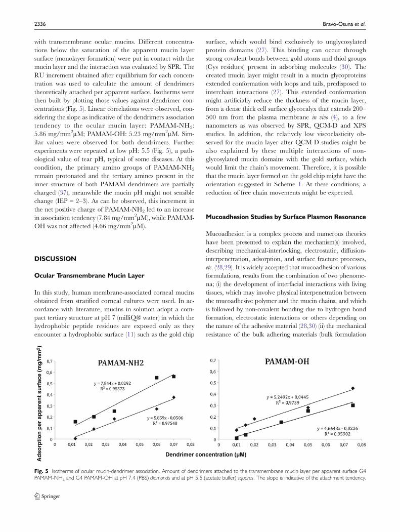
with transmembrane ocular mucins. Different concentra-tions below the saturation of the apparent mucin layersurface (monolayer formation) were put in contact with themucin layer and the interaction was evaluated by SPR. TheRU increment obtained after equilibrium for each concen-tration was used to calculate the amount of dendrimerstheoretically attached per apparent surface. Isotherms werethen built by plotting those values against dendrimer con-centrations (Fig. 5). Linear correlations were observed, con-sidering the slope as indicative of the dendrimers associationtendency to the ocular mucin layer: PAMAM-NH2:5.86 mg/mm2μM; PAMAM-OH: 5.23 mg/mm2μM. Sim-ilar values were observed for both dendrimers. Furtherexperiments were repeated at low pH: 5.5 (Fig. 5), a path-ological value of tear pH, typical of some diseases. At thiscondition, the primary amino groups of PAMAM-NH2
remain protonated and the tertiary amines present in theinner structure of both PAMAM dendrimers are partiallycharged (37), meanwhile the mucin pH might not sensiblechange (IEP 0 2–3). As can be observed, this increment inthe net positive charge of PAMAM-NH2 led to an increasein association tendency (7.84 mg/mm2μM), while PAMAM-OH was not affected (4.66 mg/mm2μM).
DISCUSSION
Ocular Transmembrane Mucin Layer
In this study, human membrane-associated corneal mucinsobtained from stratified corneal cultures were used. In ac-cordance with literature, mucins in solution adopt a com-pact tertiary structure at pH 7 (milliQ® water) in which thehydrophobic peptide residues are exposed only as theyencounter a hydrophobic surface (11) such as the gold chip
surface, which would bind exclusively to unglycosylatedprotein domains (27). This binding can occur throughstrong covalent bonds between gold atoms and thiol groups(Cys residues) present in adsorbing molecules (30). Thecreated mucin layer might result in a mucin glycoproteinsextended conformation with loops and tails, predisposed tointerchain interactions (27). This extended conformationmight artificially reduce the thickness of the mucin layer,from a dense thick cell surface glycocalyx that extends 200–500 nm from the plasma membrane in vivo (4), to a fewnanometers as was observed by SPR, QCM-D and XPSstudies. In addition, the relatively low viscoelasticity ob-served for the mucin layer after QCM-D studies might bealso explained by these multiple interactions of non-glycosylated mucin domains with the gold surface, whichwould limit the chain’s movement. Therefore, it is possiblethat the mucin layer formed on the gold chip might have theorientation suggested in Scheme 1. At these conditions, areduction of free chain movements might be expected.
Mucoadhesion Studies by Surface Plasmon Resonance
Mucoadhesion is a complex process and numerous theorieshave been presented to explain the mechanism(s) involved,describing mechanical-interlocking, electrostatic, diffusion-interpenetration, adsorption, and surface fracture processes,etc. (28,29). It is widely accepted that mucoadhesion of variousformulations, results from the combination of two phenome-na; (i) the development of interfacial interactions with livingtissues, which may involve physical interpenetration betweenthe mucoadhesive polymer and the mucin chains, and whichis followed by non-covalent bonding due to hydrogen bondformation, electrostatic interactions or others depending onthe nature of the adhesive material (28,30) (ii) the mechanicalresistance of the bulk adhering materials (bulk formulation
Ad
sorp
tio
n p
er a
pp
aren
t su
rfac
e (m
g/m
m2 )
Dendrimer concentration (µM)
Fig. 5 Isotherms of ocular mucin-dendrimer association. Amount of dendrimers attached to the transmembrane mucin layer per apparent surface G4PAMAM-NH2 and G4 PAMAM-OH at pH 7.4 (PBS) diamonds and at pH 5.5 (acetate buffer) squares. The slope is indicative of the attachment tendency.
2336 Bravo-Osuna et al.

and tissues). The SPR technique has been chosen for probingthe interfacial interaction phenomena between ocular mucinsand adhesive molecules, because they are the basis for thedevelopment of strongly bioadhesive formulations. As previ-ously suggested, the configuration of the mucins at the surfaceof the chips corresponded probably to the presence of analmost monomolecular layer of glycoproteins, making thissurface suitable for the observation of secondary bonding,but more unlikely chain interpenetration phenomena leadingto mechanical interlocking or expanded secondary bonding.Indeed, chains interdiffusion is a time-dependent diffusionphenomenon (31) where a minimum thickness of both parts(mucin and polymer) is needed (32), The penetration/diffu-sion/entanglement processes between polymers and mucinchains can enhance “interfacial” interactions, but also can bemore easily investigated through “bulk-like” mucoadhesion invitro tests (tensiometric measurements, rheological studies, etc.),where both mechanisms are inseparable. This was favourable,as the SPR method should rather allow the determination ofchemical interactions between rather well defined surfaces,therefore, can be useful to discern between the mucin-polymer chemical interactions at molecular scale.
Very well known hydrophilic polymers, especially electri-cally charged polymers, rich in OH, COOH and NH2
groups demonstrated a high mucoadhesive capacity (33).The ocular mucin-polymer interactions of two negativelycharged polymers (HA and CMC) and one positivelycharged polymer (chitosan) were evaluated. Additionally, a
non-charged polymer (HPMC) was also tested. In all cases,solutions of these polymers at the concentrations typicallyused for ocular administration (and not hydrogels) wereemployed. Chitosan is only soluble at pH lower than 6.5,even when the molecular weight is reduced, which is thecase of the one used in the present work, that is why a aceticsolution (pH 6) was used instead of PBS pH 7.4. Both pH (6and 7.4) are accepted at the ocular surface. Furthermore,considering that the isoelectric point (IEP) of mucins isaround 2–3, no significant changes can be expected in themucin layer at these conditions (34).
According to results, only chitosan developed statisticallysignificant permanent chemical adherence to the mucin layer.This behaviour could explain the promising results observedfor chitosan-based drug delivery when administered on theocular surface (19). Chitosan, a polycation (pKa 0 6.2), hasbeen reported to bind mucin, a polyanion (pKa 0 2.6), viaionic interaction between primary amino groups and the sialicand sulphonic acid substructures of glycosylated chains ofmucins. Additionally, the hydroxyl and amino groups of chi-tosan may also interact with mucin via hydrogen bonds (28).On the contrary, it may suggest that a prior chain interdiffu-sion is mandatory for HPMC, HA and CMC polymers topromote the formation of a sufficiently stable adhesive inter-face. In the case of HPMC, its ability to increase the drugretention time after ocular administration has been claimed tobe related to its viscosity enhancing capacity (1), more thanany intrinsic mucoadhesive nature, showing “negative inter-action” with mucin after rheological studies (35). For bothanionic polymers, HA andCMC, although it was claimed thatthey are able to develop strong hydrogen bonding with mucinmolecules (1,36), it seems that these interactions need to beaccompanied by chains interpenetration. In fact, it was al-ready demonstrated that changes in pH solutions of HA didnot influence the extension of its interaction with mucins.Thus, its adhesion behaviour might be mainly governed byphysical chain interpenetration, more than chemical interac-tion with mucin chains (1).
Among the different new drug delivery systems currentlyunder study, dendrimers, more precisely polyamidoaminedendrimers (PAMAM), have gained increasing attention.These new systems have potential medical applications dueto the combination of unique chemical properties with highbiocompatibility, low immunogenicity, and ease of synthesison a large scale with reasonable manufacturing cost (37). Atthe ocular level, some studies have demonstrated their utilityin the increase of drug bioavailability after topical adminis-tration in solutions (8) and as hydrogels based on PAMAMand polyethylene glycol (PEG) (37). One of the most impor-tant characteristics of dendrimers is the high density ofactive chemical groups located on their surface. In compar-ison to linear polymers, these multivalent systems mightpromote a co-operative effect leading to a large increase in
Au chip surface
HYPOTHESIS OF MUCIN DEPOSITION ON Au CHIP
Au chip surface
glycosylated non-glycosylated
MUCIN
Scheme 1 Hypothesis of transmembrane ocular mucin chains immobi-lization on gold surface. Hydrophobic peptide residues of the unglycosy-lated domains of mucin are exposed in the proximity of the gold chipsurface, developing strong covalent bonds. The created layer results in amucin glycoproteins extended conformation with a thickness of a fewnanometers.
Ocular Mucoadhesion of Polymers and Dendrimers by SPR 2337

reactivity (38). Effectively, in contrast to linear polymers, thethree different PAMAM dendrimers evaluated developedadhesion to the transmembrane ocular mucin surface what-ever their chemical surface group (NH2; OH; COOH). Inan interesting work, Griffiths et al. (39) evaluated the inter-action of PAMAM-NH2 dendrimers (generation 2.0 and4.0) with porcine gastric mucin (type III) in solution, bypulsed-gradient spin-echo NMR and small-angle neutronscattering. They also observed that these dendrimers (con-centration 0.5 % wt) experienced significant interaction withmucins at pH 7. At neutral pH all primary amino groups ofPAMAM-NH2 dendrimers are protonated (40), whichshould promote electrostatic interactions with the anionicsialic groups of mucins. According to our results, thePAMAM-mucin interactions observed were statistically sim-ilar for PAMAM-NH2 and PAMAM-OH, although thelatter can only develop weaker non-ionic interactions, suchas hydrogen bonds, with mucins. This behaviour wouldsuggest a remarkable contribution of non-ionic interactionin the development of ocular mucin-dendrimer interactions.In fact, other authors have previously suggested that poly-mers exhibiting high density of available hydrogen bondinggroups would be able to intensely interact with mucins (28).Additionally, the pH-dependent behaviour of PAMAM-NH2 suggests that at these pH conditions, this dendrimermight be able to perform even stronger interaction withocular mucins, which could be of relevance in the develop-ment of dendrimer based drug delivery systems for thetreatment of ocular surface pathologies involving reductionof tear pH, such as ocular inflammation.
Interestingly, PAMAM-COOH dendrimers also devel-oped adhesion on the mucin layer, in spite of the electrostaticrepulsion with the negatively charged mucin surface. Otherauthors also observed interaction between PAMAM-COOH(generation 3.5 and 5.5) and gastric mucins at pH 7, due to theformation of hydrogen bonds between charged carboxylicgroups of the dendrimer and the sugar residue on the mucinsside chains (39). Furthermore, the non-glycosylated domainsof mucins posses positively charged amino acid residues thatcould establish electrostatic interactions with negativelycharged groups of the polymers, as long as the repulsive forcesbetween negative charges (polymer and O-glycan chain) areefficiently screened by salt (23). This scenario could explainthe more erratic behaviour observed for the interaction ofPAMAM-COOH with ocular mucins in this work.
CONCLUSIONS
In this work, we have explored some of the many possibil-ities of SPR biosensors in the in vitro evaluation of mucoad-hesion, offering the first in vitro method specifically designedto evaluate the interactions of ocular transmembrane
mucins with polymers or other macromolecules. The pre-sented technique allows the evaluation of interfacial mucins-polymer interactions at the molecular level, which, in com-bination with “bulk-like” macroscopic studies, is a usefultool to give a better understanding of the complex mecha-nisms of mucoadhesion on the ocular surface. In this con-text, according to our findings, it can be concluded that,among the linear polymers evaluated, only the cationiclinear chitosan was able to chemically interact with thetransmembrane ocular mucin surface. In addition, themucoadhesion test developed in this work was used toevaluate the ocular mucoadhesion of one of the newestand more promising drug delivery systems currently understudy: dendrimers. Results demonstrate that PAMAM den-drimers can develop permanent chemical interactions withtransmembrane ocular mucins, especially PAMAM-NH2
and PAMAM-OH, at physiological and pathological tearpH. Furthermore, the carboxylic terminal PAMAM den-drimer was also able to perform interaction with such a mucinsto a remarkable extent. These results reinforce the idea of thehigh potential that dendritic structures could have on the de-velopment of new drug delivery systems able to increase theresidence time of drugs on the ocular surface, thanks to theirinteraction with transmembrane ocular mucins. Appendix
ACKNOWLEDGMENTS & DISCLOSURES
Dr. Bravo-Osuna would like to thank the Institute de Chimiedu Centre National de Recherche Scientifique du France(CNRS) for financial support. She is also very thankful toMrs. S. Mazzaferro (CNRS UMR 8612, Université ParisSud) for the preparation of low molecular weight chitosan,to Dr. V. Andrés andDr.M. Vicario (University Complutenseof Madrid) for their useful comments, and to Dr. J.A. García(Surface Physics and Engineering Department–CSIC) forkindly help in XPS discussion. Dr. Bravo-Osuna, Dr.Herrero-Vanrell and Dr. Molina-Martínez would like tothank to Research Group UCM 920415 (GR35/10-A) andMAT2010-18242 for financial support. Dr. Argüeso wouldlike to thank NIH/NEI Grant No. R01EY014847 (PA) forfinancial support. Authors would like to thank the IOTDYS(Université Paris VII) for XPS analysis.
APPENDIX
Chip Regeneration
Some authors have already observed the strong interactionestablished between mucins and polymers during SPRexperiments, which makes difficult, even impossible to sep-arate both ligand and analyte without the risk of alteringmucin structure (10,11). Several detergents as well as acidic
2338 Bravo-Osuna et al.

and basic solutions have been proposed to regenerate ligandsurfaces in SPR studies (41), however, all recommendedsolutions were useless in the regeneration of a ligand layerin the present experimental work (data non shown). In orderto reuse chips, it was necessary to develop a total regenera-tion method, which removed not only analyte but alsoligand from the gold surface. The method selected, knownas “basic piranha”, was able to eliminate all bound protein.This method, in combination with surface exposure to aUV-ozone chamber, was used to recondition the Au surface.XPS studies were performed on regenerated Au surfaces.According to Fig. 2b, c, the intensity of Au4f reached levelssimilar to the non-treated gold surfaces. Additionally in thecore levels, neither N1s peak nor second maximum at288 eV were recorded in the regenerated Au chip scan,indicative of the absence of proteins. Further, the cleaningprocedure does not alter the chemical state since there is notenergy displacement of peak positions.
Additionally, the baselines of “recycled” chips were mon-itored on Biacore® showing statistically similar (p>0.05)values before and after regeneration.
REFERENCES
1. Durrani AM, Farr SJ, Kellaway IW. Influence of molecular weightand formulation pH on the precorneal clearance rate of hyaluronicacid in the rabbit eye. Int J Pharmaceut. 1995;118:243–50.
2. HartmannV,Keipert S. Physico-chemical, in vitro and in vivo character-isation of polymers for ocular use. Pharmazie. 2000;55(6):440–3.
3. SnibsonGR, Greaves JL, Soper NDW, et al. Ocular surface residencetimes of artificial tear solutions. Cornea. 1992;11(4):288–93.
4. Mantelli F, Argüeso P. Functions of ocular surface mucins inhealth and disease. Curr Opin Allergy Clin Immunol. 2008;8(5):477–83.
5. Guzman-Aranguez A, Argüeso P. Structure and biological roles ofmucin-typeO-glycans at the ocular surface.Ocul Surf. 2010;8(1):8–17.
6. Gipson IK, Hori Y, Argüeso P. Character of ocular surface mucinsand their alteration in dry eye disease. Ocul Surf. 2004;2(2):131–48.
7. Bin Choy Y, Park JH, Prausnitz MR. Mucoadhesive micropar-ticles engineered of ophthalmic drug delivery. J Phys Chem Solid.2008;69(5–6):1533–6.
8. Vandamme TF, Brobeck L. Poly(amidoamine) dendrimers as oph-thalmic vehicles for ocular delivery of pilocarpine nitrate andtropicamide. J Contr Release. 2005;102(1):23–38.
9. Fan X, White IM, Shopova SI, Zhu H, Suter JD, Sun Y. Sensitiveoptical biosensors for unlabeled targets: a review. Anal Chim Acta.2008;620:8–26.
10. Takeuchi H, Thongborisute J, Matsui Y, Sugihara H, YamamotoH, Kawashima Y. Novel mucoadhesion test for polymers andpolymer-coated particles to design optimal mucoadhesive drugdelivery systems. Adv Drug Deliv Rev. 2005;57:1583–94.
11. Chayed S, Winnik M. In vitro evaluation of the mucoadhesiveproperties of polysaccharide-based nanoparticulate oral drug de-livery systems. Eur J Pharm Biopharm. 2007;65:363–70.
12. Gipson IK, Spurr-Michaud S, Argüeso P, Tisdale A, Ng TF,Russo CL. Mucin gene expression in immortalized humancorneal-limbal and conjunctival epithelial cell lines. Invest Oph-thalmol Vis Sci. 2003;44(6):2496–506.
13. Argüeso P, Gipson IK. Quantitative analysis of mucins in mucosalsecretions using indirect enzyme-linked immunosorbent assay.Meth Mol Biol. 2006;347:277–88.
14. Thornton DJ, Khan N, Mehrotra R, Howard M, Veerman E,Packer NH, et al. Salivary mucin MG1 is comprised almost entirelyof different glycosylated forms of the MUC5B gene product.Glycobiology. 1999;9(3):293–302.
15. Rodahl M, Höök F, Kasemo B. QCM operation in liquids: anexplanation of measured variations in frequency and Q factor withliquid conductivity. Anal Chem. 1996;68:2219–27.
16. Sauerbrey G. Verwendung von Schwingquarzen zurWägung dünnerSchichten und zur Mikrowägung Z. Phys. 1959;155:206–22.
17. Vionova MV, Rodahl M, Jonson M, Kasemo B. Viscoelasticaccustic response of layered polymer films at fluid–solid interfaces.Continuum mechanism approach. Phys Scr. 1999;59:391–6.
18. Andrés-Guerrero V, Molina-Martínez IT, Peral A, de las Heras B,Pintor J, Herrero-Vanrell R. The use of mucoadhesive polymers toenhance the hypotensive effect of a melatonin analog (5-MCA-NAT)in rabbit eyes. Invest Ophthalmol Vis Sci. 2011;52(3):1507–15.
19. De la Fuente M, Raviña M, Policelli P, Sanchez A, Seijo B, AlonsoMJ. Chitosan-based nanostructures: a delivery platform for oculartherapeutics. Adv Drug Deliv Rev. 2010;62:100–17.
20. Bravo-Osuna I, Vauthier C, Farabollini A, Palmieri GF, PonchelG. Mucoadhesion mechanism of chitosan and thiolated chitosan-poly(isobutylcyanoacrylate) core-shell nanoparticles. Biomat.2007;28:233–43.
21. Makky A,Michel JP, Kasselouri A, Briand E,Maillard Ph, Rosilio V.Evaluation of the specific interactions between glycodendrimericporphyrins, free or incorporated into liposomes, and concanavalinea by fluorescence spectroscopy, surface pressure, and QCM-Dmeas-urements. Langmuir. 2010;26(15):12761–8.
22. Halthur TJ, Arnebrant T, Macakova L, Feiler A. Sequentialadsorption of bovine mucin and lectoperoxidase to various sub-strates studies with quartz crystal microbalance with dissipation.Langmuir. 2010;26(7):4901–8.
23. Feldoto Z, Pettersson T, Dedinaite A. Mucin-electrolyte interac-tions at the solid–liquid interface probed by QCM-D. Langmuir.2008;24:2248–3357.
24. Komolov KE, Senin II, Philippov PP, Koch KW. Surface plasmonresonance study of G protein/receptor coupling in a lipid bilayer-freesystem. Anal Chem. 2006;78:1228–34.
25. Uchida H, Furtain K, Kawai Y, Kitazawa H, Horii A, Shiba K, et al.A new assay using surface plasmon resonance (SPR) to determinebinding of the Lactobacillus acidophilus group to human colonicmucin. Biosci Biotechnol Biochem. 2004;68(5):1004–10.
26. Russel BG, Moddeman WE, Birkbeck JC, Wright SE, MillingtonDS, Stevens RD, et al. Surface structure of human mucin using X-ray protpelectron spectroscopy. Biospectroscopy. 1998;4:257–66.
27. Kesimer M, Sheehan JK. Analyzing the functions of large glyco-conjugates through the dissipative properties of their absorbedlayers using the gel-forming mucin MUC5B as an example. Gly-cobiology. 2008;18(6):463–72.
28. Andrews GP, Laverty TP, Jones DS. Mucoadhesive polymericplatforms for controlled drug delivery. Eur J Pharm Biopharm.2009;71:505–18.
29. Ponchel G, Touchard F, Duchene D, Peppas NA. Bioadhesiveanalysis of controlled-release systems I. Fracture and interpenetrationanalysis of poly(acryl acid)-containing systems. J Contr Release.1987;5:129–41.
30. Mortazavi SA, Smart J. An investigation into the role of watermovement and mucus gel hydration in mucoadhesion. J ContrRelease. 1993;25:197–203.
31. Pimenta C, Lenaerts V, Cadieux C, Raymond P, Juhasz J,Simard MA, et al. Mucoadhesion of hydroxypropylmethacrylatenanoparticles to ral tintestinal ileal segments in vitro. Pharm Res.1990;7:49–53.
Ocular Mucoadhesion of Polymers and Dendrimers by SPR 2339

32. Rao KVR, Buri P. A novel in situ method to test polymers adcoated microparticles bioadhesion. Int J Pharm. 1990;52:265–70.
33. Ludwig A. The use of mucoadhesive polymers in ocular drugdelivery. Adv Drug Deliv Rev. 2005;57(11):1595–639.
34. Lee S, Müller M, Rezwan K, Spencer ND. Porcine gastricmucin (PGM) at the water/poly(dimethylsiloxane) (PDMS) inter-face: influence of pH and ionic strength on its conformation,adsorption, and aqueous lubrication properties. Langmuir.2005;21(18):8344–53.
35. Liu Q, Wang Y. Development of an ex vivo method for evaluationof precorneal residence of topical ophthalmic formulations. AAPSPharmaceut Sci Tech. 2009;10(3):796–805.
36. Saettone MF, Monti D, Torracca MT, Chetoni P. Mucoadhesiveophthalmic vehicle: evaluation of polymeric low-viscosity formulations.J Pharm. 1994;10(1):83–92.
37. Holden CA, Tyagi P, Thakur A, Kadam R, Jadhav G, KompellaUB, Yang H. Polyamidoamine dendrimer hydrogel for enhanceddelivery of antiglaucoma drugs. Nanomedicine Nanotechnol BiolMed. 2011 In press
38. Boas U, Heegaard MH. Dendrimers in drug research. Chem SocRev. 2004;33:43–63.
39. Griffiths PC, Occhipinti P, Morris C, Heenan RK, King SM,Gumbleton M. PSGE-NMR and SANS studies of the interactionof model polymer therapeutics with mucins. Biomacromolecules.2010;11:120–5.
40. Liu Y, Bryantsev VS, Diallo MS, Goddard III WA. PAMAM den-drimers undergo pH responsive conformational changes withoutswelling. J Am Chem Soc. 2009;131:2798–9.
41. Andersson K, Hamalainen M, Maemqvist M. Identification and opti-mization of regeneration conditions for affinity-based biosensors assays.A multivariate cocktail approach. Anal Chem. 1999;71:2475–81.
2340 Bravo-Osuna et al.