interactive simulation of diaphragm motion through muscle
TRANSCRIPT
HAL Id: hal-00430234https://hal.archives-ouvertes.fr/hal-00430234
Submitted on 6 Nov 2009
HAL is a multi-disciplinary open accessarchive for the deposit and dissemination of sci-entific research documents, whether they are pub-lished or not. The documents may come fromteaching and research institutions in France orabroad, or from public or private research centers.
L’archive ouverte pluridisciplinaire HAL, estdestinée au dépôt et à la diffusion de documentsscientifiques de niveau recherche, publiés ou non,émanant des établissements d’enseignement et derecherche français ou étrangers, des laboratoirespublics ou privés.
Interactive Simulation of Diaphragm Motion ThroughMuscle and Rib Kinematics
Pierre-Frédéric Villard, Wesley Bourne, Fernando Bello
To cite this version:Pierre-Frédéric Villard, Wesley Bourne, Fernando Bello. Interactive Simulation of Diaphragm MotionThrough Muscle and Rib Kinematics. Nadia Magnenat-Thalmann, Jian J. Zhang, David D. Feng.Recent Advances in the 3D Physiological Human, Springer London, pp.91-103, 2009, �10.1007/978-1-84882-565-9_6�. �hal-00430234�
Interactive Simulation of Diaphragm Motion Through Muscle and Rib Kinematics
Pierre-Frédéric Villard1, Wesley Bourne2 and Fernando Bello3
Abstract Modelling of diaphragm behaviour is of relevance to a number of clini-cal procedures such as lung cancer radiotherapy and liver access interventions. The heterogeneity in tissue composition of the diaphragm, as well as the various physiological phenomena influencing its behaviour, requires a complex model in order to accurately capture its motion. In this paper we present a novel methodol-ogy based on a heterogeneous model composed of mass-spring and tensegrity elements. The physiological boundary conditions have been carefully taken into account and applied to our model. Thus, it incorporates the influence of the rib kinematics, the muscle natural contraction/relaxation and the motion of the ster-num. Initial validation results show that the behaviour of the model closely fol-lows that of a real diaphragm.
Introduction
Diaphragm motion has a crucial influence on surrounding organs. Real time knowledge of its behaviour is of great significance in the context of various treat-ments for procedure rehearsal, planning and guidance. We are particularly inter-ested in lung cancer radiotherapy and liver access procedures. In the first case, the lung is subject to the up and down movement of the diaphragm, which can in turn produce tumour movement of up to 5cm [1]. Liver motion is an important factor to consider in liver access procedures. It has been estimated to be up to 3cm [2]. Such motion is directly related to diaphragm movement, which is transmitted to the liver through its ligamentous attachments.
1 Biosurgery and Surgical Technology, Imperial College London, UK [email protected] 2 Biosurgery and Surgical Technology, Imperial College London, UK [email protected] 3 Biosurgery and Surgical Technology, Imperial College London, UK [email protected]
2 Pierre-Frédéric Villard , Wesley Bourne and Fernando Bello
A possible solution to predict diaphragm motion is to apply displacement vec-tors obtained from non-rigid registration in combination with a statistical approach as in von Siebenthal et al [3]. A disadvantage of this approach is that it tends to as-sume a reproducible breathing cycle, which has been demonstrated to be not al-ways a valid assumption [1]. Our aim is to build a model that can be customized to the patient to comply with the accuracy requirements of radiotherapy. Physically-based models of the diaphragm have already been proposed in the past (e.g. [4, 5], etc.), but they are mainly based on mass-spring systems, with their motion dictated by a single parameter.
We proposed in [6] a model that takes into account the heterogeneity of the diaphragm, considering its main tissues types, as well the relevant physiological phenomena and various forces involved during normal diaphragm movement, thus introducing additional parameters to fine tune / control the motion. It consists of mass-spring elements for the muscular part and Tensegrity elements [7] for the tendinous part. Here we focus on the respiration process. Deformations are pro-duced by three different phenomena: the diaphragm contraction, the ribs kinemat-ics and the sternum motion. This paper presents our methodology and initial vali-dation results on a commercial virtual anatomy model, two radiotherapy patients and a normal volunteer.
Materials and methods
We start with a careful study of the relevant anatomy and physiology as it is important to understand the real behaviour of the diaphragm in order to be able to establish adequate boundary conditions and select suitable physical models. This section also indicates the integration technique used and the method to monitor the external forces.
Anatomy and physiology
Research on previous work about diaphragm physiology was focused on its anatomical, histological and mechanical components. The data obtained were ana-lyzed and compared to determine viability and validity for its use. Based primarily on the work presented in [8, 9, 10], we obtained the main physiological compo-nents involved in displacement of the diaphragm and studied how each one of them influences diaphragm motion.
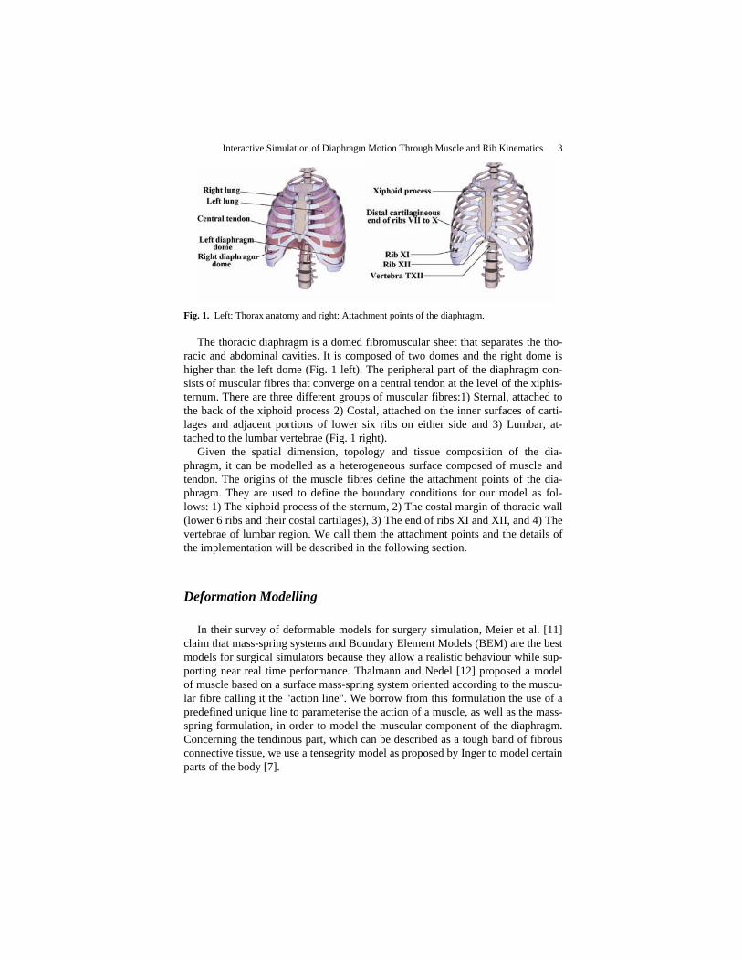
Interactive Simulation of Diaphragm Motion Through Muscle and Rib Kinematics 3
Fig. 1. Left: Thorax anatomy and right: Attachment points of the diaphragm.
The thoracic diaphragm is a domed fibromuscular sheet that separates the tho-racic and abdominal cavities. It is composed of two domes and the right dome is higher than the left dome (Fig. 1 left). The peripheral part of the diaphragm con-sists of muscular fibres that converge on a central tendon at the level of the xiphis-ternum. There are three different groups of muscular fibres:1) Sternal, attached to the back of the xiphoid process 2) Costal, attached on the inner surfaces of carti-lages and adjacent portions of lower six ribs on either side and 3) Lumbar, at-tached to the lumbar vertebrae (Fig. 1 right).
Given the spatial dimension, topology and tissue composition of the dia-phragm, it can be modelled as a heterogeneous surface composed of muscle and tendon. The origins of the muscle fibres define the attachment points of the dia-phragm. They are used to define the boundary conditions for our model as fol-lows: 1) The xiphoid process of the sternum, 2) The costal margin of thoracic wall (lower 6 ribs and their costal cartilages), 3) The end of ribs XI and XII, and 4) The vertebrae of lumbar region. We call them the attachment points and the details of the implementation will be described in the following section.
Deformation Modelling
In their survey of deformable models for surgery simulation, Meier et al. [11] claim that mass-spring systems and Boundary Element Models (BEM) are the best models for surgical simulators because they allow a realistic behaviour while sup-porting near real time performance. Thalmann and Nedel [12] proposed a model of muscle based on a surface mass-spring system oriented according to the muscu-lar fibre calling it the "action line". We borrow from this formulation the use of a predefined unique line to parameterise the action of a muscle, as well as the mass-spring formulation, in order to model the muscular component of the diaphragm. Concerning the tendinous part, which can be described as a tough band of fibrous connective tissue, we use a tensegrity model as proposed by Inger to model certain parts of the body [7].
4 Pierre-Frédéric Villard , Wesley Bourne and Fernando Bello
The word "tensegrity" comes from a contraction of "Tensional integrity". It is used to define a mechanical system with components that combine tension and compression in such a way as to enable the whole system to receive and apply forces, tensions and pressures. Tensegrity systems are composed of two kinds of elements: the elastic elements, which give the system tension, and the rigid ele-ments, which have a constant length and that will exert compression forces.
The evolution of node positions is simulated by taking into account all of its links that could either be elastic or rigid. Each point is represented at a time t by the triplet (a(t), v(t), p(t)) representing the acceleration, velocity and position. As we define the forces to apply on the nodes with elastic and rigid links, we can cal-culate their acceleration with the fundamental law of dynamics:
∑ = af .m (1)
It is then possible to compute the velocity and the position at time (t+Δt):
⎪⎪⎩
⎪⎪⎨
⎧
+ =Δ+
+ =Δ+
∫
∫ ∑Δ+
Δ+
tt
tt
)()(t)p(t
)(/t)(t
t
t
tdtt
tmdt
pv
vfv
(2)
The elastic links are simulated as in the mass-spring system with two parame-ters: their initial length l0 and their elasticity k. A force Fe exerting on an elastic link to elongate its length from l0 to l is thus expressed as follows:
)--k(e 0ll=F (3)
We add a damping force Fd to systematically and progressively decrease the velocity of each node.
vFd .-γ= (4)
The rigid links in the tensegrity part exert reaction forces to avoid changes in their length. We assume that there is no collision between rigid links. For each time step:
1. The elastic forces Fe are computed using Eq. (3) as well as the new positions of the nodes as if there were no rigid links;
Interactive Simulation of Diaphragm Motion Through Muscle and Rib Kinematics 5
2. The rigid constrains are applied to ensure that the distance between two nodes remains constant;
3. Given two nodes A and B linked by a rigid link, A' and B' are their respective positions after applying the classical mass-spring algorithm (as if there was no rigid link). The real positions A1 and B1 considering the rigid links are given by the following:
A'AB'BBBAA1 111 // = (5) such that A1 and B1 remains on the line A'B'.
4. The new velocity is computed at each node.
The resulting set of differential equations is solved using a second order
Runge-Kutta method:
⎪⎪⎩
⎪⎪⎨
⎧
ΔΔ−++Δ+=Δ+Δ++ΔΔ−=Δ+
+ΔΔ−=Δ+=Δ+ ∑
2)(2/1)()()()2/()(2/1)(
)()(2/1)2/()(
ttttttttttttttttttttt
tt
apvpvav
vavfa
(6)
We will now focus on the movement of the diaphragm during inhalation. First,
the anterior-posterior diameter increases because the sternum moves forwards as ribs are raised. Secondly, the transverse diameter increases due to a "pump han-dle" movement which elevates the ribs. Both movements can be simulated by the combined motion of the ribs, the sternum and the cartilage. We first assume that the ribs are rigid and their motion can be modelled by a kinematics law based on the finite helical axis method as in [13]. The cartilage-sternum follows the motion of the ribs.
The attachment points are modelled by a mass-spring system with a very low elasticity in order to ensure that it remains attached. A fall-off function insures that the deformation field is continuous and smooth. The ones belonging to the dia-phragm are automatically obtained such that the distance to the group {ribs, ster-num, cartilage} is under a given threshold. Then, the vertical dimension increases due to diaphragm movement during inhalation. This motion can be simulated by the muscle action generation. We define an action line as a vertical line (inferior-superior) passing through given points and with a certain radius of action. The forces of contraction during inhalation or relaxation during exhalation are com-puted such that their intensity varies according to the distance from the action line.
6 Pierre-Frédéric Villard , Wesley Bourne and Fernando Bello
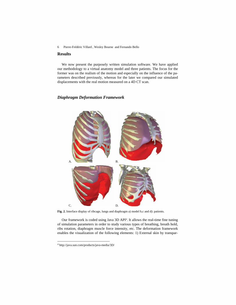
Results
We now present the purposely written simulation software. We have applied our methodology to a virtual anatomy model and three patients. The focus for the former was on the realism of the motion and especially on the influence of the pa-rameters described previously, whereas for the later we compared our simulated displacements with the real motion measured on a 4D CT scan.
Diaphragm Deformation Framework
A. B.
C. D.
Fig. 2. Interface display of ribcage, lungs and diaphragm a) model b,c and d): patients.
Our framework is coded using Java 3D API4. It allows the real-time fine tuning of simulation parameters in order to study various types of breathing, breath hold, ribs rotation, diaphragm muscle force intensity, etc. The deformation framework enables the visualization of the following elements: 1) External skin by transpar-
4 http://java.sun.com/products/java-media/3D/
Interactive Simulation of Diaphragm Motion Through Muscle and Rib Kinematics 7
ency (rigid model), 2) The spine (rigid model), 3) The ribs (modelled following a kinematics law), 4) The sternum and the ligaments attached to the ribs (modelled by a very stiff mass-spring system), and 5) The diaphragm (modelled as described above). Fig. 2 shows an illustration of the organ deformation display with exam-ples of anatomy: a commercial virtual anatomy model and three real patients. The parameters of the simulation are: Damping=-0.02, Stiffness= 15, mass point=0.1 and time step=0.01. Results of the simulation and validation using both models are now presented.
Simplified model
We used a simplified model from a detailed commercial anatomy set (Anatomium 3D by CF Lietzau 3D Special Service). The anatomical parts are shown in Fig. 1. The diaphragm was remeshed to obtain a surface mesh composed of 11359 nodes and 22722 triangles. The tendinous tissue and the muscle tissue were separated by a plane. The rigid links of the tensegrity part were created by linking the upper surface to the lower surface with a heuristic algorithm. The rea-soning behind it is as follows. Once a suitable distance has been chosen, a list of close masses is found. These masses could either be neighbouring masses on the same surface or ideally those on opposing surfaces. Testing the angle removes those on the same surface, leaving a sorted list of masses on the opposing surface.
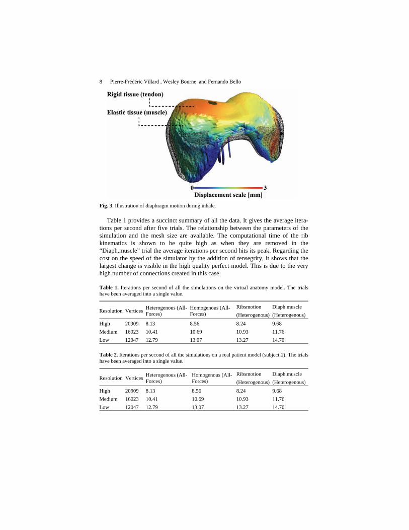
Fig. 3 is an illustration of the simulation results. The diaphragm geometry at the beginning of the simulation (end of inhale) is represented by a wireframe. The fi-nal geometry (end of exhale) is represented by the coloured surface. It can be no-ticed that the tendon has kept its smooth geometry due to the tensegrity elements, while the muscle part has extended more due to their elastic elements. The influ-ence of the three phenomena mentioned before can be seen here: the muscle re-laxation that increases the height of the domes, the ribs rotation that presses the diaphragm on each side and the sternum going backward that pushes the dia-phragm.
Now that the realism of the simulations has been evaluated, we focus on the computational cost of the simulation parameters. The tests below were performed on a dual core 2.4 ghz machine with 2 gigabytes of ram and a 256 megabyte graphics card. The operating system was Windows Vista Service Pack 1 with Java 1.6 installed.
8 Pierre-Frédéric Villard , Wesley Bourne and Fernando Bello
Fig. 3. Illustration of diaphragm motion during inhale.
Table 1 provides a succinct summary of all the data. It gives the average itera-tions per second after five trials. The relationship between the parameters of the simulation and the mesh size are available. The computational time of the rib kinematics is shown to be quite high as when they are removed in the “Diaph.muscle” trial the average iterations per second hits its peak. Regarding the cost on the speed of the simulator by the addition of tensegrity, it shows that the largest change is visible in the high quality perfect model. This is due to the very high number of connections created in this case.
Table 1. Iterations per second of all the simulations on the virtual anatomy model. The trials have been averaged into a single value.
Resolution Vertices Heterogenous (All-Forces)
Homogenous (All-Forces)
Ribsmotion (Heterogenous)
Diaph.muscle (Heterogenous)
High 20909 8.13 8.56 8.24 9.68 Medium 16023 10.41 10.69 10.93 11.76 Low 12047 12.79 13.07 13.27 14.70
Table 2. Iterations per second of all the simulations on a real patient model (subject 1). The trials have been averaged into a single value.
Resolution Vertices Heterogenous (All-Forces)
Homogenous (All-Forces)
Ribsmotion (Heterogenous)
Diaph.muscle (Heterogenous)
High 20909 8.13 8.56 8.24 9.68 Medium 16023 10.41 10.69 10.93 11.76 Low 12047 12.79 13.07 13.27 14.70

Interactive Simulation of Diaphragm Motion Through Muscle and Rib Kinematics 9
Patient specific models
Our method can be used on real patients. To test it, we first took 2 sets of 4D CT scan data acquired during normal tidal breathing, therefore the diaphragm mo-tions were not as large as in the virtual model described above. We then acquired 2 MRI datasets from a volunteer subject at two different levels of breath holding.
The diaphragms were manually segmented using ITK-SNAP5. A mesh was then generated using the CGAL library6 with a mesh angle of 5, mesh distance of 1, and mesh radius of 1. The mesh was smoothed using vtkWindow-edSincPolyDataFilter() with a pass band of 0.2, followed by a 50\% decimation, followed by another 200 iterations of Windowed Sinc smoothing. An example of a segmented and meshed diaphragm is presented on Fig. 4. The mesh is composed of 6633 nodes and 13267 triangles.
Fig.4. Left: Segmented diaphragm inside CT scan and right: Combination of segmentation and the mesh.
The rib cage and the lungs were segmented by the levelset method. Rib motion can be neglected in this case because of the normal tidal breathing and the position of the patient lying down on the scanning table. This assumption has been vali-dated by computing the displacement of the ribs. Therefore, only the diaphragm relaxation needs to be taken into account from the initial exhale to the final exhale. The diaphragm will be attached (as in the virtual model) to the locations described in §2. The simulation is applied with small forces on the previously defined action line and the lungs are linked to the diaphragm with springs.
The simulation was compared to the imaging data at the same point in the breathing cycle by exporting the resulting lung geometry from the fine-tuned simulation and creating a mesh from the segmented 4D CT diaphragm. The differ-ences were then measured and studied with the MESHDEV7 software that com- 5 http://www.itksnap.org/ 6 http://www.cgal.org/ 7 http://www.meshdev.sourceforge.net/
10 Pierre-Frédéric Villard , Wesley Bourne and Fernando Bello
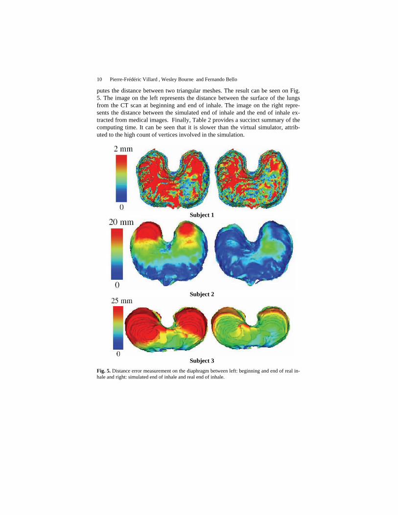
putes the distance between two triangular meshes. The result can be seen on Fig. 5. The image on the left represents the distance between the surface of the lungs from the CT scan at beginning and end of inhale. The image on the right repre-sents the distance between the simulated end of inhale and the end of inhale ex-tracted from medical images. Finally, Table 2 provides a succinct summary of the computing time. It can be seen that it is slower than the virtual simulator, attrib-uted to the high count of vertices involved in the simulation.
Subject 1
Subject 2
Subject 3
Fig. 5. Distance error measurement on the diaphragm between left: beginning and end of real in-hale and right: simulated end of inhale and real end of inhale.
Interactive Simulation of Diaphragm Motion Through Muscle and Rib Kinematics 11
Discussion
The physiological behaviour of this generic model was discussed with our clinical collaborator, who validated that the antero-posterior, transverse and verti-cal dimensions of the thorax are increased as illustrated by the model. It was also pointed out that only the domes descend when breathing lightly, whereas with a deep breath, the domes descend further and the central tendon can move from the level of vertebrae T8 to T9. In full exhale, the right dome reaches the 4th intercos-tal space and the left dome reaches the 5th rib. This complex behaviour further justifies the need for a heterogenous model such as that presented here.
Concerning the patient specific model, it can be seen that the main output of the simulation is the relaxation of the two domes. The contribution is only slightly significant on Subject 1. This is mainly due to the diaphragm displacement (be-ginning to end of inhale) which is not large enough to correctly appreciate the ad-vantage of the simulation. This displacement is much larger with the 4D CT scan of Subject 2 and with the MRI of Subject 3. The average diaphragm displacements are, respectively, 5.8mm with a standard deviation of 5mm and 16mm with a stan-dard deviation of 9.4mm. The aim of our simulator is to obtain a geometry result-ing from the simulation that is as close as possible to the final position of the dia-phragm. Fig. 5 shows a clear correlation between both meshes, with average displacements of 4.29mm with a standard deviation of 3.6mm for Subject 2, and 12mm with a standard deviation of 6.7mm for Subject 3. The diaphragm contrac-tion/relaxation modelling is good enough to simulate a realistic behaviour for liver access procedure training, but it would need to be further improved to be used in radiotherapy where precision is crucial.
The performance evaluations of Tables 1 and 2 show that the addition of the tensegrity constraint makes a slightly slower simulation, but it is still more than adequately fast. The recorded times were very similar. This demonstrates that, even in large models with hundreds of rigid links, the computational time is not massively increased.
Rib kinematics makes a considerable difference to the speed. This is a result of some large equations being solved to update the locations at each iteration. If a pa-tient is observed to only be using the diaphragm and no rib action during respira-tion (as subject 1, 2 and 3), a large computational cost can be avoided. Another in-fluencing factor on the speed of a simulation is the size of the mesh. Both virtual and patient models were tested with different resolutions. The evaluation shows an intuitive linear relationship between the number of vertices in the scene and the time taken in computation.
12 Pierre-Frédéric Villard , Wesley Bourne and Fernando Bello
Conclusions
We have presented here a novel methodology to simulate the behaviour of the diaphragm. It is based on a heterogeneous model composed of mass-spring and tensegrity elements. These latter elements are used to rigidify the tendinous tissue. The physiological boundary conditions have been carefully studied and applied to our model: the diaphragm motion follows more than one parameter, thus our model also takes into account the influence of the rib kinematics, the muscle natu-ral contraction/relaxation and the motion of the sternum.
Our implementation runs in real-time and can be fine-tuned by adjusting the simulation parameters, e.g. if the patient is holding her breath or suddenly having a normal tidal breathing. The anatomy and the physiology can also be customized by generating patient-specific segmentations, mesh models and individual dia-phragm actions. In the future, we aim to further validate our method and apply it to liver biopsy planning and rehearsal, as well as to monitoring lung motion in a radiotherapy context.
References
1. Shirato H et al (2006) Speed and amplitude of lung tumor motion precisely detected in four-dimensional setup and in real-time tumor-tracking radiotherapy. International Journal of Ra-diation Oncology, Biology, Physics 64(4):1229–1235
2. Rohlfing T et al (2004) Modeling liver motion and deformation during the respiratory cycle using intensity-based free-form registration of gated MR images. Medical Physics 31(3):427–432
3. Siebenthal M, Szekely G, Lomax AJ, Cattin P (2007) Intersubject modelling of liver defor-mation during radiation therapy. In: MICCAI (1), pp. 659–666
4. Promayon E (1997) Modelling and simulation of the respiration. Ph.D. thesis, University Jo-seph Fourier of Grenoble
5. Zordan V et al (2004) Breathe easy : Model and control of simulated respiration for anima-tion. In: ACM SIGGRAPH Symposium on Computer Animation
6. Villard PF, Bourne W, Bello F (2008) Modelling Organ Deformation Using Mass-Springs and Tensional Integrity. In: Proceedings of the 4th international Symposium on Biomedical Simulation, LNCS, vol. 5104, pp. 221–226
7. Ingber D (2000) Opposing views on tensegrity as a structural framework for understanding cell mechanics. J Appl Physiol 89:1663–1678
8. Whitelaw WA (1987) Shape and size of the human diaphragm in vivo. J Appl Physiol 62(1):180–186
9. Gauthier AP, Verbanck S, Estenne M, Segerbarth C, Macklem PT, Paiva M (1994) Three-dimensional reconstruction of the in vivo human diaphragm shape at different lung volumes. J Appl Physiol 76(2):495–506
10. Aladin B, Rodarte J (1994) Inferences on passive diaphragm mechanics from gross anatomy. J Appl Physiol 77(5):2065–2070
11. Meier M (2005) Real-time deformable models for surgery simulation: a survey. Comput Methods Programs Biomed. 77(3):183–97
12. Nedel LP, Thalmann D (1998) Real time muscle deformations using mass-spring systems. In: CGI, pp. 156–165

Interactive Simulation of Diaphragm Motion Through Muscle and Rib Kinematics 13
13. Didier AL, Villard PF, Bayle JY, Beuve M, Shariat B (2007) Breathing Thorax Simulation based on Pleura Behaviour and Rib Kinematics. In: IEEE Information Visualisation - Medi-Vis, pp. 35–40