skeletal muscle - springer · pdf filemodifiers and found that a region on chromosome 2 was...
TRANSCRIPT

The superhealing MRL background improvesmuscular dystrophyHeydemann et al.
Skeletal Muscle
Heydemann et al. Skeletal Muscle 2012, 2:26http://www.skeletalmusclejournal.com/content/2/1/26

Skeletal MuscleHeydemann et al. Skeletal Muscle 2012, 2:26http://www.skeletalmusclejournal.com/content/2/1/26
RESEARCH Open Access
The superhealing MRL background improvesmuscular dystrophyAhlke Heydemann1,3, Kayleigh A Swaggart2, Gene H Kim1, Jenan Holley-Cuthrell1, Michele Hadhazy1
and Elizabeth M McNally1,2*
Abstract
Background: Mice from the MRL or “superhealing” strain have enhanced repair after acute injury to the skin,cornea, and heart. We now tested an admixture of the MRL genome and found that it altered the course of musclepathology and cardiac function in a chronic disease model of skeletal and cardiac muscle. Mice lackingγ-sarcoglycan (Sgcg), a dystrophin-associated protein, develop muscular dystrophy and cardiomyopathy similar totheir human counterparts with limb girdle muscular dystrophy. With disruption of the dystrophin complex, themuscle plasma membrane becomes leaky and muscles develop increased fibrosis.
Methods: MRL/MpJ mice were bred with Sgcg mice, and cardiac function was measured. Muscles were assessedfor fibrosis and membrane leak using measurements of hydroxyproline and Evans blue dye. Quantitative trait locusmapping was conducted using single nucleotide polymorphisms distinct between the two parental strains.
Results: Introduction of the MRL genome reduced fibrosis but did not alter membrane leak in skeletal muscle ofthe Sgcg model. The MRL genome was also associated with improved cardiac function with reversal of depressedfractional shortening and the left ventricular ejection fraction. We conducted a genome-wide analysis of geneticmodifiers and found that a region on chromosome 2 was associated with cardiac, diaphragm muscle andabdominal muscle fibrosis.
Conclusions: These data are consistent with a model where the MRL genome acts in a dominant manner tosuppress fibrosis in this chronic disease setting of heart and muscle disease.
Keywords: Cardiomyopathy, Fibrosis, MRL, Muscular dystrophy
BackgroundMurphy Roths Large (MRL) mice are an inbred mousestrain noted to have enhanced healing ability. This MRLstrain was initially discovered because of its rapid abilityto heal ear holes [1,2]. The MRL strain’s capacity to rap-idly recover from injury has been seen for both digitwounding and corneal scarring [3,4]. The MRL strainhas been reported to reduce scar formation after acutecardiac injury, including freeze injury and coronary ar-tery ligation [5,6]. However, other studies have suggestedthat larger scale acute cardiac injury cannot be overcomeby the MRL strain’s healing capacity [7-11]. In those
* Correspondence: [email protected] of Medicine, Section of Cardiology, 5841 S. Maryland, MC 6088,Chicago, IL 60637, USA2Department of Human Genetics, The University of Chicago, Chicago, IL60637, USAFull list of author information is available at the end of the article
© 2012 Heydemann et al.; licensee BioMed CeCreative Commons Attribution License (http:/distribution, and reproduction in any medium
injury settings where the MRL background induces morerapid healing, multiple mechanisms have been impli-cated to explain this phenomenon including decreasedscar formation, altered inflammatory response, reducedapoptosis, increased proliferation, improved remodelingand, in some settings, enhanced stem cell function[4,6,12-15]. Genetic data support that many differentmechanisms account for enhanced healing since morethan 40 different genetic loci have been associated withaspects of the healing phenotype [14,16].The dystrophin complex is composed of membrane-
associated proteins that mediate membrane stability inheart and skeletal muscle. Mutations that disrupt expres-sion of dystrophin or its associated proteins the sarco-glycans proteins cause progressive cardiac and skeletalmuscle degeneration in humans and mouse models. Atthe cellular level, the loss of dystrophin or the
ntral Ltd. This is an Open Access article distributed under the terms of the/creativecommons.org/licenses/by/2.0), which permits unrestricted use,, provided the original work is properly cited.
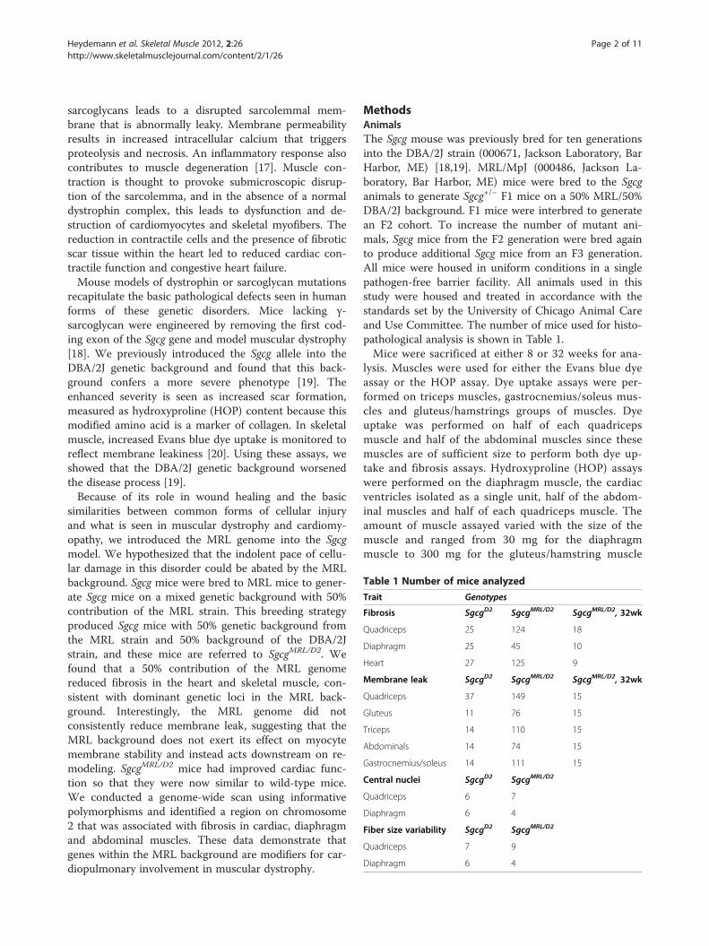
Table 1 Number of mice analyzed
Trait Genotypes
Fibrosis SgcgD2 SgcgMRL/D2 SgcgMRL/D2, 32wk
Quadriceps 25 124 18
Diaphragm 25 45 10
Heart 27 125 9
Membrane leak SgcgD2 SgcgMRL/D2 SgcgMRL/D2, 32wk
Quadriceps 37 149 15
Gluteus 11 76 15
Triceps 14 110 15
Abdominals 14 74 15
Gastrocnemius/soleus 14 111 15
Central nuclei SgcgD2 SgcgMRL/D2
Quadriceps 6 7
Diaphragm 6 4
Fiber size variability SgcgD2 SgcgMRL/D2
Quadriceps 7 9
Diaphragm 6 4
Heydemann et al. Skeletal Muscle 2012, 2:26 Page 2 of 11http://www.skeletalmusclejournal.com/content/2/1/26
sarcoglycans leads to a disrupted sarcolemmal mem-brane that is abnormally leaky. Membrane permeabilityresults in increased intracellular calcium that triggersproteolysis and necrosis. An inflammatory response alsocontributes to muscle degeneration [17]. Muscle con-traction is thought to provoke submicroscopic disrup-tion of the sarcolemma, and in the absence of a normaldystrophin complex, this leads to dysfunction and de-struction of cardiomyocytes and skeletal myofibers. Thereduction in contractile cells and the presence of fibroticscar tissue within the heart led to reduced cardiac con-tractile function and congestive heart failure.Mouse models of dystrophin or sarcoglycan mutations
recapitulate the basic pathological defects seen in humanforms of these genetic disorders. Mice lacking γ-sarcoglycan were engineered by removing the first cod-ing exon of the Sgcg gene and model muscular dystrophy[18]. We previously introduced the Sgcg allele into theDBA/2J genetic background and found that this back-ground confers a more severe phenotype [19]. Theenhanced severity is seen as increased scar formation,measured as hydroxyproline (HOP) content because thismodified amino acid is a marker of collagen. In skeletalmuscle, increased Evans blue dye uptake is monitored toreflect membrane leakiness [20]. Using these assays, weshowed that the DBA/2J genetic background worsenedthe disease process [19].Because of its role in wound healing and the basic
similarities between common forms of cellular injuryand what is seen in muscular dystrophy and cardiomy-opathy, we introduced the MRL genome into the Sgcgmodel. We hypothesized that the indolent pace of cellu-lar damage in this disorder could be abated by the MRLbackground. Sgcg mice were bred to MRL mice to gener-ate Sgcg mice on a mixed genetic background with 50%contribution of the MRL strain. This breeding strategyproduced Sgcg mice with 50% genetic background fromthe MRL strain and 50% background of the DBA/2Jstrain, and these mice are referred to SgcgMRL/D2. Wefound that a 50% contribution of the MRL genomereduced fibrosis in the heart and skeletal muscle, con-sistent with dominant genetic loci in the MRL back-ground. Interestingly, the MRL genome did notconsistently reduce membrane leak, suggesting that theMRL background does not exert its effect on myocytemembrane stability and instead acts downstream on re-modeling. SgcgMRL/D2 mice had improved cardiac func-tion so that they were now similar to wild-type mice.We conducted a genome-wide scan using informativepolymorphisms and identified a region on chromosome2 that was associated with fibrosis in cardiac, diaphragmand abdominal muscles. These data demonstrate thatgenes within the MRL background are modifiers for car-diopulmonary involvement in muscular dystrophy.
MethodsAnimalsThe Sgcg mouse was previously bred for ten generationsinto the DBA/2J strain (000671, Jackson Laboratory, BarHarbor, ME) [18,19]. MRL/MpJ (000486, Jackson La-boratory, Bar Harbor, ME) mice were bred to the Sgcganimals to generate Sgcg+/− F1 mice on a 50% MRL/50%DBA/2J background. F1 mice were interbred to generatean F2 cohort. To increase the number of mutant ani-mals, Sgcg mice from the F2 generation were bred againto produce additional Sgcg mice from an F3 generation.All mice were housed in uniform conditions in a singlepathogen-free barrier facility. All animals used in thisstudy were housed and treated in accordance with thestandards set by the University of Chicago Animal Careand Use Committee. The number of mice used for histo-pathological analysis is shown in Table 1.Mice were sacrificed at either 8 or 32 weeks for ana-
lysis. Muscles were used for either the Evans blue dyeassay or the HOP assay. Dye uptake assays were per-formed on triceps muscles, gastrocnemius/soleus mus-cles and gluteus/hamstrings groups of muscles. Dyeuptake was performed on half of each quadricepsmuscle and half of the abdominal muscles since thesemuscles are of sufficient size to perform both dye up-take and fibrosis assays. Hydroxyproline (HOP) assayswere performed on the diaphragm muscle, the cardiacventricles isolated as a single unit, half of the abdom-inal muscles and half of each quadriceps muscle. Theamount of muscle assayed varied with the size of themuscle and ranged from 30 mg for the diaphragmmuscle to 300 mg for the gluteus/hamstring muscle

Heydemann et al. Skeletal Muscle 2012, 2:26 Page 3 of 11http://www.skeletalmusclejournal.com/content/2/1/26
group that included the semimembranosus, semitendi-nosus and biceps femoris.
Evans blue dye uptake assay for membrane leakEvans blue dye (Sigma, E-2129) was performed asdescribed [19,21]. Evans blue dye (Sigma, E-2129) wasdissolved in phosphate-buffered saline at 10 mg/ml andinjected intraperitoneally at 5 μl/g body weight. Twentyto 40 h later, the tissues were harvested, finely minced,weighed and incubated at 55°C in 1 ml formamide for2 h before spectrophotometric absorbance was measuredat 620 nm [22,23]. Results are reported as absorbance/mg tissue.
Hydroxyproline assay for fibrosisThe hydroxyproline (HOP) assay was performed asdescribed [19,21,24]. The tissue was minced, weighedand hydrolyzed overnight in 2 ml of 6 M hydrochloricacid at 110°C. Ten μl of this hydrolysate was mixed with150 μl isopropanol, then 75 μl of 1.4% chloramine-T(Sigma, St Louis, MO) in citrate buffer and oxidized atroom temperature for 10 min. One ml of a 3:13 solutionof Ehrlich’s reagent (3 g of 4-(dimethylamino) benzalde-hyde, Sigma, St Louis, MO; 10 ml ethanol; 675 μl sul-furic acid) to isopropanol was added, mixed andincubated for 30 min at 55°C followed by extinctionmeasurement at 558 nm. A standard curve (0–5000 nM,trans-4-hydroxy-L-proline, Sigma, St Louis, MO) wasincluded in each assay. Results are reported as nMHOP/mg tissue.
Immunofluorescence microscopyTissues were flash frozen in liquid nitrogen-cooled iso-pentane and stored at 80°C; 7-μm sections were cut on acryostat and fixed to slides in ice-cold 100% methanol.The following antibodies were used: dystrophin NCL-DYS2 (Novocastra/Leica), PH3 04–817 (Millipore), CD3MON1003-1 (Monsanto), caspase 3 (BD Biosciences),MAC1 BD557395 (BD Biosciences) and eMHC F1.652(ATCC). The TUNEL kit was from Millipore. Centralnuclei and fiber size variability was determined blindedto genotype by analyzing ten randomly chosen fields of40× magnification. Fiber size variability was comparedusing each animal’s coefficient of variability (standarddeviation/mean).
EchocardiographyTwelve-week animals were evaluated by echocardiog-raphy as described [25,26]. Investigators were blinded togenotype. To avoid the stress associated with consciousrestraint, anesthetized animals were studied. Anesthesiawas induced by 1% isoflurane in a closed chamber(Ohmeda Fluotec 3; Matrix Medical, Orchard Park, NY)in 20% O2 delivered through a nose cone. Chest hairs
were removed with a topical depilatory agent. Limb leadswere attached for electrocardiogram gating, and the ani-mals were imaged in the left lateral decubitus position witha Visual Sonics Vevo 770 machine using a 30-MHz high-frequency transducer. Body temperature was maintainedusing a heated imaging platform and warming lamps.Anesthesia was variably delivered to maintain heart ratesthroughout the procedure at a constant 380–420 beats perminute. Two-dimensional images were recorded in para-sternal long- and short-axis projections, with guided M-mode recordings at the midventricular level in both views.LV cavity size and wall thickness were measured in at leastthree beats from each projection and averaged. LV wallthickness, interventricular septum (IVS) and posterior wall(PW) thickness, and internal dimensions at diastole andsystole (LVIDd and LVIDs, respectively) were measured.LV fractional shortening [(LVIDd – LVIDs)/LVIDd] andrelative wall thickness [(IVS thickness + PW thickness)/LVIDd] were calculated from the M-mode measurements.
Genetic and statistical analysisSingle nucleotide polymorphisms (SNPs, n = 1,701) in-formative between the parental DBA/2J and MRL/MpJstrains were genotyped in 80 SgcgMRL/D2F2-F4 Sgcg ani-mals on the Illumina GoldenGate platform using theMutation Mapping and Developmental Analysis Panel(MMDAP) [27] and the Mouse Universal GenotypingArray (GeneSeek, Neogen Corp., Lansing, MI) [28]. Rpackage QTLRel was used to perform whole-genomequantitative trait locus (QTL) mapping for each of themembrane permeability and fibrosis phenotypes andusing sex as a covariate [29,30]. Significance thresholdswere determined by 1,000 permutation tests. The 1.5-LOD drop support interval was calculated usingQTLRel. Tests of normality and other statistics were cal-culated in Prism (GraphPad). Build 37.1 was used forgenomic analysis.For HOP and dye uptake assays, data were analyzed by
one-way, unpaired ANOVA with parametric methodsfollowed by the Tukey multiple comparison post-test(Prism, Graphpad), and p < 0.05 was considered signifi-cant. Data from 197 Sgcg animals from an F3 intercrossbetween the DBA/2J and 129T2/SvEmsJ backgrounds(SgcgD2/129 ) was compared to the SgcgMRL/D2cohort.
ResultsA 50% contribution of the MRL background reducesfibrosis in Sgcg miceTo assess the MRL contribution to muscular dystrophy,we used the MRL/MpJ substrain since it contains a wild-type fas allele [1]. We also used mice lacking γ-sarcoglycan (Sgcg null) since these mice are a model oflimb girdle muscular dystrophy 2C [18] and on theDBA/2J background have a more severe phenotype,

Heydemann et al. Skeletal Muscle 2012, 2:26 Page 4 of 11http://www.skeletalmusclejournal.com/content/2/1/26
reminiscent of what is seen in humans [19,21]. MRL/MpJ mice were bred to Sgcg null and then interbred togenerate Sgcg null mice with a 50% MRL/MpJ and 50%DBA/2J contribution. In Sgcg null mice, like all dys-trophin complex-associated mutations, fibrosis and col-lagen deposition is increased in the heart and muscle,similar to what is seen in human patients with similarmutations [18,31]. A 50% contribution from the MRLbackground reduced fibrosis in Sgcg heart and muscles(Figure 1). In Sgcg hearts, fibrosis was often seen grosslyas large patchy white areas and with the introduction ofthe MRL background visible fibrosis was diminished sothat the hearts were indistinguishable from those ofwild-type mice (Additional file 1: Figure S1). Of all themuscle groups analyzed, only diaphragm muscle retainedany visible fibrosis. Diaphragm muscle is the most con-sistently damaged muscle in multiple mouse models, in-cluding this model, and the mdx model of Duchennemuscular dystrophy [32]. Therefore, it is possible thatthe degree of injury in this muscle overwhelms the heal-ing properties of the MRL background.We quantified fibrosis by measuring HOP content as
an indicator of collagen in SgcgD2 and SgcgMRL/D2 mus-cles. SgcgMRL/D2 mice have significantly reduced fibrosiscompared to SgcgD2 mice (Figure 2). We also evaluated
Figure 1 The MRL genome suppresses fibrosis in the heart andmuscles of SgcgD2 mice. Sgcg mice lack the dystrophin-associatedprotein, γ-sarcoglycan, and when in the DBA/2J (D2) backgroundhave a more severe phenotype with enhanced membrane leak andfibrosis [21]. A 50% contribution of the MRL genome, referred to asSgcgMRL/D2, suppressed fibrosis in both heart and skeletal muscle.
fibrosis in aged (32 week) animals and found a persistentreduction in fibrosis in the SgcgMRL/D2 mice (Figure 2, di-agonal bars). The 129T2/SvEmsJ strain was previouslyshown to suppress the severity of muscle pathology in Sgcgnull mice [33]. In comparison, the MRL background
Figure 2 The MRL genome quantitatively reduces fibrosis inSgcg mice. Hydroxyproline (HOP) is a measure of fibrosis and isrepresented on the y axis (mM/mg). In the heart, diaphragm andquadriceps muscles, fibrosis is significantly (p < 0.001) reduced inintercrossed SgcgMRL/D2 (D2/MRL) animals compared to Sgcg in the D2(D2) background at 8 weeks. Fibrosis remains significantly (***p < 0.001)reduced in SgcgMRL/D2 animals at the 32-week time point.

Heydemann et al. Skeletal Muscle 2012, 2:26 Page 5 of 11http://www.skeletalmusclejournal.com/content/2/1/26
suppressed fibrosis more than the 129T2/SvEmsJ back-ground (Figure 3). The MRL background dramaticallyreduced fibrosis in the heart and abdominal muscles com-pared to 129T2/SvEmsJ. The MRL background also sup-pressed fibrosis more than the 129T2/SvEmsJ backgroundin the diaphragm and quadriceps muscles, but to a lesserdegree. This finding suggests that the phenotypically bene-ficial genetic modifiers in the MRL genome may be morepotent than those in the 129T2/SvEmsJ genome.
The MRL background does not protect against membraneleakDisruption of the membrane-associated dystrophin com-plex renders the sarcolemma unusually fragile, leading toabnormal membrane leakage that is visualized by uptake ofthe nonspecific vital tracer Evans blue dye [20]. Upon grossinspection, the muscles from the SgcgMRL/D2 mice displayedhigh levels of dye uptake, comparable to what was observedin the parental Sgcg mice (Additional file 1: Figure S1). Ona microscopic level, dye-positive cells were readily detect-able in SgcgMRL/D2 muscle and heart (Figure 4). Evans bluedye levels were measured in multiple muscle groups andwere not significantly different between SgcgMRL/D2 andSgcgD2 muscles for quadriceps, triceps, gastrocnemius/soleus, gluteus and abdominals muscle groups (Figure 5).We also measured dye uptake in older animals at 32 weeksto assess disease progression. At 32 weeks, the skeletalmuscles continued to show comparable levels of membraneleak as what was seen at 8 weeks (Figure 5). The gluteus/hamstring group of muscles showed an increased of dyeuptake compared to the 8-week animals. This increase wasnot seen for other muscle groups.
Figure 3 The reduction of fibrosis in intercrossed Sgcg animals is spefibrosis and is represented on the y axis (mM/mg). Fibrosis is significantly r(*p = 0.041) and abdominal muscles (p < 0.001) of Sgcg animals intercrosseD2/129 background.
We compared suppression of dye uptake by the MRLbackground to that induced by the 129T2/SvEmsJ back-ground in Sgcg mice (Figure 6). The quadriceps and ab-dominal muscles from Sgcg null mice with a contributionfrom the 129T2/SvEmsJ background showed increasedmembrane leak compared to Sgcg mice with an MRL con-tribution. However, the triceps muscle group from theMRL background showed increased dye uptake, and thegluteus/hamstring muscle group showed no significant dif-ference. Thus, there was no consistent suppression ofmembrane leakiness by the MRL strain.
The MRL genome may promote skeletal muscleregenerationThe MRL background is thought to exert part of its effectby promoting regeneration [34]. We examined embryonicmyosin heavy chain (eMHC) expression as a reflection ofregeneration. SgcgMRL/D2 muscle shows patchy areas with aqualitative increase in eMHC-positive fibers compared toSgcg (Figure 7), and wild-type muscle showed no eMHCpositive fibers (data not shown). In dystrophic skeletalmuscle, ongoing regeneration is thought to offset degener-ation. Consistent with this, eMHC-positive regions werealso positive for Evans blue dye uptake, indicative ofmuscle damage. We also evaluated phosphorylated histone3 (PH3) as a reflection of mitotic index. No clear differ-ences for PH3 staining were seen between SgcgMRL/D2 andSgcg muscle. Regenerating skeletal muscle, whether fromtrauma or muscular dystrophy, is identified by the presenceof myofibers with centrally positioned nuclei. In musculardystrophy, ongoing regeneration is also marked byincreased fiber size variability. SgcgMRL/D2 mice have an
cific to the MRL genome. Hydroxyproline (HOP) is a measure ofeduced in the heart (***p < 0.001), diaphragm (p < 0.001), quadricepsd in the D2/MRL background compared to those intercrossed in the

Figure 4 Membrane leak is not corrected by the presence ofthe MRL genome in Sgcg heart and muscle. Evans blue dye isfound in cardiomyocytes and skeletal myofibers reflectingmembrane leakiness. Dye uptake occurs in patchy patternthroughout the heart and muscle and is seen as opacified cells thatfluoresce red. Nuclei are shown in blue and dystrophin in green.
Heydemann et al. Skeletal Muscle 2012, 2:26 Page 6 of 11http://www.skeletalmusclejournal.com/content/2/1/26
increased number of central nuclei in quadriceps and dia-phragm muscle compared to SgcgD2 mice (Figure 7). Thesesame muscles also showed increased fiber size variabilitywhen there is a contribution from the MRL background.These data are consistent with a model in which favorablematrix remodeling may support enhanced regeneration.We also assessed apoptosis using a TUNEL assay and
caspase 3 staining and again found no differences be-tween SgcgMRL/D2 and Sgcg muscle, suggesting thatgross differences in programmed cell death are unlikelyto account for the improved healing of the MRL strain(Additional file 1: Figure S2). We also characterizedwhether T cell infiltration or macrophage infiltrationdiffered qualitatively between SgcgMRL/D2 and Sgcgmuscle, but we found no clear differences betweenSgcgMRL/D2 and Sgcg muscle, suggesting other featurescontribute to the MRL background’s effect on musculardystrophy (Additional file 1: Figure S2).
Improved cardiac function from a 50% contribution ofthe MRL backgroundWe performed 2D and M mode echocardiography onSgcgMRL/D2 and SgcgD2 mice at 12 weeks of age. Both frac-tional shortening and the left ventricular ejection fractionwere significantly reduced in SgcgD2 mice, indicating thatthe increased fibrosis impairs cardiac function (Table 2).Fractional shortening and the left ventricular ejection frac-tion were similar between SgcgMRL/D2 and WTMRL/D2 mice,consistent with improved function mediated by 50% of theMRL genome. Wall thickness was also increased in Sgcgmice compared to the wild type of the same background.In contrast, wall thickness was not different betweenSgcgMRL/D2 and WTMRL/D2 mice.
Chromosome 2 associates with reduced fibrosis Sgcgheart and diaphragm muscleWe conducted a genome-wide scan using markers thatwere informative in the two parental strains DBA/2J andMRL/MpJ (Figure 8). QTLRel was used to identifyregions of association; this analysis takes into accountthe relatedness of individual animals in the cohort[29,30]. A region on chromosome 2 was identified thatassociated with fibrosis in the heart. The 1.5-LOD dropinterval of this region spans from 64.8008−73.1758 cMin mouse genome build 37.1. This same region was alsoassociated with fibrosis in the diaphragm muscle andalso for fibrosis in the abdominal muscles. It should benoted that the significance of these associations is sug-gestive (p < 0.63) when using the stringent QTLRel ana-lysis. However, the overlapping intervals found in heart,diaphragm and abdominal muscles provide additionalsupport that this interval modifies fibrosis.
Ltbp4 polymorphism does not account for the MRLhealing propertiesThe DBA/2J strain contains an insertion/deletion poly-morphism within the Ltbp4 gene [21] that modifies bothmembrane permeability and fibrosis, as two independenttraits, in Sgcg muscular dystrophy. Ltbp4 encodes latentTGFβ-binding protein 4, and TGFβ proteins have beenextensively linked to fibrosis in many disease states in-cluding muscular dystrophy [35,36]. Most murinestrains, including the MRL/MpJ strain used here, containthe protective Ltbp4 allele with an additional 12 aminoacids inserted in exon 12. Because the SgcgMRL/D2 cohortused here contained both the protective (LtbpI allele)and the disease-enhancing allele (Ltbp4D), we testedwhether Ltbp4 genotype correlated with fibrosis in theSgcgMRL/D2 cohort. Table 3 shows that neither membranepermeability nor fibrosis is correlated with the Ltbp4genotype in the SgcgMRL/DBA2J cohort. Therefore, Ltbp4does not account for the suppressive effect of the MRL

Figure 5 The MRL genome does not significantly reduce membrane damage in Sgcg muscle. Evans blue dye uptake is a measure ofmembrane damage and is represented on the y axis (absorbance/mg). In the quadriceps, triceps, abdominal and gastrocnemius/soleus muscles,membrane damage is not significantly different between SgcgD2 animals and intercrossed SgcgMRL/D2 animals at both the 8- and 32-week timepoints. In the gluteus/hamstring muscles, membrane damage is significantly (**p < 0.01) increased at the 32-week time point in intercrossed SgcgMRL/D2 animals compared to the 8-week time point.
Figure 6 The MRL genome has a variable ability to reduce membrane leak in muscular dystrophy compared to the 129T2/SvEmsJstrain. Evans blue dye uptake was measured in multiple muscle groups represented on the y axis (absorbance/mg). Membrane damage isreduced in the quadriceps (**p = 0.0053) and abdominal (**p = 0.0012) muscles of intercrossed SgcgD2/MRL animals compared to intercrossedSgcgD2/129 mice. Membrane damage is increased in the triceps muscle (*p = 0.0298) of intercrossed SgcgMRL/D2 animals. There is no difference inmembrane damage in the gluteus/hamstring muscle.
Heydemann et al. Skeletal Muscle 2012, 2:26 Page 7 of 11http://www.skeletalmusclejournal.com/content/2/1/26
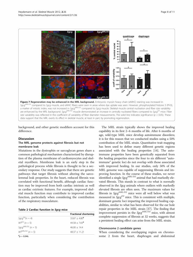
Figure 7 Regeneration may be enhanced in the MRL background. Embryonic myosin heavy chain (eMHC) staining was increased inSgcgMRL/D2 compared to Sgcg muscle, and eMHC fibers were seen in areas where dye uptake was seen. However, phosphorylated histone 3 (PH3),a marker of mitotic index, was not increased in SgcgMRL/D2 compared to Sgcg muscle. Skeletal muscle central nucleation and fiber size variabilityare enhanced by the MRL background. SgcgMRL/D2 muscle demonstrated an increase in centrally nucleated fibers compared to SgcgD2 mice. Fibersize variability was reflected in the coefficient of variability of fiber diameter measurements. The solid line indicates significance (p < 0.05). Thesedata support that the MRL exerts its effect in skeletal muscle, at least in part, by promoting regeneration.
Heydemann et al. Skeletal Muscle 2012, 2:26 Page 8 of 11http://www.skeletalmusclejournal.com/content/2/1/26
background, and other genetic modifiers account for thisdifference.
DiscussionThe MRL genome protects against fibrosis but notmembrane leakMutations in the dystrophin or sarcoglycan genes share acommon pathological mechanism characterized by disrup-tion of the plasma membrane of cardiomyocytes and skel-etal myofibers. Membrane leak is an early step in thepathological process while fibrosis is thought to be a sec-ondary response. Our study suggests that there are geneticpathways that target fibrosis without altering the sarco-lemmal leak properties. In the heart, reduced fibrosis wascorrelated with functional benefit, although cardiac func-tion may be improved from both cardiac intrinsic as wellas cardiac extrinsic features. For example, improved skel-etal muscle function may contribute to improved cardiacfunction, particularly when considering the contributionof the respiratory musculature.
Table 2 Cardiac function in Sgcg mice
Fractional shortening
SgcgD2(n = 4) 33.87 ± 4.80a
WTD2 (n = 5) 44.58 ± 7.65a
SgcgMRL/D2 (n = 5) 46.00 ± 14.4
WTMRL/D2 (n = 5) 48.37 ± 11.21ap = 0.046.
The MRL strain typically shows the improved healingcapability in its first 2–6 months of life. After 6 months ofage, wild-type MRL mice develop autoimmune disorders;it is for this reason that we conducted studies using a 50%contribution of the MRL strain. Quantitative trait mappinghas been used to define many different genetic regionsassociated with the healing properties [14]. The auto-immune properties have been genetically separated fromthe healing properties since the four to six different “auto-immune” genetic loci do not overlap with those associatedwith improved healing. In our studies, only 50% of theMRL genome was capable of suppressing fibrosis and im-proving function. In the course of these studies, we neveridentified a single SgcgMRL/D2 animal that had markedly ele-vated fibrosis. This stands in contrast to what is normallyobserved in the Sgcg animals where outliers with markedlyelevated fibrosis are often seen. The maximum values forfibrosis in SgcgMRL/D2 mice were all well below what wasmeasured in SgcgD2 mice. This is consistent with multiple,dominant genetic loci imparting the improved healing cap-abilities, similar to what has been observed for the ear holerepair properties in the MRL strain [37]. The finding thatimprovement persists in the SgcgMRL/D2 mice, with almostcomplete suppression of fibrosis at 32 weeks, suggests thata persistent healing effect can arise from the MRL strain.
Chromosome 2 candidate genesWhen considering the overlapping region on chromo-some 2 from the heart, diaphragm and abdominal

Figure 8 Fibrosis in the heart, diaphragm and abdominalmuscles is modified by a locus on chromosome 2. Genome-wideassociation was examined for fibrosis in SgcgMRL/D2 progeny in the(A) cardiac muscle (n = 65), (B) diaphragm (n = 78) and (C)abdominal muscle (n = 78). Chromosomes are plotted on the x axis,and the informative SNPs tested (n = 1,707) are displayed as opencircles that alternate color by chromosome; LOD scores arerepresented on the y axis. Overlapping regions on chromosome 2showed suggestive (p < 0.63) association with fibrosis in heart,diaphragm and abdominal muscle.
Table 3 Ltbp4 genotype in SgcgMRL/D2 mice
Dye uptake Fibrosis
Ltbp4D/D (n = 14) 3.90 ± 0.83 6.00 ± 1.81
Ltbp4D/I (n = 34) 4.18 ± 0.84 8.05 ± 3.02
LtbpI/I (n = 47) 4.00 ± 0.85 6.64 ± 2.15
Heydemann et al. Skeletal Muscle 2012, 2:26 Page 9 of 11http://www.skeletalmusclejournal.com/content/2/1/26
fibrosis data, the interval contains 49 known genes.There are additional predicted and unnamed genes andgenes encoding olfactory receptors that are not consid-ered likely candidates. Within the interval is Jag1, encod-ing jagged the ligand for the Notch receptor. Loss offunction of Notch 3 leads to muscle hyperplasia, espe-cially when subjected to repetitive injury [38]. Ex vivoactivation of Notch signaling helps maintain donor cellengraftment during myoblast transfer [39]. Another genelinked to growth and healing in the interval is Bmp2-en-coding bone morphogenetic protein 2. The BMPs medi-ate musculoskeletal regeneration [40], and given theMRL background effect on multiple cells and tissues,BMP2 is well positioned to contribute to the MRLsuperhealing response. That said, this chromosome 2interval has not previously been linked to other MRLhealing properties. The strict criteria imposed by theQTLRel algorithm makes it unlikely that these resultsderive from relatedness of animals within the cohort.
Cardiac injury and repair in the MRL strainThe ability of the heart to recover after injury has beenstudied in the MRL strain using cryoinjury, left anteriordescending ligation and ischemia reperfusion methods[5,6,8-10,14,15]. However, scar reduction has been notedafter some forms of injury while not after others. Incommon to all these studies is an acute injury modelwhere a normal heart was substantially damaged in asingle setting. The MRL’s ability to heal the heart may belimited such that larger amounts of injury may be insur-mountable, as proposed by Naseem [6]. Our data sup-port that lower, although persistent, levels of injury canbe managed by the MRL strain where there is significantreduction in fibrosis and a corresponding functional im-provement in cardiac function.
Mechanisms for MRL healingA number of mechanisms likely act in concert to achievesuppression of fibrosis and improvement of function. Inskeletal muscle, where muscle stem cells robustly regen-erate muscle after injury, there is indirect evidence ofenhanced regeneration. Specifically, there is an increasein centrally nucleated myofibers, which is thought to re-flect enhanced myoblast fusion. However, our data donot distinguish whether the MRL background exerts itseffect on muscle regenerative cells, or by creating a moresupportive extracellular matrix or both. MRL animalsheal with embryonic characteristics with enhanced blas-tema formation and metabolic and gene expression fea-tures consistent with an earlier developmental state [41].Altered protease expression has also been noted in theMRL mouse in response to damage [5]. Recent workcharacterizing the immune infiltrate in muscular dys-trophy found that reducing osteopontin was effective at

Heydemann et al. Skeletal Muscle 2012, 2:26 Page 10 of 11http://www.skeletalmusclejournal.com/content/2/1/26
suppressing fibrosis [17]. Together these data may favormatrix-associated modifications that can modulatemuscle fibrosis and function.
ConclusionsHerein the MRL background was shown to reduce fibrosisin a chronic model of muscular dystrophy and cardiomy-opathy, the Sgcg mouse. Although membrane leak was stillevident in Sgcg mice sharing a portion of the MRL gen-ome, the reduction in fibrosis was associated withimproved cardiac function. The identification of gene(s)from the MRL genome will help identify pathways import-ant for chronic repair of myopathic processes.
Additional file
Additional file 1: Figure S1. Shown are gross images from Sgcg vs.SgcgMRL/D2 mice. Evans blue dye uptake could be readily seen in thequadriceps and diaphragm muscles and did not appear grossly alteredby the presence of the MRL background. In contrast, fibrosis was visuallyreduced in the quadriceps and heart of SgcgMRL/D2 compared to Sgcgmice. The diaphragm muscle retained evidence of fibrosis in SgcgMRL/D2,but the white stripes of fibrosis were smaller, and intact diaphragmmuscle was still evident compared to the near total replacement ofdiaphragm muscle in Sgcg mice. Figure S2. Shown is staining forapoptosis with TUNEL and caspase indicating no gross differencesbetween Sgcg and SgcgMRL/D2 muscle. CD3 and MAC1 staining toexamine T cell and macrophage infiltrate also did not appear grosslyaltered by the presence of the MRL background.
AbbreviationsBMP: Bone morphogenetic protein; D2: (DBA/2J); DMD: Duchenne musculardystrophy; HOP: Hydroxyproline; LTBP4: Latent TGFβ-binding protein;MRL: Murphy Roth Large; QTL: Quantitative trait loci; SNP: Single nucleotidepolymorphism; TGFβ: Transforming growth factor β.
Competing interestThe authors have no competing interests related to this work.
Authors’ contributionsAH conducted the phenotypic analysis of muscle. KAS conducted thegenome-wide SNP analysis and phenotype analysis. GK performed theechocardiographic analysis. MH oversaw the breeding. JHC assisted withphenotypic analysis. EMM conceived the experiments, analyzed the data andwrote the manuscript. All authors read and approved the final manuscript.
AcknowledgementsSupported by NIH R01HL61322 (EMM), R01HL102322 (AH) and NIHK08HL098565 (GK).
Author details1Department of Medicine, Section of Cardiology, 5841 S. Maryland, MC 6088,Chicago, IL 60637, USA. 2Department of Human Genetics, The University ofChicago, Chicago, IL 60637, USA. 3Current address: Department of Physiologyand Biophysics, University of Illinois at Chicago, COMRB 2035, MC 901, 835South Wolcott Ave, Chicago, IL 60612-7352, USA.
Received: 24 May 2012 Accepted: 8 October 2012Published: 05 December 2012
References1. Clark LD, Clark RK, Heber-Katz E: A new murine model for mammalian wound
repair and regeneration. Clin Immunol Immunopathol 1998, 88:35–45.
2. McBrearty BA, Clark LD, Zhang XM, Blankenhorn EP, Heber-Katz E: Geneticanalysis of a mammalian wound-healing trait. Proc Natl Acad Sci U S A1998, 95:11792–11797.
3. Gourevitch DL, Clark L, Bedelbaeva K, Leferovich J, Heber-Katz E: Dynamicchanges after murine digit amputation: the MRL mouse digit showswaves of tissue remodeling, growth, and apoptosis. Wound Repair Regen2009, 17:447–455.
4. Ueno M, Lyons BL, Burzenski LM, Gott B, Shaffer DJ, Roopenian DC, ShultzLD: Accelerated wound healing of alkali-burned corneas in MRL mice isassociated with a reduced inflammatory signature. Invest Ophthalmol VisSci 2005, 46:4097–4106.
5. Leferovich JM, Bedelbaeva K, Samulewicz S, Zhang XM, Zwas D, LankfordEB, Heber-Katz E: Heart regeneration in adult MRL mice. Proc Natl Acad SciU S A 2001, 98:9830–9835.
6. Naseem RH, Meeson AP, Michael Dimaio J, White MD, Kallhoff J, HumphriesC, Goetsch SC, De Windt LJ, Williams MA, Garry MG, Garry DJ: Reparativemyocardial mechanisms in adult C57BL/6 and MRL mice followinginjury. Physiol Genomics 2007, 30:44–52.
7. Abdullah I, Lepore JJ, Epstein JA, Parmacek MS, Gruber PJ: MRL mice fail toheal the heart in response to ischemia-reperfusion injury. Wound RepairRegen 2005, 13:205–208.
8. Cimini M, Fazel S, Fujii H, Zhou S, Tang G, Weisel RD, Li RK: The MRL mouseheart does not recover ventricular function after a myocardial infarction.Cardiovasc Pathol 2008, 17:32–39.
9. Grisel P, Meinhardt A, Lehr HA, Kappenberger L, Barrandon Y, Vassalli G: TheMRL mouse repairs both cryogenic and ischemic myocardial infarctswith scar. Cardiovasc Pathol 2008, 17:14–22.
10. Robey TE, Murry CE: Absence of regeneration in the MRL/MpJ mouseheart following infarction or cryoinjury. Cardiovasc Pathol 2008, 17:6–13.
11. Oh YS, Thomson LE, Fishbein MC, Berman DS, Sharifi B, Chen PS: Scarformation after ischemic myocardial injury in MRL mice. Cardiovasc Pathol2004, 13:203–206.
12. Harty M, Neff AW, King MW, Mescher AL: Regeneration or scarring: animmunologic perspective. Dev Dyn 2003, 226:268–279.
13. Gourevitch D, Clark L, Chen P, Seitz A, Samulewicz SJ, Heber-Katz E: Matrixmetalloproteinase activity correlates with blastema formation in theregenerating MRL mouse ear hole model. Dev Dyn 2003, 226:377–387.
14. Heber-Katz E, Leferovich J, Bedelbaeva K, Gourevitch D, Clark L: The scarlessheart and the MRL mouse. Philos Trans R Soc Lond B Biol Sci 2004,359:785–793.
15. Bedelbaeva K, Gourevitch D, Clark L, Chen P, Leferovich JM, Heber-Katz E:The MRL mouse heart healing response shows donor dominance inallogeneic fetal liver chimeric mice. Cloning Stem Cells 2004, 6:352–363.
16. Heydemann A: The super super-healing MRL mouse strain. Front Biol2012, in press.
17. Spencer MJ, Montecino-Rodriguez E, Dorshkind K, Tidball JG: Helper (CD4(+)) and cytotoxic (CD8(+)) T cells promote the pathology of dystrophin-deficient muscle. Clin Immunol 2001, 98:235–243.
18. Hack AA, Ly CT, Jiang F, Clendenin CJ, Sigrist KS, Wollmann RL, McNallyEM: Gamma-sarcoglycan deficiency leads to muscle membrane defectsand apoptosis independent of dystrophin. J Cell Biol 1998,142:1279–1287.
19. Heydemann A, Huber JM, Demonbreun A, Hadhazy M, McNally EM: Geneticbackground influences muscular dystrophy. Neuromuscul Disord 2005,15:601–609.
20. Straub V, Rafael JA, Chamberlain JS, Campbell KP: Animal models formuscular dystrophy show different patterns of sarcolemmal disruption.J Cell Biol 1997, 139:375–385.
21. Heydemann A, Ceco E, Lim JE, Hadhazy M, Ryder P, Moran JL, Beier DR,Palmer AA, McNally EM: Latent TGF-beta-binding protein 4 modifiesmuscular dystrophy in mice. J Clin Invest 2009, 119:3703–3712.
22. Matsuda R, Nishikawa A, Tanaka H: Visualization of dystrophic musclefibers in mdx mouse by vital staining with Evans blue: evidence ofapoptosis in dystrophin-deficient muscle. J Biochem (Tokyo) 1995,118:959–964.
23. Thurston G, Suri C, Smith K, McClain J, Sato TN, Yancopoulos GD, McDonaldDM: Leakage-resistant blood vessels in mice transgenicallyoverexpressing angiopoietin-1. Science 1999, 286:2511–2514.
24. Flesch M, Schiffer F, Zolk O, Pinto Y, Rosenkranz S, Hirth-Dietrich C, Arnold G,Paul M, Bohm M: Contractile systolic and diastolic dysfunction in renin-induced hypertensive cardiomyopathy. Hypertension 1997, 30:383–391.

Heydemann et al. Skeletal Muscle 2012, 2:26 Page 11 of 11http://www.skeletalmusclejournal.com/content/2/1/26
25. Collins KA, Korcarz CE, Shroff SG, Bednarz JE, Fentzke RC, Lin H, Leiden JM,Lang RM: Accuracy of echocardiographic estimates of left ventricularmass in mice. Am J Physiol Heart Circ Physiol 2001, 280:H1954–H1962.
26. Wheeler MT, Korcarz CE, Collins KA, Lapidos KA, Hack AA, Lyons MR,Zarnegar S, Earley JU, Lang RM, McNally EM: Secondary coronary arteryvasospasm promotes cardiomyopathy progression. Am J Pathol 2004,164:1063–1071.
27. Moran JL, Bolton AD, Tran PV, Brown A, Dwyer ND, Manning DK, Bjork BC,Li C, Montgomery K, Siepka SM, Vitaterna MH, Takahashi JS, Wiltshire T,Kwiatkowski DJ, Kucherlapati R, Beier DR: Utilization of a whole genomeSNP panel for efficient genetic mapping in the mouse. Genome Res 2006,16:436–440.
28. Collaborative Cross Consortium: The genome architecture of theCollaborative Cross mouse genetic reference population. Genetics 2012,190:389–401.
29. Cheng R, Abney M, Palmer AA, Skol AD: QTLRel: an R package forgenome-wide association studies in which relatedness is a concern.BMC Genet 2011, 12:66.
30. Cheng R, Lim JE, Samocha KE, Sokoloff G, Abney M, Skol AD, Palmer AA:Genome-wide association studies and the problem of relatednessamong advanced intercross lines and other highly recombinantpopulations. Genetics 2010, 185:1033–1044.
31. McNally EM, Passos-Bueno MR, Bonnemann CG, Vainzof M, de Sa Moreira E,Lidov HG, Othmane KB, Denton PH, Vance JM, Zatz M, Kunkel LM: Mild andsevere muscular dystrophy caused by a single gamma-sarcoglycanmutation. Am J Hum Genet 1996, 59:1040–1047.
32. Stedman HH, Sweeney HL, Shrager JB, Maguire HC, Panettieri RA, Petrof B,Narusawa M, Leferovich JM, Sladky JT, Kelly AM: The mdx mousediaphragm reproduces the degenerative changes of Duchenne musculardystrophy. Nature 1991, 352:536–539.
33. Swaggart KA, Heydemann A, Palmer AA, McNally EM: Distinct geneticregions modify specific muscle groups in muscular dystrophy. PhysiolGenomics 2011, 43:24–31.
34. Bedelbaeva K, Snyder A, Gourevitch D, Clark L, Zhang XM, Leferovich J,Cheverud JM, Lieberman P, Heber-Katz E: Lack of p21 expression links cellcycle control and appendage regeneration in mice. Proc Natl Acad Sci U SA 2010, 107:5845–5850.
35. Bernasconi P, Torchiana E, Confalonieri P, Brugnoni R, Barresi R, Mora M,Cornelio F, Morandi L, Mantegazza R: Expression of transforming growthfactor-beta 1 in dystrophic patient muscles correlates with fibrosis.Pathogenetic role of a fibrogenic cytokine. J Clin Invest 1995, 96:1137–1144.
36. Cohn RD, Van Erp C, Habashi JP, Soleimani AA, Klein EC, Lisi MT, Gamradt M,ap Rhys CM, Holm TM, Loeys BL, Ramirez F, Judge DP, Ward CW, Dietz HC:Angiotensin II type 1 receptor blockade attenuates TGF-beta-inducedfailure of muscle regeneration in multiple myopathic states. Nat Med2007, 13:204–210.
37. Kench JA, Russell DM, Fadok VA, Young SK, Worthen GS, Jones-Carson J,Henson JE, Henson PM, Nemazee D: Aberrant wound healing and TGF-beta production in the autoimmune-prone MRL/+ mouse. Clin Immunol1999, 92:300–310.
38. Kitamoto T, Hanaoka K: Notch3 null mutation in mice causes musclehyperplasia by repetitive muscle regeneration. Stem Cells 2010, 28:2205–2216.
39. Parker MH, Loretz C, Tyler AE, Duddy WJ, Hall JK, Olwin BB, Bernstein ID,Storb R, Tapscott SJ: Activation of notch signaling during ex vivoexpansion maintains donor muscle cell engraftment. Stem Cells 2012,30:2212–20.
40. Ruschke K, Hiepen C, Becker J, Knaus P: BMPs are mediators in tissuecrosstalk of the regenerating musculoskeletal system. Cell Tissue Res 2012,347:521–544.
41. Naviaux RK, Le TP, Bedelbaeva K, Leferovich J, Gourevitch D, Sachadyn P,Zhang XM, Clark L, Heber-Katz E: Retained features of embryonicmetabolism in the adult MRL mouse. Mol Genet Metab 2009, 96:133–144.
doi:10.1186/2044-5040-2-26Cite this article as: Heydemann et al.: The superhealing MRLbackground improves muscular dystrophy. Skeletal Muscle 2012 2:26.
Submit your next manuscript to BioMed Centraland take full advantage of:
• Convenient online submission
• Thorough peer review
• No space constraints or color figure charges
• Immediate publication on acceptance
• Inclusion in PubMed, CAS, Scopus and Google Scholar
• Research which is freely available for redistribution
Submit your manuscript at www.biomedcentral.com/submit