interactions between amiodarone and the herg potassium ... · interactions between amiodarone and...
TRANSCRIPT
Zhang, Y. H., Colenso, C. K., Harchi, A. E., Cheng, H., Witchel, H. J.,Dempsey, C. E., & Hancox, J. C. (2016). Interactions between amiodaroneand the hERG potassium channel pore determined with mutagenesis and insilico docking. Biochemical Pharmacology, 113, 24-35.https://doi.org/10.1016/j.bcp.2016.05.013
Publisher's PDF, also known as Version of record
License (if available):CC BY
Link to published version (if available):10.1016/j.bcp.2016.05.013
Link to publication record in Explore Bristol ResearchPDF-document
This is the final published version of the article (version of record). It first appeared online via Elsevier athttp://www.sciencedirect.com/science/article/pii/S000629521630106X. Please refer to any applicable terms ofuse of the publisher.
University of Bristol - Explore Bristol ResearchGeneral rights
This document is made available in accordance with publisher policies. Please cite only the publishedversion using the reference above. Full terms of use are available:http://www.bristol.ac.uk/pure/about/ebr-terms
Biochemical Pharmacology 113 (2016) 24–35
Contents lists available at ScienceDirect
Biochemical Pharmacology
journal homepage: www.elsevier .com/locate /b iochempharm
Interactions between amiodarone and the hERG potassium channel poredetermined with mutagenesis and in silico docking
http://dx.doi.org/10.1016/j.bcp.2016.05.0130006-2952/� 2016 The Author(s). Published by Elsevier Inc.This is an open access article under the CC BY license (http://creativecommons.org/licenses/by/4.0/).
Abbreviations: AF, atrial fibrillation; diLQTS, drug induced long QT syndrome;DEA, desethylamiodarone; HEK, human embryonic kidney; hERG, human-Ether-à-go-go-Related Gene; IC50, half-maximal inhibitory concentration; IhERG, ioniccurrent carried by hERG potassium channels; IKr, rapid delayed rectifier potassiumcurrent; IKs, slow delayed rectifier potassium current; LQTS, long QT syndrome.⇑ Corresponding authors.
E-mail addresses: [email protected] (C.E. Dempsey), [email protected] (J.C. Hancox).
1 Current address: Universidad Austral de Chile, Instituto de Fisiologia, Valdivia511-0566, Chile.
Yihong Zhang a, Charlotte K. Colenso b,1, Aziza El Harchi a, Hongwei Cheng a, Harry J. Witchel c,Chris E. Dempsey b,⇑, Jules C. Hancox a,⇑a School of Physiology and Pharmacology and Cardiovascular Research Laboratories, Medical Sciences Building, University of Bristol, University Walk, Bristol BS8 1TD, UKb School of Biochemistry, Medical Sciences Building, University of Bristol, University Walk, Bristol BS8 1TD, UKcBrighton and Sussex Medical School, University of Sussex, Falmer BN1 9PX, UK
a r t i c l e i n f o
Article history:Received 12 April 2016Accepted 27 May 2016Available online 30 May 2016
Chemical compound studied in this article:Amiodarone hydrochloride (PubChem CID:441325)
Keywords:AmiodaroneAntiarrhythmichERGIKrLong QTQT interval
a b s t r a c t
The antiarrhythmic drug amiodarone delays cardiac repolarisation through inhibition of hERG-encodedpotassium channels responsible for the rapid delayed rectifier potassium current (IKr). This study aimedto elucidate molecular determinants of amiodarone binding to the hERG channel. Whole-cell patch-clamp recordings were made at 37 �C of ionic current (IhERG) carried by wild-type (WT) or mutanthERG channels expressed in HEK293 cells. Alanine mutagenesis and ligand docking were used to inves-tigate the roles of pore cavity amino-acid residues in amiodarone binding. Amiodarone inhibited WT out-ward IhERG tails with a half-maximal inhibitory concentration (IC50) of�45 nM, whilst inward IhERG tails ina high K+ external solution ([K+]e) of 94 mM were blocked with an IC50 of 117.8 nM. Amiodarone’s inhi-bitory action was contingent upon channel gating. Alanine-mutagenesis identified multiple residuesdirectly or indirectly involved in amiodarone binding. The IC50 for the S6 aromatic Y652A mutationwas increased to �20-fold that of WT IhERG, similar to the pore helical mutant S624A (�22-fold WT con-trol). The IC50 for F656A mutant IhERG was �17-fold its corresponding WT control. Computational dockingusing a MthK-based hERG model differentiated residues likely to interact directly with drug and thosewhose Ala mutation may affect drug block allosterically. The requirements for amiodarone block of aro-matic residues F656 and Y652 within the hERG pore cavity are smaller than for other high affinity IhERGinhibitors, with relative importance to amiodarone binding of the residues investigated beingS624A � Y652A > F656A > V659A > G648A > T623A.
� 2016 The Author(s). Published by Elsevier Inc. This is an open access article under the CC BY license(http://creativecommons.org/licenses/by/4.0/).
1. Introduction
The benzofuran-based Class III antiarrhythmic drug amiodaroneis used in the treatment of both supraventricular and ventriculararrhythmias [1,2]. It is recommended for the pharmacological car-dioversion of recent onset atrial fibrillation (AF) in patients with
structural heart disease, may enhance the effectiveness of directcurrent cardioversion of AF and can be useful for long-termrate-control in patients with a history of AF [3]. Intravenousamiodarone is the most effective pharmacological approach formanaging life-threatening ventricular arrhythmias and is valuablein the treatment of cardiac arrest [4,5]. Amiodarone’s compara-tively favourable safety profile is likely to result from the fact thatthe drug has multiple cardiac ion channel-blocking effects(on K+
, Na+ and Ca2+ channels) as well as b-adrenoceptor blockingactivity (for reviews see [1,2]).
Rapid and slow delayed rectifier K+ currents (IKr and IKs respec-tively) are important contributors to cardiac action potential repo-larisation [6,7]. Short term administration of amiodaronepreferentially inhibits ventricular IKr over IKs, a result replicatedin experiments on recombinant ‘‘hERG” and ‘‘KCNQ1 + KCNE1”channels [8], for nomenclature see [9]. Amiodarone was first
Table 1Mutagenic primers for alanine mutants in S6 helix of hERG.
Mutant Primer sequence (50–30)
L646A Forward: CATCTGCGTCATGGCCATTGGCTCCCTCReverse: GAGGGAGCCAATGGCCATGACGCAGATG
I647A Forward: CATCTGCGTCATGCTCGCTGGCTCCCTCATGTATGReverse: CATACATGAGGGAGCCAGCGAGCATGACGCAGATG
G648A Forward: CGTCATGCTCATTGCCTCCCTCATGTATGReverse: CATACATGAGGGAGGCAATGAGCATGACG
S649A Forward: GTCATGCTCATTGGCGCCCTCATGTATGCReverse: GCATACATGAGGGCGCCAATGAGCATGAC
M651A Forward: GCTCATTGGCTCCCTCGCGTATGCTAGCATCTTCGReverse: CGAAGATGCTAGCATACGCGAGGGAGCCAATGAGC
S654A Forward: CTCATGTATGCTGCCATCTTCGGReverse: CCGAAGATGGCAGCATACATGAG
G657A Forward: GCTAGCATCTTCGCCAACGTGTCGGReverse: CCGACACGTTGGCGAAGATGCTAGC
N658A Forward: GCTAGCATCTTCGGCGCAGTGTCGGCCATCATCReverse: GATGATGGCCGACACTGCGCCGAAGATGCTAGC
Y. Zhang et al. / Biochemical Pharmacology 113 (2016) 24–35 25
demonstrated to inhibit hERG (human-Ether-à-go-go-Related Gene)encoded channels in 1999 [10]. hERG current (IhERG) measurementsfrom Xenopus oocytes showed a half-maximal inhibitory concen-tration (IC50) of 9.8 lM, with suggested mixed channel-state(closed, open, inactivated channel) block [10]. Similar to otherdrugs, amiodarone’s IhERG blocking potency is greater when thedrug is tested on mammalian cell lines expressing hERG [11–15].With mammalian expression systems, IhERG IC50 values for amio-darone of between �26 and 300 nM were reported [12–14,16,17]and its metabolite desethyl-amiodarone (DEA) has been shownalso to inhibit IhERG, with an IC50 of �160 nM [14]. It is likely, there-fore, that IhERG/IKr blockade contributes to the acute clinical effectsof amiodarone administration and that an inhibitory action of DEAadditionally contributes to the chronic actions of the drug [14].
hERG channels are of particular pharmacological interest asthey have a high susceptibility to pharmacological blockade bydiverse cardiac and non-cardiac drugs, an action that is stronglyassociated with drug-induced Long QT Syndrome (diLQTS)[18,19]. The channel’s ability to interact with diverse drugs isattributed to structural features of the channel that include a com-paratively large inner cavity and the presence of aromatic amino-acid residues (Y652 and F656) in the S6 domain that favour druginteractions [18,19]. For example, alanine mutants of Y652 andF656 have been shown to increase the IC50 for hERG block by themethansulphonanilide MK-499 by 94-fold and 650-fold respec-tively [20], and they also have a profound effect on the inhibitoryactions effects of the related drugs dofetilide and E-4031 [21].For many (typically high affinity) drugs hERG channel inactivationalso appears to contribute to the drug-channel interaction[13,18,19,22].
Amiodarone appears to differ from canonical IhERG inhibitors inthe extent to which channel inactivation influences blockingpotency [13]. In a direct comparison with E-4031, amiodarone’saction was impaired less than that of E-4031 by attenuated-inactivation mutants [13]. Moreover, the effects of a profoundblocking concentration of amiodarone (10� IC50 for wild-typehERG) have been reported to be only partially attenuated by muta-tion at Y652, whilst a concentration blocking WT IhERG by �90% hasbeen suggested to be relatively little affected by mutation at F656[12]. These observations raise the possibility that binding determi-nants of amiodarone inhibition of hERG channels may be qualita-tively or quantitatively different from those for canonical highaffinity hERG inhibitors. The present study was undertaken to elu-cidate the nature of the interaction between amiodarone and thehERG channel, through mutagenesis of amino acids from the S6and pore-helix regions that line the channel’s inner cavity togetherwith in silico docking and molecular dynamics simulations. Theresults obtained show that, whilst in common with other drugsamiodarone binds within the hERG channel inner cavity, the rolesof S6 aromatic residues are quantitatively smaller than for highaffinity selective IKr/IhERG inhibitors [20,21] and that other residuescontribute significantly to amiodarone’s blocking action.
V659A Forward: CATCTTCGGCAACGCGTCGGCCATCATCCReverse: GGATGATGGCCGACGCGTTGCCGAAGATG
S660A Forward: CTTCGGCAACGTGGCGGCCATCATCCReverse: GGATGATGGCCGCCACGTTGCCGAAG
I663A: Forward: GTCGGCCATCGCCCAGCGGCTGReverse: CAGCCGCTGGGCGATGGCCGAC
Q664A Forward: GTCGGCCATCATCGCGCGGCTGTACTCGReverse: CGAGTACAGCCGCGCGATGATGGCCGAC
R665A Forward: CCATCATCCAGGCGCTGTACTCGGReverse: CCGAGTACAGCGCCTGGATGATGG
L666A Forward: CCATCATCCAGCGGGCGTACTCGGGCACAGReverse: CTGTGCCCGAGTACGCCCGCTGGATGATGG
Y667A Forward: CATCCAGCGGCTGGCCTCGGGCACAGCCReverse: GGCTGTGCCCGAGGCCAGCCGCTGGATG
2. Materials and methods
2.1. Mutagenesis
An alanine-scanning approach was used to examine most of theindividual residues from the S6 helix and the H5 pore/selectivityfilter for possible interaction with amiodarone. The residues exam-ined with the alanine scan are highlighted in Fig. 3A. Alanine waschosen because of its small size and its likely ability to minimiseinterruptions in secondary structure in tightly packed regions ofthe channel and this approach is an established one for studyingstructural determinants of hERG channel blockade [20,21,23].
Alanine mutants of hERG at the base of the pore helices near theselectivity filter (T623A, S624A, V625A) and the S6 helix (L646A,I647A, G648A, S649A, M651A, S654A, G657A, N658A, V659A,S660A, I663A, Q664A, R665A, L666A and Y667A) were constructedusing the QuickChange� site-directed mutagenesis kit (Stratagene,La Jolla, CA) as previously reported ([24–26], see Table 1 for pri-mers used). L622A, L650A, I655A, and I662A are excluded from thislist as they do not express channels that conduct currents [20,23].A pair of complementary oligonucleotide primers (forward primersand reverse primers were synthesised by Sigma-Genosys, Haver-hill, UK, see Table 1) were used in a PCR (95 �C for 1 min, 60 �Cfor 1 min, 68 �C for 16 min for 18 cycles) using hERG in a modifiedpcDNA3.0 vector as a DNA template. A DpnI (New England BiolabsLtd, Herts, UK) digest of the PCR mix was then performed for 1 h at37 �C. Competent DH5a Escherichia coli (Invitrogen, Paisley, UK)were transformed using standard procedures. Mutations were con-firmed by sequencing the entire open reading frame (EurofinsMWG Operon, Ebersberg, Germany).
2.2. Maintenance of mammalian cell lines and cell transfection
Experiments on wild-type hERG were performed on a cell line(Human Embryonic Kidney; HEK 293) stably expressing hERG(generously donated by Dr. Craig January, University of Wisconsin).HEK 293 cell lines stably expressing mutant F656A and Y652AhERG were created in our laboratory using standard techniques:appropriately mutated hERG sequences were subcloned into ahERG expression vector (based on pIRES1hyg) into the BstEII/Sse8387I sites of hERG; the expression constructs were transfected

26 Y. Zhang et al. / Biochemical Pharmacology 113 (2016) 24–35
using Fugene (Roche Diagnostics, West Sussex, UK) into HEK 293cells, selected, subcloned, and assayed for hERG expression byimmunofluorescence (using Alomone APC-016, Jerusalem, Israel)followed by electrophysiological validation [27]. Cells were pas-saged using enzyme free cell dissociation solution (Millipore, Wat-ford, UK) and plated onto sterilised 13-mm glass coverslips in 40-mm petri dishes containing a modification of Dulbecco minimumessential medium with Glutamax-1 (DMEM; Invitrogen, Paisley,UK). This was supplemented with 10% fetal bovine serum, 50 lg/mL gentamycin (Invitrogen, Paisley, UK), and 400 lg/mL geneticin(G418, Invitrogen, Paisley, UK) for WT or 100 lg/mL of hygromycinfor Y652A and F656A [14,24–27]. For other mutants, HEK293 cells(ECACC, Porton Down, UK) were transiently transfected with cDNAplasmids using Lipofectamine 2000 (Invitrogen, Paisley, UK)according to the manufacturer’s instructions. Expression plasmidencoding CD8 was also added (in pIRES, donated by Dr. I Baró,University of Nantes, France) as a marker for successful transfec-tion. Recordings were performed 12–72 h after transfection. Suc-cessfully transfected cells (positive to CD8) were identified usingDynabeads� (Invitrogen, Paisley, UK) [24–26].
2.3. Solutions, electrophysiological recordings, experimental protocoland data analysis
Once in the recording chamber, cells were superfused with nor-mal Tyrode’s solution containing (in mM): 140 NaCl, 4 KCl, 2.5CaCl2, 1 MgCl2, 10 Glucose, and 5 HEPES (titrated to pH of 7.45 withNaOH). For experiments with mutants T623A, G648A, F656A andthe corresponding WT control, the superfusate contained 94 mMKCl (with NaCl concentration correspondingly reduced) [25,26].Patch-pipettes (Corning 7052 glass, AM Systems, Carlsborg, USA)were pulled and heat-polished (Narishige MF83, Tokyo, Japan) to2.5–4 MX; pipette dialysate contained (in mM): 130 KCl, 1 MgCl2,5 EGTA, 5 MgATP, 10 HEPES (titrated to pH 7.2 using KOH) [14,24–26]. Amiodarone (Sigma–Aldrich, Gillingham, UK) was dissolved indimethyl sulfoxide to produce a stock solution of 50 mM, whichwas serially diluted to produce stock solutions ranging from50 mM to 5 lM. The amiodarone stock solutions were then diluted1:1000-fold with Tyrode solution to achieve concentrations statedin Section 3.
Measurements of hERG current (IhERG) were made at 37 ± 1 �C asdescribed previously [14,24–27]. It has already been establishedthat some of the mutant channels do not conduct adequate currentusing a traditional hERG protocol (depolarisation to +20 mV, fol-lowed by repolarisation to �40 mV), due to changes in the chan-nel’s activation/inactivation kinetics, ion selectivity/sensitivity orexpression level [20,21,28]. The selection of external [K+] andvoltage-protocol for each mutant was informed by prior studiesand experience. Activating voltage commands to +20 mV wereused, with tail currents observed at either �40 mV (for mostmutants), or �120 mV (T623A, V625A, G648A, F656A, V659A)[20,21,25,26,29,30]. High external [K+] conditions were used forcomparatively poorly expressing mutations (T623A, G648A, andF656A) [25,26,30]. For all mutants studied, block levels wereattained by repetitive stimulation for 10 min and fractional inhibi-tion of IhERG tails measured. The data for each mutant were com-pared with WT IhERG studied under comparable conditions; in allcases tail current measurements were evaluated (outward tail at�40 mV or inward tail at �120 mV with normal (4 mM) or raised(94 mM) [K+]) as in previous studies [25–27,30].
Data were shown as mean ± SEM of the number of independentexperiments indicated (n). Statistical comparisons were madeusing a Student t test or a one-way analysis of variance (ANOVA)followed by a Bonferroni post-test, as appropriate. p values <0.05were considered statistically significant.
2.4. Concentration–response data and correction for IhERG run-down
The fractional block (FB) of IhERG ‘‘tails” by the different drugconcentrations studied was determined using the equation:
Fractional block ¼ 1� ððIhERG-AMIODÞ=IhERG-CONTROLÞ ð1Þwhere ‘‘Fractional block” refers to the degree of inhibition of hERGcurrent by a given concentration of amiodarone. IhERG-AMIOD andIhERG-CONTROL represent ‘‘tail” current amplitudes in the presenceand absence of amiodarone.
Concentration–response data were fitted by a standard Hillequation of the form:
Fractional block ¼ 1=ð1þ ðIC50=½AMIOD�ÞhÞ ð2Þwhere IC50 is [AMIOD] producing half-maximal inhibition of theIhERG tail and h is the Hill coefficient for the fit.
As observed previously for amiodarone and its relatives [12,14],amiodarone exhibited a progressive development of IhERG blockade,reaching a stable level of block by �10 min of drug exposure, withcontinuous application throughout this period of the voltage proto-col shown in Fig. 1A (start-to-start interval of 12 s). During thisperiod, there was some overlying rundown of IhERG. Therefore,control experiments were performed to correct concentration–response data for IhERG rundown. To do this, WT IhERG was moni-tored during a 2–3 min stabilisation period followed by a 10-minrecording period in normal Tyrode’s solution. The mean level ofrundown of IhERG tails following pulses to +20 mV during this10 min period was 12.8% ± 1.8% of the peak outward tail magni-tude (n = 5 cells). We subtracted 12.8% of current magnitude fromthe last tail current in the control periods and used the resultingvalue to calculate fractional block following (10 min) exposure toamiodarone. All concentration response data were run-down cor-rected, except for V659A, for which a clear pattern of run-downwas absent. The correction procedure used for concentrationresponse relations is in accord with that adopted previously forthe study of amiodarone and its major metabolite desethylamio-darone [14].
2.5. Computational docking and molecular dynamics simulations
In the absence of a crystal structure for the hERG channel pore,computational docking of amiodarone to hERG was conductedusing a homology model encompassing the pore helix, selectivityfilter and S6 helix, built onto the crystal structure template ofthe MthK structure (pdb: 1LNQ) [31]. This model is described else-where [25,32]. We recently showed that this model accords wellwith experimental data on drug block for a range of structurally-diverse hERG blockers [32]. Computational docking was conductedas described in [32] using the FlexiDock module of Sybyl (Certara,St. Louis, MO, USA) which allows unrestricted sampling of sidechain bond rotations. Free side chain flexibility was sampled forthe following residues: T623, S624, V625, Y652, F656 and S660.Definition of the drug-binding pocket, construction of starting con-figurations and choice of genetic algorithm parameters were car-ried out as described previously [25,32]. A version of our hERGpore model including the S5 transmembrane helix (Dempseyet al., unpublished) was used for performing molecular dynamicssimulations in a fully-hydrated bilayer membrane model to testthe stability of amiodarone in its low energy score docked stateand to explore amiodarone block of K+ diffusion and binding withinthe pore. Molecular dynamics simulations were done in a palmitoyl-oleoyl-phosphatidylcholine (POPC) bilayer membrane patchwith 15 Å layers of water containing K+ and Na+ ions equivalentto a concentration of 140 mM above and below the membrane ina periodic boundary system with Gromacs [33] using methods

Fig. 1. Effect of amiodarone on WT IhERG. (A, B) Representative current traces show outward (A) or inward WT IhERG tail (B) in control (normal 4 mM [K+]e Tyrode’s) solutionand after 10 min application of 100 nM amiodarone (AMIOD), the current was evoked by the protocol shown in the lower panel and is shown on an expanded time-scale(denoted by the boxed area) in (B). Tail currents recorded at �40 mV or �120 mV were used to assess amiodarone inhibition. (C) Concentration response curves for outwardand inward WT IhERG tail inhibition by amiodarone in normal 4 mM [K+]e and 94 mM [K+]e Tyrode’s (high [K+]e). Data were fitted with a Hill-equation (nP 5 cells per data-point). For IC50 and h values refer to Section 3, also see [14]. (D) Representative current traces in control (normal 4 mM [K+]e Tyrode’s) solution and in 100nM amiodarone,overlying the applied AP voltage command.
Y. Zhang et al. / Biochemical Pharmacology 113 (2016) 24–35 27
described previously [34]. Structural figures and movies weremade using Pymol [35] and VMD [36] respectively.
3. Results
3.1. IhERG inhibition by amiodarone
The sensitivity of IhERG to amiodarone was determined using thevoltage protocol shown in Fig. 1A (continuously applied with astart-to-start interval of 12 s) [14,25,26]. Tail current magnitudeat �40 mV was measured relative to instantaneous currentobserved during a brief (50 ms) depolarisation to �40 mV that pre-ceded the +20 mV step depolarisation [14,25,26]. Fig. 1A showsrepresentative traces in Control and in the presence of 100 nMamiodarone (AMIOD), which resulted in �70% inhibition of theIhERG tail. The interaction of some drugs with hERG is influencedby the direction of K+ flux [12,25,26]. The effect of reversal of thedirection of K+ ion flux on potency of amiodarone action was deter-mined using the protocol shown in Fig. 1B (a 2 s depolarising stepto +20 mV followed by a 500-ms hyperpolarising step to�120 mV), measuring inward IhERG tails at �120 mV. As shown inthe inset to Fig. 1B the extent of inward IhERG tail inhibition by100 nM amiodarone was less extensive than that seen for the out-ward tail current in Fig. 1A. A range of amiodarone concentrationswas tested, for both outward and inward IhERG tail inhibition, withconcentration–response relations shown in Fig. 1C. The sensitivityto amiodarone of inward IhERG in the presence of raised [K+]e was
also examined (this was necessitated by the requirement to haveWT data under similar conditions as required to study some ala-nine mutants). The IC50 and h values derived from the fits to thedata (Fig. 1C) were: outward tail 45.0 ± 5.2 nM, 1.0 ± 0.1; inwardtail 93.3 ± 12.8 nM, 0.8 ± 0.1; inward tail with raised [K+]e117.8 ± 31.0 nM, 0.8 ± 0.2.
Sensitivity of WT IhERG to amiodarone under ventricular actionpotential (AP) clamp was also determined (Fig. 1D; with the APcommand applied at a start-to-start interval of 3 s). Maximal IhERGduring AP repolarisation was inhibited 65.5 ± 4.3% (n = 7) by100 nM AMIOD, compared with 66.5 ± 7.0% (n = 5) with the stan-dard protocol shown in Fig. 1A (p > 0.05, t test). The voltage atwhich peak IhERG during repolarisation occurred was�20.6 ± 2.7 mV in control and �23.3 ± 2.4 mV in amiodarone(p > 0.05, t test).
3.2. The time-dependence of inhibition on IhERG by amiodarone
A prior study, conducted utilising Xenopus oocyte expression,has suggested that hERG channel inhibition by amiodarone exhi-bits both gated-state and closed-state components [10]. However,we previously found that the closed-channel block component forIhERG recorded from mammalian cells at physiological temperaturewas likely to be small for the amiodarone relative dronedarone[12]. We therefore investigated the issue of gated versus non-gated block for amiodarone using a similar approach to that previ-ously adopted in studying dronedarone [12].

28 Y. Zhang et al. / Biochemical Pharmacology 113 (2016) 24–35
During a sustained depolarisation (a 10 s step to 0 mV from aholding potential of �80 mV), IhERG block showed progressivedevelopment with increased time during depolarisation, indicativeof time-dependence of inhibition (data not shown), although thisapproach does not discriminate well between gated/non-gatedinhibition over short time-periods. In order to investigate time-dependence of IhERG inhibition over comparatively short time peri-ods immediately following membrane depolarisation, the pairedpulse protocol shown in Fig. 2A was used. This was applied froma holding potential of �100 mV, which greatly favours the closedchannel state(s), and was comprised of two depolarising com-mands to +40 mV: the first of short duration (5 or 10 ms) and thesecond of longer duration (500 ms). The IhERG tail at �40 mV aftereach command was measured. The protocol was applied undercontrol conditions, was discontinued whilst the cells were exposedto 600 nM amiodarone for 3 min, and was then reapplied in themaintained presence of drug. As the channels were not gatedthrough open/inactive states during the resting period during drugexposure, any block seen after the first brief (5 ms or 10 ms) depo-larisation would be expected to result either from closed channelblock or from very rapidly developing gated channel block. Thechannels were gated for longer during the 500 ms depolarisation.Representative traces are shown in Fig. 2B. We found the current
0.2 0.4 0.6 0.8 1.0-0.5
0.0
0.5
1.0
1.5
2.0
0.5-120-80-40
040
0
1
2
Cur
rent
(nA)
Time(s)
5 ms & 500 msControl
600 nM AMIOD
Control
600 nM AMIOD
5 ms 10ms0.0
0.2
0.4
0.6
0.8
Bii
Frac
tiona
l blo
ck
Depolarization step dura
A
Bi
C
***
(7)(6)
Volta
ge(m
V)
Time(s)
5 ms10 ms
500 ms
400 ms
Fig. 2. The time-dependence of inhibition of IhERG by amiodarone. (A) Is a schematic repshows representative current traces elicited during and following both 5 ms and 500 msand presence of 600 nM amiodarone (applied for 3 min in the absence of pulsing). (C) Tduration steps to +40 mV. **p < 0.001 compared to 500 ms step, one way ANOVA follow
traces in control and after amiodarone following 5 ms or 10 mssteps to +40 mV showed negligible difference (p > 0.05), but weresubstantially smaller in amiodarone following the 500 ms step.The bar chart in Fig. 2C displays the mean fractional block of IhERGtails following 5 ms (n = 7), 10 ms (n = 6), and 500 ms (n = 13) stepsto +40 mV. For 5 ms pulses tail current was inhibited by 3.4 ± 1.7%(n = 7); inhibition was 8.3 ± 8.1% for the 10 ms pulse (p > 0.05 com-pared with 5 ms pulse), with a marked increase to 48.6 ± 7.3% forthe 500 ms pulse (p < 0.001 compared with both 5 ms and10 ms). It is important to note that the protocol was applied onlyonce in the presence of drug and so the mean values here do notrepresent steady-state block. However, the results from this exper-iment indicate clearly that IhERG block by amiodarone is very lar-gely gated-state dependent and that any component of closedchannel block with the drug is likely to be small.
3.3. Alanine-scanning of potential amiodarone binding residues
Key drug binding residues on the hERG channel reside in the S6and pore helices of the channel [20,21,37]. We therefore conductedan alanine scan of pore helix and S6 residues (shown in Fig. 3A).Initial experiments utilised an amiodarone concentration at600 nM; at steady state it produced 94.5 ± 0.0% block of WT
1.0 1.5
0.2 0.4 0.6 0.8 1.0
500ms
tion(ms)
* (13)
Time(s)
10 ms & 500 msControl
600 nM AMIOD
Control
600 nM AMIOD
resentation of paired pulse voltage protocol used to elicit currents shown in (B) (Bi)(n = 7) or (Bii) both 10 ms and 500 ms (n = 6, right) steps to +40 mV, in the absencehe bar chart displays the mean fractional block of IhERG tails following the differented by Bonferroni’s post test (5 ms, n = 7; 10 ms, n = 6; 500 ms, n = 13).

Fig. 3. Alanine-scanning mutagenesis of hERG to define binding sites for amiodarone. (A) Sequence alignment for hERG and the MthK channel, highlighting the pore helix andS6 transmembrane domains. The residues of hERG analysed in this study by Ala-scanning mutagenesis are underlined. Bottom pair highlight amino acid identities (-), strongsimilarities (:). Amino acids in red text have side chains facing the pore cavity of the MthK structure. Note that the last four residues of S6 in the MthK structure italicised(INRE) are not seen in the crystal structure and aren’t included in the hERG model. (B) Example traces showing IhERG inhibition of WT or mutants in transient transfectedHEK293 cells, IhERG was recorded before (control) and after achieving steady-state block of current with 600 nM amiodarone. Voltage protocols are shown in each lower panel.(C) Normalised current (IAMIOD/Icontrol) measured after steady-state block by 600nM amiodarone (n = 5–6 for each point; error bars, ±SEM). A value of 1 indicates no currentinhibition by amiodarone (**p < 0.001 compared to its WT, one way ANOVA followed by Bonferroni’s post test).
Y. Zhang et al. / Biochemical Pharmacology 113 (2016) 24–35 29
outward tail current (Fig. 3Bi); 82.6 ± 0.0% block of inward tail cur-rent in normal [K+]e and 75.8 ± 0.02% block of inward tail current in94 mM high [K+]e (Fig. 3Bi, Bii, and Biii, p < 0.001 compared withWT outward tail current). As shown in Fig. 3B (for S660A, V625Aand T623A) inhibition of individual alanine mutants was comparedto inhibition of WT IhERG under similar recording conditions. Themean normalised remaining currents at steady state followingdrug application were calculated and plotted in Fig. 3C (IAMIOD/IControl), with larger values indicating smaller fractional block. Asshown in Fig. 3C, IhERG inhibition for each of the S6 domain mutantsG648A, Y652A, F656A and V659A was statistically significantly dif-ferent from the corresponding WT control. Three mutant channels
located in the base of the pore helix (T623A, S624A, V625A) werealso significantly less sensitive to amiodarone. To characterise fur-ther the relative importance of specific residues to the drug bind-ing sites, we determined concentration–response relations for the6 mutant channels least affected by drug.
3.4. Concentration-dependent IhERG inhibition of the S6 domainmutations by amiodarone
Fig. 4A shows the effects of 1 lM amiodarone on Y652A hERG.This concentration, expected to produce well over 90% inhibitionof WT IhERG tails (see the concentration–response relation for

Fig. 4. Effect of S6 mutations on amiodarone inhibition of IhERG. Representative traces from Y652A (A), F656A (B), G648A (C) and V659A (D) before (Control) and afterachieving steady-state block by amiodarone, with the voltage protocol underneath. Lower panel shows concentration–response relation for the mutant (black) and itscorresponding WT control (grey), yielding the IC50 and h values in section 3. (For all, nP 5 cells per data-point). Note that for some data-points in (A), (B), (C) the SEM valuesare small and obscured by the symbols.
30 Y. Zhang et al. / Biochemical Pharmacology 113 (2016) 24–35
outward IhERG tails in Fig. 1C), produced �50% block of Y652A IhERG(upper traces); the lower panel of Fig. 4A shows the mean concen-tration–response relations for Y652A IhERG and for its WT control.The derived IC50 and h values for Y652A-hERG were912.8 ± 61.3 nM and 1.1 ± 0.1, thus the IC50 was �20-fold its WTcontrol. Fig. 4B (upper traces) shows representative traces forF656A IhERG and its WT control; the lower panel shows correspond-ing concentration response relations. The derived IC50 and h valuesfor F656A hERG were 2121.6 ± 168.6 nM and 1.4 ± 0.1: �17-fold itsWT control. Fig. 4C and D show similar data for G648A hERG (IC50
and h of 673.9 ± 2.2 nM and 1.9 ± 0.0: �5.7-fold its WT control) andV659A hERG respectively (IC50 and h of 921.9 ± 498 nM, 0.9 ± 0.4:�9.9-fold its WT control).
3.5. Concentration-dependent IhERG inhibition of the pore helixmutations by amiodarone
T623A and S624A hERG were also studied but V625A was notincluded in full concentration–response studies. This is because,although the alanine scan identified V625 to influence amiodaroneblock, under our conditions it was found to be technically difficultto maintain sufficiently sustained recordings from V625A IhERG toobtain full concentration–response data for amiodarone. Fig. 5Ashows data for T623A hERG. 1 lM amiodarone blocked inwardIhERG by 68.8 ± 6.1%, with concentration response data yieldingIC50 and h values of 765.5 ± 287.8 nM and 0.9 ± 0.4. S624A hERGcan be studied under similar conditions to WT at normal [K+]e

Fig. 5. Effect of pore helix mutations on amiodarone inhibition of IhERG. Representative traces from T623A (A) and S624A (B) before (Control) and after achieving steady-stateblock by amiodarone, with the voltage protocol underneath. Lower panel shows concentration–response relation for the mutant (black) and its corresponding WT control(grey), yielding the IC50 and h values in Section 3. (For all, nP 5 cells per data-point.) Note that for some data-points the SEM values are small and obscured by the symbols.
Y. Zhang et al. / Biochemical Pharmacology 113 (2016) 24–35 31
and Fig. 5B shows representative traces for the effect of 1 lM amio-darone and the corresponding concentration–response relation,yielding IC50 and h values of 979.2 ± 84.3 nM and 1.1 ± 0.1. TheIC50 for T623A hERG was �6.5-fold its WT control and for S624AhERG was �21.7-fold its WT control. Table 2 summarises experi-mental data from all the mutants for which full concentration–re-sponse relations were obtained.
3.6. Docking of amiodarone into a hERG pore homology model
Docking of amiodarone into the MthK-based homology modelof the hERG pore resulted in drug-bound states that are broadlyconsistent with the experimental data (Fig. 6). The predominantconformational state from docking using FlexiDock was one inwhich the drug was oriented with the tertiary aliphatic aminogroup near the top of the channel pore cavity, in or near the inter-nal binding site for a K+ ion [31,38] and the bulky iodinated aro-matic group lower down in the cavity; a representative structureis shown in Fig. 6. In this state the drug makes multiple interac-tions with the aromatic side chains of Y652 and F656, consistentwith the reduction in drug block in hERG Y652A and F656A (Figs. 3and 4). The location of the protonated tertiary aliphatic aminogroup near the internal K+ binding site is consistent with the effectof inward K+ flux in reducing amiodarone block potency (Fig. 1C) asa result of direct competition of drug and K+ for binding in the porecavity. The location of amiodarone high in the pore cavity with theprotonated tertiary amino group located just below the selectivityfilter near S624 is also consistent with the reduction in amiodaroneblock in hERG S624A (Figs. 3C and 5). These interpretations aresupported by molecular dynamics simulations of amiodarone in
the low energy score docked conformation within a membrane-embedded MthK model extended to include the S5 helix (Movies1 and 2). In the absence of drug, K+ ions were observed to diffuseinto the pore cavity through the open gate on the cytoplasmic sideof the channel and periodically to occupy the internal K+ bindingsite (Movie 1). The bound configuration of amiodarone withinthe channel pore was found to be stable (Movie 2) and in this loca-tion the drug blocked K+ ions from interacting with the internal K+
binding site, and indeed entirely blocked K+ ions from entering thepore cavity.
4. Discussion
4.1. Clinical relevance
Previous experiments using Xenopus oocytes yielded an amio-darone IC50 value for IhERG of 9.8 lM [10] whilst in mammalianexpression systems IhERG IC50 values between �26 and 300 nMwere reported [12–14,16,17]. Amiodarone is highly lipophilic andfor such agents the use of Xenopus oocytes can markedly underes-timate blocking potency due to drug accumulation in the yolk sac[39,40]. Amiodarone has also been shown to produce greater IhERGblock at physiological (37 �C) than at ambient (23 �C) temperature(IC50 of 0.30 lM versus 0.56 lM, respectively) [17]. Our IC50 of�45 nM is consistent with the potency of inhibition observed pre-viously [12–14,16,17]. The comparable levels of WT IhERG inhibitionobserved here with conventional and AP voltage clamp (Fig. 1) ispredictive of significant inhibition of IKr during physiological wave-forms within the plasma clinical concentration range (1.6–5.9 lM)[41]. Prior data from Xenopus oocyte experiments suggest that, at a

Table 2Effect of pore helix and S6 mutations on IhERG inhibition by amiodarone.
Channel Voltage step(mV)
K+
(mM)Tested amiod concentration range(n numbers per concentration)(nM)
IC50 (mean ± SEM)(nM)
h Shift in potencycompared to itsWT-control
Shift in WT potencycompared to WT-1
WT-1 �40 4 3–600(5–6)
45.0 ± 5.2 1.0 ± 0.1
WT-2 �120 4 10–600(5)
93.3 ± 12.8 0.8 ± 0.1 2.1
WT-3 �120 94 10–1000(5)
117.8 ± 31.0 0.8 ± 0.2 2.6
T623A �120 94 100–10,000(5–6)
765.5 ± 287.8 0.9 ± 0.4 6.5
S624A �40 4 100–10,000(5–6)
979.2 ± 84.3 1.1 ± 0.1 21.8
G648A �120 94 100–10,000(4–6)
673.9 ± 2.2 1.9 ± 0.0 5.7
Y652A �40 4 100–10,000(5–6)
912.8 ± 61.3 1.1 ± 0.1 20.3
F656A �120 94 10–10,000(5–8)
2021.6 ± 168.6 1.4 ± 0.1 17.2
V659A �120 4 100–10,000(5)
921.9 ± 498.0 0.9 ± 0.4 9.9
Fig. 6. Representative low energy score docking output for amiodarone in the MthK-based hERG pore homology model. (A) Amiodarone is shown in relation to the amino acidresidues described in the text: blue: F656, pink: Y652; green: T623, S624, V625. These residues are also annotated in (B) which highlights the set of interactions betweenamiodarone and specific amino acid side chains including two pi-stacking interactions between F656 and amiodarone aromatic rings, and two cation–pi interactions and onehydrogen bond involving the protonated amino group and Y656 side chains. The location of the aliphatic amino group near the internal binding site for a K+ ion is indicated bythe blue star. Stabilisation of the protonated amino group in this location may be enhanced by the hydroxyl side chain groups of S624. (For interpretation of the references tocolour in this figure legend, the reader is referred to the web version of this article.)
32 Y. Zhang et al. / Biochemical Pharmacology 113 (2016) 24–35
holding potential of �80 mV, recovery of IhERG from block betweensuccessive commands in the presence of drug would be anticipatedto be small at cycle lengths of �10 s or less [42]. Our data are con-sistent with this, as the AP and step protocols used in Fig. 1achieved similar levels of block despite differences not only inwaveform type, but also in protocol application frequency. Thus,little recovery of IhERG from block would be anticipated atphysiological heart rates. Recently, results have been reported thatsome ion channel effects of amiodarone that underlie the drug’sclinical actions may result from physical effects of the drug onthe lipid bilayer in which ion channels reside [43]. The struc-ture–functional analysis in the present study indicates that IhERGchannel inhibition (and consequently the associated Class III effectof the drug) results from a direct channel-drug interaction withinthe channel pore and not from a physical effect of the drug onthe lipid bilayer.
4.2. Mechanism of WT IhERG block
Amiodarone has been reported to block hERG channelsexpressed in Xenopus oocytes in closed, open, and inactivatedstates [10]. Whilst the electrophysiological discrimination betweenclosed and rapid open state channel block can be challenging[27,44], the use of protocols similar to that shown in Fig. 2A canprovide some clarification in discriminating gated (open/inacti-vated) from closed state inhibition [12]. Thus, we observed negligi-ble block of IhERG tails when these were elicited by 5 or 10 ms briefcommands in the presence of amiodarone. Additionally, with theprotocol employed in Fig. 2, currents in the absence and presenceof amiodarone elicited by 500 ms commands initially overlaidone another and then diverged as IhERG block developed duringthe depolarisation (Fig. 2Bi, Bii). It is likely, therefore, that anyclosed channel block component, if present, is slight. Prior studies

Y. Zhang et al. / Biochemical Pharmacology 113 (2016) 24–35 33
have demonstrated that amiodarone inhibition of IhERG shows amoderate dependence on inactivation that is intermediatebetween that of Class I antiarrhythmic drugs (low) and other ClassIII methansulphonanilides (high) [13,22]. Titration of the level ofIhERG inactivation through the use of single and double mutationsthat impair inactivation resulted in graded changes to amiodaroneIC50: the N588 K and S631A mutations each resulted in IC50 values4-fold that for WT IhERG, whilst the N588 K/S631A double mutationresulted in an IC50 value 29-fold that of WT IhERG [13]. Thus, whenthe data from the present study are considered alongside results ofprior studies [10,13,22], gated state block is likely to involve inter-actions with both activated and inactivated channels. AlthoughIhERG inactivation is reduced in high [K+]e [45], the reduced sensi-tivity (increased IC50) for IhERG block by amiodarone for inwardIhERG tail current with both normal as well as raised [K+]e suggeststhat the effect of reversing the direction of K+ flux on blockingpotency is likely to result from a direct interaction between K+ ionsand amiodarone, rather than a consequence of altered inactivation[25]. Moreover, a direct interaction involving electrostatic repul-sion or ‘‘knock-off” [45,46] is consistent with amiodarone bindingwithin the K+ ion conduction pathway, supported by the resultsof our docking analysis (Fig. 6) and MD simulations (Movies 1and 2).
4.3. Molecular determinants of block
The apparently large size of the central cavity below the selec-tivity filter, and the positioning of the aromatic side chains of Y652and F656 on S6 allow hERG to accommodate diverse drugs [18,20].Although we have previously identified a partial dependence ofamiodarone block on aromatic residues within the hERG channelcavity [12], to our knowledge, the present study is the first to makean extensive and quantitative description of the side chains in thehERG channel cavity that constitute determinants of amiodaroneblock. This study locates the binding site of the drug within theK+ permeation pathway below the selectivity filter. Mutation toalanine of T623, S624, V625 located near or within the selectivityfilter, and G648, Y652, F656, V659 in the S6 helix, all attenuatedamiodarone block (Fig. 3C). These mutations are similar to thoseattenuating block by the methanesulphonanilides E-4031, MK-499, dofetilide and ibutilide [20,21,23,47], but differ somewhatfrom those for terfenadine and cisapride, for which high affinityblock was little affected in V625A and G648A mutants [48].
Table 3Comparison of IC50 fold change to WT in pore helix and S6 mutants for some high affinity
Agents Cell line Recording temperature WT IC50 (nM) IC50
Pore
T623
Amiodarone HEK293 37 �C 45 6.5Clomipramine HEK293 36 �C 130
Oocyte Room temperature 12,400Cisapride Oocyte Room temperature 133Clofilium Oocyte Room temperature 30 12Dofetilide estimated Oocyte Room temperature 420 7E-4031 estimated Oocyte Room temperature 570 4Ibutilide Oocyte Room temperature 28 54MK-499 Oocyte Room temperature 34 5Terfenadine Oocyte Room temperature 134Verapamil HEK293 Room temperature 143
Oocyte 5100Ziprasidone HEK293 37 �C 120
Oocyte Room temperature 2800
E: estimated. This table shows the effects of mutations in the pore-helix/selectivity filteblock by some high affinity hERG inhibitors, the fold change in IC50 relative to its correspvalues are given, experimentally derived IC50 values were not given and so the estimatepaper, by using a standard Hill equation (Eq. (2)): fractional block = 1/(1 + (IC50/[drug])h
Although the binding residues for gated-state hERG inhibitors gen-erally involve combinations of those investigated here, it has beennoted previously that the relative importance of particular residuescan vary between compounds [21,48]. The results of the presentstudy agree with this notion, though direct quantitative compar-isons with previous analyses of the molecular determinants of IhERGblock by other drugs is limited by the fact that alanine scanning ofthe hERG pore cavity has often utilised only a single (profound-blocking) concentration [12,21,23,48] and/or not all the residuesstudied here have been investigated [49]. Full IC50 determinationfor drug block of a range of hERG alanine mutants has beendescribed for MK-499, terfenadine, cisapride [20], clofilium andibutilide [50] in Xenopus oocytes at room temperature.
Perhaps the most significant feature of amiodarone bindingcompared to other high affinity blockers (see Table 3) is the smallereffect of alanine replacement of either of Y652 and F656 (17–20-fold increases in IC50 for block) than has been seen previously forhigh affinity methanesulphonanilides. For example, Lees-Milleret al. reported the IC50 for dofetilide block of hERG F656V to be�120-fold that of WT [47], whilst Mitcheson et al. reported valuesof 650-fold and 94-fold WT respectively, for F656A and Y652Amutations [20]. Subsequent work identified substantial attenua-tion of IhERG block by both dofetilide and E-4031 with Y652A andF656A mutations at single (high) drug concentrations [21]. IC50
values for IhERG block by cisapride and terfenadine were alsosubstantially elevated by Y652A and F656A mutations [20,51]. Astriking feature of our results is the similar effect of pore-helix/selectivity filter mutations and S6 aromatic mutations on amio-darone inhibition of IhERG. Thus, the relative importance for amio-darone binding (based on measured IC50 values, so excludingV625A) of the residues studied here is: S624A � Y652A >F656A > V659A > G648A > T623A. This compares with F656�Y652 > G648 = V625 > T623 > S624 = V659 for MK-499 [20].
4.4. Computational docking and molecular dynamics simulations
Comparison of residues that make defined interactions withamiodarone in docked states (Fig. 6) with those having reducedamiodarone block in the alanine scan (Fig. 3; Table 2), identifiesside chains for which the effects of alanine replacement are likelyto result from direct interaction with the drug. The patch of molec-ular surface defined by residues affecting amiodarone sensitivity inthe alanine scan (Fig. 7B) is considerably larger than the molecular
hERG inhibitors.
fold increase (mutant IC50/WT IC50) Ref.
helix S6
A S624A V625A G648A Y652A F656A V659A
22 6 (E) 5.7 20 17 9.9 This paper[56]
6 122 1 100 40 [20]
381 250 1329 484 [23,50]9 130 171 25 62 3 [21]13 86 40 31 89 4 [21]93 >300 140 (E) 67 (E) 140 (E) 18 (E) [23]
54 55 94 650 [20]1.5 1.5 150 100 [20]
[57,58]16 20
[59]140 357
r region (T623A, S624A, V652A) and S6 helix (G648, Y652A, F656A, V659A) on IhERGonding WT control is given as IC50 mutant/IC50 WT. Where ‘‘estimated” fold changed values used here were calculated using single dose data available in the relevant), and assuming h = 1.
V659
V625
T623
(A653) F656
Y652
S624
A B C
G648
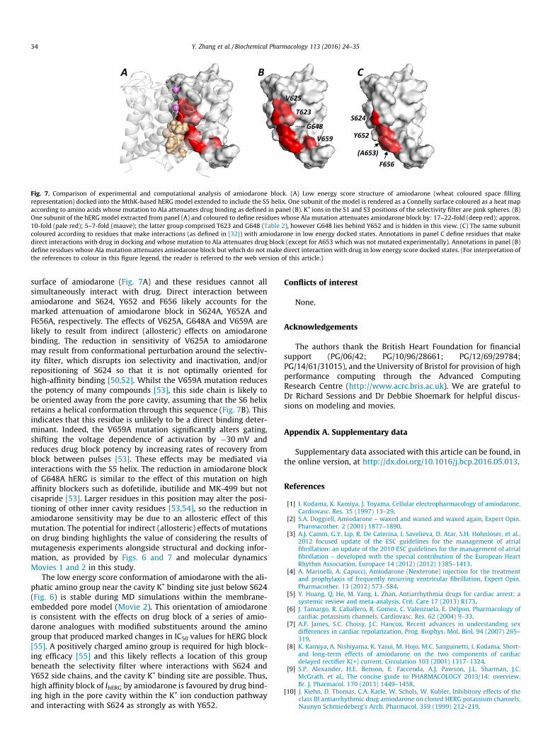
Fig. 7. Comparison of experimental and computational analysis of amiodarone block. (A) Low energy score structure of amiodarone (wheat coloured space fillingrepresentation) docked into the MthK-based hERG model extended to include the S5 helix. One subunit of the model is rendered as a Connelly surface coloured as a heat mapaccording to amino acids whose mutation to Ala attenuates drug binding as defined in panel (B). K+ ions in the S1 and S3 positions of the selectivity filter are pink spheres. (B)One subunit of the hERG model extracted from panel (A) and coloured to define residues whose Ala mutation attenuates amiodarone block by: 17–22-fold (deep red); approx.10-fold (pale red); 5–7-fold (mauve); the latter group comprised T623 and G648 (Table 2), however G648 lies behind Y652 and is hidden in this view. (C) The same subunitcoloured according to residues that make interactions (as defined in [32]) with amiodarone in low energy docked states. Annotations in panel C define residues that makedirect interactions with drug in docking and whose mutation to Ala attenuates drug block (except for A653 which was not mutated experimentally). Annotations in panel (B)define residues whose Ala mutation attenuates amiodarone block but which do not make direct interaction with drug in low energy score docked states. (For interpretation ofthe references to colour in this figure legend, the reader is referred to the web version of this article.)
34 Y. Zhang et al. / Biochemical Pharmacology 113 (2016) 24–35
surface of amiodarone (Fig. 7A) and these residues cannot allsimultaneously interact with drug. Direct interaction betweenamiodarone and S624, Y652 and F656 likely accounts for themarked attenuation of amiodarone block in S624A, Y652A andF656A, respectively. The effects of V625A, G648A and V659A arelikely to result from indirect (allosteric) effects on amiodaronebinding. The reduction in sensitivity of V625A to amiodaronemay result from conformational perturbation around the selectiv-ity filter, which disrupts ion selectivity and inactivation, and/orrepositioning of S624 so that it is not optimally oriented forhigh-affinity binding [50,52]. Whilst the V659A mutation reducesthe potency of many compounds [53], this side chain is likely tobe oriented away from the pore cavity, assuming that the S6 helixretains a helical conformation through this sequence (Fig. 7B). Thisindicates that this residue is unlikely to be a direct binding deter-minant. Indeed, the V659A mutation significantly alters gating,shifting the voltage dependence of activation by �30 mV andreduces drug block potency by increasing rates of recovery fromblock between pulses [53]. These effects may be mediated viainteractions with the S5 helix. The reduction in amiodarone blockof G648A hERG is similar to the effect of this mutation on highaffinity blockers such as dofetilide, ibutilide and MK-499 but notcisapride [53]. Larger residues in this position may alter the posi-tioning of other inner cavity residues [53,54], so the reduction inamiodarone sensitivity may be due to an allosteric effect of thismutation. The potential for indirect (allosteric) effects of mutationson drug binding highlights the value of considering the results ofmutagenesis experiments alongside structural and docking infor-mation, as provided by Figs. 6 and 7 and molecular dynamicsMovies 1 and 2 in this study.
The low energy score conformation of amiodarone with the ali-phatic amino group near the cavity K+ binding site just below S624(Fig. 6) is stable during MD simulations within the membrane-embedded pore model (Movie 2). This orientation of amiodaroneis consistent with the effects on drug block of a series of amio-darone analogues with modified substituents around the aminogroup that produced marked changes in IC50 values for hERG block[55]. A positively charged amino group is required for high block-ing efficacy [55] and this likely reflects a location of this groupbeneath the selectivity filter where interactions with S624 andY652 side chains, and the cavity K+ binding site are possible. Thus,high affinity block of IhERG by amiodarone is favoured by drug bind-ing high in the pore cavity within the K+ ion conduction pathwayand interacting with S624 as strongly as with Y652.
Conflicts of interest
None.
Acknowledgements
The authors thank the British Heart Foundation for financialsupport (PG/06/42; PG/10/96/28661; PG/12/69/29784;PG/14/61/31015), and the University of Bristol for provision of highperformance computing through the Advanced ComputingResearch Centre (http://www.acrc.bris.ac.uk). We are grateful toDr Richard Sessions and Dr Debbie Shoemark for helpful discus-sions on modeling and movies.
Appendix A. Supplementary data
Supplementary data associated with this article can be found, inthe online version, at http://dx.doi.org/10.1016/j.bcp.2016.05.013.
References
[1] I. Kodama, K. Kamiya, J. Toyama, Cellular electropharmacology of amiodarone,Cardiovasc. Res. 35 (1997) 13–29.
[2] S.A. Doggrell, Amiodarone – waxed and waned and waxed again, Expert Opin.Pharmacother. 2 (2001) 1877–1890.
[3] A.J. Camm, G.Y. Lip, R. De Caterina, I. Savelieva, D. Atar, S.H. Hohnloser, et al.,2012 focused update of the ESC guidelines for the management of atrialfibrillation: an update of the 2010 ESC guidelines for the management of atrialfibrillation – developed with the special contribution of the European HeartRhythm Association, Europace 14 (2012) (2012) 1385–1413.
[4] A. Marinelli, A. Capucci, Amiodarone (Nexterone) injection for the treatmentand prophylaxis of frequently recurring ventricular fibrillation, Expert Opin.Pharmacother. 13 (2012) 573–584.
[5] Y. Huang, Q. He, M. Yang, L. Zhan, Antiarrhythmia drugs for cardiac arrest: asystemic review and meta-analysis, Crit. Care 17 (2013) R173.
[6] J. Tamargo, R. Caballero, R. Gomez, C. Valenzuela, E. Delpon, Pharmacology ofcardiac potassium channels, Cardiovasc. Res. 62 (2004) 9–33.
[7] A.F. James, S.C. Choisy, J.C. Hancox, Recent advances in understanding sexdifferences in cardiac repolarization, Prog. Biophys. Mol. Biol. 94 (2007) 265–319.
[8] K. Kamiya, A. Nishiyama, K. Yasui, M. Hojo, M.C. Sanguinetti, I. Kodama, Short-and long-term effects of amiodarone on the two components of cardiacdelayed rectifier K(+) current, Circulation 103 (2001) 1317–1324.
[9] S.P. Alexander, H.E. Benson, E. Faccenda, A.J. Pawson, J.L. Sharman, J.C.McGrath, et al., The concise guide to PHARMACOLOGY 2013/14: overview,Br. J. Pharmacol. 170 (2013) 1449–1458.
[10] J. Kiehn, D. Thomas, C.A. Karle, W. Schols, W. Kubler, Inhibitory effects of theclass III antiarrhythmic drug amiodarone on cloned HERG potassium channels,Naunyn Schmiedeberg’s Arch. Pharmacol. 359 (1999) 212–219.

Y. Zhang et al. / Biochemical Pharmacology 113 (2016) 24–35 35
[11] T. Yang, D. Snyders, D.M. Roden, Drug block of I(kr): model systems andrelevance to human arrhythmias, J. Cardiovasc. Pharmacol. 38 (2001) 737–744.
[12] J.M. Ridley, J.T. Milnes, H.J. Witchel, J.C. Hancox, High affinity HERG K(+)channel blockade by the antiarrhythmic agent dronedarone: resistance tomutations of the S6 residues Y652 and F656, Biochem. Biophys. Res. Commun.325 (2004) 883–891.
[13] M.J. McPate, R.S. Duncan, J.C. Hancox, H.J. Witchel, Pharmacology of the shortQT syndrome N588K-hERG K+ channel mutation: differential impact onselected class I and class III antiarrhythmic drugs, Br. J. Pharmacol. 155 (2008)957–966.
[14] Y.H. Zhang, H. Cheng, V.A. Alexeenko, C.E. Dempsey, J.C. Hancox,Characterization of recombinant hERG K(+) channel inhibition by the activemetabolite of amiodarone desethyl-amiodarone, J. Electrocardiol. 43 (2010)440–448.
[15] C.Y. Du, A. El Harchi, Y.H. Zhang, C.H. Orchard, J.C. Hancox, Pharmacologicalinhibition of the hERG potassium channel is modulated by extracellular butnot intracellular acidosis, J. Cardiovasc. Electrophysiol. 22 (2011) 1163–1170.
[16] P.J. Chiu, K.F. Marcoe, S.E. Bounds, C.H. Lin, J.J. Feng, A. Lin, et al., Validation of a[3H]astemizole binding assay in HEK293 cells expressing HERG K+ channels, J.Pharmacol. Sci. 95 (2004) 311–319.
[17] R.R. Kauthale, S.S. Dadarkar, R. Husain, V.V. Karande, M.M. Gatne, Assessmentof temperature-induced hERG channel blockade variation by drugs, J. Appl.Toxicol. 35 (2015) 799–805.
[18] M.C. Sanguinetti, M. Tristani-Firouzi, HERG potassium channels and cardiacarrhythmia, Nature 440 (2006) 463–469.
[19] J.C. Hancox, M.J. McPate, A. El Harchi, Y.H. Zhang, The hERG potassium channeland hERG screening for drug-induced torsades de pointes, Pharmacol. Ther.119 (2008) 118–1132.
[20] J.S. Mitcheson, J. Chen, M. Lin, C. Culberson, M.C. Sanguinetti, A structural basisfor drug-induced long QT syndrome, Proc. Natl. Acad. Sci. U.S.A. 97 (2000)12329–12333.
[21] K. Kamiya, R. Niwa, J.S. Mitcheson, M.C. Sanguinetti, Molecular determinantsof HERG channel block, Mol. Pharmacol. 69 (2006) 1709–1716.
[22] M.J. Perrin, P.W. Kuchel, T.J. Campbell, J.I. Vandenberg, Drug binding to theinactivated state is necessary but not sufficient for high-affinity binding tohuman ether-a-go-go-related gene channels, Mol. Pharmacol. 74 (2008) 1443–1452.
[23] M. Perry, M.J. de Groot, R. Helliwell, D. Leishman, M. Tristani-Firouzi, M.C.Sanguinetti, et al., Structural determinants of HERG channel block by clofiliumand ibutilide, Mol. Pharmacol. 66 (2004) 240–249.
[24] Y.H. Zhang, C.K. Colenso, R.B. Sessions, C.E. Dempsey, J.C. Hancox, The hERG K(+) channel S4 domain L532P mutation: characterization at 37 �C, Biochim.Biophys. Acta 2011 (1808) 2477–2487.
[25] A. El Harchi, Y.H. Zhang, L. Hussein, C.E. Dempsey, J.C. Hancox, Moleculardeterminants of hERG potassium channel inhibition by disopyramide, J. Mol.Cell. Cardiol. 52 (2012) 185–195.
[26] C. Du, Y. Zhang, A. El Harchi, C.E. Dempsey, J.C. Hancox, Ranolazine inhibitionof hERG potassium channels: drug-pore interactions and reduced potencyagainst inactivation mutants, J. Mol. Cell. Cardiol. 74 (2014) 220–230.
[27] J.T. Milnes, O. Crociani, A. Arcangeli, J.C. Hancox, H.J. Witchel, Blockade ofHERG potassium currents by fluvoxamine: incomplete attenuation by S6mutations at F656 or Y652, Br. J. Pharmacol. 139 (2003) 887–898.
[28] J. Chen, G. Seebohm, M.C. Sanguinetti, Position of aromatic residues in the S6domain, not inactivation, dictates cisapride sensitivity of HERG and eagpotassium channels, Proc. Natl. Acad. Sci. U.S.A. 99 (2002) 12461–12466.
[29] H.J. Witchel, C.E. Dempsey, R.B. Sessions, M. Perry, J.T. Milnes, J.C. Hancox, J.S.Mitcheson, The low-potency, voltage-dependent HERG blocker propafenone –molecular determinants and drug trapping, Mol. Pharmacol. 66 (2004) 1201–1212.
[30] D. Melgari, Y. Zhang, A. El Harchi, C.E. Dempsey, J.C. Hancox, Molecular basis ofhERG potassium channel blockade by the class Ic antiarrhythmic flecainide, J.Mol. Cell. Cardiol. 86 (2015) 42–53.
[31] Y. Jiang, A. Lee, J. Chen, M. Cadene, B.T. Chait, R. MacKinnon, Crystal structureand mechanism of a calcium-gated potassium channel, Nature 417 (2002)515–522.
[32] C.E. Dempsey, D. Wright, C.K. Colenso, R.B. Sessions, J.C. Hancox, AssessinghERG pore models as templates for drug docking using publishedexperimental constraints: the inactivated state in the context of drug block,J. Chem. Inf. Model. 54 (2014) 601–612.
[33] B. Hess, C. Kutzner, D. van der Spoel, E. Lindahl, GROMACS 4: algorithms forhighly efficient, load-balanced, and scalable molecular simulation, J. Chem.Theory Comput. 4 (2008) 435–447.
[34] C.K. Colenso, R.B. Sessions, Y.H. Zhang, J.C. Hancox, C.E. Dempsey, Interactionsbetween voltage sensor and pore domains in a hERG K+ channel model from
molecular simulations and the effects of a voltage sensor mutation, J. Chem.Inf. Model. 53 (2013) 1358–1370.
[35] The PyMOL Molecular Graphics System, Version 1.4.1, Schrödinger LLC (2011).[36] W. Humphrey, A. Dalke, K. Schulten, VMD: visual molecular dynamics, J. Mol.
Graph. 14 (1996) 33–38.[37] M.C. Sanguinetti, J.S. Mitcheson, Predicting drug-hERG channel interactions
that cause acquired long QT syndrome, Trends Pharmacol. Sci. 26 (2005) 119–124.
[38] S.B. Long, X. Tao, E.B. Campbell, R. MacKinnon, Atomic structure of a voltage-dependent K+ channel in a lipid membrane-like environment, Nature 450(2007) 376–382.
[39] M. Weerapura, S. Nattel, D. Chartier, R. Caballero, T.E. Hebert, A comparison ofcurrents carried by HERG, with and without coexpression of MiRP1, and thenative rapid delayed rectifier current, Is MiRP1 the missing link? J. Physiol. 540(2002) 15–27.
[40] H.J. Witchel, J.T. Milnes, J.S. Mitcheson, J.C. Hancox, Troubleshooting problemswith in vitro screening of drugs for QT interval prolongation using HERG K+channels expressed in mammalian cell lines and Xenopus oocytes, J.Pharmacol. Toxicol. Methods 48 (2002) 65–80.
[41] R. Kannan, K. Nademanee, J.A. Hendrickson, H.J. Rostami, B.N. Singh,Amiodarone kinetics after oral doses, Clin. Pharmacol. Ther. 31 (1982) 438–444.
[42] D. Stork, E.N. Timin, S. Berjukow, C. Huber, A. Hohaus, M. Auer, S. Hering, Statedependent dissociation of HERG channel inhibitors, Br. J. Pharmacol. 151(2007) 1368–1376.
[43] R. Rusinova, R.E. Koeppe, O.S. Andersen, A general mechanism for drugpromiscuity: studies with amiodarone and other antiarrhythmics, J. Gen.Physiol. 146 (2015) 463–475.
[44] J.S. Mitcheson, Drug binding to HERG channels: evidence for a ‘non-aromatic’binding site for fluvoxamine, Br. J. Pharmacol. 139 (2003) 883–884.
[45] S. Wang, M.J. Morales, S. Liu, H.C. Strauss, R.L. Rasmusson, Modulation of HERGaffinity for E-4031 by [K+]o and C-type inactivation, FEBS Lett. 417 (1997) 43–47.
[46] P.J. Stansfeld, M.J. Sutcliffe, J.S. Mitcheson, Molecular mechanisms for druginteractions with hERG that cause long QT syndrome, Expert Opin. DrugMetab. Toxicol. 2 (2006) 81–94.
[47] J.P. Lees-Miller, Y. Duan, G.Q. Teng, H.J. Duff, Molecular determinant of high-affinity dofetilide binding to HERG1 expressed in Xenopus oocytes:involvement of S6 sites, Mol. Pharmacol. 57 (2000) 367–374.
[48] K. Kamiya, R. Niwa, M. Morishima, H. Honjo, M.C. Sanguinetti, Moleculardeterminants of hERG channel block by terfenadine and cisapride, J.Pharmacol. Sci. 108 (2008) 301–307.
[49] J. Guo, H. Gang, S. Zhang, Molecular determinants of cocaine block of humanether-a-go-go-related gene potassium channels, J. Pharmacol. Exp. Ther. 317(2006) 865–874.
[50] M. Perry, P.J. Stansfeld, J. Leaney, C. Wood, M.J. de Groot, D. Leishman, et al.,Drug binding interactions in the inner cavity of HERG channels: molecularinsights from structure-activity relationships of clofilium and ibutilideanalogs, Mol. Pharmacol. 69 (2006) 509–519.
[51] Y.N. Imai, S. Ryu, S. Oiki, Docking model of drug binding to the human ether-a-go-go potassium channel guided by tandem dimer mutant patch-clamp data:a synergic approach, J. Med. Chem. 52 (2009) 1630–1638.
[52] J.S. Mitcheson, M.D. Perry, Molecular determinants of high-affinity drugbinding to HERG channels, Curr. Opin. Drug Discov. Devel. 6 (2003) 667–674.
[53] J.S. Mitcheson, hERG potassium channels and the structural basis of drug-induced arrhythmias, Chem. Res. Toxicol. 21 (2008) 1005–1010.
[54] P.J. Stansfeld, P. Gedeck, M. Gosling, B. Cox, J.S. Mitcheson, M.J. Sutcliffe, Drugblock of the hERG potassium channel: insight from modelling, Proteins 68(2007) 568–580.
[55] K.M. Waldhauser, K. Brecht, S. Hebeisen, H.R. Ha, D. Konrad, D. Bur, et al.,Interaction with the hERG channel and cytotoxicity of amiodarone andamiodarone analogues, Br. J. Pharmacol. 155 (2008) 585–595.
[56] S.H. Jo, H.K. Hong, S.H. Chong, K.H. Won, S.J. Jung, H. Choe, Clomipramine blockof the hERG K+ channel: accessibility to F656 and Y652, Eur. J. Pharmacol. 592(2008) 19–25.
[57] S. Zhang, Z. Zhou, Q. Gong, J.C. Makielski, C.T. January, Mechanism of block andidentification of the verapamil binding domain to HERG potassium channels,Circ. Res. 84 (1999) 989–998.
[58] J.J. Duan, J.H. Ma, P.H. Zhang, X.P. Wang, A.R. Zou, D.N. Tu, Verapamil blocksHERG channel by the helix residue Y652 and F656 in the S6 transmembranedomain, Acta Pharmacol. Sin. 28 (2007) 959–967.
[59] Z. Su, J. Chen, R.L. Martin, J.S. McDermott, B.F. Cox, M. Gopalakrishnan, et al.,Block of hERG channel by ziprasidone: biophysical properties and moleculardeterminants, Biochem. Pharmacol. 71 (2006) 271–286.