herg channel (dys)function revealed by dynamic action...
TRANSCRIPT

HERG Channel (Dys)function Revealed by Dynamic Action PotentialClamp Technique
Geza Berecki,*y Jan G. Zegers,y Arie O. Verkerk,*y Zahurul A. Bhuiyan,z Berend de Jonge,* Marieke W.Veldkamp,* Ronald Wilders,y and Antoni C. G. van Ginneken**Experimental and Molecular Cardiology Group and the Departments of yPhysiology and zClinical Genetics, Academic Medical Center,University of Amsterdam, The Netherlands
ABSTRACT The human ether-a-go-go-related gene (HERG) encodes the rapid component of the cardiac delayed rectifierpotassium current (IKr). Per-Arnt-Sim domain mutations of the HERG channel are linked to type 2 long-QT syndrome. Westudied wild-type and/or type 2 long-QT syndrome-associated mutant (R56Q) HERG current (IHERG) in HEK-293 cells, at both23 and 36�C. Conventional voltage-clamp analysis revealed mutation-induced changes in channel kinetics. To assessfunctional implication(s) of the mutation, we introduce the dynamic action potential clamp technique. In this study, we effectivelyreplace the native IKr of a ventricular cell (either a human model cell or an isolated rabbit myocyte) with IHERG generated ina HEK-293 cell that is voltage-clamped by the free-running action potential of the ventricular cell. Action potential characteristicsof the ventricular cells were effectively reproduced with wild-type IHERG, whereas the R56Q mutation caused a frequency-dependent increase of the action potential duration in accordance with the clinical phenotype. The dynamic action potentialclamp approach also revealed a frequency-dependent transient wild-type IHERG component, which is absent with R56Qchannels. This novel electrophysiological technique allows rapid and unambiguous determination of the effects of an ionchannel mutation on the ventricular action potential and can serve as a new tool for investigating cardiac channelopathies.
INTRODUCTION
Discrete mutations in genes encoding ion channel proteins
that disrupt channel function are at present the most
commonly identified cause of heritable cardiac channelopa-
thies (Marban, 2002). Type 2 of the congenital long-QT
(LQT2) syndrome is linked tomutations in the human ether-a-
go-go-related gene (HERG), which encodes the pore-forming
a-subunit of the rapid delayed rectifier potassium channel
(Curran et al., 1995; Sanguinetti et al., 1995; Trudeau et al.,
1995). Properties of current through HERG channels (IHERG)are similar to those of the rapidly activating component of
delayed rectifier K1 current (IKr) that contributes to the final
repolarization of the ventricular action potential (AP)
(Sanguinetti and Jurkiewicz, 1990). Investigations of various
(wild-type and mutant) HERG channels in heterologous
expression systems such as Xenopus laevis oocytes or
mammalian tissue cells in culture have provided remarkable
results in understanding the congenital forms of the LQT2
syndrome. It is apparent that themechanisms bywhichHERG
mutations cause the clinically observed electrical disease are
various. For someHERGmutants, the observed differences in
HERG channel kinetics and/or IHERG density are evident and
translation into effects that these mutated channels would
have on the ventricularAP are obvious. Conversely, in several
cases, results of voltage-clamp experiments do not provide
satisfactory explanation to how structural changes of the
channel protein would affect cardiac AP repolarization and
ultimately lead to the observed clinical phenotype in affected
patients. In such cases, where the observed differences
between the wild-type and mutant channels are less clear, one
can in existing computer models of the human ventricular AP
(Priebe and Beuckelmann, 1998) modify IKr according to
what was found for the mutant and determine the resulting
change(s) in AP characteristics. It is then, often implicitly,
assumed that the mathematical description of the IKr fullycovers the properties of this current. Besides, this approach is
restrained by the lack of quantitative data on the complex
kinetics of the IKr and IHERG at physiological temperature. The
mathematical description is therefore merely an approxima-
tion, despite recent advances in modeling (Clancy and Rudy,
2001), and results from simulations in which HERG channel
properties have been changed should be interpreted circum-
spectly.
In this study, we introduce a novel electrophysiological
technique to assess the functional implications of ion channel
mutations. We hypothesize that rapid and unambiguous in-
terpretation of the altered channel function is possible with an
experimental setting in which mutant channels are allowed
to follow a natural time course of membrane potential (Vm)
change, upon being simultaneously allowed to contribute cur-
rent to theAPas theywouldhavewhen incorporated into a real
ventricular cell. With our dynamic action potential clamp
(dAPC) technique, the native IKr of a ventricular myocyte or
cell model is effectively replaced with IHERG recorded from
a transfected HEK-293 cell that is voltage-clamped by the
free-running AP of the ventricular cell. To this end, the native
Submitted June 8, 2004, and accepted for publication September 29, 2004.
Address reprint requests to G. Berecki, Dept. of Clinical and Experimental
Cardiology, Academic Medical Center, University of Amsterdam, Rm.
M01-217, Meibergdreef 9, 1105 AZ Amsterdam, PO Box 22700, 1100 DE
Amsterdam, The Netherlands. Tel.: 31-20-566-7547; Fax: 31-20-691-9319;
E-mail: [email protected].
� 2005 by the Biophysical Society
0006-3495/05/01/566/13 $2.00 doi: 10.1529/biophysj.104.047290
566 Biophysical Journal Volume 88 January 2005 566–578
IKr is pharmacologically blocked (or set to zero in case of
a model cell) and IHERG is applied to the ventricular cell as an
external current input. When wild-type (WT) IHERG is added
to the net membrane current of this ventricular cell, the
resulting AP should be considered as normal, whereas
a mutant IHERG should cause distortion of the AP.
We applied our dAPC technique to the R56Q (arginine to
glutamine) mutation, a defect known to increase the rate of
deactivation most profoundly (Chen et al., 1999). Pre-
viously, R56Q HERG channels had only been expressed in
Xenopus oocytes, and characterized at room temperature
(Chen et al., 1999). We studied WT and mutant channels in
HEK-293 cells also by conventional whole-cell voltage-
clamp technique, at both 23�C and 36�C. At physiologicaltemperature, the mutant channels showed both faster
deactivation, which would lengthen the AP, and faster
activation, which by itself would shorten the AP. However,
our dAPC experiments directly and unambiguously demon-
strate that the net effect of the mutation is an increase in
action potential duration (APD).
MATERIALS AND METHODS
Electrophysiological experiments
For details on plasmid construction, HEK-293 cell culture, and transfection
procedures, see the expanded Materials and Methods, available as Sup-
plementary Material online.
HEK-293 cells were either superfused with Tyrode’s solution containing
(mmol/L): 140 NaCl, 5.4 KCl, 1.8 CaCl2, 1 MgCl2, 5.5 glucose, 5 HEPES
(pH 7.4 with NaOH), or with a modified Tyrode’s solution with 4.5 instead
of 5.4 mmol/L KCl (see below). Membrane currents were recorded with an
Axopatch 200B amplifier (Axon Instruments, Union City, CA) in the whole-
cell configuration of the patch-clamp technique at 23 6 0.5�C and 36 6
0.5�C. Voltage control, data acquisition, and analysis were accomplished
using custom software. Patch pipettes (1.5–3 MV) were filled with solution
containing (mmol/L): 125 K-gluconate, 20 KCl, 1 MgCl2, 5 EGTA, 5
MgATP, 10 HEPES (pH 7.2 with KOH), resulting in a K1 equilibrium
potential (EK) of �87.7 mV at 36�C. To obtain a better match between the
EK of the experimental solutions and the model cell’s maximum diastolic
potential of�90.7 mV, we also used 4.5 mmol/L KCl in the Tyrode solution
(resulting in an EK of�92.5 mV). All figures showing APs in the model-cell
mode (see below) were obtained with this modified Tyrode solution. The pH
of solutions was corrected for temperature; potentials were corrected for
liquid junction potential. Membrane currents and potentials were low-pass
filtered (cutoff frequency 2 kHz) and digitized at 5 kHz. The current-voltage
(I-V) relationships, and IHERG kinetics were determined by voltage-clamp
protocols, as diagrammed in Figs. 2 and 3, and as described previously
(Sanguinetti et al., 1995; Smith et al., 1996; Snyders and Chaudhary, 1996)
and in the Supplementary Material. APs from freshly isolated rabbit left-
ventricular myocytes were measured at 36�C with the solutions described
above (5.4 mmol/L KCl in the Tyrode solution; EGTA was omitted in the
pipette solution), as described previously (Verkerk et al., 1996) and detailed
in the Supplementary Material.
Dynamic action potential clamp
Our approach is based on the coupling clamp (Tan and Joyner, 1990), model
clamp (Wilders et al., 1996), and dynamic clamp (Sharp et al., 1993)
techniques. The development of these techniques is built on the concept that
an isolated (cardiac) cell can be electrically coupled to either another isolated
cardiac cell or to a model analog that mimics the electrical properties of the
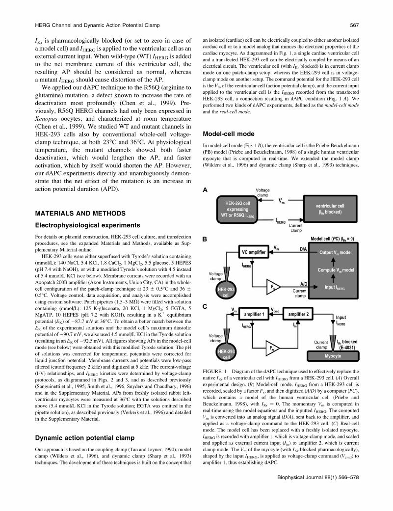
cardiac myocyte. As diagrammed in Fig. 1, a single cardiac ventricular cell
and a transfected HEK-293 cell can be electrically coupled by means of an
electrical circuit. The ventricular cell (with IKr blocked) is in current clamp
mode on one patch-clamp setup, whereas the HEK-293 cell is in voltage-
clamp mode on another setup. The command potential for the HEK-293 cell
is the Vm of the ventricular cell (action potential clamp), and the current input
applied to the ventricular cell is the IHERG recorded from the transfected
HEK-293 cell, a connection resulting in dAPC condition (Fig. 1 A). We
performed two kinds of dAPC experiments, defined as the model-cell modeand the real-cell mode.
Model-cell mode
In model-cell mode (Fig. 1 B), the ventricular cell is the Priebe-Beuckelmann
(PB) model (Priebe and Beuckelmann, 1998) of a single human ventricular
myocyte that is computed in real-time. We extended the model clamp
(Wilders et al., 1996) and dynamic clamp (Sharp et al., 1993) techniques,
FIGURE 1 Diagram of the dAPC technique used to effectively replace the
native IKr of a ventricular cell with IHERG from a HEK-293 cell. (A) Overall
experimental design. (B) Model-cell mode. IHERG from a HEK-293 cell is
recorded, scaled by a factor Fs, and then digitized (A/D) by a computer (PC),
which contains a model of the human ventricular cell (Priebe and
Beuckelmann, 1998), with IKr ¼ 0. The momentary Vm is computed in
real-time using the model equations and the inputted IHERG. The computed
Vm is converted into an analog signal (D/A), sent back to the amplifier, and
applied as a voltage-clamp command to the HEK-293 cell. (C) Real-cell
mode. The model cell has been replaced with a freshly isolated myocyte.
IHERG is recorded with amplifier 1, which is voltage-clamp mode, and scaled
and applied as external current input (Iin) to amplifier 2, which is current
clamp mode. The Vm of the myocyte (with IKr blocked pharmacologically),
shaped by the input IHERG, is applied as voltage-clamp command (Vcmd) to
amplifier 1, thus establishing dAPC.
HERG Channel and Dynamic Action Potential Clamp 567
Biophysical Journal 88(1) 566–578
implementing dAPCwith a real-timeLinux operating system (Barabanov and
Yodaiken, 1997) as a software platform according to Christini et al. (1999).
To attain simultaneous control and recording of Vm and IHERG and to resolve
the time-critical tasks of analog-to-digital conversion of IHERG, calculation of
the model, and digital-to-analog conversion of Vm, we developed a user
program (DynaClamp). This was used with a real-time module that operated
on a 1.8-GHz Pentium-4 PC with a 16-bit National Instruments PCI-6052E
data acquisition board (National Instruments, Austin, TX) under real-time
Linux, and communicated through shared memory and/or first-in, first-out
queues. This allows a guaranteed-timing real-time process (i.e., 40-ms
periodic time steps with the PB cell model). In all dAPC experiments, IKr of
the model cell is set to zero. We first determine maximal IHERG amplitude in
the HEK-293 cell in voltage-clamp configuration, with 4-s depolarizing
voltage steps to �10, 0, and 10 mV, from a holding potential of �80 mV, at
36 6 0.5�C. Considering the unusual kinetics of HERG channels (Lu et al.,
2001a), wemeasure IHERG amplitudes at the end of 4-s pulses rather than from
tail current amplitudes. The largest outward current value is then used to
estimate the scaling factor (Fs) for the IHERG input to the PBmodel cell. In our
standard protocol,WT aswell as R56Q IHERG amplitude are scaled to 47.6 pA
(equivalent to the original IKr amplitude in the PB model). After appropriate
scaling, the program establishes dAPC configuration between the model cell
and the HEK-293 cell for 10 s, during which a series of 2-ms, 4-nA, 1-Hz
suprathreshold stimuli are applied to the computer model cell. The recorded
IHERG and computed PBmodel variables (Vm and ionic currents) and settings
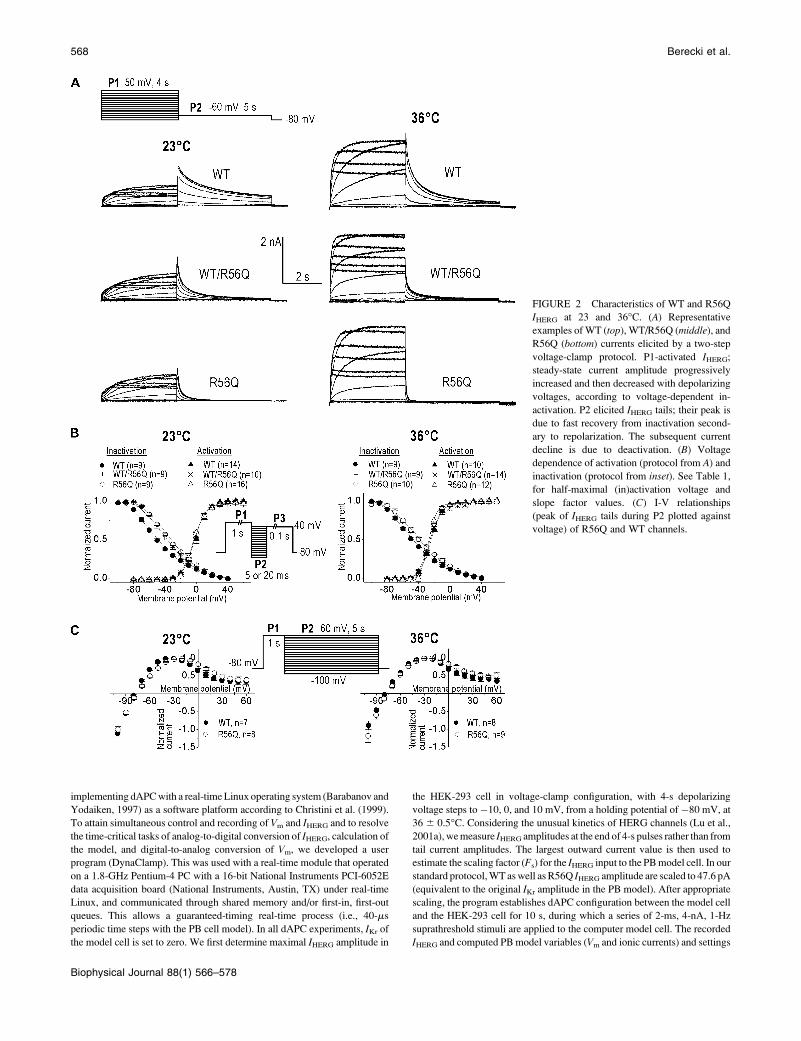
FIGURE 2 Characteristics of WT and R56Q
IHERG at 23 and 36�C. (A) Representative
examples of WT (top), WT/R56Q (middle), and
R56Q (bottom) currents elicited by a two-step
voltage-clamp protocol. P1-activated IHERG;steady-state current amplitude progressively
increased and then decreased with depolarizing
voltages, according to voltage-dependent in-
activation. P2 elicited IHERG tails; their peak is
due to fast recovery from inactivation second-
ary to repolarization. The subsequent current
decline is due to deactivation. (B) Voltage
dependence of activation (protocol from A) and
inactivation (protocol from inset). See Table 1,
for half-maximal (in)activation voltage and
slope factor values. (C) I-V relationships
(peak of IHERG tails during P2 plotted against
voltage) of R56Q and WT channels.
568 Berecki et al.
Biophysical Journal 88(1) 566–578
of the DynaClamp program are stored on disk for off-line analysis. The time-
dependent changes in Vm of the ventricular model cell are derived from WT
and/or mutant IHERG input and the model equations. The combination of the
cell model and WT IHERG will then result in a normal AP. Using the same
method for HEK-293 cells with mutant channels will reveal an AP, which
resembles the ventricular AP of the patient from which the mutant was
derived.
Real-cell mode
In real-cell mode, the model cell is replaced with a rabbit left-ventricular
myocyte (Fig. 1 C). The procedure to define Fs is as follows: we measure
IHERG amplitude in the HEK-cell (as described above) and, simultaneously,
estimate IKr density in the rabbit cell (as E-4031 sensitive current). We elicite
APs in the myocyte at 1 Hz in the presence of E-4031, and then establish
coupling between the myocyte and the HEK-293 cell, implementing scaled
WT IHERG. A proper Fs value would result in IHERG density comparable to
that of the IKr density in the myocyte and in a typical AP duration at 90%
repolarization (APD90) value of 230.8 6 4.5 ms at 1 Hz (see Table 2 in
Supplementary Material), characteristic for these cells. Ca21 loading of the
myocytes exhibiting long action potentials in the presence of E-4031 (as in
Fig. 8 B) is likely. However, this process loses its grip when the scaled IHERGis implemented and APD is shortened to its initial value (to same APD as
before the addition of E-4031) where Ca21 loading will be ruled out.
In both real-cell and model-cell modes, we can apply various stimulation
rates; Vm of the ventricular cell and IHERG of HEK-293 cell are displayed on-
TABLE 1 Parameters of WT, R56Q, and WT/R56Q IHERG activation and inactivation at 23 and 36�C
23�C 36�C
WT WT/R56Q R56Q WT WT/R56Q R56Q
Activation
V1/2 (mV) �1.1 6 1.1 �2.8 6 1.1 �3.9 6 1.0 �26.6 6 1.4 �28.1 6 1.0 �28.6 6 1.4
k (mV) 7.9 6 0.2 7.9 6 0.3 7.8 6 0.2 6.5 6 0.3 6.3 6 0.3 6.1 6 0.3
Inactivation
V1/2 (mV) �56.6 6 2.1 �40.4 6 2.3 �34.5 6 1.7* �49.6 6 2.6 �42.3 6 3.2 �39.8 6 3.2*
k (mV) �24.5 6 1.5 �22.1 6 1.5 �22.5 6 1.5 �23.5 6 0.5 �22.8 6 0.7 �23.1 6 1.0
*P , 0.05 for R56Q versus WT. Values are mean 6 SE; for n, the number of experiments, see Fig. 2 B.
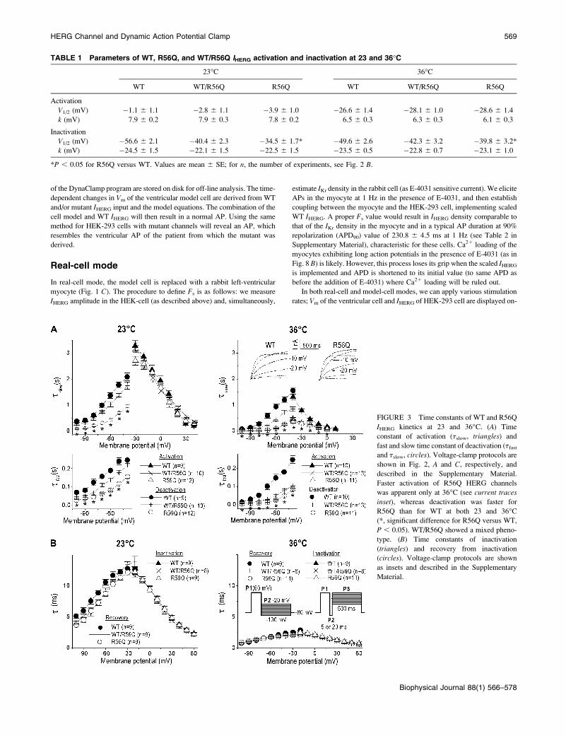
FIGURE 3 Time constants of WT and R56Q
IHERG kinetics at 23 and 36�C. (A) Time
constant of activation (tslow, triangles) and
fast and slow time constant of deactivation (tfastand tslow, circles). Voltage-clamp protocols are
shown in Fig. 2, A and C, respectively, and
described in the Supplementary Material.
Faster activation of R56Q HERG channels
was apparent only at 36�C (see current traces
inset), whereas deactivation was faster for
R56Q than for WT at both 23 and 36�C(*, significant difference for R56Q versus WT,
P , 0.05). WT/R56Q showed a mixed pheno-
type. (B) Time constants of inactivation
(triangles) and recovery from inactivation
(circles). Voltage-clamp protocols are shown
as insets and described in the Supplementary
Material.
HERG Channel and Dynamic Action Potential Clamp 569
Biophysical Journal 88(1) 566–578
line, thus providing instant information on the dAPC. DynaClamp allows
scaling of the input current to any desired magnitude and subtraction of
artifacts (e.g., endogenous HEK-293 cell currents), before IHERG is applied to
the ventricular cell. Leak subtraction, however, was not necessary as IHERG-downscaling already reduced endogenous currents to negligible levels.
Statistics
Data are expressed as mean 6 SE (n, number of cells) and considered
significantly different if P , 0.05 in ANOVA and Student’s t-test.
RESULTS
Electrophysiological characterization of WT,R56Q, and WT/R56Q HERG channels
To investigate the influence of recording temperature and
expression system on the WT and R56Q HERG channel
kinetics, we performed a series of voltage-clamp experiments
at both 23�C and 36�C. We also coexpressed WT and R56Q
cDNAs, in analogy to what is presumed to be present in
a patient with a single WT and mutant allele. Fig. 2 shows
typical WT and/or R56Q IHERG expressed in HEK-293 cells.
Increasing the recording temperature resulted in several
changes, including faster IHERG time course and larger
amplitudes (Fig. 2 A), and a negative shift in the voltage
dependence of activation (Fig. 2 B, Table 1). The R56Q
mutation caused a positive shift in the voltage dependence of
steady-state channel availability at both 23�C and 36�C (Fig.
2 B, Table 1). The normalized current-voltage (I-V) relation-
ships remained unchanged (Fig. 2 C). At 36�C, the mean
densities of IHERG, measured at the end of a 4-s pulse to �20
mV, were 2696 42 and 243 6 49 pA/pF with WT (n ¼ 17)
and R56Q channels (n ¼ 15), respectively (not significantly
different).
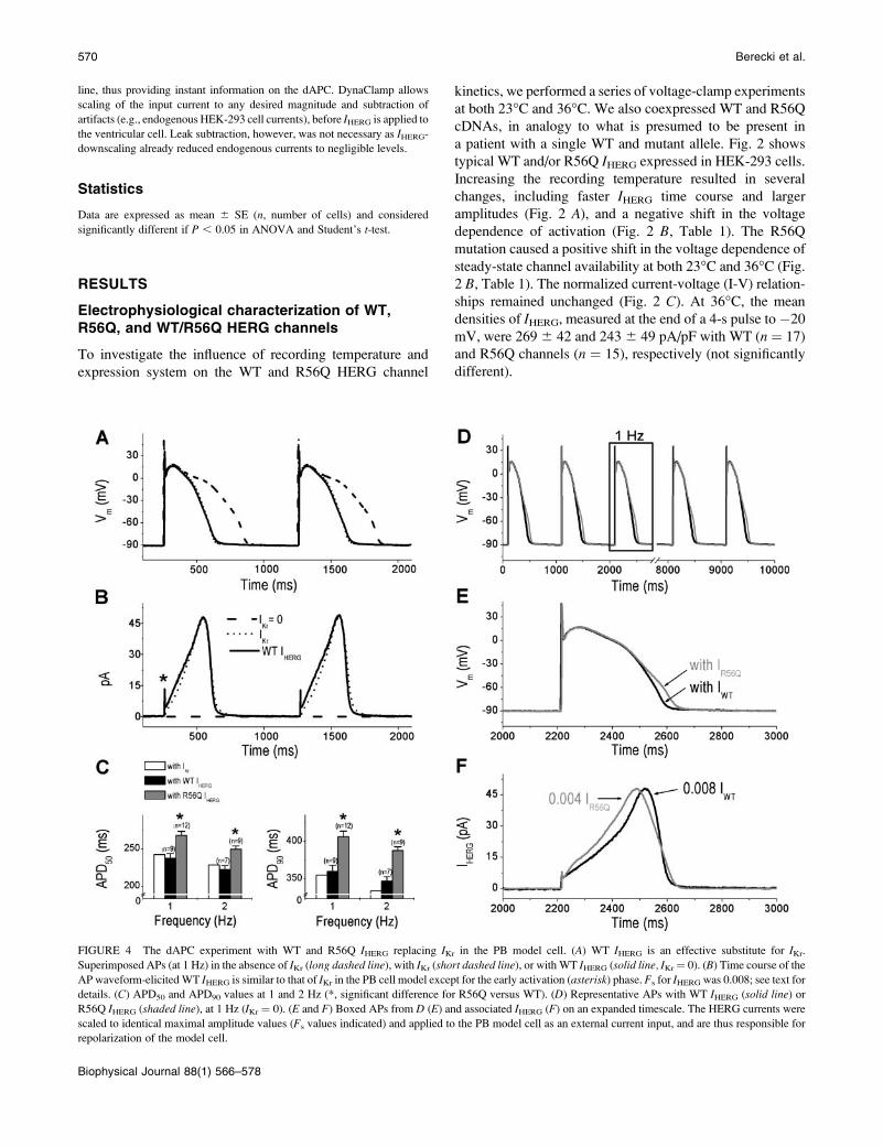
FIGURE 4 The dAPC experiment with WT and R56Q IHERG replacing IKr in the PB model cell. (A) WT IHERG is an effective substitute for IKr.
Superimposed APs (at 1 Hz) in the absence of IKr (long dashed line), with IKr (short dashed line), or with WT IHERG (solid line, IKr¼ 0). (B) Time course of the
AP waveform-elicitedWT IHERG is similar to that of IKr in the PB cell model except for the early activation (asterisk) phase. Fs for IHERG was 0.008; see text for
details. (C) APD50 and APD90 values at 1 and 2 Hz (*, significant difference for R56Q versus WT). (D) Representative APs with WT IHERG (solid line) or
R56Q IHERG (shaded line), at 1 Hz (IKr ¼ 0). (E and F) Boxed APs from D (E) and associated IHERG (F) on an expanded timescale. The HERG currents were
scaled to identical maximal amplitude values (Fs values indicated) and applied to the PB model cell as an external current input, and are thus responsible for
repolarization of the model cell.
570 Berecki et al.
Biophysical Journal 88(1) 566–578

Time constants of IHERG kinetics showed marked temper-
ature dependence (Fig. 3). At 36�C, the time course of R56Q
channel activation was approximately threefold faster at all
voltages than that ofWT channels (Fig. 3A; and see Table 1 inthe Supplementary Material). For the heteromultimer WT/
R56Q, the activation time constants were identical to those of
R56Q alone. Remarkably, in Xenopus oocytes, the time
course of R56Q channel activation was shown to be slower
than for those of WT channels (Chen et al., 1999). The
deactivation time course of R56Q channels was markedly
faster than for those of WT at both temperatures, as shown by
the diminution of both (fast and slow) time constants (Fig. 3A;and see Table 1 in the Supplementary Material). The finding
that the mutation causes faster deactivation is in agreement
with the results of Chen et al. (1999). Time constants of
inactivation and recovery from inactivation (Fig. 3 B) did notdiffer significantly betweenWT and R56Q (see Table 2 in the
Supplementary Material). Our results demonstrate that
acceleration of the R56Q HERG activation remains obscured
at 23�C and highlight the importance of investigating HERG
kinetics at physiological temperature.
Replacing IKr of the model cell with WTand R56Q IHERG
In the comprehensive human subepicardial ventricular cell
model by Priebe and Beuckelmann (1998), description of IKris based on data from human ventricular cells (Li et al.,
1996). With model-cell IKr set to zero, the AP prolongs (Fig.
4 A). When WT IHERG from a HEK-293 cell replaces IKr, APcharacteristics are restored and the AP can be considered as
normal (Fig. 4, A and C). Similar results were obtained when
the KCl content of the Tyrode solution was modified to
5.4mmol/L (see, in SupplementaryMaterial, Fig. 1 and Table
3). The time course of the scaled IHERG compares well to that
of IKr of the model cell (Fig. 4 B) except that the initial time
course of IHERG differs from that of the mathematically
described IKr, which is due to the model assumption that IKrinactivation is instantaneous. Many HERG channels are still
in the open state at �90 mV as a result of slow deactivation
(Lu et al., 2001a), and this results in an initial transient peak
(asterisk), reflecting the sudden increase of the electro-
chemical driving force for K1 during the AP upstroke. After
a fast decay of the transient peak amplitude, caused by
inactivation during the overshoot of the AP and by the
decreasing driving force for K1 at less depolarized Vm,
current increases progressively as channels rapidly recover
from inactivation, a process faster than the deactivation
(Sanguinetti et al., 1995; Trudeau et al., 1995; Smith et al.,
1996; Zhou et al., 1998). With repolarization progressing,
HERG channels dwell in a highly stable open state before
closing (Wang et al., 1998), resulting in a resurgent current.
Altered HERG channel properties in long-QT syndrome
generally reduce the magnitude of this resurgent current
(Chen et al., 1999; Sanguinetti et al., 1996). Both IKr andIHERG reach maximum value ;�40 mV, then rapidly
deactivate in a time- and voltage-dependent manner.
To study the functional consequences of the R56Q
mutation, we performed dAPC experiments with the PB
cell model and WT and/or mutant IHERG from the HEK-293
cell, in model-cell mode (Fig. 4 D). Results of these
experiments, remarkably consistent with the role of HERG
channels in cardiac repolarization, clearly show that the AP
is prolonged by the altered IHERG kinetics of the mutant (Fig.
4, C–E; see also Table 3 in the Supplementary Material). The
WT or R56Q IHERG, scaled to identical maximal amplitude
values (Fig. 4 F, Fs values indicated), was added to the PB
model cell as an external current input, and thus contributed
to repolarization of the model cell. Consistent with the results
of voltage-clamp experiments at 36�C, the input WT and
R56Q IHERG have different initial and late phases. Appar-
ently, mutant IHERG is initially larger than the WT. The faster
TABLE 2 Relative densities of selected ionic currents in the
subendocardial, M, and subepicardial cell models
Current Subendocardial
Midmyocardial
(M) Subepicardial
Ito 25%; Nabauer
et al. (1996)
87%; Liu et al.
(1993)
100%
IKs 92%; Liu and
Antzelevitch
(1995)
46%; Liu and
Antzelevitch
(1995)
100%
IK1 89%; Liu et al.
(1993)
74%; Liu et al.
(1993)
100%
All densities are percentage relative to the standard densities in the PB
model that essentially describes a human subepicardial ventricular myocyte
(Conrath et al., 2004).
FIGURE 5 Regional AP heterogeneity is reproduced in a dAPC exper-
iment. Subepicardial, M, and subendocardial APs were simulated at 1 Hz;
note the different plateau levels and repolarization phases in these model
cells (see the modified current densities in Table 2).
HERG Channel and Dynamic Action Potential Clamp 571
Biophysical Journal 88(1) 566–578

onset of the R56Q IHERG decay indicates faster deactivation
for R56Q HERG channels.
Action potential heterogeneity in the PB modelcell with WT and R56Q IHERG
The heterogeneity of the electrical properties of the myocytes
in the different layers of the human left ventricle is now well
established. As in our previous model studies (Bernus et al.,
2002; Conrath et al., 2004), we generated subendocardial,
midmyocardial (M), and subepicardial model cells by
adjusting selected membrane ionic currents in the PB model
cell (Table 2). When, in a dAPC experiment, WT IHERGreplaced model-cell IKr, APs of different shape and duration
could still be reproduced (Fig. 5). The major consequence of
the R56Q mutation on the AP characteristics of these cell
types was AP prolongation (Fig. 6). We analyzed in detail
AP characteristics of the epicardial model cell (Fig. 7),
comparing the frequency dependence of APD90 values
generated with model-cell IKr to values obtained with WT or
R56Q IHERG. These values are comparable when WT IHERGreplaces IKr, whereas R56Q IHERG causes frequency-de-
pendent APD90 prolongation (Fig. 7 A). APD90 with the
cotransfected WT/R56Q channels showed intermediate
values (not shown). The role of WT or R56Q IHERG in
shaping the AP was evaluated by phase plane analysis
(Sperelakis and Shumaker, 1968), plotting membrane
currents against membrane potential (Fig. 7 B). With input
IHERG scaled for identical amplitudes for both WT and
R56Q, the consequence of the mutation is apparent. The
FIGURE 6 AP prolongation caused
by the R56Q mutation in the three
different cell types of Fig. 5. (A)Representative APs and (B) the corre-
sponding IHERG; note the increased
inactivation of R56Q IHERG (arrow)
at the positive plateau-voltages of the
subendocardial cell; (C) averaged
APD90 values at 1 and 2 Hz (*,
significant difference for R56Q versus
WT IHERG).
572 Berecki et al.
Biophysical Journal 88(1) 566–578

most notable changes are detected during phase-3 repolar-
ization, with a reduction of the net membrane current (Itotal).
Replacing IKr of a rabbit ventricular cell with WTand R56Q IHERG
Results with IHERG replacing IKr in the model cell show that
the overall properties of the AP are well reproduced in
a dAPC experiment (Figs. 4 A and 5). Next, we used the
real-cell variant of the technique (Fig. 8). Ionic currents
underlying APs of a rabbit ventricular cell are comparable
with those in a human ventricular cell. Fig. 8 A shows typical
whole-cell currents during 4-s depolarizing prepulses to
0 mV and tail currents after returning to�60 mV. IKr may be
differentially expressed in rabbit ventricles (Cheng et al.,
1999), thus we first demonstrate IKr presence as the E-4031sensitive current (Clay et al., 1995). Currents during de-
polarization as well as tails were markedly diminished in
the presence of E-4031, resulting in prolonged repolariza-
tion and early after-depolarizations in all cells tested (n ¼ 9)
(Fig. 8 B). dAPC experiments (n ¼ 5) were performed with
a single myocyte coupled first to a HEK-293 cell with WT
IHERG (Fig. 8 C), and then to a HEK-293 cell with R56Q
IHERG (Fig. 8 D). In both cases, AP parameters were
determined at different stimulation frequencies (Fig. 9; see
also Table 4 in the Supplementary Material). The measured
resting Vm of the myocytes was �82.9 6 2.7 mV. APs were
effectively reconstituted in a dAPC experiment with WT
IHERG. APs with R56Q IHERG exhibited significant APD
prolongation at 0.2 and 1 Hz (Fig. 9 B). These experiments
also revealed that WT IHERG consists of an early fast
transient outward current followed by a sustained outward
current (Figs. 8 C and 9). Transient IHERG may contribute
importantly to AP dynamics during tachycardia (Lu et al.,
2001a). Amplitude of the transient component showed
positive frequency dependence (Fig. 9 C), whereas that of
the sustained component peaked during the terminal AP
repolarization, in a reverse frequency-dependent manner
between 1 and 5 Hz (Fig. 9 D). Although frequency
dependence of the sustained R56Q IHERG was similar to that
of WT IHERG, frequency dependence of the R56Q IHERGtransient component was absent, consistent with the
impaired deactivation kinetics of these channels.
DISCUSSION
A broad agreement prevails on the role of HERG channels in
AP repolarization. For a better understanding of the link
between LQT2 mutations and the inherent clinical pheno-
type, insight into the nature of HERG channel (dys)function
is indispensable. As a longstanding approach, the time- and
voltage-dependence of the HERG channel has most
frequently been characterized using stepwise voltage-clamp
protocols, and description of the HERG current was often
based on the extrapolation of results obtained in various
heterologous expression systems. However, it is becoming
clear that complex features of HERG channel kinetics during
the cardiac AP can best be studied during physiological
voltage waveforms (Hancox et al., 1998; Lu et al., 2001a;
Zhou et al., 1998) and, as shown in the present study, even
better during dAPC condition (i.e., by letting them shape the
ventricular action potential), in line with their normal
function.
The NH2 terminus of the a-subunit of the channel
regulates deactivation gating and represents a mechanism
by which functional diversity is generated in HERG and
related channels (Wang et al., 1998). Our electrophysiolog-
ical experiments demonstrate that the R56Q mutation
impairs not only deactivation (Chen et al., 1999) but
activation kinetics as well, the latter becoming apparent
only at 36�C (Fig. 3 A). Intriguingly, a faster activation and
a positive shift in the voltage dependence of channel
availability (Fig. 2 B), would actually act to shorten AP
duration. Characteristics of the heteromultimeric (WT/
R56Q) channels suggest that some of the functional effects
FIGURE 7 AP characteristics of the subepicardial PB model cell. (A)Frequency dependence with IKr, WT IHERG (n¼ 10), or R56Q IHERG (n¼ 8)
(*, significant difference for R56Q versus WT). (B) Phase-plane plot for the
net membrane current (Itotal) and IHERG during repolarization (starting from
;118 mV during phase-1 repolarization). APs from which these phase
planes were obtained were generated at 1 Hz and are shown in Fig. 4, D and
E. Arrows indicate progression of time.
HERG Channel and Dynamic Action Potential Clamp 573
Biophysical Journal 88(1) 566–578

are not simply combined, but that a dominant negative
interaction can also occur between the WT and R56Q HERG
channels (see activation time constants at 36�C in Fig. 3 A).Along the same lines with the impaired biophysical
properties, certain mutations in the Per-Arnt-Sim domain
might actually cause an HERG protein trafficking defect
(Paulussen et al., 2002). However, we did not find significant
differences in IHERG densities of WT and/or R56Q channels,
suggesting that the primary defect in mutant channel
properties is attributable to altered gating.
MinK-related peptide (MiRP1)/HERG complexes have
received considerable support as molecular correlates for
native IKr (Abbott et al., 1999, 2001). We did not coexpress
MiRP1 for reconstitution of native IKr by HERG, as
FIGURE 8 The dAPC experiment
with IHERG replacing IKr in rabbit
myocytes. (A) Block of IKr with
5 mmol/L E-4031 (inset, pulse pro-
tocol). Superimposed tracings of typical
recordings in absence (control) and
presence of E-4031, and difference
(E-4031 sensitive) current. Mean IKrdensity, determined from the E-4031
sensitive current, was 0.636 0.1 pA/pF
(n ¼ 9). (B) APs in a myocyte stimu-
lated at 0.2 Hz before and after applying
E-4031. Superfusion of cells with
E-4031 caused early after-depolariza-
tions. (C and D) APs from a myocyte
and associated WT (C) or R56Q IHERG(D) at different frequencies. The my-
ocyte was successively coupled to
HEK-293 cells transfected with WT or
R56Q HERG channels. Note the differ-
ent IHERG waveforms (*, transient
IHERG; arrow, sustained IHERG) and
frequency-dependent AP prolongation
with R56Q (see also Table 2 in the
Supplementary Material).
574 Berecki et al.
Biophysical Journal 88(1) 566–578

properties of IHERG in mammalian systems are similar in
many ways to those of native IKr, and discrepancies that
remain cannot be fully abolished by coexpression with
MiRP1 (Weerapura et al., 2002).
Most experimental data on cardiac ion channel (dys)func-
tion have been obtained in expression systems, away from
the cellular environment where these channels function to
generate the cardiac action potential. Table 3 shows
a comparison of IKr in the various systems: 1), PB model;
2), human ventricle; 3), rabbit ventricle; and 4), HEK-293
cells. The relatively few studies of human ventricular IKrmake it difficult to fully validate such comparison. Neverthe-
less, the study of Iost et al. (1998) provides data on IKr inhuman ventricular tissue obtained from healthy patients not
receiving medication. Despite the apparent differences
between some properties of IHERG and IKr in the present
study and previous results in the literature, mammalian cell
lines generally provide an adequate environment for HERG
channels. Here, experiments should be performed at
physiological temperatures, as HERG channel gating at
36�C more closely resembles endogenous IKr (Zhou et al,
1998; this study). Necessarily, the Xenopus system can be an
alternative when channels do not express well in a mamma-
lian cell line, although 36�C for oocytes is not physiological,
and observed differences in the behavior of the expressed
cardiac potassium channel proteins suggest that endogenous
factors in oocytes dictate channel properties to some extent
(Seebohm et al., 2001).
We have introduced the dAPC technique to investigate AP
characteristics in ventricular myocytes, by replacing IKr inthese cells by WT or mutant IHERG generated in HEK-293
cells. In both model-cell and real-cell modes, frequency
dependence of the APDs was comparable when WT IHERGreplaced IKr. AP characteristics of the ventricular cells were
effectively reproduced by WT IHERG, whereas the R56Q
IHERG caused a frequency-dependent increase in APD.
Superimposed phase plane plots of the repolarization
phases of model-cell APs indicate that the net membrane
current is severely affected by the mutation during the late
repolarization phase. APD90 values with R56Q IHERG were
FIGURE 9 Action potential characteristics of rabbit
ventricular myocytes with WT and R56Q IHERG. (A)
Superimposed APs from a single myocyte successively
coupled to HEK-293 cells expressing WT (solid line)
or R56Q IHERG (shaded line), and the corresponding
IHERG waveforms at 1 and 4 Hz. (B–D) Frequency
dependence of APD90 prolongation (B; see also Table
2 in the Supplementary Material) and transient (C) and
sustained (D) IHERG amplitudes, each normalized to
their values at 1 Hz. Asterisks indicate significant
difference for R56Q versus WT.
HERG Channel and Dynamic Action Potential Clamp 575
Biophysical Journal 88(1) 566–578

increased at lower stimulation rates and unchanged at higher
frequencies. Consistent with the role of HERG in the
suppression of arrhythmias initiated by premature beats (Lu
et al., 2001a), the technique revealed the presence of an early
fast, frequency-dependent transient WT IHERG. The fre-
quency-dependent increase of this current component was
absent with R56Q channels. APs with R56Q IHERG were
generally longer (Fig. 9 B), which can be explained by the
faster deactivation. However, the reason why the faster
activating, thus initially larger R56Q IHERG does not have
significant effect on the AP plateau is less obvious. It is likely
that the faster activation of the R56Q IHERG in the myocyte
causes a slightly modified membrane potential in the early
plateau phase of the AP, influencing activation of other
currents. Computer simulations using either the PB model or
the recently published human ventricular cell model by Ten
Tusscher et al. (2004) also predict little or no effect of
a moderate increase in IKr during the plateau phase of the
action potential (data not shown). On the other hand, even
small changes of the myocyte’s membrane potential can
cause significant changes in activation of voltage-dependent
currents, such as the transient outward current, Ito (Greensteinet al., 2000) and calcium current, ICa (Fulop et al., 2003).
In summary, both the computed model of the human
ventricular cell as well as a freshly isolated myocyte can
effectively be used in dAPC experiments. Kinetic features
that are difficult to investigate with standard voltage-clamp
protocols become apparent with the dAPC technique. The
model-cell mode offers an outstanding reproducibility of the
results during experimentation, as the input WT or mutant
IHERG is the only variable. However, the technically more
difficult real-cell mode can reveal AP waveforms and IHERGkinetics that can be considered truly physiological. Addi-
tionally, the real-cell mode offers the advantage that stimula-
tion rates above 2.5 Hz (maximal value in the model cell) can
easily be achieved. Theoretically, any individual conductance
in the model cell or in a real ventricular cell (if a specific
blocker for the investigated conductance is available) can be
replaced by a surrogate input current from an expression
system.
In the model-cell variant of the technique, it is
a straightforward operation to test the effect of interventions
directed at counteracting the effects of the mutations in
HERG, e.g., increasing the slow repolarizing component
(IKs) of the delayed rectifier K1 current.
Data presented here on the behavior of WT and R56Q
HERG channels may have implications for further studies,
where differences between WT and mutant channels are
subtle. With our approach, the contribution of (mutated)
channels to the AP is determined without making assump-
tions with regard to the kinetic properties of the channels,
and the altered shape of AP directly reflects the effect of the
TABLE 3 Biophysical properties of IKr (in the PB ventricular model cell or in freshly isolated myocytes) and IHERG (transiently
expressed in HEK-293 cells)
Idensity(pA/pF) Activation Inactivation Deactivation References
Model IKr 0.31 Priebe and Beuckelmann
(1998);
This study
V1/2 (mV) �21.0 �26.0
k (mV) 5.4 �23.0
t (ms) 194.5 (150 mV) 494.2 (�40 mV)
Human IKr ;0.3Li et al. (1996);
Iost et al. (1998)V1/2 (mV) �14.0 6 4, �5.7*y ND
k (mV) 7.7 6 2.7, 5.6* ND
t (ms) 192.0 6 53 (150 mV) 600.0 6 54 (�40 mV)*
Rabbit IKr 0.3, 0.6z
Lu et al. (2001b);
This study
V1/2 (mV) �21.9 ND
k (mV) ND ND
t1 (ms) 78.0 6 4 (150 mV) 119.0 6 25 (�50 mV)
t2 (ms) 624.0 6 42 (150 mV) 569.0 6 123 (�50 mV)
WT IHERG 269 6 42
Zhou et al. (1998);
This study
V1/2 (mV) �25.9 6 2.0§, �26.6 6 1.4 �49.6 6 2.6
k (mV) 6.0 6 0.3§, 6.5 6 0.3 �23.5 6 0.5
t1 (ms) 18.0 6 3.0 (140 mV)§ 180.0 6 20§ (�40 mV)
t2 (ms) 1299 6 118§ (�40 mV)
Values are mean6 SE;ND, not determined. All experiments were done under comparable conditions: 34–37�C, extracellular K1 concentration¼ 4–6mmol/L,
extracellular divalent cation concentration ¼ 2–3 mmol/L. Current density (Idensity) was defined as current level at the end of a �20 mV depolarizing pulse
normalized to cell capacitance.
*Iost et al. (1998).yNote that Iost et al. (1998) did not mention any correction for liquid junction potential (LJP). Taking into account an LJP of;�10 mV under the given ionic
conditions (Barry and Lynch, 1991), the actual V1/2 would be �16 mV.zThis study.§Zhou et al. (1998)
576 Berecki et al.
Biophysical Journal 88(1) 566–578

mutation. The dAPC technique allows other cardiac ion
channels than HERG (e.g., SCN5A, KvLQT1) to be studied
as well.
General considerations
The inherent limitations of the PB model and of simulations
when creating transmural AP heterogeneity on the basis of
experimental findings have been discussed before (Bernus
et al., 2002; Priebe and Beuckelmann, 1998). During dAPC
experiments, in both model-cell and real-cell modes, we
assumed that the defect in the R56Q channel is attributed to
altered gating. Accordingly, we scaled WT and mutant input
IHERG to similar magnitudes.
We are aware that it is potentially conceivable that
a mutation in an ion channel gene could result in
compensatory changes in other ion channel genes in vivo,
representing a general limitation of any heterologous
expression system. Short-term alteration of mRNA levels of
ion channels, caused by rapid pacing, is well documented
(Yamashita et al., 2000). Libbus et al. (2004) provide direct
evidence for Ito remodeling in the ventricle caused by reduced
AP upstroke amplitude, on a surprisingly short timescale.
SUPPLEMENTARY MATERIAL
An online supplement to this article can be found by visiting
BJ Online at http://www.biophysj.org.
This work was supported by Netherlands Heart Foundation grant No.
2001B155.
REFERENCES
Abbott, G. W., S. A. Goldstein, and F. Sesti. 2001. Do all voltage-gatedpotassium channels use MiRPs? Circ. Res. 88:981–993.
Abbott, G. W., F. Sesti, I. Splawski, M. E. Buck, M. H. Lehmann, K. W.Timothy, M. T. Keating, and S. A. Goldstein. 1999. MiRP1 forms IKrpotassium channels with HERG and is associated with cardiacarrhythmia. Cell. 97:175–187.
Barabanov, M., and V. Yodaiken. 1997. Introducing real-time Linux. LinuxJ. 34:19–23.
Barry, P. H., and J. W. Lynch. 1991. Liquid junction potentials and smallcell effects in patch-clamp analysis. J. Membr. Biol. 121:101–117.
Bernus, O., R. Wilders, C. W. Zemlin, H. Verschelde, and A. V. Panfilov.2002. A computationally efficient electrophysiological model of humanventricular cells. Am. J. Physiol. Heart Circ. Physiol. 282:H2296–H2308.
Chen, J., A. Zou, I. Splawski, M. T. Keating, and M. C. Sanguinetti. 1999.Long QT syndrome-associated mutations in the Per-Arnt-Sim (PAS)domain of HERG potassium channels accelerate channel deactivation.J. Biol. Chem. 274:10113–10118.
Cheng, J., K. Kamiya, W. Liu, Y. Tsuji, J. Toyama, and I. Kodama. 1999.Heterogeneous distribution of the two components of delayed rectifierK1 current: a potential mechanism of the proarrhythmic effects ofmethanesulfonanilide Class III agents. Cardiovasc. Res. 43:135–147.
Christini, D. J., K. M. Stein, S. M. Markowitz, and B. B. Lerman. 1999.Practical real-time computing system for biomedical experiment in-terface. Ann. Biomed. Eng. 27:180–186.
Clancy, C. E., and Y. Rudy. 2001. Cellular consequences of HERGmutations in the long QT syndrome: precursors to sudden cardiac death.Cardiovasc. Res. 50:301–313.
Clay, J. R., A. Ogbaghebriel, T. Paquette, B. I. Sasyniuk, and A. Shrier.1995. A quantitative description of the E-4031-sensitive repolarizationcurrent in rabbit ventricular myocytes. Biophys. J. 69:1830–1837.
Conrath, C. E., R. Wilders, R. Coronel, J. M. De Bakker, P. Taggart, J. R.De Groot, and T. Opthof. 2004. Intercellular coupling through gapjunctions masks M cells in the human heart. Cardiovasc. Res. 62:407–414.
Curran, M. E., I. Splawski, K. W. Timothy, G. M. Vincent, E. D. Green,and M. T. Keating. 1995. A molecular basis for cardiac arrhythmia:HERG mutations cause long QT syndrome. Cell. 80:795–803.
Fulop, L., T. Banyasz., J. Magyar, N. Szentandrassy, A. Varro, and P. P.Nanasi. 2003. Reopening of L-type calcium channels in humanventricular myocytes during applied epicardial action potentials. ActaPhysiol. Scand. 179:1–9.
Greenstein, J. L., R. Wu, S. Po, G. F. Tomaselli, and R. L. Winslow. 2000.Role of the calcium-independent transient outward current Ito1 in shapingaction potential morphology and duration. Circ. Res. 87:1026–1033.
Hancox, J. C., A. J. Levi, and H. J. Witchel. 1998. Time course and voltagedependence of expressed HERG current compared with native ‘‘rapid’’delayed rectifier K current during the cardiac ventricular action potential.Pflugers Arch. 436:843–853.
Iost, N., L. Virag, M. Opincariu, J. Szecsi, A. Varro, and J. G. Papp. 1998.Delayed rectifier potassium current in undiseased human ventricularmyocytes. Cardiovasc. Res. 40:508–515.
Li, G. R., J. Feng, L. Yue, M. Carrier, and S. Nattel. 1996. Evidence for twocomponents of delayed rectifier K1 current in human ventricularmyocytes. Circ. Res. 78:689–696.
Libbus, I., X. Wan, and D. S. Rosenbaum. 2004. Electrotonic load triggersremodeling of repolarizing current Ito in ventricle. Am. J. Physiol. HeartCirc. Physiol. 286:H1901–H1909.
Liu, D. W., and C. Antzelevitch. 1995. Characteristics of the delayedrectifier current (IKr and IKs) in canine ventricular epicardial, mid-myocardial, and endocardial myocytes. A weaker IKs contributes to thelonger action potential of the M cell. Circ. Res. 76:351–365.
Liu, D. W., G. A. Gintant, and C. Antzelevitch. 1993. Ionic bases forelectrophysiological distinctions among epicardial, midmyocardial, andendocardial myocytes from the free wall of the canine left ventricle. Circ.Res. 72:671–687.
Lu, Y., M. P. Mahaut-Smith, A. Varghese, C. L. Huang, P. R. Kemp, andJ. I. Vandenberg. 2001a. Effects of premature stimulation on HERG K1
channels. J. Physiol. 537:843–851.
Lu, Z., K. Kamiya, T. Opthof, K. Yasui, and I. Kodama. 2001b. Densityand kinetics of IKr and IKs in guinea pig and rabbit ventricular myocytesexplain different efficacy of IKs blockade at high heart rate in guinea pigand rabbit: implications for arrhythmogenesis in humans. Circulation.104:951–956.
Marban, E. 2002. Cardiac channelopathies. Nature. 415:213–218.
Nabauer, M., D. J. Beuckelmann, P. Uberfuhr, and G. Steinbeck. 1996.Regional differences in current density and rate-dependent properties ofthe transient outward current in subepicardial and subendocardialmyocytes of human left ventricle. Circulation. 93:168–177.
Paulussen, A., A. Raes, G. Matthijs, D. J. Snyders, N. Cohen, andJ. Aerssens. 2002. A novel mutation (T65P) in the PAS domain of thehuman potassium channel HERG results in the long QT syndrome bytrafficking deficiency. J. Biol. Chem. 277:48610–48616.
Priebe, L., and D. J. Beuckelmann. 1998. Simulation study of cellularelectric properties in heart failure. Circ. Res. 82:1206–1223.
Sanguinetti, M. C., M. E. Curran, P. S. Spector, and M. T. Keating. 1996.Spectrum of HERG K1-channel dysfunction in an inherited cardiacarrhythmia. Proc. Natl. Acad. Sci. USA. 93:2208–2212.
Sanguinetti, M. C., C. Jiang, M. E. Curran, and M. T. Keating. 1995. Amechanistic link between an inherited and an acquired cardiacarrhythmia: HERG encodes the IKr potassium channel. Cell. 81:299–307.
HERG Channel and Dynamic Action Potential Clamp 577
Biophysical Journal 88(1) 566–578

Sanguinetti, M. C., and N. K. Jurkiewicz. 1990. Two components of cardiacdelayed rectifier K1 current. Differential sensitivity to block by class IIIantiarrhythmic agents. J. Gen. Physiol. 96:195–215.
Seebohm, G., C. Lerche, A. E. Busch, and A. Bachmann. 2001.Dependence of IKs biophysical properties on the expression system.Pflugers Arch. 442:891–895.
Sharp, A. A., M. B. O’Neil, L. F. Abbott, and E. Marder. 1993. Dynamicclamp: computer-generated conductances in real neurons. J. Neuro-physiol. 69:992–995.
Smith, P. L., T. Baukrowitz, and G. Yellen. 1996. The inward rectificationmechanism of the HERG cardiac potassium channel. Nature. 379:833–836.
Snyders, D. J., and A. Chaudhary. 1996. High affinity open channel blockby dofetilide of HERG expressed in a human cell line. Mol. Pharmacol.49:949–955.
Sperelakis, N., and H. K. Shumaker. 1968. Phase-plane analysis of cardiacaction potentials. J. Electrocardiol. 1:31–41.
Tan, R. C., and R. W. Joyner. 1990. Electrotonic influences on actionpotentials from isolated ventricular cells. Circ. Res. 67:1071–1081.
ten Tusscher, K. H., D. Noble, P. J. Noble, and A. V. Panfilov.2004. Amodel for human ventricular tissue. Am. J. Physiol. Heart Circ. Physiol.286:H1573–H1589.
Trudeau, M. C., J. W. Warmke, B. Ganetzky, and G. A. Robertson. 1995.HERG, a human inward rectifier in the voltage-gated potassium channelfamily. Science. 269:92–95.
Verkerk, A. O., M. W. Veldkamp, A. C. van Ginneken, and L. N. Bouman.1996. Biphasic response of action potential duration to metabolicinhibition in rabbit and human ventricular myocytes: role of transientoutward current and ATP-regulated potassium current. J. Mol. Cell.Cardiol. 28:2443–2456.
Wang, J., M. C. Trudeau, A. M. Zappia, and G. A. Robertson. 1998.Regulation of deactivation by an amino terminal domain in human ether-a-go-go-related gene potassium channels. J. Gen. Physiol. 112:637–647.
Weerapura, M., S. Nattel, D. Chartier, R. Caballero, and T. E. Hebert. 2002.A comparison of currents carried by HERG, with and withoutcoexpression of MiRP1, and the native rapid delayed rectifier current.Is MiRP1 the missing link? J. Physiol. 540:15–27.
Wilders, R., R. Kumar, R. W. Joyner, H. J. Jongsma, E. E. Verheijck,D. Golod, A. C. van Ginneken, and W. N. Goolsby. 1996. Actionpotential conduction between a ventricular cell model and an isolatedventricular cell. Biophys. J. 70:281–295.
Yamashita, T., Y. Murakawa, N. Hayami, E. Fukui, Y. Kasaoka, M. Inoue,and M. Omata. 2000. Short-term effects of rapid pacing on mRNA levelof voltage-dependent K1 channels in rat atrium: electrical remodeling inparoxysmal atrial tachycardia. Circulation. 101:2007–2014.
Zhou, Z., Q. Gong, B. Ye, Z. Fan, J. C. Makielski, G. A. Robertson, andC. T. January. 1998. Properties of HERG channels stably expressed inHEK 293 cells studied at physiological temperature. Biophys. J. 74:230–241.
578 Berecki et al.
Biophysical Journal 88(1) 566–578