interaction drug body - uniba.it · cell surface (membrane) receptors cell-surface (or...
TRANSCRIPT

INTERACTION DRUG BODY
What the drug does to the body
What the body does to the drug

PHARMACODYNAMICS
Receptors - intracellular receptors - membrane receptors
- Channel receptors - G protein-coupled receptors - Tyrosine-kinase receptors
Drug/receptor interactions - agonists and antagonists - potency and efficacy (effectiveness) - therapeutic index
Drug effects - therapeutic effects - side effects - toxic effects

CELL SURFACE (MEMBRANE) RECEPTORS
Cell-surface (or transmembrane) receptors are membrane-anchored, or integral proteins that bind to external ligand molecules. This type of receptor spans the plasma membrane and performs signal transduction, converting an extracellular signal into an intracellular signal.

CELL SURFACE (MEMBRANE) RECEPTORS
This large group of membrane-bound receptors comprises the 7TM or 1TM receptor families. All recruit multiple intracellular signaling cascades known as “second messengers”.

G PROTEIN-COUPLED RECEPTORS (GPCRs)
Class A (Rhodopsin-like) most receptors binding monoamines and neurotransmitters Class B (Secretin receptor family) binding ligands such as secretin, glucagone, calcitonin Class C (Metabotropic glutamate/pheromone) Ca-sensitive receptors as the mGlu R Classe D (Fungal mating pheromone receptors) Classe E (Cyclic AMP receptors) Classe F (Frizzled/Smoothened)
The largest and most diverse group of membrane receptors in eukaryotes.

G PROTEIN-COUPLED RECEPTORS (GPCRs)
The largest and most diverse group of membrane receptors in eukaryotes.

G PROTEIN-COUPLED RECEPTORS (GPCRs)
They sense molecules outside the cell and activate inside signal transduction pathways and, ultimately, cellular responses.
GPCRs are also known as seven-transmembrane domain receptors, 7TM receptors because they pass through the cell membrane seven times.

GPCRs - COMMON STRUCTURE
• The extracellular part of the receptor (N terminal) holds the ligand binding site and can be glycosylated. These extracellular loops also contain two highly conserved cysteine residues that form disulfide bonds to stabilize the receptor structure.
• The intracellular parts of the receptor can be phosphorylated. This serves as additional modulatory mechanism of activity. In addition, lipid anchoring sites allow for its membrane localization.
• Similar to GPCRs, the adiponectin receptors 1 and 2 (ADIPOR1 and ADIPOR2) also possess 7 transmembrane domains. However, ADIPOR1 and ADIPOR2 are orientated oppositely to GPCRs in the membrane (i.e., extracellular C-terminus, cytoplasmic N-terminus) and do not associate with G proteins.

GPCR ACTIVATION AND SUBSEQUENT EFFECTORS
2) ENZYMES and second messengers
1) ION CHANNELS
Some others amplify the signal giving raise to biologically active
second messengers
Some GPCRs, when activated, modify the intracellular ion concentration

GPCR and G PROTEINs
• GPCR are so called because they are bound to an intracellular G protein • Guanine nucleotide-binding proteins (G proteins) act as molecular switches inside cells, and are involved in transmitting signals from a variety of stimuli.

G PROTEIN
• The heterotrimeric G protein is made up of alpha (α), beta (β) and gamma (γ) subunits.
•The alpha (α) subunit holds the catalytic GTPase activity.
• The beta (β) and gamma (γ) subunits can form a stable dimeric complex referred to as the beta-gamma complex with regulatory activity.
There are two classes of G proteins: the monomeric small GTPases, and the heterotrimeric G protein complexes.
Their activity is regulated by factors that control their ability to bind to and hydrolyze guanosine triphosphate (GTP) to guanosine diphosphate (GDP).

ACTIVATION/INACTIVATION CYCLE OF GPCR
INACTIVATION: it occurs because G-alpha has intrinsic GTPase activity. After GTP hydrolysis, G-alpha bound to GDP will reassociate with a beta-gamma complex to form an inactive G-protein that can again associate with a receptor
• G-alpha bound to GTP is active, and diffuses along the membrane surface to activate target proteins, (often enzymes that generate second messengers). • The beta-gamma complex is also able to diffuse and activate proteins, typically affecting ion channels
ACTIVATION: ligand binding results in G-protein exchange of GTP for GDP. The activated G-protein then dissociates into an alpha (G-alpha) and a beta-gamma complex.
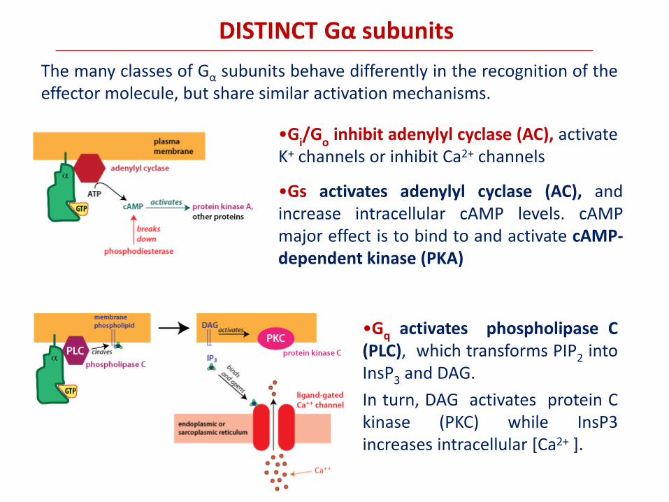
DISTINCT Gα subunits
The many classes of Gα subunits behave differently in the recognition of the effector molecule, but share similar activation mechanisms.
•Gq activates phospholipase C (PLC), which transforms PIP2 into InsP3 and DAG.
In turn, DAG activates protein C kinase (PKC) while InsP3 increases intracellular [Ca2+ ].
•Gs activates adenylyl cyclase (AC), and increase intracellular cAMP levels. cAMP major effect is to bind to and activate cAMP-dependent kinase (PKA)
•Gi/G
o inhibit adenylyl cyclase (AC), activate
K+ channels or inhibit Ca2+ channels

SUBUNIT
EFFECTOR LIGANDS AND RECEPTOR TYPES
s Adenylyl-cyclase (+)
Canali al calcio (+)
noradrenaline (1, 2, 3); dopamine (D1,D5); serotonin (5HT4); ACTH, FSH, LH, TFH, GnRH, GHRH, Vasopressin, Calcitonin, Prostaciclin
i 1,2,3
Adenylyl-cyclase (-) noradrenaline (2); dopamine (D2, D3, D4);
serotonine (5HT1); acetilcholine (M2, M4); somatostatin (SSTR1-5)
i 3
Potassium channels (GIRK) (+) acetilcholine (M2, M4); dopamine (D2,); somatostatin (SSTR1,2)
q
Phospholipase C (+) noradrenaline (1); serotonine (5HT21); TSH, TRH, GnRH, LXs, thromboxans
0 1,2
Calcium channels (-) noradrenaline (2); acetilcholine (M2, M4); somatostatin (SSTR1, 2)
SOME ENDOGENOUS LIGANDS OF GPCRs

Some GPCR undergo dimerization. This process can be a homodimerization (M3, 2, GABAB) or a more complex heterodimerization (D2/SSTR5, AT1/B2, SSTR2/µ opioid).
GPCR dimerization
Constitutive Ligand-induced
δ/κ opioid SSTR5/D2
Adenosin A1/DopamineD1 GABAB/D5
δ opioid/β2 adrenergic

GPCR desensitization mechanisms
GPCRs become desensitized when exposed to their ligand for a prolonged period of time. There are two recognized forms of desensitization:
1) homologous desensitization, in which the activated GPCR is
downregulated;
2) heterologous desensitization, wherein the activated GPCR causes
downregulation of a different GPCR. This downregulation is regulated by protein kinase-dependent phosphorylation of the intracellular (or cytoplasmic) receptor domain.

GPCR desensitization mechanisms
Arrestins are normally cytosolic proteins, but they recognise agonist-activated, phosphorylated receptors and bind them. This binding makes the receptor inaccessible for G-proteins (i.e. the arrestin-bound receptor is desensitised), and it targets the receptor for internalization. This is because arrestins do not only bind receptors, but they also bind components of clathrin-coated pits. Thus, arrestin-bound receptors move into clathrin-coated pits and are then internalized.
Agonist binding also converts the receptor into a substrate for a family of kinases, the G-protein-coupled receptor kinases (GRKs). GRKs phosphorylate only agonist-activated receptors. Subsequently, the phosphorylated receptor becomes a binding partner for arrestins.

GPCR desensitization mechanisms
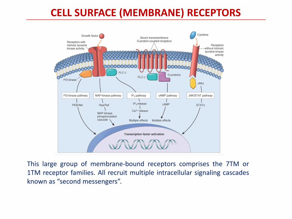
CELL SURFACE (MEMBRANE) RECEPTORS
This large group of membrane-bound receptors comprises the 7TM or 1TM receptor families. All recruit multiple intracellular signaling cascades known as “second messengers”.
Farmacologia per immagini
Atlante tascabile
II Edizione Italiana
Lullmann
Mohr
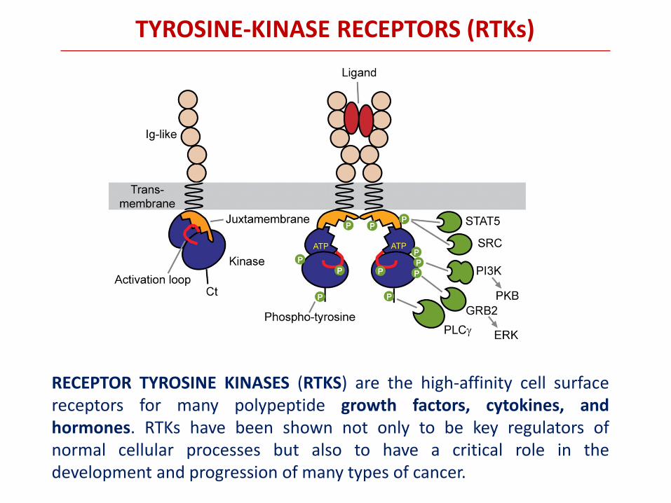
RECEPTOR TYROSINE KINASES (RTKS) are the high-affinity cell surface receptors for many polypeptide growth factors, cytokines, and hormones. RTKs have been shown not only to be key regulators of normal cellular processes but also to have a critical role in the development and progression of many types of cancer.
TYROSINE-KINASE RECEPTORS (RTKs)

extracel N
transmem iuxtaglo
intracel C
catal
TYROSINE-KINASE RECEPTORS (RTKs)
SCHEMATIC REPRESENTATION OF VARIOUS RTK SUBTYPES AND THEIR RESPECTIVE COGNATE LIGANDS

RTKs – ACTIVATION MECHANISMS
Ligand binding to the extracellular domain of the TKRs causes dimerization that results in autophosphorylation and activation of the intracellular kinase domain. The ensuing phosphorylation of docking sites on the receptor leads to the recruitment of enzymes, signal transducers and adaptor molecules activating downstream signalling pathways. The signalling pathways regulate a diverse array of processes including transcription, translation, metabolism, cell proliferation, survival, differentiation and motility.

RTKs – TRANSDUCTION SIGNALING CASCADES

RTKs – TRANSDUCTION SIGNALING CASCADES
(STAT3) - signal transducer and activator of transcription 3
(GRB2) - growth factor receptor-bound protein 2

RTKs – TRANSDUCTION SIGNALING CASCADES
The RAS/RAF/MEK/ERK pathway is the classical RAS/MAPK signaling pathway implicated in growth-factor mediated cell proliferation, differentiation and cell death. RAS activates RAF, which in turn activate mitogen-activated protein kinase kinase 1/2 (MAP2K1/2 or MEK1/2). MEK1 and MEK2 then phosphorylate their two known substrates, ERK1 and ERK2
RAS proteins are small GTPases, which serve as master regulators of a myriad of signaling cascades involved in highly diverse cellular processes. RAS oncogenes have been originally discovered as retroviral oncogenes