integrated corneal transplant care
TRANSCRIPT

Quality-Based Procedures Clinical Handbook for Integrated Corneal Transplant Care
Ministry of Health and Long-Term Care
October 20, 2017

Table of Contents 1.0 Purpose ........................................................................................................................... 3
2.0 Introduction ..................................................................................................................... 4
3.0 Description of Integrated Corneal Transplant QBP .................................................... 9
4.0 Best practices guiding the implementation of Corneal Transplantation ................ 17
5.0 Implementation of best practices ............................................................................... 32
6.0 What does it mean for multi-disciplinary teams? ...................................................... 35
7.0 Service capacity planning............................................................................................ 36
8.0 Performance evaluation and feedback ....................................................................... 39
9.0 Support for change ....................................................................................................... 44
10.0 Clinical Expert Advisory Group Membership .......................................................... 45
Appendix A: Patient Survey on Corneal Transplant Care .............................................. 47
References .......................................................................................................................... 48

3 | P a g e
Quality-Based Procedures Clinical Handbook: Integrated Corneal Transplant Care
1.0 Purpose
This Clinical Handbook has been created to serve as a compendium of the evidence-based rationale and clinical consensus driving the development of the policy framework and implementation approach for integrated corneal transplant care.
This document has been prepared for informational purposes only. This document does not mandate health care providers to provide services in accordance with the recommendations included herein. The recommendations included in this document are not intended to take the place of the professional skill and judgment of health care providers.

4 | P a g e
2.0 Introduction The Ministry of Health and Long-Term Care (Ministry) established Health System Funding Reform (HSFR) in Ontario in 2012 with a goal to develop and implement a strategic funding system that promotes the delivery of quality health care services across the continuum of care, and is driven by evidence and efficiency. HSFR is based on the key principles of quality, sustainability, access, and integration, and aligns with the four core principles of the Excellent Care for All Act (ECFAA):
• Care is organized around the person to support their health; • Quality and its continuous improvement is a critical goal across the health system; • Quality of care is supported by the best evidence and standards of care; and • Payment, policy, and planning support quality and efficient use of resources.
Since its inception in April 2012, the Ministry has shifted much of Ontario’s health care system funding away from the current global funding allocation (currently representing a large portion of funding) towards a funding model that is founded on payments for health care based on best clinical evidence-informed practices.
Principles of ECFAA have been further reinforced first by Ontario’s Action Plan for Healthcare in January 2012, and recently with Patients First: Action Plan for Healthcare in February 2015, which signals positive transformational activity which will require adaptive responses across sectors and organizational levels at a time of accelerated change. The Ministry’s commitment is to make Ontario the best healthcare system in the world.
The 2012 Action Plan identified HSFR as a lever to advance quality and ensure that the right care gets provided at the right place and at the right time. HSFR focuses on delivering better quality care and maintaining the sustainability of Ontario’s universal public health care system. Ontario is shifting the focus of its health care system away from one that has primarily been health care provider-focused, to one that is patient-centred. The 2015 Action Plan continues to put patients at the heart of the health care system by being more transparent and more accountable to provide health care in a way that maximizes both quality and value.
HSFR comprises 2 key components:
1. Organizational-level funding, which will be allocated as base funding using the Health-Based Allocation Model (HBAM); and
2. Quality-Based Procedure (QBP) funding, which will be allocated for targeted activities based on a “(price x volume) + quality” approach premised on evidence-based practices and clinical and administrative data.
2.1 ‘Money follows the patient’ Prior to the introduction of HSFR, a significant proportion of hospital funding was allocated through a global funding approach, with specific funding for select provincial programs, wait times services and other targeted activities. However, a global funding approach may not account for complexity of patients, service

5 | P a g e
levels and costs, and may reduce incentives to adopt clinical best practices that result in improved patient outcomes in a cost-effective manner. These variations in patient care evident in the global funding approach warranted the move towards a system where ‘money follows the patient”.
Under HSFR, provider funding is based on: the types and quantities of patients that providers treat, the services that they deliver, the quality of care delivered, and patient experience/outcomes. Specifically, QBPs provide incentive to health care providers to become more efficient and effective in their patient management by accepting and adopting clinical best practices that ensure Ontarians get the right care, at the right time, in the right place, and from the right provider.
QBPs were initially implemented in the acute care sector, but as implementation evolves, they are being expanded across the continuum of care, including into the community home care sector, in order to address the varying needs of different patient populations.
Internationally, similar models have been implemented since 1983. While Ontario is one of the last leading jurisdictions to move down this path, this uniquely positions the province to learn from international best practices and pitfalls, in order to create a sustainable, efficient and effective funding model that is best suited for the province and the people of Ontario.
2.2 What are Quality-Based Procedures? QBPs are clusters of patients with clinically related diagnoses or treatments that have been identified using an evidence-based framework as providing opportunity for process improvements, clinical re-design, improved patient outcomes, enhanced patient experience, and potential health system cost savings.
Initially developed in the acute (hospital) sector, QBPs were defined as “procedures”. However, as implementation evolved since the introduction of QBPs in 2012, so too has the approach. Currently, the expanded focus is on care provided in other parts of the health care sector with a focus on a more functional/programmatic/population-based approach. As a result, the definition of QBPs is expanding to include Quality-Based Procedures, Programs and Populations.
QBPs have been selected using an evidence-based framework. The framework uses data from various sources such as, but not limited to: the Discharge Abstract Database (DAD) and National Ambulatory Care Reporting System (NACRS) adapted by the ministry for its HBAM repository. The HBAM Inpatient Grouper (HIG) groups inpatients based on the diagnosis or treatment responsible for the majority of their patient stay. Additional data has been used from the Ontario Case Costing Initiative (OCCI), and Ontario Cost Distribution Methodology (OCDM). Evidence published in literature from Canada and international jurisdictions, as well as World Health Organization reports, have also assisted with the definition of patient clusters and the assessment of potential opportunities (e.g. reducing variation, improving patient outcomes, sustainability).
The evidence-based framework assesses patients using five perspectives, as presented in Figure 2.1. It is this evidence-based framework that has identified QBPs that have the potential to improve quality of care, standardize care delivery across the province and show increased cost efficiency.

6 | P a g e
Figure 2.1: Evidence-Based Framework
2.2.1 Practice Variation
Practice variation is the cornerstone of the QBP evidence-based framework. A demonstrated large practice or outcome variance across providers or regions in clinical areas, where a best practice or standard exists, represents a significant opportunity to improve patient outcomes through focusing on the delivery of standardized, evidence-informed practices. A large number of ‘Beyond Expected Length of Stay’ and a large standard deviation for length of stay and costs were flags to such variation.
2.2.2 Availability of Evidence
A significant amount of research has been conducted and collected, both nationally and internationally, to help develop and guide clinical practice. Working with clinical experts, best practice guidelines and clinical pathways can be developed for QBPs and establish appropriate evidence-informed indicators. These indicators can be used to measure the quality of care and help identify areas for improvement at the provider level, and to monitor and evaluate the impact of QBP implementation.
2.2.3 Feasibility/Infrastructure for Change
Clinical leaders play an integral role in this process. Their knowledge of the identified patient populations, and the care currently provided and/or required for these patients, represents an invaluable element in the assessment of much needed clinical delivery and clinical process improvements. Many groups of clinicians have already developed care pathways to create evidence-informed practice. There is now an opportunity for this knowledge to be transferred provincially.

7 | P a g e
2.2.4 Cost Impact
The provincial footprint from a financial perspective also impacts the selection of the QBP. This may include QBPs that are high volume and low-cost, as well as those that are low-volume and high cost (i.e. specialized procedures that demonstrate opportunity for improvement).
A selected QBP should have, as a guide, no less than 1,000 cases per year in Ontario and represent at least one percent of the provincial direct cost budget. For patient cohorts that fall below these thresholds, the resource requirements to implement a QBP can be restrictive. Even where the patient cohorts represent an opportunity for improvement, it may not be feasible, even if there are some cost efficiencies, to create a QBP.
2.2.5 Impact on Transformation
The Action Plan for Health Care was launched in January 2012 and is already making a difference to Ontarians and our health care system:
• We’ve bent the cost curve since 2011/12 • We’re improving the health of Ontarians • We’re enhancing the experience of Ontarians when they use the health system • We’re working with our health sector partners to improve the quality of health care
The next phase of Transformation will build on and deepen implementation of the Action Plan. HSFR is a key element of the Health System Transformation Agenda by ensuring sustainability and quality.
Selected QBPs should, where possible, align with the government’s transformational priorities. In addition, the impact on transformation of certain patient populations hitherto not prioritized by the framework can be included as QBPs. This will ensure that QBPs are wide ranging in their scope e.g. paediatric patient populations or patients requiring community care. QBPs with a lower cost impact but a large impact on the provincial health care system may still be a high priority for creation and implementation.
2.3 How will QBPs encourage the delivery of high quality, evidence-based care and innovation in health care delivery? The QBP methodology is driven by clinical evidence and best practice recommendations from the Clinical Expert Advisory Groups (Advisory Groups). Advisory Groups are comprised of cross-sectoral, multi-geographic and multi-disciplinary membership, including representation from patients. Members leverage their clinical experience and knowledge to define the patient populations and recommend best practices.
Once defined, these best practice recommendations are used to understand required resource utilization for QBPs and will further assist in the development of evidence-informed prices. The development of evidence-informed pricing for the QBPs is intended to incent health care providers to adopt best practices

8 | P a g e
in their care delivery models, maximize their efficiency and effectiveness, and engage in process improvements and/or clinical re-design to improve patient outcomes.
Best practice development for QBPs is intended to promote standardization of care by reducing inappropriate or unexplained variation and ensuring that patients get the right care, at the right place, at the right time, and from the right provider. Best practice standards will encourage health service providers to ensure that appropriate resources are focused on the most clinically and cost-effective approaches.
QBPs create opportunities for health system transformation where evidence-informed prices can be used as a financial lever to incent providers to:
• Adopt best practice standards; • Re-engineer their clinical processes to improve patient outcomes; • Improve coding and costing practices; and • Develop innovative care delivery models to enhance the experience of patients.
An integral part of the enhanced focus on quality patient care is the development of indicators to allow for the evaluation and monitoring of actual practice and support on-going quality improvement.
In addition, the introduction of additional QBPs such as outpatient and community-based QBPs will further help integrate care across sectors and encourage evidence-based care across the continuum.

9 | P a g e
3.0 Description of Integrated Corneal Transplant Care QBP 3.1 Description of Corneal Disease The cornea is a transparent dome-shaped structure that covers the front of the eye. The cornea consists of an outer epithelium layer and the following four inner layers: Bowman layer, stroma, Descemet’s membrane, and endothelium.
A healthy cornea protects the eye and helps focus incoming light onto the retina. The retina converts the light rays into impulses, which the brain translates into images that we see.
Damage to any of the corneal layers can cause loss of transparency and/or impact the shape of the cornea, resulting in vision loss. Damage can result from inflammation, infection, degeneration, hereditary condition (dystrophy) and/or trauma.
Treatment for corneal disease depends on the underlying problem as well as the patient’s preferences. Some conditions may resolve on their own or with medication. In advanced stages of disease, corneal surgery may be necessary in order to prevent blindness or restore a severely damaged eye.
Corneal scarring (e.g. infections, trauma) and failed graft are common indications for transplantation. The following section outlines some common corneal conditions:
• Keratoconus – a progressive eye disease that involves architectural damage to the cornea that can lead to thinning of the corneal tissue causing the cornea to bulge into a cone-like shape. This irregularity in the cornea deflects light that enters the eye causing distorted and blurred vision.
• Fuchs’ Dystrophy – a progressive eye disease that leads to the deterioration of the corneal endothelial cells. Once these cells are lost, they do not grow back. As the disease progresses with loss of endothelial cells, fluid builds up resulting in corneal swelling and (sometimes painful) vision loss.
• Pseudophakic Bullous Keratopathy – damage to the corneal endothelium can occur during cataract surgery. If the damage results in a loss of endothelial cells below a critical point, the cornea cannot pump out all the fluid from within it and begins to swell. Swelling results in loss of vision; if the front layer (epithelium) of the cornea swells, bullae (vesicles of fluid) may develop and these can cause pain if they break and expose the underlying corneal nerves.
• Pterygium – is a winged-shaped, vascular, fleshy growth that originates on the conjunctiva (the lining of the white part of the eye) and can expand onto the cornea. A pterygium arises in response to chronic exposure to the elements (UV light, wind, dust) which lead to degeneration of conjunctival tissue. A pterygium is made up of collagen and fibrovascular proliferation resulting in progressive benign growth. Significant pterygium growth can invade the cornea affecting vision by obscuring the optical center of the cornea, causing astigmatism/irritation or causing corneal scarring.

10 | P a g e
3.1.1 Keratoconus
Epidemiology
The prevalence of keratoconus has been estimated to be 5.4 per 10, 000 in the general population.1 Keratoconus is a slowly progressive condition that commonly presents in people in their teens and early twenties. The cause of keratoconus is unknown with research suggesting that a combination of environmental, genetic, and age factors play a role in the development of the condition.
Signs and Symptoms
Ocular signs and symptoms vary depending on disease severity.
Early keratoconus signs and symptoms can include: • Blurry vision that cannot be corrected with glasses • Frequent change in visual acuity • Increased light sensitivity • Difficulty driving at night • Halos and glare especially at night • Eye strain • Headaches and general eye pain • Eye irritation
Figure 3.1: Cornea Bulge in Keratoconus
Figure source: keratoconuscanada.org
Diagnosis
Diagnosis of keratoconus can be difficult. Early presentation of the disease does not produce any symptoms and can go unnoticed by the patient and practitioner. Specific tests by an ophthalmologist or optometrist are required to uncover the condition.
Three histopathological signs that typically characterize keratoconus are: (1) Stromal corneal thinning (2) Bowman’s layer breakage (3) Iron deposits within the corneal epithelium basal layer
3.1.2 Fuchs’ Dystrophy
Epidemiology
Prevalence of Fuchs’ dystrophy is difficult to estimate given the disease pathology and lack of symptoms in early stages. It is one of the most common indications for corneal transplantation. Fuchs’ dystrophy is defined as the accumulation of corneal guttata (small pigmented bumps of the back surface of the cornea) on the endothelial surface of the cornea. Prevalence estimates for guttata in eyes range from 0.18% to 3.9%. Asymptomatic guttata can progress to swelling in the cornea (corneal edema) with painful loss of vision in the more advanced stages.2

11 | P a g e
Signs and Symptoms
Fuchs’ dystrophy symptoms usually affect both eyes and may include: • Glare that reduces contrast perception or affects vision in low light • Blurred vision, predominantly in the morning • Distorted vision, sensitivity to light, difficulty seeing at night and seeing halos around light • Painful, tiny blisters on the surface of the cornea • A cornea that looks cloudy or hazy
Diagnosis
The diagnosis of Fuchs’ dystrophy is primarily made by the appearance of guttata with or without corneal edema, primarily through biomicroscopic examination by an ophthalmologist or optometrist. Tests can include:
• Slit-lamp biomicroscopy (clinical exam with a microscope) • Corneal pachymetry (corneal thickness measurement) • Noncontact specular microscopy (photography of the endothelial layer)
3.1.3 Pseudophakic Bullous Keratopathy (PBK)
Epidemiology
Prevalence and incidence of PBK is unknown. It is estimated that 0.1% of patients undergoing cataract surgery will develop PBK. Improvement in both intraocular lens design and surgical technique has reduced the occurrence of this problem. However, it remains an important cause of vision loss following surgery. Older patients with less endothelial reserve are at higher risk of developing PBK. 3
Signs and Symptoms
Signs and symptoms for PBK include: • Poor vision and haloes • Pain • Foreign body sensation • Photophobia
Diagnosis
PBK can be diagnosed through slit lamp examination, which reveals folds in Descemet’s membrane and thickening of central and peripheral cornea. Vesicles and bullae can be seen in advanced stages of PBK.
3.1.4 Pterygium
Epidemiology
Occurrence of pterygium has been found to vary with geographic location. A pooled prevalence rate of 10.2% has been reported in the general population.4 Pterygium growth has been reported to occur in

12 | P a g e
males twice as frequently as in females, and found to be associated with long-term exposure to excessive sun and chronic irritation due to wind, dust and sand. A genetic predisposition to the development of pterygia has also been reported.
Signs and Symptoms
Early in the condition, there are no symptoms. Symptoms can include redness, blurred vision, and eye irritation due to irregular wetting of the eye surface caused by the lesion. Some patients experience a feeling of a burning sensation or itching irritation. As the disease progresses, the pterygium growth becomes more apparent to the naked eye. Large pterygia can feel like a foreign object in the eye.
Diagnosis
Pterygium can be diagnosed by an optometrist or ophthalmologist by slit lamp examination. Additional tests can include:
• Corneal Topography to measure curvature changes of the cornea • External photography to track the growth rate of pterygium

13 | P a g e
3.2 QBP Inclusion and Exclusion Criteria This QBP includes day surgery cases only. Inpatient surgery is excluded from all patient groups below.
The Clinical Expert Advisory Group (Advisory Group) defined patient groups who receive corneal procedures in hospitals. The patient groups were identified from the CIHI National Ambulatory Care Reporting System (NACRS) for day surgeries.
The following section defines the Integrated Corneal Transplant QBP inclusion and exclusion criteria.
General Inclusion Criteria:
• Adults 18 years and older• Day surgeries recorded in CIHI NACRS (MOHLTC derived variable patient category = ‘DS’).• Ontario funded cases, i.e. The health card issuing province is Ontario and Ontario is responsible
for payment• Cases in CACS C051 (Reconstruction/Transplant Cornea) and CACS C052 (Other intervention of
cornea).• Surgeries with the specific intervention/diagnosis codes listed below, recorded at any position in
the abstract.
Included Procedure Groups:
1. Transplant Homograft• The procedure group includes both penetrating and
lamellar keratoplasty procedures (includes DALK,DMEK, DLEK, DSAEK, DSEK)
• Defined as:• Penetrating Keratoplasty: 1.CC.85.LA-XX-K OR• Lamellar Keratoplasty:
– 1.CC.85.LL-XX-H (DALK) OR– 1.CC.85.WJ.XX-H (DMEK) OR– 1.CC.85.PF-XX-H (DLEK, DSAEK, DSEK)
1a. Transplant Homograft with Cataract Surgery Procedure will be included in the Corneal Transplant QBP. • Defined as Transplant Homograft (1.CC.85.LA-XX-
K, 1.CC.85.LL-XX-H, 1.CC.85.WJ.XX-H OR1.CC85.PF.XX-H) AND excision total, lens(1.CL.89.^^)
Figure 3.2: Integrated Corneal Transplant QBP

14 | P a g e
Exclusion Criteria: • Acute inpatient cases identified from the Discharge Abstract Database (DAD) • Paediatric cases defined as procedures performed on patients 17 years and younger or
procedures performed in a paediatric hospital • Cases where the key interventions are abandoned or out-of-hospital. • Pterygium Surgery defined as excision partial, cornea (1.CC.87.^^) COMBINED WITH a Pterygium
diagnosis (H11.0) • Limbal Stem Cell Transplant defined as Transplant, Cornea using donor limbal stem cells
(1.CC.85.HA-U7-K) OR Transfer Cornea including limbal stem cell transplantation from contralateral eye (1.CC.83.LA-XX-A)
Note: An error in the coding definition for limbal stem cell transplant code 1.CC.85.HA-U7-K was identified by the Advisory Group. The current CIHI definition includes DALK with donor limbal stem cells. DALK cannot be performed with donor limbal stem cells. • CIHI is requested to correct coding definition for limbal stem cell transplants by removing DALK • Hospitals are encouraged to correctly code DALK using the CIHI code 1.CC.85.LA-XX-K
• Keratoprosthesis (KPro) defined as 1.CC.84.LA-AH OR 1.CC.84.LA-LC codes: o 1.CC.84.LA-AH - using a laser o 1.CC.84.LA-LC - using a scalpel or diamond blade
• Transplant homograft which meet the definition of the Integrated Retinal Care Clinical Handbook are excluded
The Advisory Group excluded Limbal Stem Cell Transplants, and KPro from the QBP due to low volume of cases across the Province. Best practice pathways have been developed for these in the clinical handbook as advancements in the field will increase use of these procedures.
There are instances where phototherapeutic keratectomy and corneal cross linking procedures can be done at the same time as a corneal transplant surgery. Although, these procedures are not included in the QBP inclusion criteria, the Advisory Group has defined best practices for these procedures in this clinical handbook.
Corneal surgery procedures excluded from the QBP will continue to remain in hospital global funding. Hospitals should maintain and manage access for patients who require these procedures.

15 | P a g e
3.3. Opportunities to Improve Corneal Transplant Care
Figure 3.3: Evidence-based Framework for Integrated Corneal Transplant Care
3.4 Objectives of Integrated Corneal Transplant Care QBP The objectives of the Integrated Corneal Transplant Care QBP are to:
• Encourage the adoption of best practices to improve outcomes and experience of patients receiving corneal transplant care.
• Maximize system capacity through integration of processes for the recovery, processing, distribution and utilization of corneal tissue for corneal transplantation.
• Reduce practice variation through the standardization of corneal transplant techniques, communication and coordination between providers, and ongoing management of complications pre- and post-surgery.
• Promote adoption of quality indicators to measure performance across the system to support delivery of high quality corneal transplant care to patients in Ontario.

16 | P a g e
3.5 Clinician and Patient Engagement
3.5.1 Clinician Engagement
A Clinical Expert Advisory Group (Advisory Group) was convened to develop the Integrated Corneal Transplant Care QBP Clinical Handbook. The purpose of the Advisory Group was to support the development and utilization of evidence-based best practice clinical pathways for the QBP and corresponding performance indicators to support measurement and monitoring of QBP implementation.
The Advisory Group was formed with multidisciplinary and provincial perspectives and included 25 members with the following expertise:
• Ophthalmology (corneal, retinal and glaucoma specialty, general ophthalmology)
• Optometry • Family Practice • Systems Researchers • Hospital Administration
• Coding, Costing and Health Analytics • Trillium Gift of Life Network • Eye Bank of Canada - Ontario Division • Local Health Integrated Network (LHIN) • Ministry of Health and Long-Term Care
(MOHLTC)
3.5.2 Patient Engagement
To support a patient-centred approach for best practice pathway development care, patients were asked about their experience for receiving Corneal Transplant care. Patients were recruited to participate in patient surveys from seven corneal surgery practices across Ontario including:
• Ottawa • Kingston • Toronto • Hamilton • London
Survey findings from 33 patient responses across the practices found that patients were highly satisfied with the explanation they received about their eye condition and proposed treatment, their experiences on the day of surgery, being able to understand instructions on how to care for their eye (including use of their eye medications), and knowing how to access after-hours care. Qualitative responses in each survey were analyzed for themes and the following care aspects were identified as being of high value to patients:
• Timely surgery to support return to work, activity, and independence • Caring, professional, and knowledgeable physician and staff • Team work between physician and staff • Having a good understanding of the treatment and process • Knowing how to care for their eye and who to follow-up with after surgery • Good outcomes to surgery
The survey findings were used to incorporate the patient’s perspectives into the development of the best-practice pathways. A full copy of the survey is attached in Appendix A

17 | P a g e
4.0 Best practicesi guiding the implementation of Corneal Transplantation
i Best practice refers to a combination of best available evidence and clinical consensus as recommended by the Clinical Expert Advisory Groups
The Advisory Group defined best practice recommendations in the form of clinical pathways for patients receiving procedures to treat damage to their cornea or corneal disease.
These pathways cover the entire continuum of care including screening, decision to treat, treatment and follow-up care. This QBP is unique in its consideration of the donor pathway for corneal tissue recovery, processing, and distribution to optimize the delivery of corneal transplant care.
Best practice clinical pathways were developed for the following corneal surgical procedures included in the QBP.
• Penetrating Keratoplasty (PK): The surgery involves transplant of a full thickness graft replacing all of the corneal layers.
• Deep Anterior Lamellar Keratoplasty (DALK): The surgery involves replacing only the damaged outer corneal tissue with the goal of preserving the inner corneal layers: Descemet’s membrane and the endothelium.
• Descemet's Membrane Endothelial Keratoplasty (DMEK): The DMEK procedure is a new type of partial-thickness corneal graft operation, where the damaged inner layers from the patient’s eye are stripped and only the innermost corneal layers are replaced (Descemet’s membrane and endothelium).
• Descemet's Stripping Automated Endothelial Keratoplasty (DSAEK): The DSAEK procedure strips damaged layers from the patient’s eye and transplants the inner layers (Posterior stroma, Descemet’s membrane, and endothelium) of the cornea.
Best practice pathways have also been defined for the following procedures that are not part of the hospital funded portion of the QBP but represent important corneal surgery procedures that warrant attention:
• Corneal Collagen Cross-Linking (CXL): is a technique that uses UV light and a photosensitizer to strengthen chemical bonds in the cornea. The goal of the treatment is to halt progressive and irregular changes in corneal shape.
• Phototherapeutic Keratectomy (PTK): PTK involves treatment of the superficial layers of the cornea with a laser to smooth irregularities and dissipate cloudiness in the cornea. It can prevent the need to perform full thickness corneal transplantation in certain clinical situations.
• Limbal Stem Cell Transplant: There are many variations in technique that can include autologous or allogenic grafts, tissue culturing and concomitant surgery with penetrating keratoplasty to treat limbal stem cell deficiency.
• Keratoprosthesis (KPro): This corneal transplant procedure uses an artificial cornea (keratoprosthesis), a clear plastic device that is coupled to a corneal graft. The graft is sutured into the patient’s cornea as in standard transplantation. A soft contact lens is applied to the surface.

18 | P a g e
The KPro is a permanent implant used when corneal transplantation and limbal stem cell transplantation are not viable.
• Pterygium Surgery: A minimally invasive corneal surgical technique that removes the excess tissue growth and uses a conjunctival graft that is harvested from the same patient to cover the removal site to minimize recurrence of pterygium growth.
Best practices to support the development of the pathways were derived using a combination of expert consensus and evaluation of standards and published evidence in corneal transplantation.5,6,7,8,9,10,11
4.1 Donor Cornea Clinical Pathway The transplantation process depends on the gift of corneal donation from one human to another. Best practices have been defined to maximize utilization of donated corneal tissue in Figure 4.1.
Figure 4.1Corneal Tissue Donor Pathway
The Trillium Gift of Life Network (TGLN) works closely with the Eye Bank of Canada -Ontario Division (Eye Bank) to obtain, and medically evaluate and distribute eyes donated by individuals for use in corneal transplantation.

19 | P a g e
TGLN is Ontario’s organ and tissue donation agency that plans, promotes, and coordinates activities related to the donation of tissue for transplant, education or research. TGLN’s scope of activities related to tissue includes:
1. Coordinate and support the work of designated facilities in connection with the donation and transplant of tissue.
2. Manage the procurement, distribution and delivery of tissue. 3. Establish and manage waiting lists for the transplant of tissue, and establish and manage a system to
fairly allocate tissue that is available. 4. Make reasonable efforts to ensure that patients and their substitutes have appropriate information and
opportunities to consider whether to consent to the donation of tissue and facilitate the provision of that information.
5. Provide education to the public and to the health-care community about matters relating to the donation and use of tissue and facilitate the provision of such education by others.
6. Collect, analyze and publish information relating to the donation and use of tissue. 7. Advise the Minister on matters relating to the provincial tissue and organ transplantation system.
The Eye Bank is ranked as the third most successful Eye Bank in North America in receiving donated eyes. Working with TGLN, the Eye Bank procures, prepares and distributes donated eyes for transplantation to over 60 surgeons across the province. It complies with strict medical standards and quality control as established by Health Canada and the Eye Bank Association of America (EBAA).
The Eye Bank is required to comply with the following regulatory schemes and standards:
• The Trillium Gift of Life Network Act (the Act) and any regulations made thereunder • Safety of Human Cells, Tissues and Organs for Transplantation Regulations, SOR/2007-118 made
under the Food and Drugs Act (Canada), as amended from time to time • The most up-to-date versions of applicable Canadian Standards Association standards, including: • National Standard of Canada CAN/CSA-Z900.1-12 entitled Cells, Tissues and Organs for
Transplantation and Assisted Reproduction: General Requirements, as amended from time to time • National Standard of Canada CAN/CSA-Z900.2.4-12 entitled Ocular Tissues for Transplantation, as
amended from time to time • Establishment licensing provisions under the Food and Drugs Act (Canada) • Applicable Health Canada guidance documents, including Guidance Document for Cell, Tissue and
Organ Establishments: Safety of Human Cells, Tissues and Organs for Transplantation (2nd ed., 2013) as amended from time to time
• Standards and requirements set by the EBAA under its voluntary accreditation program.
Recovery
The tissue donation system may begin with the front line hospital professional who is uniquely positioned to identify potential donors and their families if the potential donor is in hospital. A hospital is required to notify TGLN within 1 hour of death that an individual has died. TGLN conducts preliminary screening at the time of referral using the Eye Bank criteria.

20 | P a g e
Hospitals are required by the Act to establish policies and procedures that meet the requirements of TGLN. The Act also enables TGLN to have access to personal health information for the purposes of donation, and specify the manner of approach to families for consent.
TGLN coordinators obtain consent for donation after receiving any registered donor decisions from the Registered Person’s Database available via Service Ontario and through working with the donor’s legally authorized representative (usually next of kin). TGLN coordinators also complete the medical and social history to help the Eye Bank determine donor eligibility.
A coordinated recovery plan is made with the hospital and a certified Eye Bank enucleator or TGLN coordinator to recover the eyes. The eyes are then transported to the Eye Bank.
If the donor is not in hospital (for example – person dies in a long term care or retirement home, or at home) contact may be made with TGLN or the Eye Bank directly by the family, attending physician or funeral home to offer the donation. Acceptable time intervals from death, enucleation, or excision to preservation may vary according to the circumstances of death, patient demographics and health of the corneal tissue. It is recommended that preservation occur as soon as possible after death. Usually this process is completed within 14 hours of donor death.
The costs for recovery, processing, banking, and distribution of donor corneal tissue for transplantation are not included in this pathway. Therefore, these costs are excluded from the QBP as it relates to funding.
Processing
Assessment of the quality and viability of the donor cornea tissue is an essential pre-requisite for successful outcome of corneal transplantation. The Eye Bank conducts an extensive review of the donor’s medical, family, and social history, and a tissue quality assessment prior to distribution of the cornea for surgery.
The following quality assessments12 take place:
• Assessment of indications and contraindications to use of donor cornea which include o donor diseases with potential for transmission (infections, neoplasms, and corneal
disorders) o donor age o death to enucleation, excision and preservation time o cause of death o health of cornea endothelium
• Bacteriological evaluation of donor eye and surface decontamination • Transfer of corneoscleral button into preservation media (OPTISOL) as soon as possible • Serology testing to screen for HIV and hepatitis • Slit lamp examination before corneoscleral rim excision, or examination after in situ excision
provides an overall status of the cornea and endothelium • Specular count provides an added dimension of the endothelium status in terms of cell density,
pleomorphism and presence of guttata and supports better selection of corneas

21 | P a g e
• Precise documentation of assessments using standardized Eye Bank chart review forms.
Tissue that is determined to be ineligible for transplantation can be distributed for teaching and/or research purposes provided the donor/donor family consents to such use. Clear labelling and separate storage is required for such use and to prevent improper release.
Tissue determined to be eligible is stored using appropriate storage and labelling methods as defined by the Eye Bank. These requirements are in compliance with the regulatory schemes and standards.
Distribution
Once the tissue has been cleared for release, the Eye Bank assigns the tissue to the corneal surgeon who will transplant the tissue in a recipient. Prior to release of the tissue, the Eye Bank Medical Director or appropriate designate must review and document the medical and laboratory information.
A tissue report form must accompany the tissue to the operating site and include the following information: • Name of (source) eye bank, location of eye bank, telephone number of eye bank, eye bank
identification number unique to each tissue graft • Type of preservation medium • If cornea is pre-cut, clearly indicate the type of pre-cut method performed or the indicated use (e.g.
endothelial keratoplasty, posterior lamellar keratoplasty, anterior lamellar keratoplasty, laser assisted keratoplasty etc)
• If prepared for laser assisted penetrating keratoplasty: a. morphology and dimensions of cut, b. pre- and post-cut slit lamp reports, c. pre- and post-cut specular microscopy reports
• If prepared for lamellar anterior or endothelial keratoplasty: a. estimate thickness of transplant portion; b. diameter of cut; c. pre- and post-cut slit lamp reports; d. pre- and post-cut specular microscopy reports for tissue intended for endothelial keratoplasty use
• Age of donor, cause of death, date and time of death • Preservation date and time, additional tissue processing date and time, the time that cooling of
ocular tissues and/or refrigeration of the body was begun, name of technician who enucleated, excised, processed, and evaluated the tissue
• Slit lamp report/date of each evaluation • Specular microscopy report/date of each evaluation • Health Canada or EBAA Accreditation Status of each operating site that performs any of the
following steps in the preparation of tissue: recovery, processing, tissue storage, evaluation, donor eligibility determination and final distribution
• A summary of records reviewed regarding the eligibility of tissue for transplant
The operating site must submit information to the Eye Bank on the use of the tissue for transplantation. The Eye Bank collects post-operative outcomes and adverse events information at six months on all recipients from the transplanting surgeon. Information for each recipient chart is filed and kept for a minimum of 20 years. The Eye Bank follows up with all physicians who fail to send the information.

22 | P a g e
4.2 Referral Pathway for Corneal Surgical Treatment Procedures to assess and refer for corneal opacification, ectasia and pterygium are outlined in Figure 4.2. Clinical findings that warrant referral to a treating ophthalmologist for further diagnosis and treatment are highlighted in red.
Signs and symptoms suggestive of reduced vision related to corneal disease should be referred directly to an ophthalmologist. Referring to emergency room is to be discouraged whenever possible.
Figure 4.2 Referral Pathway for Corneal Surgical Treatment
Education
Corneal ectasias are progressive conditions, which tend to be asymptomatic in early stages of the disease. Appropriate exams and assessment can identify these conditions early and support referral for timely diagnosis and treatment.
Education of the public and family physicians should be provided on the key risk factors for eye diseases and the need for regular eye exams in populations who are at greater risk for eye disease. Patients should be educated about when to seek emergency treatment and how best to access care for their conditions.
Annual Eye Exams
Annual vision exams and best corrected visual acuity tests should be performed in those individuals over 65 years of age, for those with keratoconus and other corneal diseases, or those presenting with blurred

23 | P a g e
vision, chronic redness, pain, and/or irritation. Exams can be performed by an optometrist or ophthalmologist.
Although everyone should receive regular comprehensive eye exams, it is important to note that OHIP only insures an annual eye exam in patients:
• If they under 20 years of age or older than 65 • If they have one or more specific ocular/medical conditions requiring regular monitoring, or • If their primary care provider identifies the patient as needing regular monitoring
Comprehensive eye examinations include:
• Current vision and best corrected visual acuity tests • Measurement of intraocular pressure • Anterior segment and lens exam • Dilated fundus exam • Other possible specialized testing can include Topography, Keratometry, Specular Microscopy
A report on exam findings by the optometrist or ophthalmologist should ideally be provided to the referring provider sharing management of the patient. The report should include documentation of the patient’s best corrected visual acuity, findings of the anterior and posterior segment exams, the general impression and management as well as recommended follow-up.
If regular eye exams findings show corneal opacification, ectasia, or pterygium requiring surgical management, the patient should be referred to the treating ophthalmologist (in most cases a corneal surgeon).
Referral for further assessment and possible surgical treatment is warranted for the following clinical findings:
• Pterygium causing significant visual reduction, astigmatism, or of significant size • Eye exam findings suggesting corneal ectasia with decreased visual acuity or progressive
keratoconus • Eye exam showing opacification of the cornea with reduced vision • Eye exam showing ocular surface disease • Eye exam showing corneal edema with reduced vision
If symptoms have not progressed to point of treatment or if patient preference is not to have surgery, the appropriate eye care provider should continue ongoing monitoring and screening for new signs and symptoms or disease progression, while providing medical treatment when possible.

24 | P a g e
4.3 Surgical Decision to Treat Pathway Processes to support treating ophthalmologist with decision to treat with surgical intervention are highlighted in Figure 4.3.
Figure 4.3 Surgical Decision Pathway
Assessments
Careful patient assessments should be conducted by the treating ophthalmologist prior to surgical intervention to achieve an accurate diagnosis and determine appropriate treatment options for patients presenting with signs and symptoms of corneal damage. Assessments will include a complete medical and family history, as well as history of visual change, best corrected visual acuity and a full anterior and posterior segment examination. Diagnostic exams can include anterior segment optical coherence tomography (OCT), topography, Pentacam, pachymetry, and specular microscopy.
Discussion of Treatment Options
It is recognized that each patient’s presentation is unique and the physician must consider patient preferences for management, level of pain experienced, and disease progression in addition to diagnosis in determining appropriate treatment approaches. Informed patient consent must include a discussion of risk and benefits of available options, including options not provided by their cornea specialist, and the potential damage to the eye if treatment is not instituted.

25 | P a g e
Decision to Treat with Surgery
Once the patient provides informed consent for surgery, the patient will follow a surgical pathway to receive the appropriate corneal transplant procedure. The treating ophthalmologist should contact the Eye Bank for availability of corneal tissue for transplantation, if required. Some procedures (e.g. pterygium surgery, corneal cross-linking) can be provided by the treating ophthalmologist in an ophthalmology clinic while others will occur in the hospital/ambulatory surgical facility.
4.4 Corneal Surgery Clinical Pathways This section describes the surgical pathways to treat corneal damage. Best practice evidence and expert consensus guided the development of these pathways. Processes of care are outlined across common features in each pathway.
Suitability for Surgery
Clinical indications that necessitate surgery and assessment of patient’s ability to follow post-surgical routine and complete clinic follow-up visits are provided and vary for each procedure.
Pre-operative Care
Considerations for preparation of donor cornea, type of anesthesia and use of surgical checklist are provided and vary by procedure.
Follow-up Care
Follow-up care processes are outlined to ensure patient-centred care. Follow-up includes post-operative follow-up immediately after surgery, short-term follow-up within 48 hours of surgery and ongoing shared care follow-up.
Post-operative Follow-up Care is common across all pathways and requires the operating surgeon to provide written instructions to the patient on the following:
• Potential early complications and expectations following surgery. The patient must be able to understand the instructions for the care of their eye, including the use of their eye medications
• Emergency contact information must be provided to the patient to access after-hours assistance or emergent care.
• Follow-up time and place for their follow-up visit, usually carried out by the operating surgeon. If returning to an operating surgeon is not feasible, follow-up care may be delegated to a qualified eye care professional with demonstrated competence to detect complications.
• An operative report must be prepared and ideally sent to the referring eye care professional and/or family physician.
Corneal surgery procedures require removal of tissue from the eye. For these procedures, post-operative care requires that all specimens of the cornea or pterygium should be submitted for histopathological examination based on Public Hospitals Act Regulations 13, which outlines:

26 | P a g e
• 31.(1) Where tissues are removed from a patient during an operation or curettage, the surgeon performing the operation or curettage shall cause all tissues removed from the patient to be sent, together with a short history of the case and a statement of the findings of the operation, to a laboratory for examination and report. R.R.O. 1990, Reg. 965, s. 31 (1).
• 31.(1)(2) Despite subsection (1), where the tissue removed is an arm, finger, foot, hand, hemorrhoid, lens, leg, prepuce, tonsil, toe, toenail, tooth, the tissue shall not be sent to a laboratory unless the surgeon conducting the operation requests an examination and report on the tissue. R.R.O. 1990, Reg. 965, s. 31 (2).
Short-Term Follow-up Care is required with each surgical patient within 1 week or sooner after the initial post-operative visit (which ideally occurs within 48 hours) from the date of the procedure by the operating surgeon to check the status of healing of the surgical site, and follow resolution of post-operative inflammation. If ongoing treatment is delegated to a qualified eye care professional, appropriate communication must occur regarding the treatment plan. A summary note should also be completed at follow-up and ideally sent to the delegate and referring eye care professional, and/or family physician.
Ongoing Shared-Care Follow-up: The patient should continue to have ongoing follow-up for their condition by the operating surgeon or delegate until the cornea is stable. The results of the histopathology examination of the tissue sample should be reviewed by the operating surgeon. Additional tests and exams should be performed by the operating surgeon/delegate or referring eye care provider to monitor disease progression, treatment rejection/failure, or recurrence of signs and symptoms. These considerations vary by pathway.
The specific surgical procedure performed will be based on the disease process, patient preferences and characteristics, and surgeon preference informed by clinical indications.
4.4.1 Corneal Transplant Clinical Pathway Corneal transplant is a surgical procedure that is often referred to as corneal grafting or keratoplasty. The type of technique used to perform the procedure further differentiates the types of corneal transplant procedures.
Penetrating keratoplasty (PK) or ‘full thickness’ transplantation replaces all the layers of the cornea. Lamellar keratoplasty targets partial replacement of diseased corneal tissue and includes DALK, DSAEK, and DMEK procedures.
Corneal transplantation is indicated in patients with confirmed corneal opacification in the anterior cornea and/or evidence of endothelial disease/compromise accompanied by visual symptoms and a desire by the patient for correction.
The processes of care for corneal transplantation are common across all types of corneal transplantation techniques and outlined in Figure 4.4.

27 | P a g e
** Usually carried out by operating surgeon but may be delegated to a qualified eye care professional with demonstrated competence to detect complications if returning to operating surgeon is not feasible
Figure 4.4 Corneal Transplant Clinical Pathway
• For DSAEK, pre-cut tissue can be ordered from Eye Bank. If pre-cut tissue is not being ordered from the Eye Bank then the operating surgeon must arrange additional OR time to allow for tissue preparation.
• For DMEK, operating surgeon can request DMEK tissue from the Eye Bank. Pre-stripped tissue is currenty not available but may be available in the future; the Eye Bank has insufficient resources to prepare DMEK tissue at the present time.
• Phototherapeutic keratectomy (PTK) may be used to avoid a DALK or PKP to remove anterior scarring. • In some cases procedures may need to be performed with adjunct procedures such as intraoperative
lens insertion, iris reconstruction etc.
CIHI is requested to correct coding definition for limbal stem cell transplants by removing DALK from its current definition of donor limbal stem cells. DALK cannot be performed with donor limbal stem cells. Hospitals are encouraged to correctly code DALK using the CIHI code 1.CC.85.LA-XX-K.
4.4.2 Pterygium Surgery Clinical Pathway Pterygium surgery in indicated in patients with increasing pterygium growth size, induced corneal changes, visual acuity changes, and/or chronic irritation/redness.
The processes of care for Pterygium Surgery are outlined in Figure 4.5.

28 | P a g e
** Usually carried out by operating surgeon but may be delegated to a qualified eye care practitioner with demonstrated competence to detect complications if returning to operating surgeon is not feasible
Figure 4.5 Pterygium Surgery Clinical Pathway
• The standard of care is to cover the bare sclera when performing Pterygium Surgery. Conjunctival or limbal autograft have been proven to reduce pterygium recurrence compared with bare sclera excision for both primary and recurrent pterygia.14
• Pterygium specimens must be sent for histopathological examination. Unexpected ocular surface squamous neoplasia has been found in some pterygium specimens.15,16
• The procedure will often entail use of antimetabolite adjuvant to achieve desired results. Adjuvants can be used based on surgeon preference that is informed by clinical indications of the patient.
If cataract surgery is also required, pterygium surgery should usually be performed initially. Sufficient time should be allowed to ensure accurate K readings are obtained for IOL calculations prior to cataract surgery at another visit.
4.4.3 Limbal Stem Cell Transplant Clinical Pathway Limbal stem cell deficiency develops when an underlying condition destroys the stem cells in the eye. The depleted stem cells are replaced with abnormal conjunctival cells and scar tissue, resulting in symptoms including pain and poor vision.
Limbal stem cell transplantation uses a variety of techniques to restore limbal stem cells in the eye from donor cells that can be obtained from patient’s other eye, living relative or a cadaver allogenic donor.
The processes of care for Limbal Stem Cell Transplant are outlined in Figure 4.6.

29 | P a g e
** Usually carried out by operating surgeon but may be delegated to a qualified eye care practitioner with demonstrated competence to detect complications if returning to operating surgeon is not feasible
Figure 4.6 Limbal Stem Cell Transplant Clinical Pathway
• Autologous Limbal Stem Cell Transplant (LSCT) is the treatment option of choice when possible. If autologous LSCT is not possible, living relative or cadaveric allogeneic LSCT can be used. 17
• For LSCTs with living relative or donor cadaveric tissue: • Pre-operative processes of care can require liaison with medical/transplant team for
immunosuppression therapy management prior to performing LSCT • Post-operatively, a multi-disciplinary follow-up approach may be required using anti-rejection protocol
CIHI is requested to correct coding definition for Limbal Stem Cell Transplant by removing DALK from its current definition of donor limbal stem cells. DALK cannot be performed with donor limbal stem cells. Hospitals are encouraged to correctly code DALK using the CIHI code 1.CC.85.LA-XX-K.
4.4.4 Keratoprosthesis (KPro) Clinical Pathway This procedure involves transplanting an artificial cornea (the Keratoprosthesis [KPro]) to replace the damaged cornea. The procedure is indicated for patients who have had multiple graft failure or corneal opacification secondary to limbal stem cell deficiency and who are not amenable to limbal stem cell transplantation due to such processes as Stevens-Johnson syndrome, ocular cicatricial pemphigoid, or chemical injury.
The processes of care for transplant with KPro are outlined in Figure 4.7

30 | P a g e
CE = Cataract extraction; IOL = Intraocular lens, VF= Visual fields, IOP= Intraocular pressure
Figure 4.7 Keratoprosthesis (KPro) Clinical Pathway
• Patient must have adequate glaucoma control to be suitable for surgery • Pre-operative process requires Special Access Program approval and ordering of KPro. • Patient must attend at least 2 post-operative visits within 1 week of surgery with the operating surgeon or
delegate. o The first visit should be completed within 36 hours of the procedure. o Another visit is required 1 week after surgery. o Additional follow-up visits with the treating surgeon may be required depending on clinical need.
• Ongoing, shared care follow-up is usually completed by the operating surgeon until KPro is stable. Patients receiving KPro will require life-long follow-up by the operating surgeon or delegate. To manage the high risk of glaucoma and endophthalmitis noted in KPro patients, shared care management between a corneal specialist, glaucoma specialist, and retinal specialist is highly recommended.
• Due to high risk of post-operative complications, KPro patients require close monitoring. At minimum, quarterly eye exams are required to be performed by the operating surgeon or delegate.
4.4.5 Corneal Collagen Cross-Linking Clinical Pathway The primary purpose of Corneal Collagen Cross-Linking (CXL) is to halt the progression of keratoconus. CXL is a technique that uses UV light and a photosensitizer to strengthen chemical bonds in the cornea.
Parameters to consider suitability for CXL include changes to corneal shape (topography and tomography), and historical progression in visual acuity.
The processes of care for CXL are outlined in Figure 4.8

31 | P a g e
Figure 4.8 Corneal Collagen Cross-Linking Clinical Pathway
• The procedure may be performed with adjunct procedures to achieve desired results. • Adjuvants can be used by the surgeon based on the surgeon’s preference as informed by clinical
indications.

32 | P a g e
5.0 Implementation of best practices The Advisory Group identified key partners to support implementation of best practices outlined in this QBP:
• The Trillium Gift of Life Network (TGLN) Working Group on Corneal Transplant Care • The Eye Bank of Canada - Ontario Division (Eye Bank) • The Provincial Vision Strategy Task Force
The Advisory Group has identified key considerations for effective implementation for this QBP:
1. Hospitals resources should be matched to available donated corneal tissue to meet demand for corneal transplantation in the system.
A number of factors are increasing corneal tissue availability for corneal transplantation.
Figure 5.1 Tissue Donation: Five Year Review
Source: Trillium Gift of Life Network
• There has been significant growth in lamellar keratoplasty procedures (90%):18 these newer techniques now make it possible to transplant only a part of the cornea, which shortens the procedure time, supports faster recovery, and improves outcomes.
• Advancements in recovery and surgical procedure techniques also allow ‘corneoscleral rim excision’ rather than whole eye donation that will increase overall donation of cornea tissue for transplantation.
• The Eye Bank is currently establishing processes to provide pre-cut corneal tissue for DSAEK corneal transplant procedures.

33 | P a g e
• The TGLN Corneal Transplant Working Group has been working on several initiatives aimed at improving access and quality of corneal transplant services. Key initiatives have focused on improving data quality, standardization of practices, performance measurement and improving matching of corneal tissue availability with surgeon and operating room availability.
• The Eye Bank is establishing electronic systems to enhance information capture on tissue availability and OR scheduling. The system will also support surgeons with receiving individualized reports on their wait times for corneal transplantation.
• Provincial efforts are currently underway to redesign the donor cornea recovery system. TGLN is working closely with the Ministry, the Eye Bank and its other stakeholders to address gaps and develop a coordinated system to enhance the recovery of corneal tissue across Ontario.
• This QBP outlines best practices for tissue recovery, processing and distribution that will support engagement of donor sites (hospitals, funeral homes, coroner’s offices) to enhance recovery of donor tissue as well as use of available donor cornea tissues for transplantation by hospitals.
2. TGLN and the Eye Bank will work with system partners to support the redesign of the ocular recovery system that is aligned with best practices and volumes.
The current ocular recovery model in Ontario involves:
1) The Eye Bank of Canada – Ontario Division • Responsible for the recruitment, training, and payment of locally based recovery staff across the
province • Payment to non-physician enucleators is at a fixed rate • Physicians receive payment through OHIP billing
2) Trillium Gift of Life Network • Initiated ocular recovery in 2008 to close the gap in the number of cases not recovered due to a
lack of recovery resources, particularly in the Greater Toronto Area (GTA) • Responsible for recruitment, training, and payment of recovery staff • Teams established in the GTA, Eastern, and Southwest regions • Payment to TGLN recovery coordinator on a per hour basis
The Ministry has requested that TGLN lead the implementation of redesign of Ontario’s Tissue System. TGLN will work with the Eye Bank and key stakeholders to support planning and coordination to better organize ocular tissue recovery in Ontario as per best practices outlined in this clinical handbook.
The costs for recovery, processing, banking and distribution of corneal tissue for transplantation are excluded from the QBP as it relates to funding.
34 | P a g e
3. Individual surgical centres should determine the appropriate setting for their facility to offer pterygium surgery and ensure that sufficient patient care supports are provided.
Provincial analysis identified that pterygium surgery procedures are performed in a variety of clinical and surgical settings: hospital operating rooms, outpatient treatment rooms, minor operating rooms, outpatient ambulatory care units, and freestanding surgical centres. Best practices would suggest that individual centres determine the appropriate setting for their facility and ensure that sufficient patient care supports are provided.
4. Implementation of QBP will require accurate data entry and coding for reimbursement and quality indicator measurement.
An error in coding definition for limbal stem cell transplant code 1.CC.85.HA-U7-K was identified by the QBP Advisory Group. The current CIHI definition includes DALK with donor limbal stem cells. DALK cannot be performed with donor limbal stem cells.
• CIHI is requested to correct coding definition for limbal stem cell transplants by removing DALK • Hospitals are encouraged to correctly code DALK using the CIHI code 1.CC.85.LA-XX-K
Currently there are no codes to capture Phototherapeutic Keratectomy (PTK). • CIHI is requested to create a code for PTK. The use of this procedure is expected to increase due
to its role in preventing the need for more invasive corneal transplant surgery.
5. Corneal surgery procedures excluded from the QBP will continue to remain in hospital global funding. Hospitals should manage and maintain access to these procedures.
The Advisory Group excluded inpatient corneal transplant procedures, Limbal Stem Cell Transplants, and corneal transplants with keratoprosthesis from the QBP. Best practice pathways have been developed for these procedures in this clinical handbook as advancements in the field will increase their use. These procedures and other non-QBP corneal procedures will remain in the hospitals’ global budgets. Hospitals should manage and maintain access to these procedures.
6. The Ontario Hospital Association has developed a Toolkit to support implementation of QBPs that is available online.
The Toolkit provides:
• Organizational structure overview required to support successful implementation of QBPs • Patient engagement approaches to identify improvements that positively impact patient experience • Change management considerations, including senior leadership support, clinical engagement and
high quality clinical, financial and statistical data. • Approaches to monitoring and measuring process and outcomes related to QBP implementation
Toolkit Link: http://www.oha.com/CurrentIssues/keyinitiatives/PatientSafety/Documents/QBP/QBP%20Toolkit_toolkit%20only.pdf

35 | P a g e
6.0 What does it mean for multi-disciplinary teams?
A new funding model for corneal transplantation will require collaboration between ophthalmology and optometry providers involved in patient care delivery as well as require maximization of system capacity through integration of processes for the recovery, processing and distribution of donor corneal tissue. Optometrists and general ophthalmologists will play a critical role in assessing patients for corneal opacification, ectasia and pterygium and referring to a treating ophthalmologist (i.e. corneal surgeon) for clinical indications that warrant surgical treatment. Given that most corneal transplant procedures are performed in centres for corneal transplantation, family physicians, general ophthalmologists and optometrists may individually or together play an important role in ensuring that the patients can receive their post-operative care in their local community. It is recognized that the role of family physician is to direct the patient to the most appropriate eye care provider if the patient is unable to return to the operating surgeon. Implementation of the handbook best practices on corneal tissue recovery, processing and distribution will require coordination between TGLN, the Eye Bank and corneal surgeons and hospitals to ensure that donated tissue volumes are matched to operating room and surgeon availability, thereby maximizing utilization of donor corneal tissue for transplantation.

36 | P a g e
7.0 Service capacity planning Centres of corneal transplant specialization in Ontario are:
Toronto • UHN - Toronto Western Hospital • Toronto East General Hospital • Sunnybrook Health Sciences • St. Michael’s Hospital • Kensington Eye Institute
Hamilton • Hamilton Health Sciences Centre
Ottawa: • The Ottawa Hospital
Kingston • Hotel Dieu Hospital
London • St. Joseph’s Health Care London
The Provincial Vision Strategy Task Force has identified that current corneal transplant centres have the capacity to meet current corneal transplant needs in the province.
The current Wait 2 for corneal transplantation in Ontario at 160 days, below the 182 days provincial target for ophthalmology surgery (See Table 7.1).
Table 7.1: Corneal Transplant Wait Times - FY13/14 and FY14/15 FY 2013/2014 FY 2014/2015
Volume % Completed within Access
Target (182 days)
90th Percentile
Wait 2 Volume
% Completed within Access
Target (182 days)
90th Percentile
Wait 2
Erie St. Clair (Hotel-Dieu Grace Hospital, Windsor Regional Hospital) 29 100 91 30 100 72
Waterloo Wellington (St. Mary's General Hospital) 30 100 83 37 100 92
Central (North York General Hospital) 18 94 94 NR* NR* NR*Champlain (The Ottawa Hospital) 160 96 167 180 96 146 South East (Hotel Dieu Hospital Kingston) 25 96 169 28 89 178 South West (St. Joseph's Health Care London) 119 89 191 96 88 217
Toronto Central (St. Michael's Hospital, Toronto East General Hospital, University Health Network, Sunnybrook Health Sciences, Kensington Eye Institute)
588 76 302 617 91 166
Hamilton Niagara Haldimand Brant (Brantford General Hospital, St. Joseph's Community Health Centre Hamilton, Niagara Health System)
67 81 370 63 94 150
Province (Ontario) 1,036 83 254 1,051 92 160 *Not Reportable < 5 cell count Source: iPort, Wait Time Information System (WTIS) – Abstracted on July 29, 2015,
There is a need to address a care gap in northern access to corneal transplant services, which has been identified through the LHIN Vision Care Plans, the Provincial Vision Strategy Task Force, and the QBP Advisory Group.

37 | P a g e
Aging demographics may increase the demand for corneal transplant procedures in the future.
For the next 15 years, the average annual growth in adult ophthalmology is projected to be 2.6% per year, with a more significant growth rate of 3.4-3.5% per year for specialty procedures 18 based on population demographics.
New Corneal Transplant Specialty Programs
Should hospitals want to establish new corneal transplant programs, they should work in conjunction with existing centres and the LHINs to establish need and ensure existing programs across the province are not destabilized.
Establishment of new corneal transplant specialty programs in Ontario requires evaluation with respect to planning parameters across LHIN boundaries, expertise of practicing ophthalmologists, and critical mass to maintain an adequate surgeon skill level and support good patient outcomes. The following should be considered:
• New corneal specialty programs should be developed using a LHIN-led process that accounts for local geography, considers adjacent LHIN ophthalmology program services, and assesses regional volumes and whether current capacity addresses needs in the LHIN.
• Sufficient procedure volumes are needed to ensure surgeons maintain requisite skill level and achieve low surgical complication rates. The Provincial Vision Task Force has recommended that surgeons should perform a minimum of 40 Cornea surgeries per year to support delivery of high quality services.
• Performance should be measured for pre and post-operative visual acuity, surgical wait times, appropriateness, and efficiency and quality with respect to rates of post-operative infection, patient satisfaction and patient outcomes.
The service capacity planning for the Integrated Corneal Transplant Care QBP will require that hospitals maintain their corneal transplant volumes.
We expect that the number of corneal surgery procedures included in the QBP will increase due to patient demand and availability of donated corneal tissue for transplantation. We encourage the QBP process to increase volumes with respect to patient demand and tissue availability to allow appropriate care for the patients presenting with corneal disease in any given year.
QBP volumes over three years are shown in Table 7.2.

38 | P a g e
Table 7.2: QBP case volumes over 3 years (FY 11/12- FY 13/14)
QBP Included Cases 2011/12 2012/13 2013/14
1. Corneal Transplant with no Cataract Procedure 508 664 689 1a. Corneal Transplant with Cataract Procedure 102 140 158 1b. Corrected DALK codes Not available Not available 150*TOTAL CORNEAL TRANSPLANT QBP CASES 610 804 997
* Table 7.2 Note: The Advisory Group identified an error in CIHI coding definition for limbal stem cells, which has resulted in hospitals miscoding DALK transplant homograft procedures as limbal stem cell cases in FY 2013/2014. The Advisory Group worked with hospitals with high volume limbal stem cell cases to validate these cases. Chart review with the hospitals’ decision support and clinical program corrected coding for these cases. This has resulted in identification of an additional 150 corneal transplant QBP cases performed in the province in 2013/14.
Source: Ministry of Health and Long-Term Care (NACRS)
It should also be noted that the Kensington Eye Institute provides corneal transplantation. These cases are not part of the QBP. A total of 406 corneal transplant surgery cases were performed at the Kensington Eye Institute from January 2014 to December 2014 (See Table 7.3).
Table 7.3: Kensington Eye Institute Corneal Transplant Volumes, January 2014 to December 2014 Procedure Type # of Cases PK/DALK alone 132 PK/DALK combined with cataract surgery 12 DSAEK alone 177 DSAEK combined with cataract surgery 85 TOTAL KENSINGTON Volume 406
Source: Ministry of Health and Long-Term Care

39 | P a g e
8.0 Performance evaluation and feedback
The Ministry requested that the Advisory Group recommend performance indicators as part of the Integrated Corneal Transplant Care QBP work. The Advisory Group recommended indicators that are aligned with best practices outlined for the episode of care. The indicators include existing as well as developmental measures to support measurement and monitoring of QBP implementation. In introducing the QBPs the Ministry has a strong interest in:
1. Supporting monitoring and evaluation of the impact (intended and unintended) of the introduction of QBPs
2. Providing benchmark information for clinicians and administrators that will enable mutual learning and promote on-going quality improvement
3. Providing performance-based information back to Advisory Groups to evaluate the impact of their work and update as required in real time
There was recognition that reporting on a few system-level indicators alone would not be sufficient to meet the Ministry’s aim of informing and enabling quality improvement initiatives at the provider-level. Therefore measures meaningful to hospitals and clinicians that are interpretable and have demonstrable value in improving the quality of care provided to patients are also of utmost importance. To guide the selection and development of relevant indicators for each QBP, the Ministry, in consultation with experts in evaluation and performance measurement, developed an approach based on the policy objectives of the QBPs and a set of guiding principles. This resulted in the creation of an integrated scorecard with the following six quality domains:
• Effectiveness (including safety) • Appropriateness • Integration • Efficiency • Access • Patient-centeredness
The scorecard is based on the following guiding principles: • Relevance – the scorecard should accurately measure the response of the system to introducing
QBPs • Importance – to facilitate improvement, the indicators should be meaningful for all potential
stakeholders (patients, clinicians, administrators, LHINs and the Ministry) • Alignment – the scorecard should align with other indicator-related initiatives where appropriate • Evidence – the indicators in the integrated scorecard need to be scientifically sound or at least
measure what is intended and accepted by the respective community (clinicians, administrators and/or policy-decision makers)

40 | P a g e
A set of evaluation questions was identified for each of the QBP policy objectives outlining what the Ministry would need to know in order to understand the intended and unintended impact of the introduction of QBPs. These questions were translated into key provincial indicators resulting in a QBP scorecard (see table below).
The Advisory Group recommended indicators across the six quality domains of the scorecard to provide a foundation that ensures corneal transplant programs provide care that is aligned with best practice principles. The Panel considered feasibility for collecting indicators using existing data sources. Some indicators are currently available and being measured. Other indicators were proposed for future development using existing data sources.
Quality Domain What is being measured?
Key provincial indicators
Advisory Group Recommended indicators
Data Source(s)
Effectiveness What are the outcomes of care received by patients? Do results vary across providers? Can any variance be explained by population characteristics? Is care provided without causing harm?
1. Proportion of QBP patients with improved outcomes
2. Proportion of QBPs that reduced variation in outcome
3. Proportion of QBP patients who avoided adverse events and infections
Proportion of corneal transplant surgery performed that achieve therapeutic goals at 6-12 months post-transplantation
Trillium Gift of Life Eye Bank of Canada-Ontario Division
Proportion of adult patients who developed post-operative complications after corneal transplant surgery:
• Infectious Endophthalmitis
• Primary Graft Failure • Microbial Keratitis
Eye Bank of Canada-Ontario Division
Appropriateness Is patient care being provided according to scientific knowledge and in a way that avoids overuse, underuse or misuse?
4. Proportion of patients who received care aligned with standard QBP pathway
5. Proportion of QBP patients that saw a substitution from inpatient to outpatient/day surgery (where appropriate)
6. Proportion of QBP patients who received less invasive procedures (where appropriate)
7. Proportion of QBP
Standardized corneal QBP procedure rate per 100 000 by patient LHIN and hospital LHIN
NACRS
Standardized corneal QBP procedure rate per 100 000 by patient population (e.g. Aboriginal population, Northern population)
NACRS
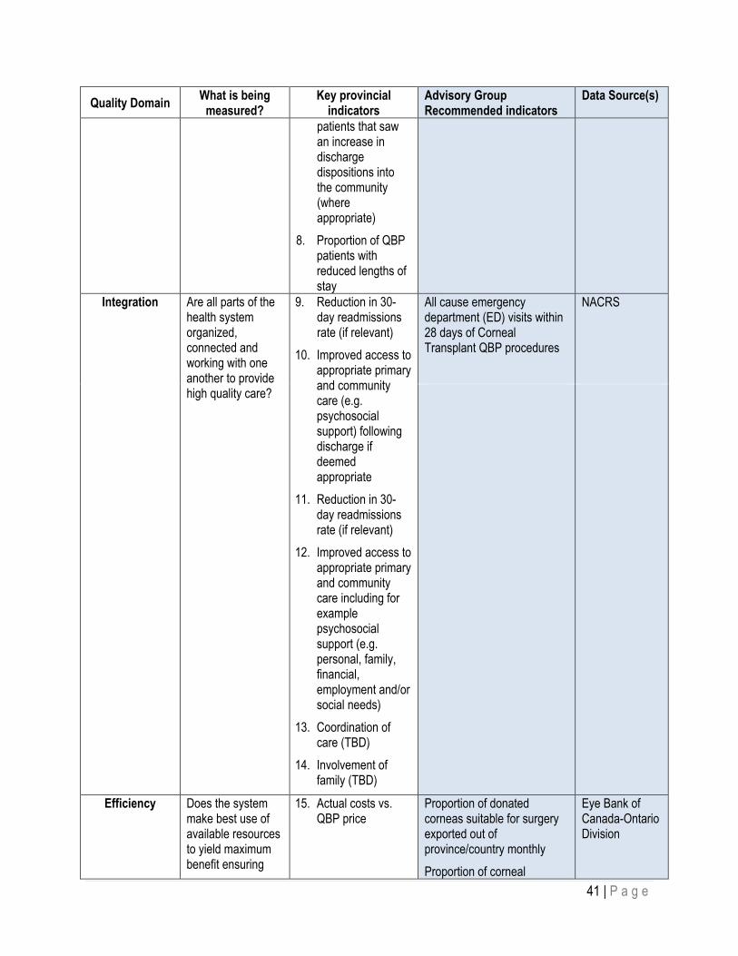
41 | P a g e
Quality Domain What is being measured?
Key provincial indicators
Advisory Group Recommended indicators
Data Source(s)
patients that saw an increase in discharge dispositions into the community (where appropriate)
8. Proportion of QBP patients with reduced lengths of stay
Integration Are all parts of the health system organized, connected and working with one another to provide high quality care?
9. Reduction in 30-day readmissions rate (if relevant)
10. Improved access to appropriate primary and community care (e.g. psychosocial support) following discharge if deemed appropriate
11. Reduction in 30-day readmissions rate (if relevant)
12. Improved access to appropriate primary and community care including for example psychosocial support (e.g. personal, family, financial, employment and/or social needs)
13. Coordination of care (TBD)
14. Involvement of family (TBD)
All cause emergency department (ED) visits within 28 days of Corneal Transplant QBP procedures
NACRS
Efficiency Does the system make best use of available resources to yield maximum benefit ensuring
15. Actual costs vs. QBP price
Proportion of donated corneas suitable for surgery exported out of province/country monthly Proportion of corneal
Eye Bank of Canada-Ontario Division

42 | P a g e
Quality Domain What is being measured?
Key provincial indicators
Advisory Group Recommended indicators
Data Source(s)
that the system is sustainable for the long term?
transplants performed using imported tissue monthly Proportion of corneal surgeon requests for pre-cut corneal tissue not met
Access Are those in need of care able to access services when needed?
16. Increase in wait times for QBPs / for specific QBP populations
17. Increase in wait times for other procedures
18. Increase in distance patients have to travel to receive the appropriate care related to the QBP
19. Proportion of providers with a significant change in resource intensity weights (RIW)
90th percentile corneal transplant Wait 1 90th percentile corneal transplant Wait 2
Cancer Care Ontario Access to Care Wait Time Strategy
Proportion of corneal transplant surgeries cancelled due to lack of donor corneal tissue
Eye Bank of Canada-Ontario Division
Distance traveled by patients from their homes to the hospital where QBP surgery procedures are performed
Wait Time Information System (WTIS), Cancer Care Ontario (CCO), Canada Postal Code Conversion File
Patient- Centeredness (to be further developed)
Is the patient/user at the centre of the care delivery and is there respect for and involvement of patients’ values, preferences and expressed needs in the care they receive?
20. Increased rate of patients being involved in treatment decision
21. Coordination of care
22. Involvement of family
Proportion of adult patients who received a corneal transplant surgery and were satisfied with:
• Explanation received about eye condition and proposed treatment
• Experience on day of surgery
• Ability to understand instructions for care of eye including use of eye medications
• Knowing how to access after-hours or emergent care
Hospital Survey (Developmental)

43 | P a g e
It should be noted that although not explicitly mentioned as a separate domain, the equity component of quality of care is reflected across the six domains of the scorecard and will be assessed by stratifying indicator results by key demographic variables and assessing comparability of findings across sub-groups. Where appropriate, the indicators will be risk-adjusted for important markers of patient complexity so that they will provide an accurate representation of the quality of care being provided to patients.
The Ministry and experts recognized that to be meaningful for clinicians and administrators, it is important to tie indicators to clinical guidelines and care standards. Hence, advisory groups that developed the best practices were asked to translate the provincial-level indicators into QBP-specific indicators. In consulting the advisory groups for this purpose, the Ministry was interested in identifying indicators both for which provincial data is readily available to calculate and those for which new information would be required. Measures in the latter category are intended to guide future discussion with Ministry partners regarding how identified data gaps might be addressed.
In developing the integrated scorecard approach, the Ministry recognized the different users of the indicators and envisioned each distinct set of measures as an inter-related cascade of information. That is, the sets of indicators each contain a number of system- or provincial-level measures that are impacted by other indicators or driving factors that are most relevant at the Local Health Integration Networks (LHINs), hospital or individual clinician level. The indicators will enable the Province and its partners to monitor and evaluate the quality of care and allow for benchmarking across organizations and clinicians. This will in turn support quality improvement and enable target setting for each QBP to ensure that the focus is on providing high quality care, as opposed to solely reducing costs.
It is important to note that process-related indicators selected by the Advisory Groups will be most relevant at the provider level. The full list of these measures is intended to function as a ‘menu’ of information that can assist administrators and clinicians in identifying areas for quality improvement. For example, individual providers can review patient-level results in conjunction with supplementary demographic, financial and other statistical information to help target care processes that might be re-engineered to help ensure that high-quality care is provided to patients.
Baseline reports and regular updates on QBP specific indicators will be included as appendices to each QBP Clinical Handbook. Reports will be supplemented with technical information outlining how results were calculated along with LHIN and provincial-level results that contextualize relative performance. Baseline reports will also be accompanied by facility-level information that will enable sharing of best practices and target setting at the provider-level.
The Ministry recognizes that the evaluation process will be ongoing and will require extensive collaboration with researchers, clinicians, administrators and other relevant stakeholders to develop, measure, report, evaluate and, if required, revise and/or include additional indicators to ensure that the information needs of its users are met.

44 | P a g e
9.0 Support for Change The Ministry, in collaboration with its partners, will deploy a number of field supports to support adoption of the funding policy. These supports include:
• Committed clinical engagement with representation from cross-sectoral health sector leadership and clinicians to champion change through the development of standards of care and the development of evidence-informed patient clinical pathways for the QBPs.
• Dedicated multidisciplinary clinical expert groups that seek clearly defined purposes, structures, processes and tools that are fundamental for helping to navigate the course of change.
• Strengthened relationships with Ministry partners and supporting agencies to seek input on the development and implementation of QBP policy, disseminate quality improvement tools, and support service capacity planning.
• Alignment with quality levers such as the Quality Improvement Plans (QIPs). QIPs strengthen the linkage between quality and funding and facilitate communication between the hospital board, administration, providers and public on the hospitals’ plans for quality improvement and enhancement of patient-centered care.
• Deployment of a Provincial Scale Applied Learning Strategy known as IDEAS (Improving the Delivery of Excellence Across Sectors). IDEAS is Ontario’s investment in field-driven capacity building for improvement. Its mission is to help build a high-performing health system by training a cadre of health system change agents that can support an approach to improvement of quality and value in Ontario.
We hope that these supports, including this Clinical Handbook, will help facilitate a sustainable dialogue between hospital administration, clinicians, and staff on the underlying evidence guiding QBP implementation. The field supports are intended to complement the quality improvement processes currently underway in your organization.

45 | P a g e
10.0 Clinical Expert Advisory Group Membership Member Name Affiliation Region
1. Dr. Sherif El-Defrawy (Co-Chair)
Chair, Department of Ophthalmology and Vision Sciences, University of Toronto; Medical Director, Kensington Eye Institute Toronto
2. Dr. David Rootman (Co-Chair)
Corneal Surgeon, University Health Network; Professor, Department of Ophthalmology and Vision Sciences, University of Toronto Toronto
3. Dr. Kashif Baig Director of the Cornea, Anterior Segment, and Refractive Surgery Fellowship Program; Cornea, Anterior Segment, and Refractive Surgeon at the University of Ottawa Eye Institute and The Ottawa Hospital
Ottawa
4. Dr. Stephanie Baxter Eye Physician and Surgeon Cornea and External Disease, Hotel Dieu Hospital, Kingston; Assistant Professor Residency Program Director, Department of Ophthalmology, Queen's University
Kingston
5. Dr. Chaim Bell Adjunct scientist at the Institute for Clinical Evaluative Sciences (ICES); Associate Professor, Institute of Health Policy, Management & Evaluation, University of Toronto
Provincial
6. Dr. Robert Campbell Glaucoma specialist, Deputy-chair of Ophthalmology, Queen’s University; ICES Scientist Ophthalmology Kingston
7. Dr. Varun Chaudhary Chief, Department of Eye Medicine & Surgery; Hamilton Regional Eye Institute; McMaster University & St. Joseph's Healthcare Hamilton; Associate Professor, Ophthalmology and Vitreoretinal Surgery
Hamilton
8. Elizabeth Chiu Manager of Coding and Abstracting, Decision Support, University Health Network Toronto
9. Ginette Daigle (Ex – Officio)
Senior Program Consultant -Specialized Programs and Agency Liaison Unit, Ministry of Health and Long-Term Care Provincial
10. Dr. William (Bill) Dixon Medical Director, The Eye Bank of Canada, Ontario Division, Professor, Department of Ophthalmology and Vision Sciences, University of Toronto Provincial
11. Dr. Jennifer Everson Family Physician, Physician Lead-Clinical Planning and Integration, Hamilton Niagara Haldimand Brant LHIN HNHB LHIN
12. Dr. Mark Fava Eye Physician and Surgeon Cornea and External Disease, St. Joseph’s Hospital, Hamilton Hamilton
13. Dr. Phil Hooper Clinical Provincial Lead, Ophthalmology, Access to Care; Associate Professor, Ophthalmology, Western University London
14. Joanna Li Health Analyst, Decision Support, University Health Network Toronto
15. Dr. Rookaya Mather Ivey Eye Institute, St Joseph’s Health Care London; Associate Professor of Ophthalmology, University of Western Ontario London

46 | P a g e
Member Name Affiliation Region
16. Anne Marie McIlmoyle Director, Perioperative Services, St. Joseph's Health Care, London London
17. Dr. Kylen McReelis General ophthalmology; Tariff Chair; Eye Physicians and Surgeons Ontario Peterborough
18. Samra Mian (Project Lead) Corporate Planner, Strategic Developments, University Health Network Toronto
19. Dr. Thomas Noël Optometrist, Ottawa; Co-chair, EHCO; President of the College of Optometrists of Ontario Ottawa
20. Audrey Pereira Health Analyst, Methods and Modeling, Health Analytics Branch, Ministry of Health and Long-Term Care Provincial
21. Dr. Neera Singal Corneal and Refractive Surgeon, Yonge Eglinton Laser Eye and Cosmetic Centre and the Kensington Eye Institute Toronto
22. Michael Stewart (Ex – Officio)
Executive lead, Decision Support and Knowledge Transfer, Ministry of Health and Long-Term Care Provincial
23. Julie Trpkovski Vice President, Clinical Transplant Systems, Trillium Gift of Life Network Provincial
24. Marnie Weber Executive Director, Strategic Developments, University Health Network Toronto
25. Dr. Charlotte Wedge Chief, Department of Ophthalmology, Toronto East General Hospital; Associate Medical Director, Eye Bank of Canada, Ontario Division. Toronto

47 | P a g e
Appendix A: Patient Survey on Corneal Transplant Care INSTRUCTIONS- Please place an x in the box that best describes your experience
How satisfied were you with: Very dissatisfied Dissatisfied Satisfied Very
satisfied Comment
1. The explanation you received about your eye condition and the treatment that was proposed
2. Your experience on the day of surgery
3. Being able to understand the instructions for the care of your eye including the use of your eye medications
4. Knowing how to access after-hours assistance or emergent care
Please provide any additional feedback that would have improved the experience and care you received:
Once again thank you for your input

48 | P a g e
.
.
.
.
.
.
.
.
.
.
.
.
.
.
.
.
.
.
References 1 Romero-Jiménez M1, Santodomingo-Rubido J, Wolffsohn JS. Keratoconus: a review. Cont Lens Anterior Eye. 2010
Aug;33(4):157-66; quiz 205. doi: 10.1016/j.clae.2010.04.006.
2 Leejee H. Suh, M. Vaughn Emerson. Fuchs Endothelial Dystrophy: Pathogenesis and Management. Cornea and External Eye Disease, Essentials in Ophthalmology 2008, pp 1-13.
3 Taylor DM, Atlas BF, Romanchuk KG, Stern AL. Pseudophakic bullous keratopathy. Ophthalmology. 1983 Jan;90(1):19-24.
4 Liu L, Wu J, Geng J, Yuan Z, Huang D. Geographical prevalence and risk factors for pterygium: a systematic review and meta-analysis.. BMJ Open. 2013 Nov 19;3(11):e003787. doi: 10.1136/bmjopen-2013-003787
5 Coster DJ, Lowe MT, Keane MC, Williams KA A comparison of lamellar and penetrating keratoplasty outcomes: a registry study. Ophthalmology. 2014 May;121(5):979-87. doi: 10.1016/j.ophtha.2013.12.017.
6 Reinhart WJ, Musch DC, Jacobs DS, Lee WB, Kaufman SC, Shtein RM.Deep anterior lamellar keratoplasty as an alternative to penetrating keratoplasty a report by the American academy of ophthalmology. Ophthalmology. 2011 Jan;118(1):209-18. doi: 10.1016/j.ophtha.2010.11.002.
7 Gomaa A, Comyn O, Liu C. Keratoprostheses in clinical practice - a review. Clin Experiment Ophthalmol. 2010 Mar;38(2):211-24. doi: 10.1111/j.1442-9071.2010.02231.x.
8 Avadhanam VS1, Smith HE2, Liu C3.Keratoprostheses for corneal blindness: a review of contemporary devices. Clin Ophthalmol. 2015 Apr 16;9:697-720. doi: 10.2147/OPTH.S27083. eCollection 2015.
9 Li LM, Zhao LQ, Qu LH, Li P. Excimer laser phototherapeutic keratectomy for the treatment of clinically presumed fungal keratitis. J Ophthalmol. 2014;2014:963287. doi: 10.1155/2014/963287. Epub 2014 May 7.
10 Frank MH, Frank NY. Restoring the cornea from limbal stem cells. Regen Med. 2015;10(1):1-4. doi: 10.2217/rme.14.66.
11 O'Brart DP. Corneal collagen cross-linking: a review. J Optom. 2014 Jul-Sep;7(3):113-24. doi:10.1016/j.optom.2013.12.001.
12 Eye Bank Association of America. Medical Standards: These standards have the approval of the Eye Banking Committee of the American Academy of Ophthalmology. October 2011 (Report). Accessed online on August 12, 2015 from: http://www.restoresight.org/wp-content/uploads/2011/11/Medical-Standards-October-2011.pdf
13 R.R.O. 1990, Reg. 965: HOSPITAL MANAGEMENT. under Public Hospitals Act, R.S.O. 1990, c. P.40. Available online: http://www.ontario.ca/laws/regulation/900965/v1
14 Kaufman SC, Jacobs DS, Lee WB, Deng SX, Rosenblatt MI, Shtein RM. Options and adjuvants in surgery for pterygium: a report by the American Academy of Ophthalmology. Ophthalmology. 2013 Jan;120(1):201-8. doi:10.1016/j.ophtha.2012.06.066.
15 Hirst LW, Axelsen RA, Schwab I. Pterygium and associated ocular surface squamous neoplasia. Arch Ophthalmol. 2009 Jan;127(1):31-2. doi: 10.1001/archophthalmol.2008.531.
16 Rudkin AK, Dodd T, Muecke JS.The differential diagnosis of localised amelanotic limbal lesions: a review of 162 consecutive excisions. Br J Ophthalmol. 2011 Mar;95(3):350-4. doi: 10.1136/bjo.2009.172189.
17 Ontario Health Technology Advisory Committee (OHTAC). OHTAC Report - Limbal Stem Cell Transplantation. 2008 http://www.hqontario.ca/english/providers/program/ohtac/tech/recommend/rec_lsc_20080413.pdf
18 The Provincial Vision Strategy Task Force. A Vision for Ontario. Strategic Recommendations for Ophthalmology in Ontario, May 31, 2013. http://www.health.gov.on.ca/en/common/ministry/publications/reports/docs/ontario_vision_strategy_report.pdf

This page has been intentionally left blank.