int. j. dev. biol. 54: 1075-1078 (2010) the international ...§oise/otherinformation/ft1075.pdf ·...
TRANSCRIPT
The quest for hematopoietic stem cells in the embryo
- an interview with Françoise Dieterlen-Lièvre
THIERRY JAFFREDO*,1 and CHARLES DURAND2
1CNRS UMR7622 and 2UPMC UMR7622, Laboratoire de Biologie du Développement, Paris, France
ABSTRACT Françoise Dieterlen-Lièvre is probably the scientist who has most contributed to our
basic knowledge of developmental hematopoiesis. She has dedicated her career to answering
cutting edge questions on the origin of hematopoietic stem cells in the embryo. Her seminal
contributions, widely recognized by the scientific community, have paved the way for generations
of developmental hematologists questioning the origins of hematopoietic stem cells. After having
demonstrated the intra-embryonic origin of hematopoietic stem cells, established the dual origin
of the endothelial network in the embryo and revealed the hematopoietic function of the allantois
in birds, she has switched to mammals and contributed to demonstrating that the aorta and
allantois/placenta are new sites of hematopoietic production in the mouse embryo. The manifold
insights generated by the pivotal work of Françoise Dieterlen-Lièvre have created multiple
paradigm shifts which continue to challenge the field of developmental hematopoiesis.
KEY WORDS: embryo,chick, quail, mouse, aorta, allantois, yolk sac, placenta, hematopoiesis
Introduction
Dr. Françoise Dieterlen-Lièvre has dedicated her career toexperimental avian and mouse embryology. From the sixties tothe mid seventies, she studied in depth the formation of thepancreas in birds and the emergence of Langherans islets, thesubject of her PhD thesis. Soon after the discovery of the quail-chicken system by Pr. Nicole le Douarin, she became interestedin Developmental Hematopoiesis and demonstrated in 1975,using the avian model, the existence of an intra-embryonic sourcefor Hematopoietic Stem Cells (HSC). Pursuing the quest for HSC,she demonstrated in 1986 with her team that the aorta was ableto produce multipotent hematopoietic progenitors in culture, longbefore similar questions were asked for the mouse embryo, thusdemonstrating the great possibilities offered by the use of theavian embryo. At the same time, she also became interested inthe origin of the vascular system in the avian embryo anddeveloped several tools to follow endothelial precursors in theembryo. This interest lasted until the end of the nineties, bringingseveral major contributions, one of the most well known being theexistence of two different endothelial lineages in the embryo, onebeing associated with hematopoiesis. From the beginning of thenineties, she turned her attention to the mouse embryo and theorigin of HSC in this species. In 1993, she unravelled an unex-
Int. J. Dev. Biol. 54: 1075-1078 (2010)doi: 10.1387/ijdb.103153tj
THE INTERNATIONAL JOURNAL OF
DEVELOPMENTAL
BIOLOGYwww.intjdevbiol.com
*Address correspondence to: Thierry Jaffredo. CNRS UMR7622, Laboratoire de Biologie du Développement, Bat C, 6ème étage; Case 24; 75252 Paris Cedex05; France. Fax: +33-14-427-3497. e-mail: [email protected]
Final author corrected PDF published online: 16 July 2010.
ISSN: Online 1696-3547, Print 0214-6282© 2010 UBC PressPrinted in Spain
Abbreviations used in this paper: AGM, aorta-gonad-mesonephros; HSC,hematopoietic stem cell; P-Sp; para-aortic splanchnopleura.
pected hematopoietic activity in the aortic region of the earlymouse embryo. In the next few years, she published severalseminal papers on the mouse aorta and HSC production. At thesame time, she demonstrated, using the avian model, the produc-tion of hematopoietic cells by endothelial cells. Last but not least,from 1998, she unravelled the existence of a new hematopoieticsite and organ i.e. the allantois. First investigated in the avianmodel, the question of the mouse allantois rapidly came underscrutiny and she demonstrated that the mouse allantois wascapable of autonomously producing multipotent progenitors andthat the placenta was an organ where HSC expanded. FrançoiseDieterlen-Lièvre was co-director of the Nogent Institute from 1981to 2000 (see Le Douarin, 2005) . She retired in 2003 but continuesher research.
First of all, could you tell us when your interest in develop-mental biology arose and why you decided to study theembryo?
My interest in biology in general, and more specifically inembryology, was largely incidental. When I was 16, I passed the

1076 T. Jaffredo and C. Durand
baccalauréat, which in France opens the door to University, andhad no precise calling. My parents were both physicians, but didnot particularly encourage their progeny to adopt this track. Isuppose studying biology was a close make-believe. Then whenI finished my undergraduate studies, I went to see Pr. EtienneWolff, originator of the French school of avian embryology, whowas an acquaintance. He was moving from Strasbourg andcreating a laboratory in the College de France in Paris, where hehad recently been elected. He accepted me as a junior investiga-tor in the new team he was creating. This turn of events wasdeterminant, as developing embryos are fascinating and theexperimental possibilities provided by the avian model turned meinto an enthusiast.
How did you come up with the idea that the embryo harbouredan intra-embryonic source of HSC? At that time, Moore andOwen’s hypothesis of the yolk sac origin of HSC was preva-lent, if not actually a dogma.
My interest in hematopoietic development arose as a follow-upof my PhD work on pancreatic development. This was in the earlysixties, that is, the pre-molecular era of developmental biology.Many scientists were trying to understand the mechanisms ofepithelio-mesenchymal interactions. E. Wolff had designed aculture system that allowed organ rudiments to develop ex vivo,while preserving their three-dimensional structure (Wolff andHaffen, 1952a). In the laboratory, each person worked on adifferent rudiment. Seminal contributions were made at that time,for instance by Katty Haffen about gonadal differenciation (Wolffand Haffen, 1951, 1952b), by Philippe Sengel about skin devel-opment (Sengel, 1955 a,b), by Nicole Le Douarin, who discoveredat that time the sequential inductions required for liver develop-ment (Le Douarin, 1964a; Le Douarin, 1964b; see interview withNicole Le Douarin in this issue: Durand and Jaffredo, 2010).
Against this background, I was assigned the pancreas. I spenta lot of time establishing the ratio between endocrine and exocrinepancreas (the so-called ‘Richardson and Young ratio’) and foundthat the islets of Langerhans are prevalent in early developmentand synthesize glucagon and insulin long before the exocrinetissue produces enzymes (Dieterlen-Lièvre, 1970 a,b).
During ontogeny, the spleen rudiment is a mesodermal capcontinuous with pancreatic mesoderm. As several hematopoieticorgan rudiments are built from endoderm and mesoderm, forinstance the thymus, I wondered whether the pancreatic endo-derm had a role in the formation of the spleen. This was thestarting point of my interest in blood-forming organs. At that time(1972), Claude Martin, who was a micro-surgery champion,invented sophisticated new techniques, including the ‘yolk sacchimera’. She published the technique (Martin, 1972), but shewas involved in analyzing the developmental cascade of the threeembryonic kidneys with Yvon Croisille and Madeline Pinot, so thatshe did not exploit these chimeras.
Could you explain what these chimeras are?First a point of history: it was in 1969 that Nicole Le Douarin
imagined transplanting quail rudiments into chick embryos, anidea which was based on her noticing a marked differencebetween the cell nuclei of the two species (see Coutinho, 2005).She thus devised the labelling method which allowed her tounravel many developmental processes, relating notably to the
nervous and the immune systems (Le Douarin, 1969). The ‘yolksac chimeras’ devised by Claude are built surgically by graftingthe central area (the future body) of a quail blastoderm onto theperipheral area of a chick yolk embryo (the future yolk sac). Thisis done in the egg at two days of incubation and the operatedembryo can successfully pursue its development in about ten percent of the cases. These heterospecific chimeras are raised untilday 13 of incubation; later the yolk sac, disproportionate inrelationship to the embryo, fails to incorporate into the body wall(Dieterlen-Lièvre, 1975). A couple of years later, homospecificchimeras built between two strains of chicken were raised suc-cessfully to adulthood (Lassila et al., 1978).
When I began speculating about the hematopoietic system,this novel type of chimera was an obvious tool. At that time, I hadno preconceived idea; it was more or less unstated in my mind thatthis approach would validate Moore and Owen’s experiments.Their interpretation, according to which all Hematopoietic StemCells (HSCs) were born in the yolk sac (Moore & Owen, 1965;Moore & Owen, 1967), had immediately become a dogma in thescientific community.
In a first set of experiments I produced about 10 chimeraswhich gave very homogeneous results: the thymus, bursa ofFabricius and spleen were all populated by quail cells, whichhence did not originate from the yolk sac. I was thrilled, so I sentthe paper to Nature. The reply received within a week was thefollowing: “Moore and Owen have already proved the contrary, itis preposterous to return back to this”. I decided to try the Journalof Embryology and Experimental Morphology (now Develop-ment), where it was readily accepted (Dieterlen-Lièvre, 1975).This 1975 paper has recently been elected as the first of a seriesof ‘JEEM Classics’ (Dzierzak & Medvinsky, 2008).
In 1978, you published a Nature paper on the origin oflymphoid stem cells analysed by means of a chicken yolksac/chicken embryo chimera. How was the paper receivedand does it mean that the intra-embryonic source of HSC wasaccepted by the scientific community?
This piece of work resulted from a collaboration with the finnishteam of Paavo Toivanen (Lassila et al., 1978, 1982). In thesechimeras, the markers distinguishing the two chicken embryoscomposing the chimera were either different immunoglobulinallotypes or different Major Histocompatibility Complex haplotypes.This work confirmed and extended the conclusions drawn fromthe analysis of heterospecific chimeras, as the chicken-chickenchimeras hatched and were raised to adulthood. In this make up,where no differences in size and developmental rhythm, norimmune acute rejection could interfere, red cells of yolk sac origindisappeared before hatching and the cells identified through thetwo markers were all of embryo origin.
The turn of opinions was driven by the acceptance of this paperin Nature and, very soon afterwards, of another in Blood (1979).The latter, carried out with Claude Martin and Denise Beaupain,described the replacement of circulating chicken red cells (de-rived from yolk sac HSCs) by quail red cells (derived fromembryonic body HSCs) (Beaupain et al., 1979). Amazingly, for tenyears, many papers in the field of immunology stated that ‘HSCscome from the yolk sac, even though we know it is different in theavian embryo’. The avian paradigm however had soon beenextended to amphibians.

Interview with Françoise Dieterlen-Lièvre 1077
Your demonstration in a 1993 Nature paper that the aorticregion of the early mouse embryo harbours HSCs has cer-tainly changed the common view?
Nearly twenty years elapsed however before experimentscarried out in mice were published back to back in this issue ofNature, that you mention, by Alexander Medvinsky, NinaSamoylina, Albrecht Müller and Elaine Dzierzak on the one hand(Medvinsky et al., 1993) and by ourselves on the other (Godin etal., 1993). It is clear that these papers became acceptablebecause of their identical conclusion, and also because the wayof thinking had been modified by the concurring data obtained inother classes of vertebrates. The article by Medvinsky et al.examined CFU-S (Colony Forming Unit in the Spleen) activity (i.eshort term hematopoietic progenitors) in the tissues from the trunkregion of the embryo corresponding to the aorta plus the gonadsand the mesonephros. The authors called these tissues ‘AGM’, aterm which became adopted, even though it has no anatomical orembryological meaning, but denotes the impossibility of dissect-ing these three organs from one another in the very early embryo.We know now that the endothelium of the aorta is responsible foremitting the intra-embryonic HSCs, and I feel strongly that the
term ‘AGM’ should be replaced by ‘aorta’. Cells capable of givingrise to colonies in the spleen of irradiated mice were found in thisregion at days 10-11 of gestation. Our own work, whose concep-tion and methods were proposed and driven by Miguel Marcosand carried out with Isabelle Godin and Juan Garcia-Porrero, boreon earlier tissues (days 8.5-9) in the same region. They comprisedthe hindgut, the paired rudiments of the dorsal aorta, the vitellinearteries and the mesoderm surrounding these rudiments. Wecalled these tissues ‘Para-aortic Splanchnopleura’ (P-Sp), thesplanchnopleura being the association of mesoderm and endo-derm, which is going to give rise to visceral organs. These tissueswere tested for their capacity to restore B lymphocytes in immu-nodeficient (SCID) mice. Interestingly a particular subpopulation,the CD5+ B cells, which reside in the peritoneum, and are notreconstituted by adult bone marrow in irradiated adults, wererestored by grafts of P-Sp under the kidney capsule of the SCIDadults.
One of your most relevant discoveries may be the role of theallantois/placenta in the generation of HSC. You have openedan unexplored field that has now major implications for basicbiology but also for regenerative medicine. Could you ex-plain how you came up with this idea?
First, I would like to remark on your comment about implica-tions for regenerative medicine. I do not think that pinning downthe hematopoietic potential of the placenta will revolutionize thestate of the art (see Dieterlen-Lèvre et al., 2010). Cord blood stemcells, discovered and promoted by Elaine Gluckmann in Paris, arenow major actors in regenerative medicine. As they are relativelyeasy to obtain, I do not think they will be superseded by cells fromthe placenta. But they probably are the product of placentalactivity, an important point to establish for the sake of knowledge.In that area again, the avian model gave the lead. In the 1979Blood paper (Beaupain et al., 1979), a picture of the sectionedallantois of a quail/chicken chimera clearly showed a blood island-like structure, which encased quail blood-like cells. I decided topursue this lead, when cultures of whole chicken allantoises inliquid medium gave rise to clouds of red cells. Our classical modusoperandi was repeated, namely unvascularized allantois rudi-ments from the quail embryo were grafted (ectopically) into non-compromised chicken hosts, with the result that the host bonemarrow was heavily populated by quail HSCs and endothelialcells, a story that you, Thierry, know well, since this was your entryinto the world of hemangioblasts. It is fair to mention here that theseminal experiments in my group about the relationship betweenthe ‘hemogenic’ endothelium of the aorta and HSCs were carriedout by Luc Pardanaud. Soon we addressed the issue successfullyin the mouse embryo with Marcio Alvarez Silva and JosselyneSalaün (Alvarez-Silva et al., 2003), continuing into the matter withHanna Mikkola and Stu Orkin (Developmental Cell, 2005) andCatherine Corbel and Josselyne Salaün for the allantois (Corbelet al., 2008). Several groups entered the field along the process,but this is not the place for an exhaustive review!
I know from our frequent discussions that, despite the factthat you spent more than half of a century working ondevelopmental hematopoiesis, you are still fascinated bythis field and have revolutionary ideas to explore hematopoi-etic production in the embryo. What are according to you the
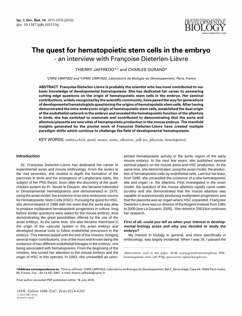
Fig. 1. Françoise Dieterlen-Lièvre at a party at Nogent Institute in
1980.

1078 T. Jaffredo and C. Durand
most relevant and still unanswered questions regardinghematopoietic development?
The hemogenic capacity of the endothelium is the next chal-lenge. Is this striking potential restricted to the embryonic aorta,where it is very visible and was experimentally pinned down or isit a generalized potential? If it is, the role of the niche (formerly themicroenvironment) must be critical in allowing its expression.
References
ALVAREZ-SILVA, M., BELO-DIABANGOUAYA, P., SALAUN, J. and DIETERLEN-LIEVRE, F. (2003). Mouse placenta is a major hematopoietic organ. Develop-ment 130: 5437-5444.
BEAUPAIN, D., MARTIN, C. and DIETERLEN-LIEVRE, F. (1979). Are develop-mental hemoglobin changes related to the origin of stem cells and site oferythropoiesis? Blood 53: 212-225.
CORBEL, C., SALAUN, J., BELO-DIABANGOUAYA, P. and DIETERLEN-LIEVRE,F. (2007). Hematopoietic potential of the pre-fusion allantois. Dev Biol 301: 478-488.
COUTINHO, A. (2005). The Le Douarin phenomenon: a shift in the paradigm ofdevelopmental self-tolerance. Int. J. Dev. Biol. 49: 131-136.
DIETERLEN-LIEVRE, F. (1960). Influence of embryonic pancreas on the formationof liver glycogen in chicks studied by the method of in vitro parabiosis. C R HebdSeances Acad Sci 250: 1349-1351.
DIETERLEN-LIEVRE, F. (1965). Morphological and experimental study of thedifferentiation of the pancreas in the chick embryo. Bull Biol Fr Belg 99: 3-116.
DIETERLEN-LIEVRE, F. (1969). Experimental analysis of the origin of pancreastissue components in chick embryo and conditions of their differentiation. C RAcad Sci Hebd Seances Acad Sci D 269: 2392-2395.
DIETERLEN-LIEVRE, F. (1970a). Epithelio-mesenchymatous relations in differen-tiation of the pancreas in chick embryo. Annee Biol 9: 261-266.
DIETERLEN-LIEVRE, F. (1970b). Exocrine and endocrine tissue in the pancreasof the chic embryo: origin and tissue interactions in differentiation. Dev Biol 22:138-156.
DIETERLEN-LIEVRE, F. (1975). On the origin of haemopoietic stem cells in theavian embryo: an experimental approach. J Embryol Exp Morphol 33: 607-619.
DIETERLEN-LIEVRE, F., CORBEL, C. and SALAÜN, J. (2010). Allantois andplacenta as developmental sources of hematopoietic stem cells. Int. J. Dev.Biol. 54: 1079-1087.
DURAND, C. and JAFFREDO, T. (2010). Developmental hematopoiesis: historicalbackground and perspectives - an interview with Nicole Le Douarin. Int. J. Dev.Biol. 54: 951-954.
DZIERZAK, E. and MEDVINSKY, A. (2008). The discovery of a source of adulthematopoietic cells in the embryo. Development 135: 2343-2346.
GEKAS, C., DIETERLEN-LIEVRE, F., ORKIN, S.H. and MIKKOLA, H.K. (2005).The placenta is a niche for hematopoietic stem cells. Dev Cell 8: 365-375.
GODIN, I.E., GARCIA-PORRERO, J.A., COUTINHO, A., DIETERLEN-LIEVRE, F.and MARCOS, M.A. (1993). Para-aortic splanchnopleura from early mouseembryos contains B1a cell progenitors. Nature 364: 67-70.
LASSILA, O., ESKOLA, J., TOIVANEN, P., MARTIN, C. and DIETERLEN-LIEVRE,F. (1978). The origin of lymphoid stem cells studied in chick yolk sac-embryochimaeras. Nature 272: 353-354.
LASSILA, O., MARTIN, C., TOIVANEN, P. and DIETERLEN-LIEVRE, F. (1982).Erythropoiesis and lymphopoiesis in the chick yolk-sac-embryo chimeras:contribution of yolk sac and intraembryonic stem cells. Blood 59: 377-381.
LE DOUARIN, N. (1964a). Induction de l’endoderme pré-hépatique par le mésodermede l’aire cardiaque chez l’embryon de Poulet. J. Embryol. Exp. Morph. 12: 651-676.
LE DOUARIN, N. (1964b). Isolement expérimental du mésenchyme propre du foieet rôle morphogène e la composante mésodermique dans l’organogenèsehépatique.. J. Embryol. Exp. Morph. 12: 141-160.
LE DOUARIN, N. (1969). Details of the interphase nucleus in Japanese quail(Coturnix coturnix japonica). Bull Biol Fr Belg 103: 435-452.
LE DOUARIN, N. (2005). The Nogent Institute - 50 Years of Embryology. Int. J. Dev.Biol. 49: 85-103.
MARTIN, C. (1972). Method of explantation in ovo of the blastoderm of birdembryos. C R Seances Soc Biol Fil 166: 283-285.
MEDVINSKY, A.L., SAMOYLINA, N.L., MULLER, A.M. and’DZIERZAK, E.A. (1993).An early pre-liver intraembryonic source of CFU-S in the developing mouse.Nature 364: 64-67.
MOORE, M.A. and OWEN, J.J. (1965). Chromosome marker studies on thedevelopment of the haemopoietic system in the chick embryo. Nature 208: 956passim.
MOORE, M.A. and OWEN, J.J. (1967). Chromosome marker studies in theirradiated chick embryo. Nature 215: 1081-1082.
SENGEL, P. (1955a). Effect of tyrosine on pigmentation of feather germ ofembryonic chick skin cultured in vitro. C R Seances Soc Biol Fil 149: 1479-1482.
SENGEL, P. (1955b). Influence of certain amino acids on the growth and pigmen-tation of the feather papilla from chick embryo skin cultured on synthetic media.C R Seances Soc Biol Fil 149: 1032-1035.
WOLFF, E. and HAFFEN, K. (1951). Culture in vitro of the genital glands of birdembryos; obtaining normal sex differentiation and experimental intersexuality ofthe explanted gonads. C R Hebd Seances Acad Sci 233: 439-441.
WOLFF, E. and HAFFEN, K. (1952a). Method for culture of embryonic organs. TexRep Biol Med 10: 463-472.
WOLFF, E. and HAFFEN, K. (1952b). Sexual differentiation of embryonic gonadsin chicks in vitro. Ann Endocrinol (Paris) 13: 345.
5 yr ISI Impact Factor (2009) = 3.253

Interview with Françoise Dieterlen-Lièvre 1079
Further Related Reading, published previously in the Int. J. Dev. Biol.
See our recent Special Issue Placenta edited by Joan S. Hunt and Kent L. Thornburg at:http://www.ijdb.ehu.es/web/contents.php?vol=54&issue=2-3
Allantois and placenta as developmental sources of hematopoietic stem cellsFrançoise Dieterlen-Lièvre Catherine Corbel and Josselyne SalaünInt. J. Dev. Biol.(doi: 10.1387/ijdb.093047fd)
Challenges and strategies for generating therapeutic patient-specific hemangioblasts and hematopoietic stem cells from humanpluripotent stem cellsAnn Peters, Paul W. Burridge, Marina V. Pryzhkova, Michal A. Levine,Tea-Soon Park, Christopher Roxbury, Xuan Yuan, Bruno Péault andElias T. ZambidisInt. J. Dev. Biol. (doi: 10.1387/ijdb.093043ap)
Trophoblast phagocytic program: roles in different placental systemsEstela Bevilacqua, Mara-Sandra Hoshida, Andrea Amarante-Paffaro, Andrea Albieri-Borges and Sara Zago-GomesInt. J. Dev. Biol. (2010) 54: 495-505
On the role of placental major histocompatibility complex and decidual leukocytes in implantation and pregnancy success using non-human primate modelsThaddeus G. Golos, Gennadiy I. Bondarenko, Svetlana V. Dambaeva, Edith E. Breburda, and Maureen Durning
5 yr ISI Impact Factor (2009) = 3.253
Int. J. Dev. Biol. (2010) 54: 431-443
Development and function of trophoblast giant cells in the rodent placentaDong Hu and James C. CrossInt. J. Dev. Biol. (2010) 54: 341-354
Experimental study of early olfactory neuron differentiation and nerve formation usingquail-chick chimerasFabrice L. Lalloué and Christiane S. Ayer-Le LièvreInt. J. Dev. Biol. (2005) 49: 193-200
Cellular dynamics and molecular control of the development of organizer-derived cells inquail-chick chimerasJean-Baptiste Charrier, Martin Catala, Françoise Lapointe, Nicole Le Douarin and Marie-AiméeTeilletInt. J. Dev. Biol. (2005) 49: 181-191
Tracing the hemangioblast during embryogenesis: developmental relationships betweenendothelial and hematopoietic cellsThierry Jaffredo, Karine Bollerot, Daisuke Sugiyama, Rodolphe Gautier and Cécile DrevonInt. J. Dev. Biol. (2005) 49: 269-277
Of birds and mice: hematopoietic stem cell developmentIsabelle Godin and Ana CumanoInt. J. Dev. Biol. (2005) 49: 251-257
Embryonic development of the human hematopoietic systemManuela Tavian and Bruno PéaultInt. J. Dev. Biol. (2005) 49: 243-250
Commitment of hematopoietic stem cells in avian and mammalian embryos: an ongoingstoryFrançoise Dieterlen-LièvreInt. J. Dev. Biol. (2005) 49: 125-130
Multilineage hematopoietic progenitor activity generated autonomously in the mouse yolksac: analysis using angiogenesis-defective embryos.Christine Rampon and Philippe HuberInt. J. Dev. Biol. (2003) 47: 273-280