insulin trafficking in a glucose responsive engineered...
TRANSCRIPT

Chapter 29
Insulin Trafficking in a Glucose Responsive EngineeredHuman Liver Cell Line is Regulated by the Interactionof ATP-Sensitive Potassium Channels and Voltage-Gated Calcium Channels
Ann M. Simpson, M. Anne Swan, Guo Jun Liu,Chang Tao, Bronwyn A O’Brien, Edwin Ch’ng,Leticia M. Castro, Julia Ting, Zehra Elgundi, Tony An,Mark Lutherborrow, Fraser Torpy, Donald K. Martin,Bernard E. Tuch and Graham M. Nicholson
Additional information is available at the end of the chapter
http://dx.doi.org/10.5772/52839
1. Introduction
Type I diabetes is caused by the autoimmune destruction of pancreatic beta (β) cells [1]. Cur‐rent treatment requires multiple daily injections of insulin to control blood glucose levels.Tight glucose control lowers, but does not eliminate, the onset of diabetic complications,which greatly reduce the quality and longevity of life for patients. Transplantation of pan‐creatic tissue as a treatment is restricted by the scarcity of donors and the requirement forlifelong immunosuppression to preserve the graft, which carries adverse side-effects. This isof particular concern as Type 1 diabetes predominantly affects children. Lack of glucose con‐trol could be overcome by genetically engineering "an artificial β-cell" that is capable of syn‐thesising, storing and secreting insulin in response to metabolic signals. The donor cell typemust be readily accessible and capable of being engineered to synthesise, process, store andsecrete insulin under physiological conditions.
The cell type of choice for the gene therapy of diabetes is not the β-cell. β-cells are greatlyreduced or absent in people with Type I diabetes because of their autoimmune destruction.This fact will actively work against gene therapists trying to derive surrogate β-cells from
© 2013 Simpson et al.; licensee InTech. This is an open access article distributed under the terms of theCreative Commons Attribution License (http://creativecommons.org/licenses/by/3.0), which permitsunrestricted use, distribution, and reproduction in any medium, provided the original work is properly cited.

stem cells. There are innumerable theories describing putative mechanisms for preventing apatient’s immune system from re-attacking transplanted β-cells, but the fact that the basicprocesses of islet cell attack have not been fully elucidated makes the search for relevantgenes problematic. Thus, the engineering of non-pancreatic β-cells to synthesise, process,store and secrete insulin has several advantages, the most important of which is the readyavailability of donor cells. If non β-cells from a diabetic individual can be engineered to pro‐duce insulin, then cellular rejection is less likely to occur since donor and recipient are autol‐ogous. In pursuit of this goal, hepatocytes have been shown to be suitable target cells for thegeneration of artificial β-cells [2-9]. Moreover, liver cells that produce insulin may not beprone to autoimmune attack [10]. The suitability of hepatocytes as a β-cell replacement is at‐tributable, in part, to their inherent glucose responsiveness and their embryonic origin fromthe same endodermal precursor cells as the β-cell. Most importantly, liver cells express thehigh capacity glucose transporter, GLUT 2 [11], and the high capacity phosphorylation en‐zyme, glucokinase [12], which constitute the key elements of the “glucose sensing system”that regulates insulin release from pancreatic β-cells in response to small extracellular nu‐trient changes.
In pancreatic β-cells, a small increase in plasma glucose concentration stimulates significantinsulin secretion. Therefore, glucose is the major modulator of β-cell function and this behav‐iour must be mimicked in insulin-secreting liver cells. In pancreatic β-cells, KATP channels,which are composed of four sulphonylurea receptor (SUR) subunits and four inwardly-recti‐fying potassium channel (KIR6.2) subunits [13-18], maintain resting membrane potentials andlink plasma glucose concentrations to the insulin secretory machinery. The triggering path‐way for insulin release begins with the uptake of glucose via the glucose carrier, GLUT2, andan acceleration of metabolism, such that glucose is used to generate ATP. An increase in theabsolute intracellular concentration of ATP, with respect to ADP, stimulates the closure of KATP
channels [19, 20]. Potassium conductance of the plasma membrane decreases, allowing abackground current to shift the membrane potential away from the equilibrium potential forK+, thus depolarising the membrane. Consequently, the pancreatic β-cell is able to translatemetabolic signals to electrical signals, the latter regulating insulin secretion. Lack of function‐al KATP channels in insulin-secreting NES2Y cells resulted in the unregulated release of insu‐lin, which was restored by expression of both KIR6.2 and SUR1 [21].
When depolarisation of the pancreatic β-cell reaches the threshold for activation of L-type(CaV1.3), and to a lesser extent P/Q (CaV1.2) and T-type (CaV3.x) voltage-gated calcium chan‐nels, these open allowing Ca2+ influx down their electrochemical gradient [22]. The openingof CaV channels is intermittent, fluctuating with the membrane potential, therefore generat‐ing oscillations in the intracellular (cytosolic) calcium concentration ([Ca2+]i), which, in turn,triggers pulsatile insulin secretion. In β-cells, elevation of [Ca2+]i occurs via the release of Ca2+
from intracellular stores (endoplasmic reticulum, mitochondria and secretory granules)and/or influx of extracellular Ca2+ through CaV channels [23, 24]. No functional CaV channelshave been previously described in liver cells, however the presence of an α1-subunit lackingthe voltage sensor has been reported in the rat liver cell line H4IIE [25] and an L-type α1-subunit has been detected at low levels in rat liver by RT-PCR [26].
Gene Therapy - Tools and Potential Applications704
For an insulin-producing liver cell to be of maximum benefit in vivo it must be capable ofrapid responsive secretion of biologically active insulin. This characteristic demands that ar‐tificial β-cells process proinsulin to insulin and store it in granules. Our previous studieshave shown that the insertion of genes encoding for insulin and the glucose transporter,GLUT2, into the HEPG2 human hepatoma cell line, resulted in synthesis and storage of(pro)insulin in structures resembling the secretory granules of pancreatic β-cells(HEPG2/ins/g), and the near physiological secretion of (pro)insulin in response to glucose [2,3]. Similar to pancreatic β-cells, HEPG2ins/g cells responded to glucose via signalling path‐ways dependent upon KATP channels [27]. Therefore, expression of both insulin and GLUT2in HEPG2 liver cells appeared to be sufficient for the generation of functional KATP channels,unlike the parental cell line that required pharmacological stimulation to activate the KATP
channels [28]. It has previously been shown that stable transfection of the insulin gene intothe human liver cell line, Huh7 (which endogenously expresses GLUT2), resulted in synthe‐sis, storage, and regulated release of insulin to the physiological stimulus glucose (Huh7inscells) [7]. Huh7ins cells are more akin to pancreatic β-cells than HEPG2/ins/g cells. They ex‐press a range of β-cell transcription factors [7, 29] and possess storage granules that cleaveproinsulin to biologically active diarginyl insulin, due to the expression of the proconvertas‐es PC1 and PC2 [7]. As Huh7ins cells also rapidly secrete insulin in a tightly regulated man‐ner in response to glucose, the Huh7ins cells were able to reverse chemically induceddiabetes when transplanted into an animal model [7], which HEPG2ins/g cells [3] failed toachieve [Tuch, unpublished results].
This chapter will detail the use of electrophysiological and biochemical techniques to showthat Huh7ins cells respond to a glucose stimulus by closure of KATP channels and activationof CaV channels, which is an analogous mechanism to pancreatic β-cells. Patch-clamp elec‐trophysiology of Huh7ins cells yielded current-voltage (I-V) curves that indicated the pres‐ence of potassium-selective currents; in contrast, currents recorded from Huh7 cells werenon-selective. The presence of functional ATP-sensitive potassium (KATP) channels and volt‐age-gated calcium (CaV) channels was further validated by measurement of acute insulin se‐cretion by Huh7ins cells in response to pharmacological channel inhibitors and activatorsand by calcium imaging and patch-clamp electrophysiology experiments. Molecular analy‐ses were used to confirm that the Huh7ins cells express CaV and all the subunits of KATP
channels. The secretion of insulin from granules in live Huh7ins cells was revealed by confo‐cal microscopy which allowed visualization of secretion of insulin to a zinquin probe or aninsulin-enhanced green fluorescent protein (EGFP) fusion protein (EGFP-ins). The glucoseresponsive mechanism that we observed in the Huh7ins cells was the same as that reportedfor the pancreatic β-cell line, MIN6 [30]. Prior to this study, the physiological interaction ofKATP channels and CaV channels had never been shown in liver cells engineered to secreteinsulin. As the biochemical properties of Huh7ins cells are akin to those of pancreatic β-cells,engineering hepatocytes in this way opens a promising avenue for the ultimate replacementof the endogenous β-cell function that is lost in Type I diabetes, by modifying a patient’sown liver cells to become artificial β-cells. This is the first study that clearly delineates thecontrol of insulin trafficking in a functioning artificial β-cell line that was derived from a hu‐man liver cell.
Insulin Trafficking in a Glucose Responsive Engineered Human Liver Cell Line is Regulated by the Interaction of ATP-Sensitive Potassium Channels and Voltage-Gated Calcium Channels
http://dx.doi.org/10.5772/52839
705

2. Understanding the mechanism by which liver-derived artificial betacells respond to glucose and pharmacological stimulators and inhibitorsof insulin secretion:
The mechanisms by which liver-derived artificial β-cells respond to glucose are poorly un‐derstood. Indeed, the majority of engineered insulin-secreting liver cells lack a truly regulat‐ed pathway of insulin release [31]. As pancreas and liver are derived from the sameendodermal origin, the capacity of liver cells to differentiate into cells bearing pancreaticcharacteristics is well documented. A number of studies have shown that the expression ofβ-cell transcription factors in liver cells leads to pancreatic transdifferentiation, glucose-regulated insulin secretion and reversal of diabetes [4-7, 9, 32, 33]. Spontaneous pancreatictransdifferentiation and glucose-regulated insulin secretion have also been shown in dedif‐ferentiated liver cells that express β-cell transcription factors such as the HEPG2ins/g andHuh7ins liver cell lines [3, 7, 9], as well as liver cells that have experienced a metabolic insultsuch as hepatic oval cells cultured in high glucose [34]. Consistent with this, our laboratoryhas shown spontaneous pancreatic transdifferentiation in hyperglycaemic rat livers and re‐versal of diabetes following the delivery of the insulin gene using a lentiviral vector [8]. Oth‐er recent studies in our laboratory have employed the H4IIE liver cell line, which does notexpress β-cell transcription factors and lacks a regulated pathway of insulin release [31].When engineered to express the β-cell transcription factor Neurod1 and rat insulin (H4IIEins/ND), H4IIE cells underwent pancreatic transdifferentiation and glucose-regulated insulin se‐cretion from secretory granules. However, when Neurod1 alone was expressed, an array ofβ-cell transcription factors and pancreatic hormones were expressed, but glucose-regulatedinsulin-secretion was not observed [9]. The Huh7 parent cell line, from which the insulin-secreting Huh7ins cells were derived, represents an ideal candidate for the engineering of anartificial β-cell. These cells possess several characteristics inherent to β-cells but not intrinsicto primary hepatocytes, such as the expression of β-cell transcription factors Neurod1 [7],Pdx1, Nkx2-2, Nkx6-1, Neurog3 and Pax 6 [29]. Importantly, however, the process of transfec‐tion with insulin resulted in the formation of insulin secretory granules and the develop‐ment of a regulated insulin secretory pathway [7] as was observed in the rodentH4IIEins/ND cells. Results of a mechanistic microarray analysis comparing Huh andHuh7ins cells following insulin transfection indicated that the formation of secretory gran‐ules and the development of a regulated secretory pathway was likely related to a proteininteraction or posttranslational effect in combination with increased gene expression of se‐cretory granule proteins such as chromgranin A [29]..
2.1. Huh7ins cells possess potassium-selective plasma membranechannels
Huh7 (parental human liver cell line), Huh7ins (parental human liver cell line transfectedwith human insulin cDNA) [7] were maintained in Dulbecco’s Modified Eagle’s Medium
Gene Therapy - Tools and Potential Applications706

(DMEM) supplemented with 10% v/v fetal calf serum (FCS) (Trace Biosciences, Australia) in5% CO2 at 37°C. Although of murine origin, MIN6 cells are one of the few β-cell lines thatare responsive to glucose in the physiological range, and, accordingly provide an establish‐ed β-cell-like cell line for comparative purposes [30]. MIN6 cells were grown in DMEM sup‐plemented with 15% v/v FCS (37°C, 5% CO2). For the Huh7ins cell line, the selectiveantibiotic G418 (0.55 mg/ml) was added to maintain stable transfectants.
Figure 1. Sensitivity of potassium channels to glucose and diazoxide in Huh7ins cells. The upper three sets of currenttraces in panels A and B show superimposed families of whole-cell K+ currents elicited by 450 ms test pulses from –80to +80 mV in 10-mV steps. Lower graphs show the I-V relationship of the late current measured at the end of the testpulse and shows the mean ± SEM at each potential. (A) Glucose (20 mM) reversibly inhibited the potassium currents ofMIN6 and Huh7ins cells (left and centre columns; n = 4), however glucose did not affect the non-selective currents ofHuh7 cells (right column; n = 3). (B) The channel opener diazoxide (100 μM) reversibly increased the potassium cur‐rents of MIN6 and Huh7ins cells (left and centre columns; n = 9), but did not effect the non-selective currents of Huh7cells (right column; n = 3).
To determine if functional KATP channels were present in Huh7ins or Huh7 cells, KATP chan‐nel currents were recorded using whole-cell patch-clamp electrophysiology, with MIN6 cells
Insulin Trafficking in a Glucose Responsive Engineered Human Liver Cell Line is Regulated by the Interaction of ATP-Sensitive Potassium Channels and Voltage-Gated Calcium Channels
http://dx.doi.org/10.5772/52839
707

being included as the positive control. Whole-cell patch-clamp recordings from potassiumchannels were made as previously described [27]. Cells grown on coverslips were transfer‐red to a recording chamber and were perfused with a bath solution of the following compo‐sition (in mM): 140 Na acetate, 1 CaCl2, 1 MgCl2, 10 HEPES (pH 7.4). Patch pipettes were filledwith an internal solution containing (in mM): 136 K acetate, 5 CsF, 5 KCl, 1 EGTA, 10 HEPES(pH 7.3). For inside-out patch-clamp recordings, the patch pipette was filled with (in mM):135 NaCl, 5 KCl, 5 CaCl2, 2 MgSO4, 5 HEPES or a high K+ extracellular solution in which KClreplaced NaCl. The bath solution contained (in mM): 107 KCl, 11 EGTA, 2 MgSO4, 1 CaCl2, 11HEPES (pH 7.2). For CaV channel analyses the bath solution contained (in mM): 115 NaCl, 5KCl, 10 CaCl2, 10 HEPES, 2 D-glucose and 100 µM tetrodotoxin (pH 7.4) and the internalsolution contained (in mM): 10 CsCl, 115 Cs aspartate, 2.5 EGTA, 10 HEPES (pH 7.2). Chan‐nel currents were amplified and filtered using a MultiClamp amplifier (Molecular Devices,MDS Analytical Technologies, Toronto, Canada) and sampled on-line using a Digidata 1322(A/D converter) and pClamp 8.2 software program (Molecular Devices).
The electrophysiological properties of the KATP channel in the Huh7ins cells closely resemblethose reported for normal pancreatic β-cells [19]. The outward potassium currents of MIN6and Huh7ins cells were sensitive to glucose and inhibited by perfusing 20 mM glucose for 5min, with partial recovery of current amplitude after the washout of glucose for 10 min. Incontrast, the non-selective outward and inward currents of Huh7 cells were not altered bythe addition of 20 mM glucose (Figure 1A). The outward potassium currents of MIN6 andHuh7ins cells were also reversibly increased by perfusing with the KATP channel opener, di‐azoxide, 100 µM (Figure 1B), whereas the non-selective currents of Huh7 cells were unaffect‐ed by diazoxide.
Figure 2. I-V curves of Huh7ins and Huh7 cells. (A) Mean current-voltage relations for inside-out patches of Huh7inscells exposed to an external K+ concentration of either 140 mM or 5 mM K+ (n = 6). (B) Using an internal and external K+ concentration of 5 mM and 140 mM respectively, the reversal potential (Erev) of Huh7 whole-cell currents (n = 6), wasapproximately 0 mV, indicating a non-selective current. Values represent means ± SEM.
Further support for the presence of functional KATP channels in Huh7ins cells was obtainedby analysis of current-voltage (I-V) relationships of single channel currents, which had simi‐lar kinetics to that of pancreatic β-cells. Recordings were made from inside-out patches ex‐posed to 140 mM [K+]i and either 140 mM K+ [K+]o or 5 mM K+ [K+]o. As would be expected
Gene Therapy - Tools and Potential Applications708

for a K+-selective channel, the single channel currents recorded with symmetrical [K+] re‐versed close to 0 mV, with a mean slope conductance of 48.5 pS (–80 to –10 mV). In compari‐son, the slope conductance was reduced to 12.4 pS (0 to +60 mV) when the [K+]o was reducedto 5 mM, indicating that the channel was K+-selective (Figure 2A). In contrast the I-V curvefor KATP channels in Huh7 cells indicated that currents from these cells were non-selective asthe reversal potential was closer to 0 mV (Figure 2B). As secretory granules require KATP
channels for the appropriate release of insulin [35, 36], it is likely that Huh7ins cells also con‐tained KATP channels located intracellularly at the secretory granule membrane.
2.2. Secretion of insulin observed in real time in response to glucose andKATP channel blockers
In order to observe, in real time, the secretion of insulin from granules in response to stimu‐lators and inhibitors of insulin secretion by confocal microscopy, Huh7 and MIN 6 cellswere engineered to express insulin fused to EGFP. To accomplish this, human insulin cDNApC2 (a gift from Dr. M. Walker, Weizmann Institute, Israel) [7] was cloned into the multi-cloning site of the pEGFP-N1 vector (Clontech, CA, USA). As there were no intervening stopcodons, EGFP/insulin (EGFPins) was expressed as a fusion protein, which allowed visuali‐zation and localization of the fusion protein in cells. The construct (20 µg) or vector alonewas introduced into Huh7 and MIN6 cells using Lipofectamine 2000 (Invitrogen, Carlsbad,CA), following the instructions of the manufacturer. To obtain stable transfectants, contain‐ing the construct (EGFPins) or empty vector (EGFP), G418 antibiotic (0.55 mg/ml) (GibcoLaboratories, Grand Island, NY) was added to the culture medium after 48 h. Media andG418 were changed every 2–3 days. After 3–4 weeks of selection, 25 colonies were chosenand screened for production of insulin by radioimmunoassay (RIA) [7] and EGFP by fluo‐rescence microscopy. Human c-peptide was measured as previously described [8]. Cloneswere expanded into mass cultures and maintained in G418 selection media (37°C, 5% CO2).Huh7-EGFP (parental human liver cell line expressing EGFP) and Huh7-EGFPins (parentalhuman liver cell line expressing EGFPins) cells were maintained in DMEM supplementedwith 10% v/v fetal calf serum (FCS) (Trace Biosciences, Australia) in 5% CO2 at 37°C. MIN6-EGFP (EGFP-expressing MIN6 cells) and MIN6-EGFPins (EGFPins-expressing MIN6 cells)cells were grown in DMEM supplemented with 15% v/v FCS (37°C, 5% CO2). For thesetransfected cell lines, the selective antibiotic G418 (0.55 mg/ml) was added.
To compare the function of Huh7-EGFPins and Huh7ins cells, chronic insulin secretion, in‐sulin storage, and glucose-responsiveness were assessed. Acute insulin secretion was meas‐ured by static stimulation in basal medium consisting of PBS supplemented with (in mM): 1CaCl2, 20 HEPES, 2 mg/ml BSA, 1.0 D-glucose; pH 7.4, as previously described [7]. Insulinwas measured by RIA using human or rodent standards as previously described [7]. To as‐say insulin content, insulin was extracted from cells using 0.18 N HCl in 70% ethanol for 18h at 4°C, as previously described [7]. To assess the quantity of human as compared to rodentinsulin secreted by MIN6-EGFPins cells, a commercial RIA for human insulin (Linco Re‐
Insulin Trafficking in a Glucose Responsive Engineered Human Liver Cell Line is Regulated by the Interaction of ATP-Sensitive Potassium Channels and Voltage-Gated Calcium Channels
http://dx.doi.org/10.5772/52839
709

search, MO, USA), was used. This has less than 1% and 6% cross-reactivity with rodent insu‐lin and human proinsulin, respectively.
Of the 25 clones of Huh7-EGFPins isolated for analysis, insulin secretion differed 3-fold (0.11± 0.2 vs. 0.32 ± 0.2 pmol insulin/106 cells/24 h; n = 6) and insulin storage varied 2-fold (3.4 ±1.2 vs. 7.1 ± 0.3 pmol insulin/106 cells; n = 6). Subsequently, six clones which secreted andstored the highest levels of insulin and exhibited consistently bright EGFP fluorescence,were examined for glucose responsiveness. Whilst all clones were glucose responsive, oneclone (clone 16) was most comparable to Huh7ins cells [7] as it secreted equal amounts ofinsulin over a 24 h period (0.32 ± 0.2 vs. 0.30 ± 0.1 pmol insulin/106 cells for Huh7ins cells; n =6). Insulin storage was also comparable between the two cell lines with Huh7-EGFPins(clone 16) and Huh7ins cells storing 7.1 ± 0.3 and 7.0 ± 0.2 pmol/106 cells (n = 6), respectively.Glucose concentration-response curves for the Huh7-EGFPins (clone 16) and Huh7ins celllines were also determined and revealed that there was no significant difference to previous‐ly published values [7] (data not shown). Levels of human proinsulin (not insulin) were 11.4± 1.2% of total insulin (n = 6). Human c-peptide levels were 1.0 ± 0.4% of total insulin activity(n = 6). Therefore clone 16 was used for all subsequent analyses, and is referred to as Huh7-EGFPins hereafter. As expected, Huh7-EGFP cells did not synthesize, store nor secrete insu‐lin. Examination of the insulin secreted chronically by MIN6-EGFPins cells revealed that20.5 ± 2.3% (n = 6) was of human origin, the remainder being rodent insulin. Of the insulinstored by MIN6-EGFPins cells, 17.9 ± 2.4% (n = 6) was human insulin. As expected, all of theinsulin stored and secreted by MIN6-EGFP cells was of rodent origin. These data suggestthat MIN6 cells handled EGFPins in a similar fashion to native rodent insulin.
In order to perform confocal microscopy, cells were plated on coverslips (Marienfeld superi‐or 22 mm diameter) and grown for 2–4 days. Each coverslip was inserted into a Perspex cellchamber, sealed with silicone grease, and overlaid with 1 ml DMEM containing 5 mM glu‐cose (confocal scanning laser microscope [CSLM] medium). For Zinquin-E (zinquin ester,ethyl[2-methyl-8-p-toluenesulphonamido-6-quinolyloxy]acetate) staining, cells were incu‐bated at 37°C for 30 min with CSLM medium containing 25 µM zinquin E (Luminis Pty Ltd,Australia), as previously described [27]. After incubation, cells were rinsed with CSLM me‐dium before recording confocal images with a Leica TCSNT (Wetzlar, Germany) with an in‐verted microscope (Leica DMRBE). Cells were imaged with a UV laser, oil 100x (N.A.1.4UV-corrected Planapo) or oil 63x (N.A.1.32 UV-corrected Planapo). Emissions were collectedwith a BP490/440 filter. For analyses of stable transfectants expressing EGFP, incubationwith a fluorescent probe was not required. These cells were imaged with an Ar/Kr laser andDP488/568 dichroic and emissions were collected with a BP525/550 filter.
CSLM medium or test solutions containing glibenclamide (20 µM), or diazoxide (150 µM) inCSLM medium, or DMEM containing 20 mM glucose, were exchanged at 37°C. Densitymeasurements on images were performed using the public domain NIH Image program [37].
Defined regions of interest (ROI) for individual cells (10–30 cells per experiment) were fol‐lowed through a time series before, and after, addition of test solutions. All values were nor‐malized by subtracting the initial density, before addition of the test solution, from all themeasurements in the series for each individual ROI to give a value of zero density for the
Gene Therapy - Tools and Potential Applications710

initial time point. Confocal microscopy detected intracellular EGFP-ins or Zinquin-E aspunctuate fluorescence, which was indicative of insulin stored within secretion granules.
Figure 3. Confocal microscopic visualization of HuH7-EGFPins cells after exposure to glucose and diazoxide. (A) Huh7-EGFPins and (B) MIN6-EGFPins cells were incubated in DMEM containing 5 mM glucose (CLSM medium), then stimu‐lated with glucose (20 mM, G) and diazoxide (150 μM, D). Images were recorded in CLSM medium at 0, 10 and 20 minafter glucose addition. At 20 min, cells were placed in CLSM medium containing diazoxide and images were recordedat 10 and 20 min after diazoxide exposure (bars = 10 μm). Normalized EGFP density indicated that (C) Huh7-EGFPins(n = 60) and (D) MIN6-EGFPins cells (n = 42) showed decreasing EGFP density after addition of glucose, whereas (E)Huh7-EGFP (n = 19) and (F) MIN6-EGFP cells (n = 12) showed increasing EGFP density after addition of glucose. Valuesrepresent the mean ± SEM.
For statistical analysis of all the confocal measurements described below SPSS version 11.5(SPSS Inc) was used to determine a one-way analysis of variance after testing for homogene‐ity of variance using the Levene statistic. Huh7-EGFPins and MIN6-EGFPins cells respond‐ed in the same way to 20 mM glucose after 10 and 15 min, with loss of fluorescence from theROI indicative of insulin secretion (Figure 3A-D). There was no significant difference be‐tween the response of Huh7-EGFPins and MIN6-EGFPins cells at 10 min (p> 0.5, n = 60) and15 min (p ≥ 0.7, n = 42). The MIN6-EGFPins cells responded more rapidly to the glucosestimulus than the Huh7ins-EGFP cells, with close to maximum loss of fluorescence achievedat 5 min, but after 10 min the two cell lines had achieved the same response level (Figure 3C-D). When diazoxide (150 µM) was added to cells that had been stimulated by 20 mM glu‐cose for 20 min, cytoplasmic fluorescence accumulated, as the release of insulin from
Insulin Trafficking in a Glucose Responsive Engineered Human Liver Cell Line is Regulated by the Interaction of ATP-Sensitive Potassium Channels and Voltage-Gated Calcium Channels
http://dx.doi.org/10.5772/52839
711

secretory granules was blocked (Figure 3A-B). The response of the control cell lines, Huh7-EGFP and Min6-EGFP to 20 mM glucose was significantly different (p< 0.00001) at all timepoints from that of the engineered cell lines. In the control cell lines fluorescence increasedover time (Figure 3E-F) since cells accumulated considerable amounts of EGFP within theircytoplasm and they were unresponsive to 20 mM glucose. Presumably this phenomenon isattributable to an inability of the parental cell lines to direct EGFP to secretory granules,whereas the cells engineered to synthesize insulin responded by releasing insulin from se‐cretory granules so that their fluorescence decreased (Figure 3C-D).
Huh7ins and Min6 cells stained with the zinquin-E probe responded to 20 mM glucose withdecreasing fluorescence. There was no significant difference between Huh7ins cells labelledwith zinquin-E and Huh7-EGFPins cells in their response to 20 mM glucose after 5 min (p>0.8, n = 41) and 15 min (P> 0.1, n = 59). After incubation with the KATP channel blocking sul‐phonylurea, glibenclamide (20 µM), the two cell lines responded as they did in the presenceof glucose (i.e. decreasing fluorescence was observed). Conversely, treatment with 150 µMdiazoxide, which inhibits glucose-activated β-cell depolarisation by suppressing closure ofKATP channels, caused increased fluorescence, showing that secretion of insulin was blockedin Huh7ins-EGFP cells (Figure 3A) and Huh7ins cells (with zinquin-E probe).
Huh7ins-EGFP and MIN6ins-EGFP cells responded to either glucose or glibenclamide witha decrease in fluorescence (indicative of insulin secretion). The same secretory response toglucose or glibenclamide was seen in MIN6 and Huh7ins cells using the zinquin probe.Through its high affinity for the sulphonylurea subunit of the KATP channel, glibenclamiderenders the KATP channel inactive and calcium influx through CaV channels ensues due todepolarisation of the cell membrane. The release of insulin from intracellular storage gran‐ules is the net result of these processes. As this response to glibenclamide was observed forHuh7ins, Huh7-EGFPins, MIN6 and MIN6-EGFPins cells, insulin secretion likely occurredvia the classic insulin triggering pathway utilized by pancreatic β-cells. In contrast, the nega‐tive controls (Huh7-EGFP and MIN6-EGFP cells) or Huh7 cells in the case of Zinquin-E la‐belling, were unresponsive to glucose or glibenclamide. The increased fluorescence of thenegative control cells MIN6-EGFP and Huh7-EGFP after glucose stimulation showed thatthere was no trafficking of EGFP to secretory granules. In an earlier publication, Arvan andHalban [38] questioned the specificity of the trans Golgi network sorting process, but thefact that in our cell lines the secretion of EGFP-ins was regulated while EGFP was not,shows that the sorting of EGFP was specific, with only EGFP-insulin being trafficked to se‐cretory granules.
2.3. Huh7ins cells express the KATP channel subunits, KIR6.2, SUR2A andSUR2B, and the α1-subunit of the CaV1.3 channel
Primers were designed to the cDNA sequences encoding the human KATP subunits, KIR6.2 (F:AGCCCAAGTTCAGCATCTCTCC, R:CCAGAAATAGCATAGTGACAAGTGCC), SUR1 (F:TCAGGGTTGTGAACCGCA, R: GTTTCTGCGAAGCATAGGC), SUR2A (F:
Gene Therapy - Tools and Potential Applications712

GGCAGGTGGGAAATCATCGTTA, R: TCCCCACCTTCAGTGACAA’) and SUR2B (F:GATGCGGTTGTCACTGAA, R: ACTCCTTCACATGTCTGC). Primers were also designedto amplify the α1-subunit of the CaV1.3 channel of pancreatic β-cells (F: TGGCAGGAGAT‐CATGCTGG, R: CTAATCTCTTGCTCGCTACC). RT-PCR analyses were performed usingthe cDNA synthesised from RNA isolated from Huh7 and Huh7ins cells using TRIzol® Re‐agent (Invitrogen). Positive controls were HEPG2 cells that express the human KIR6.2 andSUR2A subunits [27], or human pancreatic islets.
Immunoblot analyses were performed using protein extracted from Huh7 and Huh7ins cellsand human pancreatic islets to detect the human KATP subunits, SUR1, SUR2A and SUR2B,and the α1-subunit of the CaV1.3 channel. Detection of the KIR6.2 subunit was determined aspreviously described [27]. For detection of KIR6.2, SUR1, SUR2A, SUR2B and the α1-subunitof the CaV1.3 channel, cell supernatants were suspended in buffer I containing (in mM): 10Tris, 20 NaH2PO4, 1 EDTA, 0.1 PMSF, 10 µg/ml pepstatin, 10 µl/ml leupeptin (pH 7.8), sub‐jected to three freeze-thaw cycles, and then incubated for 20 min at 4°C. The protein concen‐tration of the supernatant was determined using a Micro Bicinchoninic Protein AssayReagent Kit (PIERCE, Thermo Fisher Scientific, Rockford, Il, USA). Protein samples (15 µg)were electrophoresed in 10% polyacrylamide gels (100 V) and then transferred to nitrocellu‐lose membranes (Millipore Corporation, USA) for immunoblot analyses. Nitrocellulosemembranes were blocked in PBS with 5% w/v skim milk overnight at 4°C. Immunoblottingwas performed using a 1:1000 dilution of goat anti-human KIR6.2, SUR1, SUR2A, SUR2B andthe α1-subunit of the CaV1.3 channel polyclonal IgGs (Santa Cruz Biotech. USA) and detec‐tion was achieved using monoclonal (mouse) anti-goat/sheep horseradish peroxidase IgGconjugate (1:800 dilution) (Sigma).
RT-PCR analysis revealed that the Huh7 and Huh7ins cells expressed the human KATP chan‐nel subunit, KIR6.2, and the β-cell sulfonylurea receptor subunits, SUR2A and SUR2B (Figure4A-C), together with the human α1-subunit of the CaV1.3 channel (Figure 4E). SUR1 was on‐ly detected in the Huh7ins liver cell line (Figure 4D). Immunoblot analysis for the presenceof KIR6.2, SUR1, SUR2A and SUR2B, revealed strong expression in Huh7ins cells and humanpancreatic islets, with no detectable expression in Huh7 cells (Figure 4F-I). The presence ofprotein product for the α1-subunit of the CaV1.3 channel was confirmed by immunoblotanalysis of protein extracted from Huh7ins cells, with only low expression in Huh7 cells(Figure 4J).
Thus, unlike the glucose-responsive insulin-secreting cell line, HEPG2ins/g [26], theHuh7ins cells expressed the SUR1 receptor as do pancreatic β-cells. The functional recordingof KATP activity in Huh7ins cells are supported by the immunoblot blot analyses, which sug‐gests that KIR6.2, SUR1, SUR2A and SUR2B are strongly expressed in Huh7ins cells. Therewas no detectable expression of KIR6.2, SUR1, SUR2A and SUR2B in Huh7 cells, which issupported by the absence of KATP currents in the patch-clamp recordings (Figure 2B). Ex‐pression of KIR6.2 and SUR1, the two relevant subunits of the pancreatic β-cell KATP channel,is commonly seen in primary hepatocytes, although dedifferentiated cell lines such asHEPG2 [28] and Huh7 cells appear to have lost expression of SUR1 at the mRNA level. It isapparent that the process of pancreatic transdifferentiation, which has caused the formation
Insulin Trafficking in a Glucose Responsive Engineered Human Liver Cell Line is Regulated by the Interaction of ATP-Sensitive Potassium Channels and Voltage-Gated Calcium Channels
http://dx.doi.org/10.5772/52839
713

of secretory granules, has resulted in expression of KIR6.2 protein and SUR1 at the mRNA
level and protein expression in Huh7ins cells.
Figure 4. RT-PCR and immunoblot analysis for KATP and CaV channel subunits. RT-PCR analysis of liver cell lines for (A)human KIR6.2: HuH7 (lane 1), Huh7ins (lane 2), Huh7-EGFPins (lane 3), HEPG2 (lane 4, positive control), and no cDNAcontrol (lane 5); (B) human SUR2A: Huh7 (lane 1), Huh7ins (lane 2), Huh7-EGFPins (lane 3), HEPG2 (lane 4, positivecontrol), and no cDNA control (lane 5); (C) human SUR2B: no cDNA (lane 1), human pancreas (lane 2, positive control),Huh7 (lane 3), Huh7ins (lane 4), Huh7-EGFPins (lane 5), (D) Human SUR1: no cDNA (lane 1), Huh7 (lane 2), Huh7ins(lane 3), Huh7-EGFPins (lane 4), human pancreas (lane 5, positive control), (E) human α1-subunit of the CaV1.3 chan‐nel: no cDNA (lane 1), human pancreas (lane 2, positive control), Huh7 (lane 3), Huh7ins (lane 4), Huh7-EGFPins (lane5). Immunoblot analysis for (F) human KIR6.2, (G) human SUR2A, (H) SUR2B, (I) SUR1 and (J) the α1-subunit of theCaV1.3 channel in Huh7 (lane 1), Huh7ins (lane 2) and human islet (lane 3).
Gene Therapy - Tools and Potential Applications714

2.4. Huh7ins cells possess CaV channels
The level of intracellular free Ca2+ was measured using Fluo4-AM and pluronic F-127 with aZeiss microscope (Axiovert 200M; Zeiss, Germany). Cells were grown on coverslips until 50–70% confluent and were then incubated in culture medium containing 8 µM Fluo4-AM (Invi‐trogen, Carlsbad, CA) and 0.1% pluronic F-127 (Invitrogen) at 37°C for 60 min. To removeexcess Fluo4-AM and F-127, the cells were incubated with HEPES buffer containing (in mM):140 NaCl, 5 KCl, 2 CaCl2, 1 MgCl2, 5 D-glucose, 10 HEPES (pH 7.4), for 30 min images werecaptured. The coverslips were then placed in a chamber containing HEPES buffer. After controlimages were taken (before addition of glucose or glibenclamide), the cells were exposed to 20mM glucose or 20 µM glibenclamide until the completion of experiments. For the experi‐ments in the presence of CaV channel blocker, the cells were incubated with 10 µM verapa‐mil for 30 min before the addition of glucose or glibenclamide. Fluorescence intensity wasobserved under a Zeiss microscope and images were captured with a digital camera (Axio‐Cam, Zeiss) and the Axiovision program (Zeiss). Images were taken every 20 s and ana‐lyzed using ImageJ software [39]. Results were presented as relative fluorescence values (F/F0), where F 0 represents the fluorescence of controls (before addition of glucose or glibenclamide).
While the expression of CaV channels in pancreatic β-cells has been well documented [23,40], their precise role in hepatocytes is yet to be elucidated. It has been reported that CaV1channels are found in endocrine (pancreatic), cardiac and neural cells [41], but no physiolog‐ically-active CaV1 channels have been identified in hepatocytes prior to this study. Calciumimaging revealed that an increase in the extracellular glucose concentration from 5 to 20 mMimmediately stimulated an elevated level of free [Ca2+]i in Huh7ins cells, which peaked with‐in 2 min and then gradually recovered to the level observed prior to application of 20 mMglucose (Figure 5A). The F/F 0 value at 2 min after the application of 20 mM glucose inHuh7ins cells was 1.14 ± 0.038 (n = 33, Figure 5B). However, 20 mM glucose did not signifi‐cantly increase the level of free [Ca2+]i in Huh7 cells (F/F o = 1.02 ± 0.01, n = 19), which wassignificantly lower than that of Huh7ins cells (Fig 5A-B). To examine if blockade of KATP
channels mimicked the effect of 20 mM glucose, glibenclamide (20 µM) was applied in thebath solution containing 5 mM glucose. Glibenclamide dramatically increased the level ofintracellular free Ca2+ (F/F o = 1.87 ± 0.24, n = 25), which had a similar time course to thatobserved in the presence of 20 mM glucose, but with a greater peak amplitude. Similar tothe effects of 20 mM glucose on Huh7 cells, glibenclamide did not alter calcium flux in Huh7cells (Figure 5A-B). It should be noted that both 20 mM glucose and 20 µM glibenclamideproduced a more delayed increase in the [Ca2+]i in Huh7 cells in comparison with data re‐corded in Huh7ins cells (Figure 5A).
Verapamil (10 µM), a phenylalkylamine CaV1.x channel blocker, inhibited the increase in[Ca2+]I in Huh7ins cells produced by 20 mM glucose (1.04 ± 0.02, n = 31) and glibenclamide(0.99 ± 0.02, n = 31; Figure 5A and C). This indicated that the observed glucose-induced blockand diazoxide-induced increase in free [Ca2+]i was mediated by CaV channels. To further val‐idate this interpretation, we used the whole-cell patch-clamp technique to measure the effectof increased glucose on membrane currents in Huh7ins and Huh7 cells. The resultant I-Vcurve indicated that increasing the concentration of glucose from 2 to 20 mM resulted in ac‐
Insulin Trafficking in a Glucose Responsive Engineered Human Liver Cell Line is Regulated by the Interaction of ATP-Sensitive Potassium Channels and Voltage-Gated Calcium Channels
http://dx.doi.org/10.5772/52839
715

tivation of an inwardly-rectifying current in Huh7ins cells (Figure 5D). This current wasblocked by the addition of CsCl thereby lending further support to the premise that it wasmediated via K+ channels. No activation was seen when Huh7 cells were used in these ex‐periments (results not shown). CaV channel currents recorded from Huh7ins cells were in‐hibited by verapamil (10 µM), indicating that CaV1.x channels were involved in the response(Figure 5E). This further corroborates the calcium imaging data described above.
Figure 5. Calcium imaging and patch-clamp electrophysiology of Huh7ins and Huh7 cells. High glucose and blockadeof KATP channels elevated levels of intracellular free Ca2+ in Huh7ins cells. (A) Averaged time courses of relative fluores‐cence intensity (F/F0) induced by 20 mM glucose (gluc) and 20 μM glibenclamide (gliben) in the presence, and ab‐sence, of 10 μM verapamil (verap, CaV1.x channel blocker) in Huh7ins and Huh7 cells. The black bar at the base ofpanel A represents the time of application of glucose or glibenclamide. Each trace represents an average F/F0 value ofthe cells investigated. (B) Glucose and glibenclamide increased the level of free [Ca2+]i in Huh7ins cells, but not in Huh7cells. (C) Glucose- and glibenclamide-induced increases in intracellular free Ca2+ in Huh7ins cells were significantly in‐hibited by 10 μM verapamil. The values shown in B and C were taken 2 min after application of glucose or glibencla‐mide. * p < 0.05 and *** p < 0.001. (D) Mean I-V relationship in Huh7ins cells under low (2 mM) and high (20 mM)glucose conditions (n = 4). (E) I-V curves for CaV channel currents in Huh7ins cells in the presence of 20 mM glucoseand following the addition of 10 μM verapamil (n = 6). Values are expressed as means ± SEM
The CaV1.3 α1 subunit (Figure 4J), expressed in pancreatic β-cells [42], was detected in bothHuh7ins cells and the parental Huh7 cells, at both the mRNA and the protein level, suggest‐
Gene Therapy - Tools and Potential Applications716
ing that Huh7ins and Huh7 cells possess CaV1.3 channels that are similar to those found inpancreatic β-cells. Ca2+ imaging and patch-clamp electrophysiology experiments further de‐tected a CaV channel current in Huh7ins cells, which was stimulated by glucose and inhibit‐ed by verapamil. The expression of functional CaV channels in Huh7ins cells may explain, inpart, the acute secretion of insulin in response to glucose stimulation. The mechanism of in‐sulin secretion depends upon the activities of ion channels in the plasma membrane, and,more critically, upon the activation of CaV channels, caused indirectly by increased glucosemetabolism. Influx of Ca2+, through open CaV channels, is responsible for the exocytosis ofinsulin storage granules, emphasising the importance of CaV channels in glucose-stimulatedinsulin secretion [41]. The lack of functional CaV channels in Huh7 cells is likely related tothe low level of expression of the CaV1.3 α1-subunit. Once it was determined that Huh7inscells possessed functional CaV channels, static stimulation experiments using the inhibitorverapamil, and the activator BayK8644, established that CaV channels in Huh7ins cells func‐tion in a similar manner to CaV channels in pancreatic β-cells.
2.5. Huh7ins cells appear to be glucose-responsive through the presenceof functional KATP channels and CaV channels
To measure insulin secretion, monolayers of cells were incubated with KATP channel modu‐lators, using concentrations determined from concentration-response curves in the corre‐sponding cell lines. These included the KATP channel activators tolbutamide (100 µM) ordiazoxide (150 µM) and the KATP channel blocker glibenclamide (20 µM) with or without 20mM glucose for 1 h. The effects of the CaV channel blocker verapamil (10 µM), the CaV chan‐nel activator Bay K8644 (1 µM), the sarcoplasmic and endoplasmic reticulum family of Ca2+-ATPases (SERCA) blocker ryanodine (20 µM), the SERCA stimulator thapsigargin (1 µM),and the hemi-channel blocker oleic acid (20 µM) were also assessed. Inhibitors and activa‐tors were purchased from Sigma, Sydney, Australia. Results were expressed as means ±standard error of the mean (SEM). The statistical analysis of insulin RIA results was by uni‐variate repeated measures analysis of variance using Systat™ version 9. Post-hoc compari‐sons were made using Tukey’s HSD test (Minitab™ version 13, Minitab Inc).
Stimulation with 20 mM glucose resulted in a 3.6- and 5.2-fold increase in insulin secretion byHuh7ins and MIN6 cells, respectively (Figures 6 and 7). Incubation of Huh7-EGFPins cellswith the KATP channel blocker, glibenclamide, significantly increased insulin secretion by Huh7-EGFPins from 0.06 ± 0.01 to 0.26 ± 0.03 pmol/106 cells (p< 0.001, n = 6). The KATP activator,diazoxide, completely inhibited glucose-stimulated insulin release from Huh7-EGFPins (0.05± 0.02 pmol/106 cells, n = 6) and MIN6-EGFPins cells (data not shown). It was also noted that,diazoxide treatment prevented glucose-induced insulin secretion in Huh7ins and Huh7-EGF‐Pins cells. Diazoxide causes sustained opening of KATP channels causing hyperpolarisation ofthe cell membrane, thereby preventing the voltage-dependant calcium response and inhibit‐ing insulin exocytosis [43]. Static glucose stimulation experiments demonstrated that the insulinsecretory response of Huh7ins and Huh7-EGFPins cells functioned via the channel-depend‐ant pathway of insulin secretion. The responses of Huh7ins and MIN6 cells to diazoxide and
Insulin Trafficking in a Glucose Responsive Engineered Human Liver Cell Line is Regulated by the Interaction of ATP-Sensitive Potassium Channels and Voltage-Gated Calcium Channels
http://dx.doi.org/10.5772/52839
717
glibenclamide treatment were identical to that observed in each of the cell lines in which insulinwas fused to EGFP (data not shown). Therefore, fusion of EGFP to insulin did not alter thephysiological mechanism of insulin secretion.
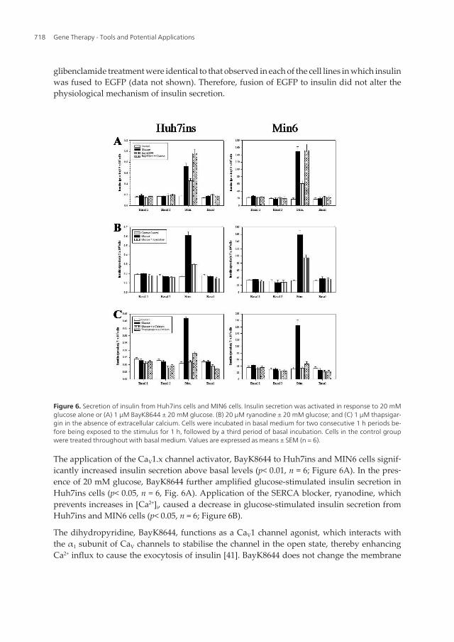
Figure 6. Secretion of insulin from Huh7ins cells and MIN6 cells. Insulin secretion was activated in response to 20 mMglucose alone or (A) 1 µM BayK8644 ± 20 mM glucose. (B) 20 µM ryanodine ± 20 mM glucose; and (C) 1 µM thapsigar‐gin in the absence of extracellular calcium. Cells were incubated in basal medium for two consecutive 1 h periods be‐fore being exposed to the stimulus for 1 h, followed by a third period of basal incubation. Cells in the control groupwere treated throughout with basal medium. Values are expressed as means ± SEM (n = 6).
The application of the CaV1.x channel activator, BayK8644 to Huh7ins and MIN6 cells signif‐icantly increased insulin secretion above basal levels (p< 0.01, n = 6; Figure 6A). In the pres‐ence of 20 mM glucose, BayK8644 further amplified glucose-stimulated insulin secretion inHuh7ins cells (p< 0.05, n = 6, Fig. 6A). Application of the SERCA blocker, ryanodine, whichprevents increases in [Ca2+]i, caused a decrease in glucose-stimulated insulin secretion fromHuh7ins and MIN6 cells (p< 0.05, n = 6; Figure 6B).
The dihydropyridine, BayK8644, functions as a CaV1 channel agonist, which interacts withthe α1 subunit of CaV channels to stabilise the channel in the open state, thereby enhancingCa2+ influx to cause the exocytosis of insulin [41]. BayK8644 does not change the membrane
Gene Therapy - Tools and Potential Applications718

potential of resting β-cells [43]. Rather, it acts on the CaV channel in the open state, failing toaffect basal insulin secretion at non-stimulatory glucose concentrations [43], but exaggerat‐ing glucose-stimulated insulin secretion [44, 45]. The addition of BayK8644 increased insulinsecretion by both the Huh7ins and MIN6 cells. However, the amount of insulin secreted inthe presence of BayK8644 was lower than that released in response to 20 mM glucose alone.Putatively, this concentration of glucose may have stimulated the influx of extracellular Ca2+,the release of [Ca2+]i from intracellular stores and increased Ca2+ via other Ca2+-related path‐ways to such an extent that the total increase of Ca2+ in the cell was higher in the presence of20 mM glucose as compared to BayK8644 alone.
Consistent with reports that BayK8644 is known to stimulate the opening of CaV channels inpancreatic β-cells without altering the membrane potential [44], static stimulation ofHuh7ins cells with 1 µM BayK8644 plus 20 mM glucose amplified glucose-stimulated insu‐lin release. However, BayK8644 failed to amplify glucose-stimulated insulin secretion inMIN6 cells. This finding may be attributable to the ability of 20 mM glucose alone to causethe maximum threshold in the activation of insulin release in MIN6 cells, such that the addi‐tion of BayK8644 was unable to exert any additional stimulatory effects. Nevertheless, theseresults demonstrate that the insulin secretory response of the Huh7ins cells is dependentupon the activation of CaV channels, as is the case for pancreatic β-cells.
Static stimulation of Huh7ins cells with the highly specific SERCA blocker thapsigargin,which induces the release of Ca2+ from intracellular stores resulted in a significant increase(two-fold increase over basal levels), in insulin secretion in the absence of extracellular Ca2+
(p< 0.05, n = 6; Figure 6C). Consistent with results from Tuch et al. [7], the response of theHuh7ins cells to glucose was abolished when Ca2+ was removed from the basal medium be‐fore 20 mM glucose was added (p> 0.05 vs. control, n = 6; Figure 6C). MIN6 cells showed asimilar response; namely, in the absence of extracellular Ca2+ the glucose-responsivenesswas abolished (p> 0.05 vs. control, n = 6; Figure 6C), and the presence of 1 µM thapsigarginsignificantly increased insulin secretion 1.8-fold over basal levels (P<0.05, n = 6; Figure 6C).
The connexon (hemi-channel blocker), oleic acid, significantly reduced acute insulin secre‐tion by 1.4-fold (p< 0.05, n = 6; Figure 7A), while verapamil (10 µM) resulted in a significantdecrease in insulin secretion to glucose in both cell lines (p< 0.05, n = 6; Figure 7B). However,the combination of verapamil and ryanodine did not exert an additive effect on insulin se‐cretion, compared to treatment with verapamil alone (p< 0.05, n = 6; Fig. 7B). Nevertheless, agreater decrease in insulin secretion was observed after the addition of verapamil, ryano‐dine and oleic acid in both Huh7ins (p< 0.05, n = 6) and MIN6 cells (p< 0.05, n = 6; Figure 7C).
SERCA operate to restore diminished intracellular endoplasmic and sarcoplasmic reticulumCa2+ stores, thereby decreasing cytoplasmic Ca2+ levels [46-50]. Thapsigargin is a highly se‐lective inhibitor of SERCA. Stimulation of β-cells with glucose causes an initial, thapsigar‐gin-inhibitable, drop in [Ca2+]i that precedes the increase in [Ca2+]i due to the pumping ofCa2+ into the endoplasmic reticulum [51, 52]. Blocking of SERCA by thapsigargin augmentsthe glucose-induced [Ca2+]i increase by activating a depolarising store-operated current,which then facilitates the opening of CaV channels [51, 53, 54]. Consistent with the resultsreported by Tuch et al, [7], in the absence of extracellular Ca2+, the glucose responsiveness of
Insulin Trafficking in a Glucose Responsive Engineered Human Liver Cell Line is Regulated by the Interaction of ATP-Sensitive Potassium Channels and Voltage-Gated Calcium Channels
http://dx.doi.org/10.5772/52839
719

both Huh7ins and MIN6 cells in the absence of extracellular Ca2+, was lost, while normalglucose responsiveness was seen when Ca2+ was present in the medium. However, thapsi‐gargin, which raises cytosolic Ca2+, stimulated insulin secretion by both Huh7ins and MIN6cells in the absence of extracellular Ca2+. This finding further supports the role of intracellu‐lar Ca2+ storage in insulin secretion in both pancreatic β-cells and in the insulin-secreting liv‐er cell line, Huh7ins.
Figure 7. Secretion of insulin from Huh7ins cells and MIN6 cells. Insulin secretion was activated in response to 20 mMglucose alone or in the presence of (A) 20 µM oleic acid; (B) 10 µM verapamil ± 10 µM ryanodine and (C) 10 µM vera‐pamil, 20 µM ryanodine and 20 µM oleic acid. Cells were incubated in basal medium for two consecutive 1 h periodsbefore being exposed to the stimulus for 1 h, followed by a third period of basal incubation. Cells in the control groupwere treated throughout with basal medium. Values are expressed as means ± SEM (n = 6).
The presence of 20 µM ryanodine, which blocks CaV channels at concentrations ≥ 10 µM [55]and prevents the release of Ca2+ from the endoplasmic reticulum, reduced the glucose-re‐sponsiveness of both Huh7ins cells and MIN6 cells, although to a lesser extent than was ob‐served in the presence of 10 µM verapamil. This finding is consistent with previous reportsthat intracellular Ca2+ stores (and therefore SERCA) contribute to the intracellular Ca2+ re‐sponse during insulin secretion. The application of verapamil to Huh7ins cells caused a
Gene Therapy - Tools and Potential Applications720

complete abrogation of glucose-responsiveness upon extracellular Ca2+ levels has been pre‐viously reported for pancreatic β-cells [40, 43, 56]. As expected the addition of oleic acid toHuh7ins and MIN6 cells resulted in reduced glucose responsiveness, due to the blockage ofhemi-channels, similar to what has been reported in pancreatic β-cells [57].
3. Conclusion
The results described in this chapter indicate that insulin secretion in engineered hepato‐cytes (Huh7ins cells) was controlled, as precisely as in the pancreatic β-cell, by a fully func‐tional KATP and CaV channel system. The results clearly document that Huh7ins cellsrespond to glucose via insulin secretion from secretory granules by the same mechanism ob‐served in pancreatic β-cells. This is the first study to demonstrate a clear physiological andbiochemical interaction of KATP channels and CaV channels in liver cells, and as such revealsthat hepatocytes are ideal candidates for the engineering of artificial β-cells. Testament tothis, we have successfully engineered a liver cell line to synthesize, store and secrete insulin.Regardless of whether this hepatoma cell line will be a viable β-cell alternative for trans‐plantation into patients, the present study provides valuable information with regards to thefuture engineering of glucose-responsive insulin-secreting liver cells. Elucidation of the min‐imal molecular modifications required for the creation of an artificial β-cell from a hepato‐cyte may one day provide therapeutic avenues to engineer a patient’s own liver cells tosynthesize, store and secrete insulin in response to metabolic stimuli.
Acknowledgements
This work was supported by grants from Diabetes Australia Research Trust, Rebecca L.Cooper Medical Research Foundation and the University of Technology Sydney. We wouldlike to thank Wayne Hawthorne and Philip O’Connell from the Westmead Millennium Insti‐tute for human pancreatic islets and Richard Limburg for IT support.
Author details
Ann M. Simpson1*, M. Anne Swan2, Guo Jun Liu3, Chang Tao1, Bronwyn A O’Brien1,Edwin Ch’ng1, Leticia M. Castro1, Julia Ting1, Zehra Elgundi1, Tony An2,Mark Lutherborrow4, Fraser Torpy5, Donald K. Martin1, Bernard E. Tuch6 andGraham M. Nicholson1
*Address all correspondence to: [email protected]
1 School of Medical & Molecular Biosciences, University of Technology Sydney, Sydney,Australia
Insulin Trafficking in a Glucose Responsive Engineered Human Liver Cell Line is Regulated by the Interaction of ATP-Sensitive Potassium Channels and Voltage-Gated Calcium Channels
http://dx.doi.org/10.5772/52839
721

2 School of Medical Sciences (Anatomy & Histology) and Bosch Institute, University of Syd‐ney, Australia
3 Brain and Mind Research Institute, Faculty of Health Sciences, University of Sydney andLife Sciences, Australian Nuclear Science and Technology Organization, Sydney, Australia
4 Australian Foundation for Diabetes Research & Diabetes Transplant Unit, Sydney, Aus‐tralia
5 School of the Environment, University of Technology Sydney, Sydney, Australia
6 Australian Foundation for Diabetes Research & Diabetes Transplant Unit, Prince of WalesHospital, and CSIRO, Division of Materials Science and Engineering, Sydney, Australia
References
[1] Eisenbarth, G. S. (1986). Type I diabetes mellitus: a chronic autoimmune disease. NEngl J Med, 4-1360.
[2] Simpson, A. M., Tuch, B. E., Swan, Tu. J., & Marshall, G. M. (1995). Functional ex‐pression of the human insulin gene in a human hepatoma cell line (HEP G2). GeneTherapy, 2-231.
[3] Simpson, A. M., Marshall, G. M., Tuch, B. E., Maxwell, L., Swan, MA, Tu, J., Beynon,S., Szymanska, B., & Camacho, M. (1997). Gene therapy of diabetes: glucose-stimulat‐ed insulin secretion in a human hepatoma cell line. Gene Therapy, 4-1202.
[4] Ber, I., Shternhall, K., Perl, S., Ohanuna, Z., Goldberg, I., Barshack, I., Benvenisti-Za‐rum, L., Meivar-Levy, I., & Ferber, S. (2003). Functional, persistent, and extended liv‐er to pancreas transdifferentiation. J Biol Chem, 278-31950.
[5] Ferber, S., Halkin, A., Cohen, H., Ber, I., Einav, Y., Goldberg, I., Barshack, I., Seijffers,R., Kopolovic, J., Kaiser, N., & Karasik, A. (2000). Pancreatic and duodenal homeoboxgene 1 induces expression of insulin genes in liver and ameliorates streptozotocin-in‐duced hyperglycaemia. Nature Med, 6-568.
[6] Kojima, H., Fujimiya, M., Matsumara, K., Yunan, P., Imaeda, H., Maeda, M., & Chan,L. (2003). NeuroD-betacellulin gene therapy induces islet neogenesis in the liver andreverses diabetes in mice. Nature Med, 9-596.
[7] Tuch, B. E., Szymanska, B., Yao, M., Tabiin, M., Gross, D., Holman, S., Swan, MA,Humphrey, R., Marshall, G. M., & Simpson, A. M. (2003). Function of a geneticallymodified human liver cell line that stores, processes and secretes insulin. Gene Thera‐py, 10-490.
Gene Therapy - Tools and Potential Applications722

[8] Ren, B. H., O’Brien, B. A., Swan, M. A., Kiona, M. E., Nassif, N., Wei, M. Q., & Simp‐son, A. M. (2007). Long-term correction of diabetes in rats following lentiviral hepaticinsulin gene therapy. Diabetologia, 50, 1910-1920.
[9] Simpson, A. M., Tao, C., Swan, M. A., Ren, B., & O’Brien, B. A. (2008). Glucose regu‐lated production of human insulin in H4IIE rat liver cells. Diabetes, 56(1), A120.
[10] Tabiin, M. T., Tuch, B. E., Bai, L., Han-G, X., & Simpson, A. M. (2001). Susceptibilityof insulin-secreting hepatocytes to the toxicity of pro-inflammatory cytokines. J Auto‐immunity, 17-229.
[11] Permutt, MA, Koranyi, L., Keller, K., Lacy, P. E., & Scharp, D. W. (1989). Cloning andfunctional expression of a human pancreatic islet glucose-transporter cDNA. ProcNatl Acad Sci USA, 86(22), 8688-8692.
[12] Weinhouse, S. (1976). In: Current topics in Cellular regulation. BL Horecker & ER Stadt‐man., editors. Academic Press.
[13] Aguilar-Bryan, L., Nichols, C. G., Wechsler, S. W., Clement, J. P., Boyd, A. E., Gonzá‐lez, G., Herrera-Sosa, H., Nguy, K., Bryan, J., & Nelson, D. A. (1995). Cloning of thebeta cell high-affinity sulfonylurea receptor: a regulator of insulin secretion. Science,268-423.
[14] Inagaki, N., Gonoi, T., Clement, J. P., Namba, N., Inazawa, J., Gonzalez, G., Aguilar-Bryan, L., Seino, S., & Bryan, J. (1995). Reconstitution of IKATP: an inward rectifiersubunit plus the sulfonylurea receptor. Science, 270-1166.
[15] Inagaki, N., Gonoi, T., & Seino, S. (1997). Subunit stoichiometry of the pancreatic be‐ta-cell ATP-sensitive K+ channel. FEBS Lett, 409, 232-236.
[16] Clement, J. P., Kunjilwar, K., Gonzalez, G., Schwanstecher, M., Panten, U., Aguilar-Bryan, L., & Bryan, J. (1997). Association and stoichiometry of KATP channel subunits.Neuron, 18, 827-838.
[17] Shyng, S., & Nichols, C. G. (1997). Octameric stoichiometry of the KATP channel com‐plex. J Gen Physiol, 110, 655-664.
[18] Aguilar-Bryan, L., Clement, J. P., Gonzalez, G., Kunjilwar, K., Babenko, A., & Bryan,J. (1998). Toward understanding the assembly and structure of KATP channels. PhysiolRev, 78-227.
[19] Ashcroft, F. M., & Rorsman, P. (1989). Electrophysiology of the pancreatic beta-cell.Prog Biophys Mol Biol, 54-87.
[20] Lang, J. (1999). Molecular mechanisms and regulation of insulin exocytosis as a para‐digm of endocrine secretion. Eur J Biochem, 259-3.
[21] Macfarlane, W. M., O’Brien, R. E., Barnes, P. D., Shepherd, R. M., Cosgrove, K. E.,Lindley, K. J., Aynsley-Green, A., James, R. F., Docherty, K., & Dunne, MJ. (2000).Sulfonylurea receptor 1 and Kir6.2 expression in the novel human insulin-secretingcell line NES2Y. Diabetes, 49-953.
Insulin Trafficking in a Glucose Responsive Engineered Human Liver Cell Line is Regulated by the Interaction of ATP-Sensitive Potassium Channels and Voltage-Gated Calcium Channels
http://dx.doi.org/10.5772/52839
723

[22] Braun, M., Ramracheya, R., Zhang, Q., Karanauskaite, J., Partridge, C., Johnson, P. R.,& Rorsman, P. (2008). Voltage-gated ion channels in human pancreatic β-cells: Elec‐trophysiological characterization and role in insulin secretion. Diabetes, 57-1618.
[23] Wollheim, C. B., & Sharp, G. W. (1981). Regulation of insulin release by calcium.Physiol Rev, 61-914.
[24] Gilon, P., & Henquin, J. C. (2001). Mechanisms and physiological significance of thecholinergic control of pancreatic beta-cell function. Endocr Rev, 22-565.
[25] Bereton, H. M., Harland, M. L., Froscio, M., Petronijevic, T., & Barrit, G. J. (1997).Novel variants of voltage-operated calcium channel alpha 1-subunit transcripts in arat liver-derived cell line: deletion in the IVS4 voltage sensing region. Cell Calcium,22, 39-52.
[26] Snutch, T. P., Tomlinson, W. J., Leonard, J. P., & Gilbert, M. M. (1991). Distinct calci‐um channels are generated by alternative splicing and are differentially expressed inthe mammalian CNS. Neuron, 7, 45-57.
[27] Liu, G. J., Simpson, A. M., Swan, Tao. C., Tuch, B. E., Crawford, R. M., Jovanovic, A.,& Martin, D. K. (2003). ATP-sensitive potassium channels induced in liver cells aftertransfection with insulin cDNA and the GLUT 2 transporter regulate glucose-stimu‐lated insulin secretion. FASEB J, 17-1682.
[28] Malhi, H., Irani, A. N., Rajvanshi, P., Suadicani, S. O., Spray, D. C., Mc Donald, T. V.,& Gupta, S. (2000). KATP channels regulate mitogenically induced proliferation in pri‐mary rat hepatocytes and human liver cell lines. J Biol Chem, 275-26050.
[29] Lutherborrow, M. A., Appavoo, M., Simpson, A. M., & Tuch, B. E. (2009). Gene ex‐pression profiling of Huh7ins lack of a granulogenic function for chromagranin A.Islets, 1, 60-70.
[30] Miyazaki-I, J., Araki, K., Yamato, E., Ikegami, H., Asano, T., Shibasaki, Y., Oka, Y., &Yamamura, K. (1990). Establishment of a pancreatic beta cell line that retains glucose-inducible insulin secretion: Special reference to expression of glucose transporter. En‐docrinology, 127-126.
[31] Sapir, T., Shternhall, K., Meivar-Levy, I., Blumenfeld, I., Cohen, H., Skutelsky, E.,Eventov-Friedman, S., Barshack, I., Goldberg, I., Pri-Chen, S., Ben-Dor, L., Polak-Charcon, S., Karasik, A., Shimon, I., Mor, E., & Ferber, S. (2005). Cell-replacementtherapy for diabetes: generating functional insulin-producing tissue from adult hu‐man liver cells. Proc Natl Acad Sci USA, 102-7964.
[32] Fodor, A., Harel, C., Fodor, L., Armoni, M., Salmon, P., Trono, D., & Karnielli, E.(2007). Adult rat liver cells transdifferentiated with lentiviral IPF1 vectors reversediabetes in mice: an ex vivo gene therapy approach. Diabetologia, 50-121.
[33] Vollenweider, F., Irminger, J. C., Gross, D. J., Villa-Komaroff, L., & Halban, P. A.(1992). Processing of proinsulin by transfected hepatoma (FAO) cells. J Biol Chem,267-14629.
Gene Therapy - Tools and Potential Applications724

[34] Yang, L., Li, S., Hatch, H., Ahrens, K., Cornelius, J. G., Petersen, B. E., & Peck, A. B.(2002). In vitro trans-differentiation of adult hepatic stem cells into pancreatic endo‐crine hormone-producing cells. Proc Natl Acad Sci USA, 99-8078.
[35] Nguyen, T., Chin, W. C., & Verdugo, P. (1998). Role of Ca2+/K+ ion exchange in intra‐cellular storage and release of Ca2+. Nature, 395-908.
[36] Quesada, I., Chin, W. C., Steed, J., Campos-Bedolla, P., & Verdugo, P. (2001). Mousemast cell secretory granules can function as intracellular ionic oscillators. Biophys J,80, 2133-2139.
[37] National Institutes of Health. (2008). NIH Image. http://rsb.info.hih.gov/nih-image/Accessed 1 July,).
[38] Arvan, P., & Halban, P. A. (2004). Sorting ourselves out: seeking consensus on traf‐ficking in the beta-cell. Traffic , 5, 53-61.
[39] National Institutes of Health. (2009). Image J. http://rsb.info.nih.gov/ij/Accessed 20September).
[40] Yoon, N., Nataliya, S., Jeong-J, M., Lee, T., Lee-S, M., Kim-L, H., Chin, H., Suh-G, P.,Kim, S., & Shin-S, H. (2003). Requirement for the L-type Ca2+ channel α1D subunit inpostnatal pancreatic β-cell generation. J Clin Inves, 108, 1015-1022.
[41] Catterall, W. A., & Striessnig, J. (1992). Receptor sites for Ca2+ channel antagonists.Trends Pharm Sci; , 13-256.
[42] Henquin, J. C. (2000). Triggering and amplifying pathways of regulation of insulinsecretion by glucose. Diabetes, 49, 1751-1760.
[43] Ammälä, C., Moorhouse, A., & Ashcroft, F. M. (1996). The sulphonylurea receptorconfers diazoxide sensitivity on the inwardly rectifying K+ channel Kir6.1 expressedin human embryonic kidney cells. J Physiol, 494, 709-714.
[44] Larsson-Nyren, G., & Sehlin, J. (1996). Comparison of the effects of perchlorate andBay K 8644 on the dynamics of cytoplasmic Ca2+ concentration and insulin secretionin mouse β-cells. Biochem J, 314-167.
[45] Malaisse-Lagae, F., Matthias, P. C. F., & Malaisse, W. J. (1984). Gating and blocking ofcalcium channels by dihydropyridines in the pancreatic β-cell. Biochem Biophys ResComm, 123-1062.
[46] Lytton, J., Westlin, M., & Hanley, M. R. (1991). Thapsigargin inhibits the sarcoplas‐mic or endoplasmic reticulum Ca-ATPase family of calcium pumps. J Biol Chem,266-17067.
[47] Kirby, M. S., Sagara, Y., Gaa, S., Inesi, G., Lederer, W. J., & Rogers, T. B. (1992). Thap‐sigargin inhibits contraction and Ca2+ transient in cardiac cells by specific inhibitionof the sarcoplasmic reticulum Ca2+ pump. J Biol Chem, 267, 12545-12551.
Insulin Trafficking in a Glucose Responsive Engineered Human Liver Cell Line is Regulated by the Interaction of ATP-Sensitive Potassium Channels and Voltage-Gated Calcium Channels
http://dx.doi.org/10.5772/52839
725

[48] Gericke, M., Droogmans, G., & Nilius, B. (1993). Thapsigargin discharges intracellu‐lar calcium stores and induces transmembrane currents in human endothelial cells.Pflügers Arch, 422-552.
[49] Parekh, A. B., Terlau, H., & Stühmer, W. (1993). Depletion of InsP3 stores activates aCa2+ and K+ current by means of a phosphatase and a diffusible messenger. Nature,364-814.
[50] Randriamampita, C., & Tsien, R. Y. (1993). Emptying of intracellular Ca2+ stores re‐leases a novel small messenger that stimulates Ca2+ influx. Nature, 364-809.
[51] Roe, M. W., Mertz, R. J., Lancaster, M. E., Worley, J. F. 3rd, & Dukes, I. D. (1994).Thapsigargin inhibits the glucose-induced decrease of intracellular Ca2+ in mouse is‐lets of Langerhans. Am J Physiol, 266, E 852-862.
[52] Miura, Y., Henquin, J. C., & Gilon, P. (1997). Emptying of intracellular Ca2+ storesstimulates Ca2+ entry in mouse pancreatic beta-cells by both direct and indirect mech‐anisms. J Physiol, 503-387.
[53] Worley, J. F., Mc Intyre, M. S., Spencer, B., & Dukes, I. D. (1994a). Depletion of intra‐cellular Ca2+ stores activates a maitotoxin-sensitive nonselective cationic current inbeta-cells. J Biol Chem, 269, 32055-32058.
[54] Worley, J. F., Mc Intyre, M. S., Spencer, B., Mertz, R. J., Roe, M. W., & Dukes, I. D.(1994b). Endoplasmic reticulum calcium store regulates membrane potential inmouse islet beta-cells. J Biol Chem, 269, 14359-14362.
[55] Meissner, G. (1986). Ryanodine activation of the Ca2+ release channel of sarcoplasmicreticulum. J Biol Chem, 261-6300.
[56] Nevins, A. K., & Thurmond, D. C. (2003). Glucose regulates the cortical actin net‐work through modulation of Cdc42 cycling to stimulate insulin secretion. Am J Physi‐ol Cell Physiol, 285, C698-710.
[57] Meda, P., Bosco, D., Chanson, M., Giordano, E., Vallar, L., Wollheim, C., & Orci, L.(1990). Rapid and reversible secretion changes during uncoupling of rat insulin-pro‐ducing cells. J Clin Invest, 86-759.
Gene Therapy - Tools and Potential Applications726