initial insights into the structure-activity...
TRANSCRIPT

LUND UNIVERSITY
PO Box 117221 00 Lund+46 46-222 00 00
INITIAL INSIGHTS INTO THE STRUCTURE-ACTIVITY RELATIONSHIPS OF AVIANDEFENSINS.
Derache, Chrystelle; Meudal, Herve; Aucagne, Vincent; Mark, Kevin J; Cadene, Martine;Delmas, Agnes F; Lalmanach, Anne-Christine; Landon, CelinePublished in:Journal of Biological Chemistry
DOI:10.1074/jbc.M111.312108
Published: 2011-01-01
Link to publication
Citation for published version (APA):Derache, C., Meudal, H., Aucagne, V., Mark, K. J., Cadene, M., Delmas, A. F., ... Landon, C. (2011). INITIALINSIGHTS INTO THE STRUCTURE-ACTIVITY RELATIONSHIPS OF AVIAN DEFENSINS. Journal of BiologicalChemistry. DOI: 10.1074/jbc.M111.312108
General rightsCopyright and moral rights for the publications made accessible in the public portal are retained by the authorsand/or other copyright owners and it is a condition of accessing publications that users recognise and abide by thelegal requirements associated with these rights.
• Users may download and print one copy of any publication from the public portal for the purpose of privatestudy or research. • You may not further distribute the material or use it for any profit-making activity or commercial gain • You may freely distribute the URL identifying the publication in the public portal

Take down policyIf you believe that this document breaches copyright please contact us providing details, and we will removeaccess to the work immediately and investigate your claim.
Download date: 30. May. 2018

1
INITIAL INSIGHTS INTO THE STRUCTURE-ACTIVITY RELATIONSHIPS
OF AVIAN DEFENSINS
Chrystelle Derache1,2, #
, Hervé Meudal1, Vincent Aucagne
1, Kevin J. Mark
1, $, Martine Cadène
1,
Agnès F. Delmas1, Anne-Christine Lalmanach
2*, Céline Landon
1*
1 Centre de Biophysique Moléculaire. CNRS UPR4301. Rue Charles Sadron, 45071 Orléans, France.
2 Unité Infectiologie Animale et Sante Publique, INRA UR1282, Centre de Recherche de Tours, 37380
Nouzilly, France. # Present address: Medical Microbiology, Department of Laboratory Medicine Malmö, Lund
University, Skåne University Hospital, Malmö, Sweden. $ Present address: Chemistry Department, York College, Jamaica, NY 111451, USA.
Running title: Structural determinants for the AvBD2 antibacterial activity
*To whom correspondence should be addressed: Céline Landon, Centre de biophysique moléculaire,
CNRS UPR4301. Rue Charles Sadron, 45071 Orléans, France, phone number +332 38 25 55 74, fax
number : +332 38 63 15 17, Email address : [email protected]; Anne-Christine
Lalmanach, INRA, UR1282, IASP 213, Centre de recherche de Tours, 37380 Nouzilly, France, phone
number : +332 47 42 77 00, fax number : +332 47 42 77 74, Email address : Anne-
Keywords: Avian β-defensin, NMR structure, antimicrobial activity, peptide synthesis, all-D
enantiomer
Background: Avian defensins are antimicrobial
peptides of bird’s immunity.
Results: The target of chicken AvBD2 defensin
is not chiral. Its structure is not amphipathic. The
reduced and AvBD2-K31A forms dramatically
decrease antibacterial activity.
Conclusion: AvBD2 may disrupt the bacterial
membrane through a non-chiral non-specific
interaction.
Significance: Knowledge of the structure-
function relationships of avian defensins is a
prerequisite for their use as alternatives to
antibiotics.
SUMMARY
Numerous β-defensins have been
identified in birds and the potential use of
these peptides as alternatives to antibiotics
has been proposed, in particular to fight
antibiotic-resistant and zoonotic bacterial
species. Little is known about the mechanism
of antibacterial activity of avian β-defensins
(AvBDs), and the present work was carried
out to obtain initial insights into the
involvement of structural features or specific
residues in the antimicrobial activity of
chicken AvBD2. Chicken AvBD2 and its
enantiomeric counterpart were chemically
synthesized. Peptide elongation and oxidative
folding were both optimized. The similar
antimicrobial activity measured for both L-
and D- proteins clearly indicates that there is
no chiral partner. Therefore the bacterial
membrane is in all likelihood the primary
target. Moreover, this work evidences that the
three-dimensional fold is required for an
optimal antimicrobial activity, in particular
for Gram-positive bacterial strains. The
three-dimensional NMR structure of chicken
AvBD2 defensin displays the structural 3-
stranded antiparallel β-sheet characteristic of
β-defensins. The surface of the molecule does
not display any amphipathic character. In
light of this new structure and of the king
penguin AvBD103b defensin structure, the
consensus sequence of avian β-defensin’s
family was analyzed. Well conserved residues
were highlighted and the potential strategic
role of the lysine 31 residue of AvBD2
emphasized. The synthetic AvBD2-K31A
variant displayed substantial N-terminal
structural modifications and a dramatic
decrease in activity. Taken together, these
results demonstrate the structural as well as
the functional role of the critical lysine 31
residue in antimicrobial activity.

2
Defensins belong to a family of
antimicrobial peptides characterized by
cationicity, small size, β-sheet structure and the
presence of three disulfide bonds (1). Three
subclasses (α, β, and θ) have been defined
depending on the disulfide arrangement and the
positioning of the six conserved cysteines. The
α- and θ-defensin families have been considered
to evolve by duplication and divergence from β-
defensin ancestor genes since the former are not
reported in evolutionary old vertebrates such as
fish and bird classes. Defensins play a major role
in both innate and adaptive immunity (2). They
have been found to be constitutively or inducibly
expressed by neutrophils and epithelial cells
from many mammals and birds, including
chicken (1,3,4). They display a wide range of
microbicidal or microbistatic activities against
Gram-negative and Gram-positive bacteria, fungi
and viruses (4). A substantial body of evidence
indicates that the mechanism of action of
defensins mainly relies on several structural
features such as cationicity and amphipathy,
which drive the antimicrobial peptide to interact
with bacterial membranes and tend to divide
peptides into two mechanistic classes: membrane
disruptive and non-membrane disruptive (5,6). In
the latter case, there is growing evidence that
defensins induce killing by acting on chiral
anionic intracellular targets (see (7,8) for a
review).
Interest in defensins as therapeutic drugs
is growing because defensins may constitute an
alternative to the controversial use of antibiotics.
In birds, a potential use of these peptides has
been proposed in particular to fight antibiotic-
resistant bacteria including Salmonella, a major
zoonotic agent that causes food poisoning (9).
Numerous β-defensins were identified in birds
from isolated peptides or gene sequences (see (4)
for a review). In a previous study, it was shown
that chicken β-defensin genes (avBD1 and 2)
were highly expressed in the intestinal tissue of
birds that are resistant to Salmonella colonisation
(10). Three defensins (AvBD1, AvBD2 and
AvBD7) were therefore purified from chicken
bone marrow and their antimicrobial activity was
tested on a series of Gram-positive and Gram-
negative bacteria (11). Only chicken AvBD2 was
shown to be more active against Gram-positive
than Gram-negative strains, as reported for the
king penguin spheniscin (AvBD103b) (12), the
only other avian β-defensin whose three-
dimensional structure has been determined to
date.
Concerning the molecular patterns
involved in the activity of avian defensins, the
sole data currently available refer to ostrich
AvBD1 and AvBD2 defensins, which
respectively share 39 and 78% of identity with
chicken AvBD2. Ostrich defensins were shown
to create a slow and partial depolarization of the
Escherichia coli membrane, but were unable to
provoke bacterium death by membrane
disruption (13). This indicated that the ostrich
defensins could cross the bacterial membrane to
target a cytoplasmic molecule. Considering that
the ostrich defensins were efficient in shifting the
mobility of bacterial DNA in a gel
electrophoresis assay, it has been proposed that
DNA could be the defensin’s target (13). In the
context of the long-term objective of improving
knowledge of immunity in birds, this work was
carried out to gain information on structure-
activity relationships of the chicken AvBD2
defensin, at the atomic level, which is an
essential first step to understanding how avian β-
defensins function.
EXPERIMENTAL PROCEDURES Reversed Phase High Performance Liquid
Chromatography - HPLC analyses were carried
out on either an Elite LaChrom system
composed of a Hitachi L-2130 pump, a Hitachi
L-2455 diode array detector and a Hitachi L-
2200 autosampler, or a LaChrom 7000 system
composed of a Merck-Hitachi L-7100 pump, a
Merck-Hitachi L-7455 diode array detector and a
Merck-Hitachi D-7000 interface, which was also
used for semi-preparative purification. The
machines were equipped with C18 reversed
phase columns, Nucleosil, 300 Å, 5 μm, 250
4.6 mm for the analytical separations, or 250
10.5 mm for purification. Solvents A and B
containing 0.1% of TFA (trifluoroacetic acid)
were H2O and MeCN, respectively.
Synthesis of the linear, S-alkylated defensins –
Solid-phase peptide synthesis (SPPS) was run on
an automated synthesizer 433A from Applied
Biosystem using Fmoc/t-Bu chemistry at a 0.1
mmol scale with HBTU (O-(benzotriazol-1-yl)-
N,N,N′,N′-tetramethyluronium hexafluorophos-
phate) / HOBt (1-hydroxybenzotriazole hydrate)
as the coupling reagent. Fmoc-Ala-
methylphenoxypropionic acid (Polypeptide
group, France) (122 mg, 0.25 mmol) was
manually coupled onto the aminomethyl PEGA
(polyethylene glycol polyacrylamide) resin (3 g
wet, 0.1 mmol) in the presence of HATU (O-(7-
aza-benzotriazol-1-yl)-N,N,N′,N′-tetramethyl-

3
uronium hexafluorophosphate) (95 mg, 0.25
mmol) and DIEA (N,N-diisopropylethylamine)
(86 µl, 0.5 mmol) for 2 h. The elongation was
then carried out automatically using a 10-fold
excess of protected amino acids and coupling
reagents. The protecting groups used for the
side-chains were Arg(Pbf), Asn(Trt), Cys(Acm),
His(Trt), Lys(Boc), Ser(t-Bu), Trp(Boc), Tyr(t-
Bu). A 0.1 mmol scale program was used, and
each coupling step was followed by capping with
acetic anhydride. The coupling step was
performed twice from Cys30 to Leu1. The
dipeptides Gly7-Ser8 and Gly31-Ser32 were also
coupled twice, as the Fmoc-Gly-Ser(ΨMe,Me
pro)-
OH pseudoproline derivative (Merck). After
completion of the peptide elongation, the peptide
resin was treated for 3 h at room temperature
with TFA/H2O/i-Pr3SiH/PhOH, 87.5:5:2.5:5, and
the linear S-Acm-alkylated peptide was
precipitated by dilution into ice-cold diethyl
ether.
Synthesis of the oxidized defensins - In a syringe
fitted with a frit, the S-Acm-protected peptide
resin (15 µmol) was swollen in NMP (2 x 5 mL
for 1 min). Silver tetrafluoroborate (58.4 mg per
Acm group, 20 equiv.) in NMP/H2O 9:1 mixture
(4 ml) was transferred to the resin by suction,
and the resulting suspension was stirred by
rotation for 5 min at RT, in the absence of light,
followed by washes with NMP/H2O 9:1 then
DMF. This treatment was repeated once (60 min
stirring), and the resin was further washed with
pyridine (5 x 6 ml), then treated alternatively
with sodium diethyldithiocarbamate (25 mM in
NMP) and pyridine hydrochloride (1M in
CH2Cl2/MeOH 95:5) (3 x 2 x 5 ml), followed by
extensive washes with DMF. The peptide resin
was then treated for 3 h at room temperature with
TFA/H2O/i-Pr3SiH/PhOH, 87.5:5:2.5:5 and the
linear peptide was precipitated by dilution with
ice-cold diethyl ether. The crude reduced form of
AvBD2 was dissolved in 20% acetic acid
(AcOH) and purified by semi-preparative C18
reversed phase HPLC.
The oxidative folding was performed at a
peptide/GSH/GSSG molar ratio of 1/100/10 in
deoxygenated MeCN/200 mM Tris-HCl buffer
pH 8.5 (50/50, v/v) containing 1 mM EDTA.
The peptide concentration (50 µg/mL) was
measured using UV spectrophotometry at 280
nm (εTrp: 5579 M-1
cm-1
). The kinetics of the
oxidative folding were monitored by quenching,
at regular time intervals, aliquots from the
reaction mixture through the addition of TFA
(final concentration 2%), and then analyzing the
sample by analytic C18 reversed phase HPLC.
The oxidative folding was quantitative over 30
min. The peptide was purified on to a Resource S
column (GE Healthcare Biosciences) using a
linear gradient of 0–0.5M NaCl in 50 mM Tris
pH7.5. The fractions corresponding to the pure
peptide were loaded on a Sep-Pak® C18 (6ml
column, Waters) followed by washings with 5%
aqueous AcOH, and eluted by
MeCN/H2O/AcOH 5:4:1 and lyophilized.
Mapping of disulfide bridges by proteolytic
cleavage and mass spectrometry
Proteolytic cleavage - Protein cleavages were
performed in a total volume of 20 µL. To avoid
the scrambling of disulfide bridges known to
occur at basic pH, cleavages were performed in
30 mM ammonium acetate buffer adjusted to pH
6.5. Trypsin (Roche Diagnostics) cleavage of
AvBD2 was performed at an enzyme:substrate
ratio of 1:20 (w/w) for 4 hours at 37°C. Papain
(Roche Diagnostics) was incubated with AvBD2
for 4 hours at 25°C using an enzyme:substrate
ratio of 1:5 (w/w). For papain cleavage, the
following amino acids were considered for
proteinase specificity: Arg, Ala, Asn, Asp, Glu,
Gln, Gly, His, Lys, Phe, Leu, and Tyr.
Mass spectrometry - Intact and proteolyzed
synthetic L-AvBD2 were analyzed by Matrix
Assisted Laser Desorption Ionization-Time of
Flight (MALDI-TOF) using an Autoflex
instrument (BrukerDaltonics, Bremen, Germany)
equipped with a 337-nm nitrogen laser and a
gridless delayed extraction ion source. Sample
deposition on the MALDI plate was performed
using the ultrathin layer method as previously
described (14,15). Samples were diluted at a
ratio of 1:20 with a matrix solution consisting of
4-hydroxy α-cyanocinnamic acid (4HCCA,
Bruker) saturated in a solution of 66.5% water,
33.3% MeCN and 0.1% TFA. A 0.5 µL aliquot
of this analyte-matrix solution was spotted onto
the ultrathin layer plate. The MALDI spot was
irradiated using a 4 Hz laser pulse to produce
ions. At least 200 laser shots were accumulated
for each spectrum. Ions were analyzed in
positive ion reflector mode with a 150 ns delay
and an accelerating voltage of 19 kV. The
measured m/z values correspond to the a0 peak
as determined by the SNAP algorithm on the
isotopic ion distribution. The spectra were
calibrated externally and internally using the
Pepmix calibrant mixture (Bruker) consisting of
bradykinin, angiotensin, substance P, bombesin,
renin substrate, adrenocorticotropic hormone 19-
38 and somatostatin. Instrument parameters were

4
adjusted using FlexControl (Bruker). Data
analysis and internal calibration were performed
using FlexAnalysis (Bruker). The disulfide-
bridged cleavage peptides were mapped to the
known AvBD2 sequence using the PeptideMap
software tool from the PROWL website at
http://prowl.rockefeller.edu (The Rockefeller
University, New York, USA). Given the amino
acid sequence of the protein and the proteinase
cleavage specificity, PeptideMap automatically
computes all theoretically possible combinations
of bridged peptides and matches the observed
masses to the corresponding theoretical masses.
Antimicrobial activity test - The antibacterial
activities of the peptides were measured by radial
diffusion assay (16) as described in Derache et
al. (11) in gel containing either one of the
following Gram-positive bacterial strains:
Bacillus cereus ATCC 14579, Staphylococcus
aureus ATCC 29740, and Listeria
monocytogenes strain EGD, or one of the
following Gram-negative bacterial strains:
Escherichia coli ATCC 25922, Salmonella
enterica serovar Enteritidis ATCC 13076, and
Salmonella enterica serovar Typhimurium
ATCC 14028. For each bacterial strain, three
identical independent measurements of the
antibacterial activity were performed. The
minimal inhibitory concentration (MIC) of each
peptide was determined from a graph constructed
by plotting the log peptide concentration against
the diameter of the clear zone on the plate minus
the diameter of the well. The best-fit straight-line
was determined using linear regression with
GraphPad Prism 5 software (GraphPad
Software). The MIC was calculated by finding
the x-intercept of the line, indicating the peptide
concentration at which no clear zone is obtained.
For each bacterial strain, the statistical difference
between native and variant peptide MICs was
assessed by comparing the slope and intercepts
of both regression lines with GraphPad Prism 5
software (GraphPad Software). The level of
significance was set at P < 0.05.
Circular Dicroism experiments - The CD
experiments were carried out on a Jasco J-810
spectropolarimeter. Solutions of 30µM (10mM
Phosphate buffer pH 7.2) of both L-AvBD2 and
D-AvBD2 enantiomers were compared.
Three-dimensional NMR structure - A standard
set of 2D 1H-NMR experiments (COSY, 80ms
TOCSY, and 160ms NOESY) was performed, on
a 0.1 mM aqueous solution of the synthetic L-
AvBD2 (H2O/D2O 90/10 and 100% D2O) at pH
4.1, and at 293K. An additional set of data,
recorded at 303K, was used to resolve
assignment ambiguities due to spin system
overlaps. All spectra were recorded on a
BRUKER 800 MHz spectrometer (NMR
facilities, Gif-sur-Yvette, France). The NMR
data sets were processed using the
NMRPipe/NMRDraw software package (17,18). 1H chemical shifts were assigned according to
classical procedures (19). NOE cross-peaks were
integrated and assigned within the NMRView
software (17). Covalent bonds were built
between the sulfur atoms of the paired cysteines.
Structure calculations were performed with the
ARIA 1.1 software (20). The calculations were
initiated using the default parameters of ARIA
and a first set of easily assigned NOEs. At the
end of each run, the new assignments proposed
by ARIA were checked manually and introduced
(or not) in the following calculation. This
iterative process was repeated until complete
assignment of the NOESY map. A last run of
1000 structures was then performed with the
final list of NOE derived distance restraints, and
200 structures were submitted to the last step on
ARIA. The 10 structures without residual NOE
violation and with the lowest residual NOE
energy were selected and considered as
characteristic of the peptide structures.
Representation and quantitative analysis of the
calculated structures were performed using
MOLMOL (21) and in-house programs.
The same sets of experiments were recorded on a
VARIAN 600MHz spectrometer for the variant
AvBD2-K31A (2.4 mM of the synthetic peptide
in aqueous solution at pH 4.2, and at 293K). The
same protocol was followed except that
ambiguous constraints were introduced between
cysteine residues, using the “ambiguous disulfide
bridges” protocol of the ARIA 1.1 software (20).
RESULTS
Chemical synthesis of AvBD2: chemical and
functional characterization versus extracted
AvBD2.
The peptide elongation of AvBD2 was carried
out by solid phase peptide synthesis (SPPS)
following the Fmoc/t-Bu strategy. Besides
repeating most of the coupling steps twice,
optimisation of the elongation yield required the
combined use of pseudoproline dipeptide
derivatives and a polar resin (22). Our synthetic
strategy also involved the use of the
acetamidomethyl (Acm) group as a TFA-stable
protection of cysteinyl residues to obtain the
linear S-Acm-alkylated AvBD2 (AvBD2-Acm).

5
To obtain the linear non-alkylated AvBD2 from
the same batch of peptide resin, we developed
conditions for the removal of the Acm groups on
the peptide resin before the final TFA treatment.
After HPLC-purification of the reduced form of
AvBD2, the oxidative folding was carried out
using a procedure based on a thermodynamically
controlled disulfide shuffling, in the presence of
reduced and oxidized glutathione at pH 8.5. The
folding kinetics was followed by quantitative
analytical HPLC and the reaction was shown to
be complete in 30 min (Fig. S1 in supplementary
data). MALDI-TOF MS analysis of oxidized
AvBD2 showed a 6 Da difference in mass
compared to the reduced form, consistent with
the fully oxidized form of this peptide (data not
shown). The oxidized AvBD2 was then purified
to homogeneity by cation exchange
chromatography. Reversed phase HPLC analysis
showed that synthetic AvBD2 co-eluted with the
natural product extracted from chicken bone
marrow (Fig. S2 in supplementary data). As
further evidence of the identity of the synthetic
and natural peptides, their activities measured in
minimal inhibitory concentration (MIC) assays
were in the same range for every bacterial strain
tested (supplementary material Table S3).
Altogether, our data validated an efficient
optimized protocol for the production of highly
pure and biologically active synthetic AvBD2. It
was successfully applied to the synthesis of the
all-D enantiomeric homologue of AvBD2 (D-
AvBD2) and the AvBD2-K31A variant (Fig.
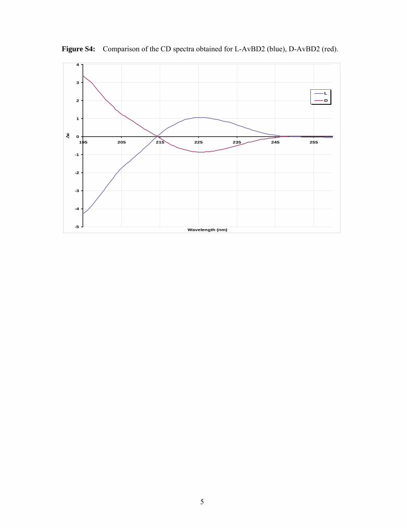
S4). The all-D form was checked by circular
dichroïsm where CD spectra of the two
enatiomers show equal and opposite spectra (Fig.
S5). In the case of the AvBD2-K31A variant, the
increase in hydrophobicity led to a poor folding
yield. Organic solvents were then screened as
folding additives (Table S6 in supplementary
data) and MeCN, which greatly enhanced the
yields, was selected for preparative scale
oxidative foldings. Our optimized protocol
including an efficient peptide elongation and the
use of a co-solvent for the folding step enabled
an enhanced production yield up to 30-40% for
all the AvBD2 peptides.
Antibacterial activity
The antimicrobial activities of D-AvBD2 and L-
AvBD2 were tested on a selection of three
Gram-positive (B. cereus, L. monocytogenes and
S. aureus) and three Gram-negative (E. coli, S.
Enteritidis and S. Typhimurium) bacterial
strains. As shown on Table 1, the MICs
measured for the two enantiomers are identical
for every tested strain.
To investigate the role of the well-conserved
three-dimensional frame of β-defensin in AvBD2
functionality, the antibacterial activities of the
linear S-alkylated AvBD2 (AvBD2-Acm) were
compared with the activities of its oxidatively
folded counterpart (Table 1). The linear AvBD2-
Acm is less active than the folded AvBD2 for
every bacterial strain tested except for E. coli
(P=0.06), as shown by the dramatic increase in
the MIC of AvBD2 when linear. In particular,
the linear form of AvBD2 is 10 and 16 times less
efficient than the folded peptide against the
Gram-positive strains B. cereus (P=0.0002) and
L. monocytogenes (P=0.0002), respectively. The
linear form is even ineffective in our conditions
towards the Gram-positive strain S. aureus,
showing the strict requirement of the three-
dimensional fold for an optimal antimicrobial
activity. The effect of the three-dimensional
structure on the activity is more limited for the
Gram-negative strains. Indeed, for S. Enteritidis
and S. Typhimurium, the linear form of AvBD2
displays an activity one and a half (P<0.0001) to
three times (P=0.0002) lower than that of the
folded AvBD2 peptide.
AvBD2 solution structure
Partial determination of AvBD2 disulfide
bridges array
The determination of the correct disulfide pairing
is generally achieved using enzymatic
proteolysis of proteins and mass spectrometry
analysis of the obtained cleavage products. These
data can thus be introduced as additional
constraints in structure calculations, allowing the
three-dimensional models to converge more
efficiently. For the chicken AvBD2 defensin, the
trypsin proteolysis experiment produced
cleavage peptides which were identified by
MALDI-TOF MS. The observed masses were
matched to four sets of disulfide-connected
peptides using the PeptideMap software (Table
S7 in supplementary data). While peptides
Leu1-Lys4 and Val20-Arg27 each contain one
connectable cysteine, peptides Gly5-Lys19 and
Ser28-Lys31 each have two cysteines which can
participate in disulfide bridges. The connection
of Leu1-Lys4 to Ser28- Lys31 shows that Cys3
is connected to Cys29 or Cys30. Similarly,
Cys23 has to be linked to either Cys8 or Cys13.
These first two connected products were
observed with one linked cysteine and one free
thiol. The products containing three connected
peptides show that the remaining cysteine, either

6
8 or 13, forms a bond with the remaining
cysteine 29 or 30. Trypsin cleavage thus
narrowed down the number of possible disulfide
bridges combinations to four (Fig. S8 in
supplementary data). Papain proteolysis
produced one set of connected peptides, defining
the Cys8-Cys23 bridge for certain.
Consequently, in keeping with the trypsin result
showing that Cys13 does not connect with Cys3,
Cys13 can only connect with Cys29 or Cys30.
However, these adjacent cysteines could not be
differentiated. It is a known limitation of this
method that, because of the impossibility to
cleave between adjacent half-cystinyl residues,
connected peptides containing a single disulfide
bond cannot be obtained in such cases (22).
Following analyses by enzymatic proteolysis
combined with mass spectrometry, software
computation and logical deduction, among the
fifteen possibilities for the disulfide bridges
array, only two remained: 3-29, 8-23, 13- 30 or
3-30, 8-23, 13-29. On NMR NOESY maps, one
connectivity was observed between one of the β-
protons of Cys8 and the β-protons of Cys23,
confirming the Cys8-Cys23 pairing determined
from mass spectrometry data. The chemical
shifts of β-protons of Cys3, Cys13, Cys29 and
Cys30 were very close, therefore NOE peak
superimpositions and/or proximity of diagonal
peaks hampered unambiguous assignments. The
sole unambiguous observed connectivity was
between the β-protons of Cys13 and one of the
β-protons of Cys30, even if very close to the
diagonal, arguing for the 13-30 disulfide bridge.
AvBD2 three-dimensional NMR structure
The quality of the NMR spectra acquired with
the 280 µg of synthetic L-AvBD2 (see sequence
Fig.1) allowed the assignment of all proton
resonances. Chemical shifts have been deposited
in the BioMagResBank
(http://www.bmrb.wisc.edu/) with the entry code
17797. NOE peaks were picked and integrated in
NMRView. A first set of about 200 intra-
residues, sequential and easily-determined long-
range peaks were assigned. Additional
assignments were progressively proposed during
the ARIA runs (20), and manually validated. The
use of ambiguous intersulphur distances, an
option assuming that a given half-cystine is part
of a bridge without supposing a particular
partner, could not be successfully applied for
AvBD2. This method is considered as a reliable
and robust method for disulfide-rich proteins
(23), but the calculations did not converge
satisfactorily enough, even based on a very
convenient set of NOEs (around 15 NOEs –
including 3 long-range restraints each).
Therefore it was more convenient to add the
disulfide bridges as constraints. The two
remaining possibilities were compared: 3-29, 8-
23, 13-30 or 3-30, 8-23, 13-29. Convergence to a
well-formed 3-stranded beta-sheet was only
obtained in the first case, with a convenient
residual number of NOE violations, and
satisfactory energies. The last iterations to refine
the structure were then performed with the 3-29,
8-23, 13-30 disulfide bridges array. The final
numbers of distance restraints used in the last run
of ARIA calculations are detailed in Table 2. The
solution structures of AvBD2 were represented
by ten conformers refined in a shell of water
(Fig. 2 A), and were deposited in the Protein
Data Bank (http://www.pdb.org) with the 2gl5
entry code. The three-dimensional structure of
AvBD2 displays the structural characteristics of
β-defensins: a three-stranded antiparallel β-sheet,
(7-9; 18-22; 28-31) stabilized by a conserved
array of three disulfide bridges. The sequential
Cys29 and Cys30 belong to the middle strand of
the β-sheet, therefore their side chains point in
opposite directions. The measured distances
greater than 8 Å between the sulfur atoms of
Cys13 and Cys29, or between Cys3 and Cys30,
definitively preclude the possibility of C1-C6;
C2-C4; C3-C5 pairing that could match NOE
NMR data. The structures were in very good
agreement with the experimental data; there was
no violation of distance restraints larger than
0.3 Å. Most of the residues (92.7%) were found
in the most favorable regions of the
Ramachandran plot. On the whole, the secondary
structure elements were well defined, and the
RMSD value calculated for secondary structures
was 0.62 Å (table 2). The analysis of the surface
properties clearly evidenced that the positive and
hydrophobic residues were well distributed on
the three-dimensional structure of the molecules
(Fig. 2B and 2C). Contrary to many antibacterial
molecules, AvBD2 did not display any
amphipathic character, neither along the primary
structure (Fig. 1) nor on the three-dimensional
structure of the molecules (Fig. 2).
Three-dimensional NMR structure and
antibacterial activity of AvBD2-K31 variant
The protocol applied for AvBD2 was followed
for AVBD2-K31A except that ambiguous
constraints were introduced between cysteine
residues, using the “ambiguous disulfide
bridges” option. In the first calculations each
half-cystine was allowed to be linked to one of

7
the 5 others, leading to 15 possibilities of
pairing. During the calculations, each disulphide
bridge is then allowed to float freely and the
protocol is driven to the most compatible
disulfide bridges array, under the influence of the
other NMR restraints. Once aberrant
conformations (bridging more than 2 sulfur
atoms) were discarded, a majority (72%) of
structures correspond to the “3-29, 8-23, 13-30”
disulfide bridges array, the residual 28%
corresponding to the “3-30, 8-23, 13-29” pairing.
At this stage, comparing two parrallel
calculations differing only by the disulfide
bridges array imposed: 3-29, 8-23, 13-30 or 3-
30, 8-23, 13-29, only the first calculations
converged to a well-formed 3-stranded beta-
sheet. In the second calculations, the strands
could not form properly. There was 25% more
NOE violations, and the total energy was
multiplied by 2. The refinement of the structure
in the last iterations was then performed with the
3-29, 8-23, 13-30 disulfide bridges array.
A very accurate model of AvBD2-K31A variant
was determined by NMR (the RMSD value
calculated for secondary structures is 0.19 Å;
Table 2). The structures were in very good
agreement with the experimental data, and most
of the residues (96.2%) were found in the most
favorable regions of the Ramachandran plot.
AvBD2-K31A displays the typical three-
stranded anti-parallel β-sheet of β-defensins (6-
10; 18-22; 27-31) stabilized by the conserved
array of 3 disulfide bridges C1-C5, C2-C4, C3-
C6. An additional short anti-parallel β-strand
was observed in the N-terminal part (Fig. 3).
Finally, the C-terminal extremity systematically
formed a final 310 helix turn at the end of the
molecule, whereas this turn was observed on
only two of the ten AvBD2 solution structures.
The assignment of all proton resonances has
been deposited in the BioMagResBank (entry
code 17798). Ten conformers representative of
the AvBD2-K31A variant in solution have been
deposited in the Protein Data Bank (2gl6 entry
code).
The effects of this point mutation on the
antimicrobial activity were measured. By
comparison with the wild type AvBD2, the
AvBD2-K31A variant exhibited a dramatic
decrease in antimicrobial activity against the six
bacterial strains tested, as shown by a significant
increase of the MICs (Table 1). This result
demonstrates the essential role played by this
lysine residue at position 31 in antimicrobial
activity. Furthermore, when comparing the effect
of AvBD2-K31A on the Gram-positive and
Gram-negative bacteria, the folded AvBD2-
K31A variant was globally more damaging than
the linear AvBD2 for the Gram-positive bacteria,
while the opposite was observed for the Gram-
negative ones. The linear form of the variant
AvBD2-K31A was almost completely inactive in
our conditions against all the bacterial strains
(Table 1).
DISCUSSION
The aim of this work was to gain initial
insights into the structure-activity relationships
of avian β-defensins. We thus focused on the
following three crucial questions: 1) Does
AvBD2 require a chiral partner for its
antimicrobial activity? 2) Is the three-
dimensional structure of AvBD2 essential for the
antimicrobial activity? 3) Can specific residues
or features be pointed out as playing a role in the
antimicrobial activity? In order to address these
questions, chicken native AvBD2 and various
peptides derived from its sequence were
successfully synthesized in their linear or fully
oxidized forms.
Involvement of a chiral partner in AvBD2
antimicrobial activity
To address the requirement of a chirality-
dependent target in the antimicrobial activity, we
synthesized and tested the all-D enantiomer of
AvBD2. It is well established that the D-
enantiomer of a native protein does not recognize
the protein partners of the L-enantiomer or vice
versa due to steric incompatibility (24,25). In the
field of antimicrobial peptides, it was early
shown that the all-D enantiomer homologues of
magainins and cecropins exert antimicrobial
potency comparable to the naturally occurring
all-L peptides, which indicates the absence of a
specific receptor-mediated mechanism and the
achiral lipid chains of the cell membrane as the
main target (26-29). By contrast, during the
discovery process of the outer-membrane protein
LptD as the chiral target of the peptidomimetic
L27-11, it was shown that the all-D enantiomer
was essentially inactive (30). The measured
MICs for both D- and L-AvBD2 enantiomers are
identical towards various bacterial strains, either
Gram-positives or Gram-negatives (Table 1).
This clearly indicates that there is no chiral
requirement for the antimicrobial activity.
However AvBD2 interacts with DNA in a gel
shift assay (see S9 supplementary data) as
reported for ostrich AvBD2 defensins (13),
which share 78% of identity with chicken

8
AvBD2 (Fig. 1). A similar behaviour of AvBD2
and of its linear AvBD2-Acm form was observed
in gel shift assays. These data suggested an
unspecific interaction owing to the cationic
nature of the molecules, as already noticed for
most antimicrobial peptides tested in vitro for
their binding to nucleic acids (7). Therefore the
bacterial membrane appears to be the AvBD2
target.
Importance of the three-dimensional
structure in AvBD2 antimicrobial activity
While the structural organization of most
defensins stabilized by a network of disulfide
bonds is crucial to maintain the antimicrobial
activity, some linearized defensins retain their
antimicrobial activity (31). On the series of
bacterial strains used in our studies, the linear
AvBD2-Acm peptide always proved to be less
active than the fully oxidized form, indicating
the requirement of the three-dimensional fold for
optimal antimicrobial activity. This requirement
is particularly critical for Gram-positive strains.
In order to draw the first structure-activity
relationships for bird defensins, we determined
the three-dimensional NMR structure of chicken
AvBD2. It displays the structural characteristics
of β-defensins, that is a three-stranded
antiparallel β-sheet, stabilized by the conserved
array of three disulfide bridges (C1-C5, C2-C4,
C3-C6). Most mammalian β-defensins display an
additional N-terminal helix. The king penguin
AvBD103b, the only avian defensin three-
dimensional structure that is currently available
displays a high propensity of the N-terminal part
to form a helix in aqueous solution (32). By
contrast, chicken AvBD2 lacks the possibility to
form an N-terminal α-helix due to its shorter
sequence (Fig. 1 and Fig. 2A). Hence, this helix
appears to be non essential for antibacterial
activity. While other studies have shown that
helix conformation is essential for the action on
zwitterionic lipid membranes, this structural
feature appears less significant for the
permeabilization of negatively charged bilayers
(33-35). This helix may be involved in activity
against fungi or host-cell membrane and indeed
involved in selectivity, as suggested by the
fungicidal activity of AvBD103b (12) compared
to the lack of AvBD2 potency against Candida
albicans (36).
Structural features The ability of antimicrobial peptides to cross
bacterial membranes and/or disrupt them is often
governed by amphipathy (37-40). The analysis
of the surface properties of chicken AvBD2
clearly showed that, contrary to many
antibacterial molecules, AvBD2 did not display
any amphipathic character (Fig. 2B and 2C),
even though it contains positively charged and
hydrophobic residues. This organization of
positive and hydrophobic residues, which are
well distributed on the three-dimensional
structure of the molecule (Fig. 2), certainly
provides an appropriate equilibrium to interact
with bacterial membranes.
In order to determine if some of these
positive and/or hydrophobic residues could play
a role in this charge/hydrophobic equilibrium,
the consensus sequence of the 32 avian β-
defensins currently known was analyzed. As
conserved residues often display a structural
and/or a functional role in a given protein family,
the consensus sequence was analyzed (Fig.1) in
the light of chicken AvBD2 and king penguin
AvBD103b three-dimensional structures (32),
which share 33% of sequence identity with
AvBD2. Globally, the amino acid composition of
avian β-defensins is highly variable, and only the
six cysteines were strictly conserved (Fig. 1).
These six cysteine residues, involved in a
conserved array of three disulfide bridges, ensure
the high stability of the molecule and the high
resistance to enzymatic degradation, and
therefore undoubtedly have a structural role. For
AvBD2, three half-cystines – one for each bridge
- were totally embedded in the core of the
protein. Their accessibility to the solvent
calculated with NACCESS software (41) was
8.4, 0.0 and 0.2% for Cys8, Cys29 and Cys30,
respectively. (For AvBD103b, the corresponding
Cys5, Cys33 and Cys34 cysteine residues were
totally embedded, with a solvent accessibility of
4.8, 0.1 and 0.3%, respectively). Subsequently,
the consensus sequence highlighted two very
well – but not strictly- conserved glycine
residues (Gly6 and Gly21), belonging to Gly-
Xaa-Cys motifs. Their role is most likely not
only structural, but their presence could impact
the neighboring residues: 1) Due to their small
side-chain, glycines are known to be highly
flexible and to have a small steric size. At
position 6, the short side-chain of Gly6
prevented steric “clashes”, in particular with the
bulky well conserved Lys31 side chain of the
“Cys-Cys-positive” motif (similarly, the totally
embedded Gly10 of AvBD103b prevented steric
clashes with the bulky well conserved Arg35).
For two of the three exceptions not containing
Gly at position 6 (Mallard Duck AvBD10 and
Chicken AvBD10, see Fig. 1), the Gly-Xaa-Cys
9
and “Cys-Cys-positive” motifs are respectively
replaced by Gly-Xaa-Xaa-Cys and “Cys-Cys-
Xaa-positive” (Fig.1), which could ensure the
same steric function; 2) The flexible and short
side-chain of Gly21 (Gly25 for AvBD103b),
conserved in all 32 avian defensins except turkey
AvBD3, chicken AvBD12 and chicken Gallins,
was involved in a bulge where Val20-Gly21 in
the second strand of the β-sheet are facing Cys29
in the third one (Ile24-Gly25 facing Cys33 for
AvBD103b). This bulge could assist in placing
the neighboring Val20 residue (or Ile24 in
AvBD103b) in a favorable position, and/or it
could ensure the proper folding of the protein
(42) and/or it could give flexibility to this part of
the protein (43). It is noticeable that this bulge is
present in all the mammal β-defensin three-
dimensional structures presently known: human
hBD1-6, bovine BD12 and mouse mBD7-8
(PDB codes 1kj5, 1fd3, 1kj6, 1zmm, 1zmp,
1zmq, 1bnb, 1e4t and 1e4r, respectively).
Moreover, the consensus sequence depicted in
Fig. 1 highlighted well conserved positive
residues at position 4 and 31, and well conserved
hydrophobic residues at position 7, 10, 18, 20
and 26 (AvBD2 numbering) which did not seem
to be involved in the fold itself, and
consequently could have a functional role. The
role of the well conserved hydrophobic residues
at position 7 (but replaced by Ser in AvBD2), or
at position 10 (but replaced by Arg in
AvBD103b) is tricky to extrapolate with the only
two three-dimensional structures available. They
probably participate in the global
hydrophobic/positive properties at the surface of
the protein, as do the exposed Lys4 and Phe26
(Fig. 2D). Lysine 31 was pointed out (Arg35 in
AvBD103b). This positive residue keeps only its
charged extremity accessible to the solvent,
whereas its hydrophobic side chain is surrounded
by the hydrophobic N-terminal Leu1, and the
well conserved Ile18 and Val20 residues,
pointing toward the solvent (Fig. 2D). A similar
feature is observed for AvBD103b, where the
hydrophobic part of Arg35 lies in a hydrophobic
environment provided by Ile22, Ile24 and Val37
pointing toward the solvent. In the case of
AvBD103b, the positively charged extremity of
Arg35, accessible to the solvent, was reinforced
in the three-dimensional structure by two close
additional positive charges: Arg8 and Arg9.
Role of Lys31 in the antibacterial activity and
the structure of AvBD2
In order to assess the structural role and to
confirm - or reject - the functional role of the
positively charged Lys31 in the mechanism of
bacterial killing by AvBD2 and/or in its
specificity towards different bacterial strains, the
AvBD2-K31A variant was synthesized and
studied. The point mutation of lysine 31 by an
alanine residue (K31A) caused a dramatic
decrease in activity (Table 1), showing the
critical functional role of Lys31. However, this
point mutation also causes a large structural
modification in the N-terminal part of the
molecule (Fig. 3), where an additional N-
terminal β-stand is formed. A fine analysis of the
three-dimensional models showed that the side
chain interactions between the hydrophobic parts
of Leu1 and Lys31, holding these residues in
contact in AvBD2, are lost in the AvBD2-K31A
variant. At this juncture it is not possible to
precisely evaluate the contribution of these
structural modifications to the decrease in
activity. However, the critical functional and
structural role of Lys31 has been evidenced.
Global cationicity versus structural
distribution of charges
A common feature of most antimicrobial
peptides/proteins is their net positive charge,
which is essential for the initial association with
bacterial membranes, through electrostatic
interactions with the anionic surface of bacteria.
However, the specificity of each defensin is
certainly linked to its own distribution of
charged and hydrophobic residues on one hand,
and to the differences in the membrane
composition of bacteria cell membranes on the
other hand (Gram-positive versus Gram-
negative, or between species). From our results,
the global cationicity of the molecule, which is
reduced in the variant form of AvBD2-K31A,
appears to be more critical for Gram-negative
strains. This could be explained by the higher
exposure of negative charges on the Gram-
negative bacterial surface due to the
lipopolysaccharide. Moreover, the discrepancy
we have observed in the present study between
Gram-positive and Gram-negative susceptibility
to linear AvBD2 might thus come from their
difference in bacterial membrane accessibility
and composition (44,45). However, the positive
net charge of AvBD2 is one of the lowest
amongst the avian -defensins, and the AvBD2-
K31A variant is charged only with three positive
residues without losing all of its activity. In that
variant, the loss of the three-dimensional
structure has a dramatic effect on activity, as
shown by the MIC of AvBD2-K31A-Acm,
which was almost above the concentration range

10
(Table 1). Thus, even if cationicity seems to be
more important in the mechanism of action of
AvBD2 against the Gram-negative bacteria than
against the Gram-positive ones, the role of the
three-dimensional structure – and the associated
distribution of positive and hydrophobic residues
at the surface - predominates in the activity of
this avian -defensin. In the absence of any
amphipathic character the interaction of AvBD2
with the bacterial membrane may be governed by
an adequate distribution of positive and
hydrophobic residues at the surface, which could
be described as an appropriate partition constant
(46). Recently, it has been proposed that
synthetic alpha-helical amphiphilic antimicrobial
peptides (47), and the amphiphilic human hBD3
(48), may act like “sand-in-a-gearbox”. This
mechanism of action may be based on the ability
of antimicrobial peptides to disrupt over space
and/or time the highly dynamics membrane-
bound protein complexes involved in essential
processes of bacterial life. Even if not
amphiphilic, AvBD2 could show an adequate
partition constant to insert into the membrane,
through non-chiral non-specific interaction, and
could disrupt the membrane equilibrium like
sand in a gearbox.
CONCLUSION:
The similar antimicrobial activity measured for
both L- and D-enantiomeric chicken AvBD2
proteins clearly indicates that there is no chiral
partner for the antimicrobial activity. While the
membrane emerges as the target, the resolution
of the three-dimentional structure and the
analysis of the AvBD2 surface revealed no
amphiphilic distribution of its positively charged
and hydrophobic residues. Thus, we propose that
chicken AvBD2 antimicrobial activity may be
based on a disorganisation of the membrane
through non-chiral non-specific interaction.
Moreover, we highlighted a series of well - but
not strictly - conserved residues that could be
involved in the antimicrobial properties and/or in
the bacterial strain specificity of bird defensins.
In particular, we pointed out lysine 31 of chicken
AvBD2, lying in the hydrophobic environment
provided by well conserved, accessible,
hydrophobic residues. The present study
demonstrates the critical functional, as well as
structural, role of Lys31 in antimicrobial activity.
REFERENCES
1. Selsted, M. E., and Ouellette, A. J. (2005) Nat Immunol 6, 551-557
2. Yang, D., Biragyn, A., Kwak, L. W., and Oppenheim, J. J. (2002) Trends Immunol 23,
291-296
3. Derache, C., Esnault, E., Bonsergent, C., Le Vern, Y., Quere, P., and Lalmanach, A.
C. (2009) Dev Comp Immunol 33, 959-966
4. van Dijk, A., Veldhuizen, E. J., and Haagsman, H. P. (2008) Vet Immunol
Immunopathol 124, 1-18
5. Powers, J. P., and Hancock, R. E. (2003) Peptides 24, 1681-1691
6. Hancock, R. E., and Sahl, H. G. (2006) Nat Biotechnol 24, 1551-1557
7. Marcos, J. F., and Gandia, M. (2009) expert opin. Drug Discov. 4, 659-671
8. Yeung, A. T., Gellatly, S. L., and Hancock, R. E. (2011) Cell Mol Life Sci 68, 2161-
2176
9. European Food Safety Authority (2007) EFSA J 98
10. Sadeyen, J. R., Trotereau, J., Protais, J., Beaumont, C., Sellier, N., Salvat, G., Velge,
P., and Lalmanach, A. C. (2006) Microbes Infect 8, 1308-1314
11. Derache, C., Labas, V., Aucagne, V., Meudal, H., Landon, C., Delmas, A. F.,
Magallon, T., and Lalmanach, A.-C. (2009) Antimicrob. Agents Chemother. 53, 4647-
4655
12. Thouzeau, C., Le Maho, Y., Froget, G., Sabatier, L., Le Bohec, C., Hoffmann, J. A.,
and Bulet, P. (2003) J Biol Chem 278, 51053-51058
13. Sugiarto, H., and Yu, P. L. (2007) FEMS Microbiol Lett 270, 195-200
14. Cadene, M., and Chait, B. T. (2000) Anal Chem 72, 5655-5658
15. Gabant, G., and Cadene, M. (2008) Methods 46, 54-61
11
16. Lehrer, R. I., Rosenman, M., Harwig, S. S., Jackson, R., and Eisenhauer, P. (1991) J
Immunol Methods 137, 167-173
17. Delaglio, F., Grzesiek, S., Vuister, G. W., Zhu, G., Pfeifer, J., and Bax, A. (1995) J
Biomol NMR 6, 277-293.
18. Johnson, B. A., and Blevins, R. A. (1994) J Biomol NMR 4, 603-614
19. Wüthrich, K. (1986) NMR of proteins and nucleic acids., New York
20. Linge, J. P., O'Donoghue, S. I., and Nilges, M. (2001) Methods Enzymol 339, 71-90
21. Koradi, R., Billeter, M., and Wuthrich, K. (1996) J Mol Graph 14, 51-55, 29-32
22. Cremer, G. A., Tariq, H., and Delmas, A. F. (2006) J Pept Sci 12, 437-442
23. Boisbouvier, J., Blackledge, M., Sollier, A., and Marion, D. (2000) J Biomol NMR 16,
197-208
24. Milton, R. C., Milton, S. C., and Kent, S. B. (1992) Science 256, 1445-1448
25. Wei, G., de Leeuw, E., Pazgier, M., Yuan, W., Zou, G., Wang, J., Ericksen, B., Lu, W.
Y., Lehrer, R. I., and Lu, W. (2009) J Biol Chem 284, 29180-29192
26. Wade, D., Boman, A., Wahlin, B., Drain, C. M., Andreu, D., Boman, H. G., and
Merrifield, R. B. (1990) Proc Natl Acad Sci U S A 87, 4761-4765
27. Casteels, P., and Tempst, P. (1994) Biochem Biophys Res Commun 199, 339-345
28. Castle, M., Nazarian, A., Yi, S. S., and Tempst, P. (1999) J Biol Chem 274, 32555-
32564
29. Chen, J., Falla, T. J., Liu, H., Hurst, M. A., Fujii, C. A., Mosca, D. A., Embree, J. R.,
Loury, D. J., Radel, P. A., Cheng Chang, C., Gu, L., and Fiddes, J. C. (2000)
Biopolymers 55, 88-98
30. Srinivas, N., Jetter, P., Ueberbacher, B. J., Werneburg, M., Zerbe, K., Steinmann, J.,
Van der Meijden, B., Bernardini, F., Lederer, A., Dias, R. L., Misson, P. E., Henze,
H., Zumbrunn, J., Gombert, F. O., Obrecht, D., Hunziker, P., Schauer, S., Ziegler, U.,
Kach, A., Eberl, L., Riedel, K., DeMarco, S. J., and Robinson, J. A. (2010) Science
327, 1010-1013
31. Hoover, D. M., Wu, Z., Tucker, K., Lu, W., and Lubkowski, J. (2003) Antimicrob
Agents Chemother 47, 2804-2809
32. Landon, C., Barbault, F., Legrain, M., Menin, L., Guenneugues, M., Schott, V.,
Vovelle, F., and Dimarcq, J. L. (2004) Protein Sci 13, 703-713
33. Dathe, M., and Wieprecht, T. (1999) Biochim Biophys Acta 1462, 71-87
34. Epand, R. F., Lehrer, R. I., Waring, A., Wang, W., Maget-Dana, R., Lelievre, D., and
Epand, R. M. (2003) Biopolymers 71, 2-16
35. Papo, N., and Shai, Y. (2003) Peptides 24, 1693-1703
36. Harwig, S. S., Swiderek, K. M., Kokryakov, V. N., Tan, L., Lee, T. D., Panyutich, E.
A., Aleshina, G. M., Shamova, O. V., and Lehrer, R. I. (1994) FEBS Lett 342, 281-
285
37. Papo, N., and Shai, Y. (2005) J Biol Chem 280, 10378-10387
38. Rosenfeld, Y., Sahl, H. G., and Shai, Y. (2008) Biochemistry 47, 6468-6478
39. Yount, N. Y., Bayer, A. S., Xiong, Y. Q., and Yeaman, M. R. (2006) Biopolymers 84,
435-458
40. Brogden, K. A. (2005) Nat Rev Microbiol 3, 238-250
41. Hubbard, S. J., and Thornton, J. M. (1993) NACCESS. (of, D., and Biochemistry and
Molecular Biology, U. C. L. eds.)
42. Xie, C., Prahl, A., Ericksen, B., Wu, Z., Zeng, P., Li, X., Lu, W. Y., Lubkowski, J.,
and Lu, W. (2005) J Biol Chem 280, 32921-32929
43. Paquet, F., Loth, K., Meudal, H., Culard, F., Genest, D., and Lancelot, G. (2010)
FEBS J 277, 5133-5145
44. Nikaido, H. (1994) Science 264, 382-388

12
45. Raetz, C. (1987) Structure and biosynthesis of lipid A in E.coli. in Escherichia coli
and Salmonella typhimurium: Cellular and Molecular Biology (Neidhart, F. C. ed.,
ASM Press, Washington D.C.
46. Melo, M. N., Ferre, R., and Castanho, M. A. (2009) Nat Rev Microbiol 7, 245-250
47. Pag, U., Oedenkoven, M., Sass, V., Shai, Y., Shamova, O., Antcheva, N., Tossi, A.,
and Sahl, H. G. (2008) J Antimicrob Chemother 61, 341-352
48. Sass, V., Pag, U., Tossi, A., Bierbaum, G., and Sahl, H. G. (2008) Int J Med Microbiol
298, 619-633
FOOTNOTES
The authors thank E. Kut, P. Marceau, and N Birlirakis for technical assistance on antimicrobial
assays, peptide synthesis, and NMR acquisitions, respectively. We thank F. Vovelle for having
initiated this study; F. Culard for fruitful discussions on DNA/protein interactions; V. Labas for
preliminary work on the mapping of disulfide bridges by mass spectrometry; and S. Bourg for
providing access to SYBYL software. The work was supported by a grant “Biotechnocentre” (#
3200068) from the Région Centre, France, by a doctoral fellowship from INRA and Région Centre,
and by a postdoctoral fellowship from CNRS. Financial support from the TGE RMN THC Fr3050 for
conducting the research is gratefully acknowledged.

13
FIGURE LEGENDS
Fig. 1: Alignment of the 32 avian defensin sequences currently referenced in Uniprot. Dots were
inserted for alignment purpose. Left: protein name; right: Uniprot entry name and accession number;
Top: Consensus sequence. Conserved residues are indicated in the consensus sequence, “h” standing
for conserved hydrophobic residues, “+” standing for conserved positive residues.
Fig. 2: Chicken AvBD2 Global fold and surface potentials. A: Superimposition of the 10 models
representative of chicken AvBD2 solution structure with the three-stranded antiparallel β-sheet drawn
in blue (drawn with MOLMOL software (21); B: Hydrophobic and hydrophilic potential areas,
calculated with the MOLCAD option of SYBYL software (TRIPOS Inc., St. Louis, MO) at the
Connolly surfaces, are displayed in brown and blue, respectively. Green surfaces represent an
intermediate hydrophobicity (scale -0.18, +0.18); C: Electrostatic positive and negative areas,
calculated with the SYBYL software at the Connolly surfaces, are displayed in red and blue,
respectively. Intermediate areas are in green (scale -230, +230 kcal.mol-1); D: Schematic
representation of one structure: disulfide bridges in yellow; hydrophobic (Phe10, Ile18, Val20, Phe26)
and positive (Lys4, Lys31) well conserved residues in blue and red, respectively. The last 2 residues of
the consensus sequence (Gly6 and Gly21) were omitted for clarity.
Fig. 3: Left: Schematic representation of chicken AvBD2-K31A variant backbone and disulfide
bridges. The three typical strands of β-defensin (in grey) are numbered β1 to β3. The additional N-
terminal β-strand and the C-terminal turn are drawn in black; Right : Superimposition of AvBD2 (light
grey) and AvBD2-K31A (dark grey) structures (drawn with MOLMOL Software)
Table 1: Antibacterial activity of AvBD2 peptides. The minimal inhibitory concentration (MIC) was
determined by radial diffusion assay for every bacterial strain. The statistical difference between L-
AvBD2 and each peptide MIC was assessed by comparing the slope and intercepts of both regression
lines with GraphPad Prism 5 software (GraphPad Software).
Table 2: Structural statistics for the ten final models of chicken AvBD2, and AvBD2-K31A variant.

14
Fig. 1

15
Fig. 2

16
Fig. 3
N
C
β2
β1
β3

17
Table1
Bacterial strains
MIC in µM
95% confidence interval
L-AvBD2
D-AvBD2 L-AvBD2-Acm AvBD2-K31A AvBD2-K31A-Acm
Gram +
B. cereus 0.31
0.19-0.47 0.34
0.19-0.53 3.47
a
2.12-4.87
0.75b
0.56-0.97
>58.26
L. monocytogenes 0.20
0.09-0.36 0.20
0.11-0.34 3.21
a
0.98-6.13
1.55c
1.05-2.14
23.3-58.26
S. aureus 0.58
0.05-1.79 1.37
0.35-3.03 >114.99 6.56
c
0-32.5
>116.52
Gram -
E. coli 1.09
0.68-1.57 1.05
0.73-1.40 1.23
0.32-2.49 2.07
d
0.11-5.20
23.3-58.26
S. Enteritidis 0.48
0.20-0.86 0.52
0.26-0.87 1.31
c
0.64-2.13
2.55c
0.77-4.64
>58.26
S. Typhimurium 3.67
1.73-5.99 1.59
1.83-5.96 5.18
a
0-6.06
7.08c
0.57-10.23
>58.26
a , P=0.0002, b , P=0.0003, c , P<0.0001, d , P=0.016.

18
Table 2
AvBD2 AvBD2-K31A
Noe restraints Total
Intraresidue (|i-j| = 0)
Sequential (|i-j| = 1)
Medium range (2 ≤ |i-j| ≤ 4)
Long range (|i-j| ≥ 5)
Disulfide bridges
530
278.7
112.3
29.2
109.8
Introduced as
constraints
1069.1
295.7
261.1
170.5
341.8
Ambiguous disulfide
bridges option
RMSD on backbone C atoms (pairwise, Å)
Global (2-35)
Triple-stranded -sheet
1
2
3
1.88 ± 0.48
0.62 ± 0.13
0.32 ± 0.20
0.35 ± 0.12
0.28 ± 0.11
0.37 ± 0.11
0.19 ± 0.04
0.14 ± 0.05
0.11 ± 0.03
0.09 ± 0.03
Ramachandran plot1
(%)
Most favored & additional allowed regions
Generously allowed regions
Disallowed regions
92.7
6.2
1.1
96.2
3.8
0
Energies2
(kcal.mol-1)
Electrostatic
van der Waals
ENOE
Total energy
-1103 ± 90
-102 ± 19
11 ± 5
-1024 ± 100
-1033 ± 66
-76 ± 6
36 ± 4
-738 ± 46 1 Determined by PROCHECK
2 Calculated with the standard parameters of ARIA

1
Figure S1: Folding kinetics of L-AvBD2 monitored by HPLC with a linear gradient (25-40%, 1ml/min) of MeCN in 0.1% TFA over 30 min. Absorbance at 214 nm. R: reduced; O: oxidized

2
Figure S2: Co-injection of the natural and synthetic AvBD2 peptides in RP-HPLC. Gradient : 25-40% B over 30 min ; Absorbance at 214 nm. Natural AvBD2 was obtained from the chicken bone marrow (11).
M i n u t e s
1 0 1 2 1 4 1 6 1 8 2 0 2 2 2 4 2 6 2 8 3 0 3 2
mA
U
- 5 0
0
5 0
1 0 0
1 5 0
2 0 0
2 5 0
3 0 0
3 5 0
4 0 0
4 5 0
5 0 0
5 5 0
6 0 0
6 5 0
7 0 0
7 5 0
mA
U
- 5 0
0
5 0
1 0 0
1 5 0
2 0 0
2 5 0
3 0 0
3 5 0
4 0 0
4 5 0
5 0 0
5 5 0
6 0 0
6 5 0
7 0 0
7 5 0
synthetic AvBD2
bonemarrow-extracted AvBD2
co-injection

3
Table S1: Antimicrobial activity of natural AvBD2 and synthetic AvBD2
Bacterial strains
Radial diffusion assay MIC in µM
95% confidence interval
Natural AvBD2 Synthetic AvBD2 Gram+
B. subtilis 0.28 0.19-0.39
0.31 0.17-0.51
B. cereus 0.47 0.29-0.70
0.51 0.25-087
S. aureus 0.42 0.14-0.86
0.53 0.06-1.41
S. haemolyticus 0.25 0.12-0.43
0.38 0.26-0.53
S. saprophyticus 0.22 0.09-0.42
0.24 0.09-0.46
L. monocytogenes 0.22 0.08-0.43
0.20 0.09-0.37
Gram-
S. Enteritidis ATCC14026 0.80
0.47-1.22 0.60
0.25-1.08
S. Enteritidis LA5 6.05
3.45-8.885.33
2.24-8.92
S. Typhimurium 2.39
0.75-4.66 3.07
1.28-5.33 E. cloacae > 64 > 64 K. pneumoniae 0.71
0.26-1.40 0.83
0.35-1.53 E. coli 0.72
0.50-1.00 0.68
0.50-0.89 P. aeruginosa 1.86
0.75-3.36 2.18
0.97-3.74
4
Minutes
0.0 2.5 5.0 7.5 10.0 12.5 15.0 17.5 20.0 22.5
Abs
orba
nce
(220
nm
)
0
100
200
300
400
500
Minutes
0 5 10 15 20 25 30 35
Ab
sorb
anc
e (2
20
nm)
0
250
500
750
1000
1250
1500
Minutes
0 5 10 15 20 25 30 35
Ab
sorb
anc
e (2
20 n
m)
-100
0
100
200
300
400
500
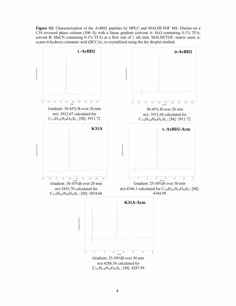
Figure S3: Characterization of the AvBD2 peptides by HPLC and MALDI-TOF MS. Elution on a C18 reversed phase column (300 Å) with a linear gradient (solvent A: H2O containing 0.1% TFA; solvent B: MeCN containing 0.1% TFA) at a flow rate of 1 mL/min. MALDI/TOF: matrix used; α-cyano-4-hydroxy-cinnamic acid (HCCA), co-crystallized using the dry droplet method.
Minutes
0.0 2.5 5.0 7.5 10.0 12.5 15.0 17.5 20.0 22.5
Abs
orba
nce
(220
nm
)
0
100
200
300
400
500
600
Minutes
0.0 2.5 5.0 7.5 10.0 12.5 15.0 17.5 20.0 22.5
Abs
orba
nce
(220
nm
)
0
50
100
150
200
250
Gradient: 30-45% B over 20 min m/z: 3912.67 calculated for
C176H249N49O42S6 ; [M]: 3911.72
30-45% B over 20 min m/z: 3912.68 calculated for
C176H249N49O42S6 ; [M]: 3911.72
D-AvBD2L-AvBD2
K31A-Acm
K31A
Gradient: 25-50%B over 30 min m/z:4288.56 calculated for
C191H278N54O48S6 ; [M]: 4287.99
L-AvBD2-Acm
Gradient: 25-50%B over 30 min m/z:4346.1 calculated for C194H285N55O48S6 ; [M]:
4344.99
Gradient: 30-45%B over 20 min m/z:3855.70 calculated for
C173H242N48O42S6 ; [M]: 3854.66
5
Figure S4: Comparison of the CD spectra obtained for L-AvBD2 (blue), D-AvBD2 (red).
-5
-4
-3
-2
-1
0
1
2
3
4
195 205 215 225 235 245 255
Wavelength (nm)
L
D

6
Table S2: Influence of different co-solvents on the folding yield of AvBD2 peptides. The oxidative foldings (50 µg/ml peptide concentration) were performed at a peptide/GSH/GSSG molar ratio of 1/100/10 in deoxygenated 100 mM Tris-HCl buffer pH 8.5 containing 1 mM EDTA, in 50% iPrOH, MeOH or MeCN, or without co-solvent. The conversion yields were determined by integrating the HPLC peaks (=280nm) corresponding to linear AvBD2 (t = 0) and folded AvBD2 (t= 60 min), keeping constant the injection volumes. The linear/folded ratio is expressed as a percentage.
Co-solvent
Peptides none iPrOH MeOH MeCN
L-AvBD2 and D-AvBD2 59 74 74 74
AvBD2-K31A 8 47 39 42

7
Table S3: Determination of disulfide-connected peptides using PeptideMap. The tolerance over statistical error on mass/charge values was set at 45 ppm.
Proteinase Observed mass/charge
Interpretation Links - Free SH
Theoretical mass/charge for charge equal to 1
Measure-ment error
Papain 1641.807 [G5- G12] — [L17- G25] 1 - 0 1641.816 -0.009
Trypsin 947.369 [L1- K4] — [S28- K31] 1 - 1 947.396 -0.019
2369.036 [G5- K19] — [V20- R27] 1 - 1 2369.074 -0.038
2444.015 [L1- K4], [G5- K19] and [S28- K31] 2 - 1 2444.080 -0.065
2806.219 [G5- K19], [V20- R27], and [S28- K31] 2 - 1 2806.214 0.005

8
Figure S5: Disulfide bridges as deduced from mass spectrometry data (see Table S6). Links in blue are deduced from possibilities remaining after deductions from individual proteolysis experiments.

9
Figure S6: Interactions between DNA (100ng/lane) and AvBD2 peptides. Lane 1, DNA:Peptide ratio1 : 0; lane 2, DNA:Peptide ratio 1 : 0.5; lane 3,DNA:peptide ratio 1 : 1; lane 4, DNA:peptide ratio 1 : 2; lane 5, DNA:peptide ratio 1 : 4 ;lane 6, DNA:peptide ratio 1 : 6; lane 7, DNA:peptide ratio 1 : 8; lane 8, DNA:peptide ratio 1 : 0.1; lane 9, DNA:peptide ratio 1 : 0.5; lane 10, DNA:peptide ratio 1 : 1; lane 11, DNA:peptide ratio 1 : 1.5. DNA used for this test was the plasmid pET14b (4671 pb, Novagen). The plasmid was mixed with 10 X buffer comprising of 100 mM Tris pH 8, 200 mM KCl, 10 mM EDTA, glycerol, water and peptides at increasing concentrations. The archaeal MC1 protein (De Vuyst, G., Aci, S., Genest, D., and Culard, F., Atypical recognition of particular DNA sequences by the archaeal chromosomal MC1 protein. Biochemistry 44 (30), 10369 (2005) was used for comparison because this peptide had previously been shown to bind to DNA fragments. The solutions were incubated at room temperature for an hour. To separate the DNA fragments, the samples were loaded onto a 1% agarose/TBE (89 mM Tris Base, 89 mM Boric acid, 2 mM EDTA) gel and run at 100 V for 55 min. The gel was then stained with 1µg/mL ethidium bromide in TBE.
AvBD2 MC1
1 2 3 4 5 6 7 8 9 10 11
AvBD2acm MC1
1 2 3 4 5 6 7 8 9 10 11