production of b-defensin antimicrobial peptides by the ...iai.asm.org/content/67/6/2740.full.pdf ·...
TRANSCRIPT
INFECTION AND IMMUNITY,0019-9567/99/$04.0010
June 1999, p. 2740–2745 Vol. 67, No. 6
Copyright © 1999, American Society for Microbiology. All Rights Reserved.
Production of b-Defensin Antimicrobial Peptides by the OralMucosa and Salivary Glands
MICHAEL MATHEWS,1 HONG PENG JIA,2 JANET M. GUTHMILLER,1 GARRETT LOSH,3
SCOTT GRAHAM,3 GEORGIA K. JOHNSON,1 BRIAN F. TACK,4
AND PAUL B. MCCRAY, JR.2*
Department of Periodontics and Dows Institute for Dental Research1 and Departments of Microbiology,4
Otolaryngology,3 and Pediatrics,2 University of Iowa Colleges of Medicineand Dentistry, Iowa City, Iowa
Received 24 November 1998/Returned for modification 8 January 1999/Accepted 3 March 1999
b-Defensins are cationic peptides with broad-spectrum antimicrobial activity that are produced by epitheliaat mucosal surfaces. Two human b-defensins, HBD-1 and HBD-2, were discovered in 1995 and 1997, respec-tively. However, little is known about the expression of HBD-1 or HBD-2 in tissues of the oral cavity andwhether these proteins are secreted. In this study, we characterized the expression of HBD-1 and HBD-2mRNAs within the major salivary glands, tongue, gingiva, and buccal mucosa and detected b-defensin peptidesin salivary secretions. Defensin mRNA expression was quantitated by RNase protection assays. HBD-1 mRNAexpression was detected in the gingiva, parotid gland, buccal mucosa, and tongue. Expression of HBD-2 mRNAwas detected only in the gingival mucosa and was most abundant in tissues with associated inflammation. Totest whether b-defensin expression was inducible, gingival keratinocyte cell cultures were treated with inter-leukin-1b (IL-1b) or bacterial lipopolysaccharide (LPS) for 24 h. HBD-2 expression increased ;16-fold withIL-1b treatment and ;5-fold in the presence of LPS. Western immunoblotting, liquid chromatography, andmass spectrometry were used to identify the HBD-1 and HBD-2 peptides in human saliva. Human b-defensinsare expressed in oral tissues, and the proteins are secreted in saliva; HBD-1 expression was constitutive, whileHBD-2 expression was induced by IL-1b and LPS. Human b-defensins may play an important role in theinnate defenses against oral microorganisms.
Defenses at mucosal surfaces are multifaceted and includeboth adaptive immunity and the innate immune system. Thelatter includes a number of broad-spectrum antimicrobial pro-teins and peptides which may protect mucosal surfaces fromthe continuous exposure to microbes. To date, many of thesepeptides have been studied in extraoral sites. Characterizationof antimicrobial factors in the oral cavity may permit newinsights into oral defense mechanisms and the pathogenesis ofdisease (21). For example, inactivation of these factors couldlead to increased microbial colonization or invasion of the oralsoft tissues, increasing the risk for candidiasis and periodontaldisease. Additionally, they may play a role in preventing viralinfections (16). The b-defensin family of antimicrobial pep-tides may contribute to oral defenses but until recently hasreceived little attention (6, 14, 20).
Mammalian defensins are cationic antimicrobial peptides,ranging from 3.5 to 4.5 kDa, which are stabilized by threeintramolecular disulfide bonds (6). There are two families ofhuman defensins, the a- and b-defensins. The b-defensins dif-fer from the classical or a-defensins by the ordering of thethree disulfide bonds between the six cysteine residues of themature peptides (6, 27). The b-defensins contribute to a pro-tective barrier on mucosal surfaces including the tongue, nasal,and intrapulmonary airways and the small intestine (10, 12, 26,29). In vitro, defensins exhibit broad-spectrum antimicrobialactivity against gram-positive and gram-negative bacteria,fungi, and enveloped viruses (6). Bovine lingual antimicrobialpeptide (LAP) is expressed in the bovine tongue epithelia and
shows a marked induction of mRNA expression in epitheliasurrounding areas of inflammation (26). The expression ofLAP is induced, in part, by bacterial lipopolysaccharide (LPS)and proinflammatory cytokines (4). Native bovine b-defensinpeptides exhibit antimicrobial activity against both gram-neg-ative and gram-positive bacteria and fungi (5). Although b-de-fensin peptides in human oral epithelial cells have receivedlittle study, it is likely that human oral tissues produce b-de-fensin peptides similar to those found in other animals.
To date, two human b-defensins (HBD-1 and HBD-2) havebeen identified. Bensch et al. isolated a 36-amino-acid b-de-fensin peptide (HBD-1) from dialysate hemofiltrate andcloned a partial cDNA fragment from human kidney RNA (1).HBD-1 is abundantly expressed in the urogenital tract (30) andhas also been detected in respiratory and other epithelia (7, 19,28, 31). Harder et al. isolated and purified a second humanb-defensin peptide, HBD-2, from psoriatic scale extracts byusing a whole Escherichia coli affinity column (9). The HBD-2sequence had significant homology to bovine LAP and bovinetracheal antimicrobial peptide (TAP). HBD-2 was highly ef-fective in killing gram-negative bacteria (E. coli and Pseudo-monas aeruginosa) and yeasts (Candida albicans) and had abacteriostatic effect on the gram-positive bacterium Staphylo-coccus aureus (9). In addition, HBD-2 mRNA expression, likethat of TAP and LAP, was induced by gram-negative bacteriaand proinflammatory cytokines (9). Recently, Krisanaprakorn-kit et al. reported the constitutive expression of HBD-1 mRNAin cultured gingival epithelial cells and noninflamed and in-flamed gingival tissues (14).
If HBD-1 and HBD-2 play a role in oral mucosal defenses,we hypothesized that they would be expressed in a number oforal epithelia, including the salivary glands, and that theywould be present in saliva. In this study, we analyzed the
* Corresponding author. Mailing address: Department of Pediatrics,University of Iowa Hospitals and Clinics, 200 Hawkins Dr., Iowa City,IA 52242. Phone: (319) 356-4866. Fax: (319) 356-7171. E-mail: [email protected].
2740
on July 13, 2018 by guesthttp://iai.asm
.org/D
ownloaded from
expression of HBD-1 and HBD-2 mRNAs in selected oraltissues by the RNase protection assay (RPA). We also testedthe hypothesis that the expression of HBD-1 and/or HBD-2was induced by a proinflammatory cytokine or bacterial LPS.Finally, we analyzed the expression of HBD-1 and HBD-2peptides in saliva by immunoblotting, capillary electrophoresis(CE), liquid chromatography (LC), and mass spectrometry(MS).
MATERIALS AND METHODS
Tissue specimens. Tissues were obtained from surgical discard and postmor-tem specimens. Samples were obtained from the tongue, gingiva, and parotidgland. The samples were immediately flash frozen in liquid nitrogen and storedat 280°C for future RNA studies. Tissue procurement procedures were ap-proved by the Institutional Review Board at the University of Iowa.
Primary culture of gingival keratinocytes. Healthy gingival tissue samplesfrom crown-lengthening procedures were obtained for keratinocyte culture asdescribed previously (11). Tissue fragments were washed in Dulbecco’s phos-phate-buffered saline containing penicillin (200 IU/ml), streptomycin (200 mg/ml), amphotericin B (5 mg/ml), and gentamicin (0.1 mg/ml). To separate theepithelium from the underlying connective tissue, the specimens were incubatedin Dispase II (2.4 U/ml; Boehringer Mannheim, Indianapolis, Ind.) overnight at5°C. The epithelium was mechanically separated from the underlying connectivetissue (22), and the epithelial sheets were placed in 0.25% trypsin–1 mM EDTAand vigorously pipetted to produce a cell suspension. Following trypsin neutral-ization by medium containing 10% fetal bovine serum, the suspension wascentrifuged (208 3 g for 5 min). The pellet was suspended in medium and seededinto a T-25 tissue culture flask (Corning, Corning, N.Y.) containing a feeder layerof mitomycin-treated NIH 3T3 murine fibroblasts (23). The cells were culturedin medium consisting of 3 parts Dulbecco’s modified Eagle’s medium (SigmaChemical Co., St. Louis, Mo.) and 1 part Ham’s F12 medium (Sigma ChemicalCo.) containing 10% fetal bovine serum (Intergen, Purchase, N.Y.). The mediumwas supplemented with penicillin (100 IU/ml), streptomycin (100 mg/ml), am-photericin B (2.5 mg/ml), epidermal growth factor (10 ng/ml), cholera toxin (0.1nM), and hydrocortisone (400 ng/ml). It was changed every 2 to 3 days, and thecultures were kept at 37°C in a humidified environment containing 5% CO2.
When the cultures reached 75 to 80% confluency, the feeder layer was re-moved by treatment with 0.5 mM EDTA for 5 min at 37°C and the cultures wereexposed to trypsin-EDTA for 1 to 2 min (37°C). Adherent keratinocytes wereincubated in trypsin-EDTA for 4 to 5 min at 37°C or until microscopic inspectiondemonstrated detachment of the majority of the cells. After trypsin inactivation,cell counts were determined with a hemocytometer and the cells were seeded forthe experimental procedures described below. The keratinocyte nature of thecells was confirmed by immunohistochemical staining for high-molecular-mass(50-, 56.5-, 57-, 58-, 66-, and 68-kDa) cytokeratins (Dako Corp., Carpinteria,Calif.) and by histologic and ultrastructural features.
RPA. Specific oral tissues expressing HBD-1 and HBD-2 mRNAs were iden-tified by RPA methods previously described for quantitative assessment ofHBD-1 mRNA in the lung (19). Total RNA was isolated from frozen tissuesamples and human cell cultures by a single-step guanidine thiocyanate-phenol-chloroform extraction (2) and stored in RNase-free water at 280°C. The[a-32P]UTP (Amersham Corp., Arlington Heights, Ill.) random-labeled HBD-1,HBD-2, and 18S ribosomal subunit (as the internal standard [Ambion Co.,Austin, Tex.]) antisense riboprobes were transcribed with cDNA templates sub-cloned into a plasmid vector containing the T7 promoter. The radiolabeledriboprobes were hybridized to tissue-specific mRNA by using a Hybspeed RPAkit (Ambion Co.). The unprotected probes were 270 and 432 nucleotides forHBD-1 and HBD-2, respectively. The protected probes were 158 and 328 nu-cleotides for HBD-1 and HBD-2, respectively. In the absence of RNA, theunprotected probes were completely digested by RNases. The RNA-RNA hy-brids were separated by denaturing Tris-borate-EDTA (TBE) vertical gel elec-trophoresis and visualized by autoradiographic methods as previously described(19).
Induction of b-defensins. We tested for inducible b-defensin mRNA expres-sion in primary cultures of human gingival keratinocytes (11). The cells weretreated with recombinant human interleukin-1-b (IL-1b) (R & D Systems, Min-neapolis, Minn.) or E. coli-derived bacterial LPS (Sigma). Cultures from fourdifferent individuals were grown on six-well plates in serum-free modified MCDB153 medium containing 0.15 mM calcium, 50 mg of gentamicin per ml, 50 ng ofamphotericin B per ml, 5 mg of insulin per ml, 0.5 mg of hydrocortisone per ml,30 mg of bovine pituitary extract per ml, and 0.1 ng of epidermal growth factorper ml (keratinocyte growth medium; Clonetics Corp., San Diego, Calif.) bymethods described previously (11). Cells (passages 2 and 3) from each subjectwere seeded at 4.0 3 106 cells per well into six-well tissue culture plates (Costar,Cambridge, Mass.). These cultures were maintained in the absence of a feederlayer in keratinocyte growth medium. When confluence was reached, two wells ofcells from each subject were treated for 24 h with normal medium (control) ormedium containing 100 ng of IL-1b per ml or 10 mg of LPS per ml. Cell viability,as assessed by commercial assays for mitochondrial dehydrogenase (MTS, Cell-
Titer aqueous nonradioactive cell proliferation assay; Promega Corp., Madison,Wis.) was not affected by either IL-1b or LPS treatments. RNA was extractedimmediately after the treatment and analyzed by RPA (19).
Purification of cationic peptides and proteins from human saliva. Saliva wascollected in 10-ml volumes from normal adult volunteers. Samples were probe-sonicated and evaluated individually by Western blotting or pooled. Cationicmaterials were batch-extracted from individual (10-ml) or pooled (100-ml) salivasamples with a 50% slurry of Macro-Prep CM beads (Bio-Rad, Richmond,Calif.) as previously described for other biological fluids (30). Following contin-uous mixing of the gel beads with the saliva overnight at 4°C, the beads werecollected by centrifugation and washed with 25 mM ammonium acetate (pH 7.3).Elution of bound material was affected by addition of an equal volume of 10%acetic acid. This was repeated three times with equal volumes of 5% acetic acid.Pooled eluates were lyophilized, resuspended in 1 ml of 1% acetic acid, andfurther purified by reversed-phase high-pressure liquid chromatography (RP-HPLC) on a 2.1- by 250-mm Vydac C18 column (218TP52) with a mobile phasesystem consisting of 0.025% trifluoroacetic acid in water (solution A) and 0.021%trifluoroacetic acid–80% acetonitrile in water (solution B). Gradient conditionswere 1 to 48% solution B in 60 min, 48 to 88% solution B in 30 min, and 88 to98% solution B in 10 min at a flow rate of 0.15 ml/min. Fractions were collectedat 2-min intervals, dried by vacuum centrifugation, and resuspended in 50 ml of0.1% acetic acid.
Immunodetection of HBD-1 and HBD-2. Cationic material from individualsamples and selected fractions from RP-HPLC were electrophoresed on urea-acetic acid polyacrylamide gels together with known quantities of purified re-combinant HBD standards. Recombinant HBD-1 and HBD-2 were prepared inbaculovirus as reported previously (18, 30). The conditions for acid-urea poly-acrylamide gel electrophoresis included 12.5% acrylamide and 4.6 M urea (pH4). Western immunoblotting was performed with rabbit antisera to HBD-1 orHBD-2 (diluted 1:1,000) as previously described (18, 30). The secondary anti-body was a horseradish peroxidase-conjugated donkey anti-rabbit immunoglob-ulin G diluted 1:10,000. The substrate was Pierce SuperSignal.
ESI-LC-MS, CE, and Edman degradation. Immunoblot-positive fractionsfrom RP-HPLC were analyzed by LC-MS with a Hewlett-Packard 1100 MSDequipped with an electrospray ionization (ESI) source, a Hewlett-Packard 1100series HPLC apparatus, and a 0.3- by 250-mm LC Packings capillary columnpacked with Vydac 218TPC18 RP material. CE was performed with a Hewlett-Packard 3D CE apparatus equipped with a Hewlett-Packard extended-light-pathfused-silicate capillary column (75 mm [inner diameter] by 80.5 cm [totallength]). The experiments were performed at 20,000 V in 0.1 M sodium phos-phate (pH 2.9). Edman degradation was conducted on a polyvinylidene difluo-ride membrane in the gas phase with an Applied Biosystems Procise sequencer.
RESULTS
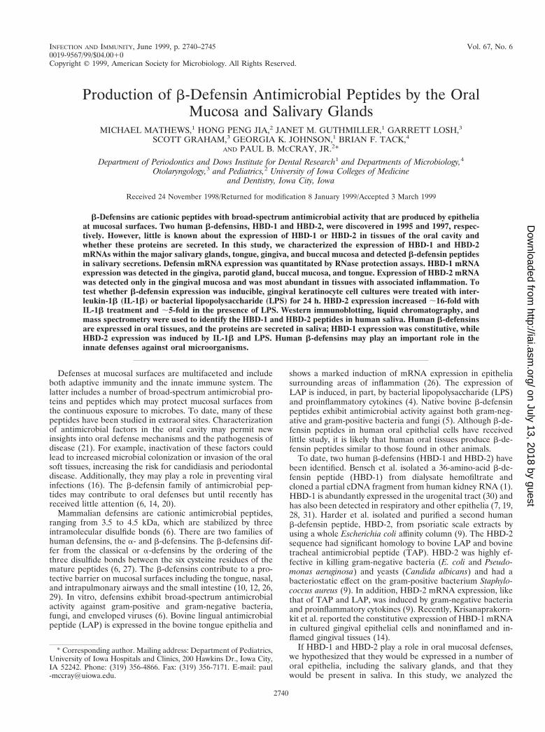
b-Defensin mRNA expression in oral tissues. HBD-1mRNA expression was detected in gingival, parotid gland, andlateral tongue tissue (Fig. 1). In contrast, expression of HBD-2was detectable only in gingival tissue (Fig. 1) and at low levels
FIG. 1. b-Defensin expression in oral tissues by RPA. HBD-1 mRNA, rep-resented by a single protected fragment of 158 bp, was detected in the lateraltongue, gingival tissue, and the parotid gland. In contrast, expression of HBD-2,represented by a single protected fragment of 328 bp, was detectable by RPAonly in gingival tissue and was most abundant in gingival tissues associated withinflammation (data not shown).
VOL. 67, 1999 b-DEFENSIN EXPRESSION IN ORAL TISSUES 2741
on July 13, 2018 by guesthttp://iai.asm
.org/D
ownloaded from
in gingival keratinocytes (see Fig. 2A). The expression ofHBD-2 mRNA was most abundant in gingival tissues withassociated inflammation (data not shown). To determinewhether b-defensin expression in oral epithelia was inducible,further experiments were performed with a gingival keratino-cyte cell culture model.
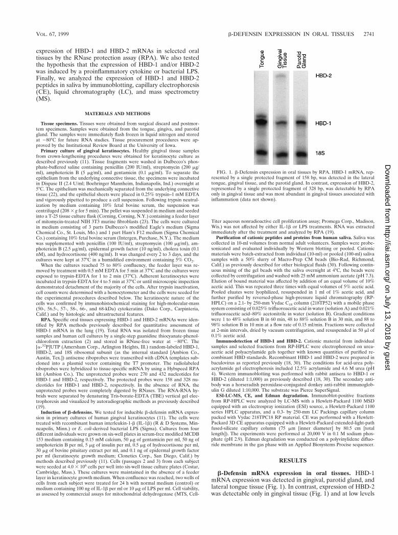
Regulation of b-defensin expression in cultured gingivalkeratinocytes. As shown in Fig. 2A, using a quantitative RPA,we found moderate HBD-1 and low HBD-2 mRNA expressionin gingival keratinocyte cultures under basal conditions. Fol-lowing a 24-h treatment of separate wells of the same culturewith 100 ng of IL-1b per ml, there was a ;16-fold increase(n 5 4) in HBD-2 mRNA expression above the control (Fig.2). Treatment of cells with 10 mg of LPS per ml for 24 h alsoinduced a ;fivefold increase in HBD-2 expression (Fig. 2). Incontrast, HBD-1 mRNA levels were unchanged in the pres-ence of either IL-1b or LPS (Fig. 2A).
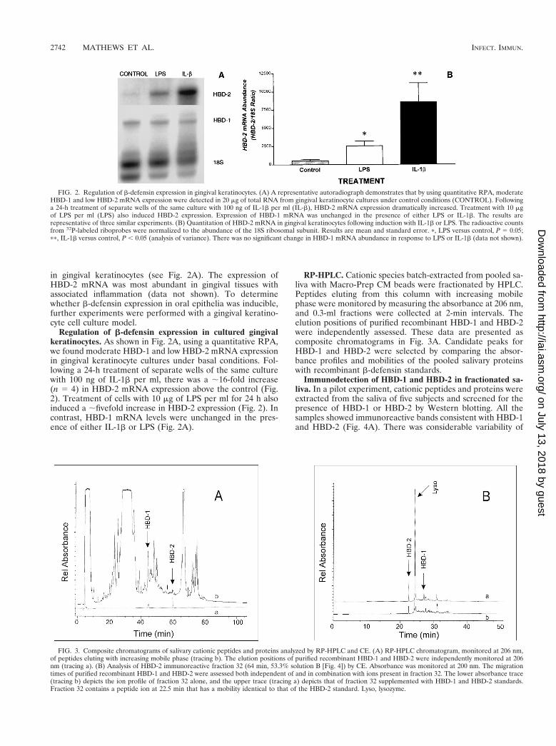
RP-HPLC. Cationic species batch-extracted from pooled sa-liva with Macro-Prep CM beads were fractionated by HPLC.Peptides eluting from this column with increasing mobilephase were monitored by measuring the absorbance at 206 nm,and 0.3-ml fractions were collected at 2-min intervals. Theelution positions of purified recombinant HBD-1 and HBD-2were independently assessed. These data are presented ascomposite chromatograms in Fig. 3A. Candidate peaks forHBD-1 and HBD-2 were selected by comparing the absor-bance profiles and mobilities of the pooled salivary proteinswith recombinant b-defensin standards.
Immunodetection of HBD-1 and HBD-2 in fractionated sa-liva. In a pilot experiment, cationic peptides and proteins wereextracted from the saliva of five subjects and screened for thepresence of HBD-1 or HBD-2 by Western blotting. All thesamples showed immunoreactive bands consistent with HBD-1and HBD-2 (Fig. 4A). There was considerable variability of
FIG. 2. Regulation of b-defensin expression in gingival keratinocytes. (A) A representative autoradiograph demonstrates that by using quantitative RPA, moderateHBD-1 and low HBD-2 mRNA expression were detected in 20 mg of total RNA from gingival keratinocyte cultures under control conditions (CONTROL). Followinga 24-h treatment of separate wells of the same culture with 100 ng of IL-1b per ml (IL-b), HBD-2 mRNA expression dramatically increased. Treatment with 10 mgof LPS per ml (LPS) also induced HBD-2 expression. Expression of HBD-1 mRNA was unchanged in the presence of either LPS or IL-1b. The results arerepresentative of three similar experiments. (B) Quantitation of HBD-2 mRNA in gingival keratinocytes following induction with IL-1b or LPS. The radioactive countsfrom 32P-labeled riboprobes were normalized to the abundance of the 18S ribosomal subunit. Results are mean and standard error. p, LPS versus control, P 5 0.05;pp, IL-1b versus control, P , 0.05 (analysis of variance). There was no significant change in HBD-1 mRNA abundance in response to LPS or IL-1b (data not shown).
FIG. 3. Composite chromatograms of salivary cationic peptides and proteins analyzed by RP-HPLC and CE. (A) RP-HPLC chromatogram, monitored at 206 nm,of peptides eluting with increasing mobile phase (tracing b). The elution positions of purified recombinant HBD-1 and HBD-2 were independently monitored at 206nm (tracing a). (B) Analysis of HBD-2 immunoreactive fraction 32 (64 min, 53.3% solution B [Fig. 4]) by CE. Absorbance was monitored at 200 nm. The migrationtimes of purified recombinant HBD-1 and HBD-2 were assessed both independent of and in combination with ions present in fraction 32. The lower absorbance trace(tracing b) depicts the ion profile of fraction 32 alone, and the upper trace (tracing a) depicts that of fraction 32 supplemented with HBD-1 and HBD-2 standards.Fraction 32 contains a peptide ion at 22.5 min that has a mobility identical to that of the HBD-2 standard. Lyso, lysozyme.
2742 MATHEWS ET AL. INFECT. IMMUN.
on July 13, 2018 by guesthttp://iai.asm
.org/D
ownloaded from
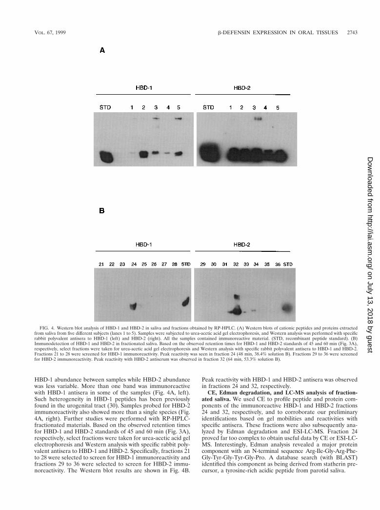
HBD-1 abundance between samples while HBD-2 abundancewas less variable. More than one band was immunoreactivewith HBD-1 antisera in some of the samples (Fig. 4A, left).Such heterogeneity in HBD-1 peptides has been previouslyfound in the urogenital tract (30). Samples probed for HBD-2immunoreactivity also showed more than a single species (Fig.4A, right). Further studies were performed with RP-HPLC-fractionated materials. Based on the observed retention timesfor HBD-1 and HBD-2 standards of 45 and 60 min (Fig. 3A),respectively, select fractions were taken for urea-acetic acid gelelectrophoresis and Western analysis with specific rabbit poly-valent antisera to HBD-1 and HBD-2. Specifically, fractions 21to 28 were selected to screen for HBD-1 immunoreactivity andfractions 29 to 36 were selected to screen for HBD-2 immu-noreactivity. The Western blot results are shown in Fig. 4B.
Peak reactivity with HBD-1 and HBD-2 antisera was observedin fractions 24 and 32, respectively.
CE, Edman degradation, and LC-MS analysis of fraction-ated saliva. We used CE to profile peptide and protein com-ponents of the immunoreactive HBD-1 and HBD-2 fractions24 and 32, respectively, and to corroborate our preliminaryidentifications based on gel mobilities and reactivities withspecific antisera. These fractions were also subsequently ana-lyzed by Edman degradation and ESI-LC-MS. Fraction 24proved far too complex to obtain useful data by CE or ESI-LC-MS. Interestingly, Edman analysis revealed a major proteincomponent with an N-terminal sequence Arg-Ile-Gly-Arg-Phe-Gly-Tyr-Gly-Tyr-Gly-Pro. A database search (with BLAST)identified this component as being derived from statherin pre-cursor, a tyrosine-rich acidic peptide from parotid saliva.
FIG. 4. Western blot analysis of HBD-1 and HBD-2 in saliva and fractions obtained by RP-HPLC. (A) Western blots of cationic peptides and proteins extractedfrom saliva from five different subjects (lanes 1 to 5). Samples were subjected to urea-acetic acid gel electrophoresis, and Western analysis was performed with specificrabbit polyvalent antisera to HBD-1 (left) and HBD-2 (right). All the samples contained immunoreactive material. (STD, recombinant peptide standard). (B)Immunodetection of HBD-1 and HBD-2 in fractionated saliva. Based on the observed retention times for HBD-1 and HBD-2 standards of 45 and 60 min (Fig. 3A),respectively, select fractions were taken for urea-acetic acid gel electrophoresis and Western analysis with specific rabbit polyvalent antisera to HBD-1 and HBD-2.Fractions 21 to 28 were screened for HBD-1 immunoreactivity. Peak reactivity was seen in fraction 24 (48 min, 38.4% solution B). Fractions 29 to 36 were screenedfor HBD-2 immunoreactivity. Peak reactivity with HBD-2 antiserum was observed in fraction 32 (64 min, 53.3% solution B).
VOL. 67, 1999 b-DEFENSIN EXPRESSION IN ORAL TISSUES 2743
on July 13, 2018 by guesthttp://iai.asm
.org/D
ownloaded from

In contrast, HBD-2 fraction 32, by virtue of its position in aless trafficked position of the chromatogram, was amenable toanalysis by CE. These data are shown in Fig. 3B. Peptide ionspresent in fraction 32 were separated according to their differ-ent migration velocities under conditions of uniform fieldstrength at pH 2.9 and monitored by measurement of absor-bance at 200 nm. The migration times of purified recombinantHBD-1 and HBD-2 were assessed both independent of and incombination with the ions present in fraction 32. The lowerabsorbance trace depicts the ion profile of fraction 32 alone,and the upper trace depicts that of fraction 32 supplementedwith HBD-1 and HBD-2 standards in a ratio of 8:2 (vol/vol).The results of this study clearly indicate the presence in frac-tion 32 of a peptide ion at 22.5 min which has a mobilityidentical to that of the HBD-2 standard. Subsequent LC-MSand Edman analysis of fraction 32 confirmed the presence ofthe 41-amino-acid form of HBD-2 (observed molecular weight,4,327.56; calculated molecular weight, 4328.06) with the N-terminal sequence Gly-Ile-Gly-Asp-Pro-Val-Thr. Edman anal-ysis further revealed the major peptide ion observed by CE at24.3 min to be the mature form of lysozyme C (data notshown). From these data, we estimate the concentration ofHBD-2 in saliva to be about 150 ng/ml. This is undoubtedly aconservative estimate, since we have no means of quantita-tively assessing the efficiency of recovery at each step of thepurification.
DISCUSSION
The above studies demonstrate the distribution of humanb-defensin mRNAs in oral epithelial tissues and the parotidgland. Expression of HBD-1 mRNA was observed in tongueand parotid gland tissues, and both HBD-1 and HBD-2mRNAs were detected in the gingiva and cultured gingivalkeratinocytes. Of note, HBD-2 expression was induced byIL-1b and LPS whereas HBD-1 expression remained un-changed in the presence of inflammatory stimuli. Both HBD-1and HBD-2 peptides were detected in saliva. The widespreadexpression of b-defensins in oral tissues suggests that theycontribute to host defenses in the oral cavity.
In our studies of cultured gingival keratinocytes, an interest-ing contrast was noted between HBD-1 and HBD-2. Consis-tent with previous reports (14, 31), HBD-1 mRNA expressionshowed no significant change in response to IL-1b or LPS. Incontrast to HBD-1, HBD-2 expression was induced markedlyby IL-1b and to a lesser extent by LPS. This suggests differentroles for these peptides in oral defenses. The induction ofHBD-2 by IL-1b and LPS is similar to that of bovine LAP andTAP (5, 24, 26). Harder et al. showed that HBD-2 expressionwas induced in skin keratinocytes in the presence of tumornecrosis factor alpha, gram-negative and gram-positive bacte-ria, and C. albicans (9). Similarly, HBD-2 expression is induc-ible in airway epithelia (9, 28). We speculate that HBD-1 playsa constitutive role in oral defenses while HBD-2 expression isinduced in response to local infection or inflammation.
Both human b-defensin peptides were readily detected insaliva. A conservative estimate for the concentrations ofHBD-1 and HBD-2 in saliva is ;150 ng/ml. We speculate thatthese concentrations may be sufficient to be microbicidal forsome organisms, especially considering that they may act syn-ergistically with other microbicidal factors present in saliva.Such factors include human salivary histatin 5, lactoferrin, mu-cin glycoprotein, and lysozyme (15, 25). Further studies of theactivity of HBD-1 and HBD-2 against a spectrum of oral mi-croorganisms including pathogenic species are needed. Theconcentrations of HBD-1 and HBD-2 peptides in saliva are
somewhat at odds with data obtained with the cultured gingivalkeratinocytes. Perhaps the constant exposure of the oral mu-cosa to microorganisms serves to induce the production ofHBD-2, even in the absence of overt oral disease. Because theparotid glands expressed HBD-1 mRNA but not HBD-2mRNA, we speculate that the HBD-2 peptide detected in thesaliva arose from induced expression by keratinocytes in themouth. Alternatively, HBD-2 may also be secreted from othersalivary glands. Zhao et al. previously reported the expressionof HBD-1 mRNA in salivary epithelia (31), and Harder et al.observed HBD-2 mRNA expression in the salivary gland, al-though the gland of origin was not stated (9). We did notdetect HBD-2 mRNA expression in the parotid gland. Thedifferences in these results may reflect the sensitivity of thedetection system (RPA versus reverse transcription-PCR), thedegree of inflammation present in the tissue sampled, or thespecific salivary gland studied (major or minor).
These results suggest that epithelial b-defenses may play animportant role in the mucosal defenses of the mouth. HBD-1may provide a basal antimicrobial activity at mucosal surfacesto guard against infections at and away from the site of inva-sion. This might explain the greater expression of HBD-1 thanHBD-2 in the kidney and salivary glands, where epithelialsurface fluids are excreted away from site of origin, preventingascending infections (30). However, further studies are neededto understand the antimicrobial activity of HBD-1 and HBD-2peptides against relevant oral microorganisms and how theactivity or expression may be altered in states such as peri-odontal disease or immunosuppression. In addition to theirantimicrobial properties, b-defensins attract monocytes (17),suggesting a possible interaction between antimicrobial expres-sion and inflammation. This relationship is an example of theability of the peptide to generate a robust local response tomicrobial and viral infections. Inducible antibiotics like de-fensins may work to repair injured mucosal sites, given thatdefensins exhibit growth factor activity, in addition to micro-bicidal activity, in vitro and in vivo (17).
Finally, b-defensins and other antimicrobial peptides mayhave therapeutic applications for the treatment of diseases inoral tissues (8, 13). For example, defensins inactivate manyenveloped viruses that can penetrate mucosal surfaces (3, 16).Thus, topical application of antimicrobial peptides may haveutility in the treatment of oral diseases including periodontitisor candidiasis (21). Clarification of the mechanisms of b-de-fensin induction may also prove useful for therapeutic appli-cations designed to enhance innate immunity.
ACKNOWLEDGMENTS
We thank Tom Ganz for many helpful discussions and for providingthe HBD-1 and HBD-2 antisera. We are grateful to Elena Rus, ProteinStructure Facility, University of Iowa, for assistance with protein se-quence analysis. We thank Connie C. Organ for technical assistanceand Larry McCray for assistance in obtaining clinical tissue samples.
REFERENCES
1. Bensch, K. W., M. Raida, H. J. Magert, P. Schulz-Knappe, and W. G.Forssmann. 1995. hBD-1: a novel beta-defensin from human plasma. FEBSLett. 368:331–335.
2. Chomczynski, P., and N. Sacchi. 1987. Single-step method of RNA isolationby acid guanidinium thiocyanate-phenol-chloroform extraction. Anal. Bio-chem. 162:156–159.
3. Daher, K. A., M. E. Selsted, and R. I. Lehrer. 1986. Direct inactivation ofviruses by human granulocyte defensins. J. Virol. 60:1068–1074.
4. Diamond, G., J. P. Russell, and C. L. Bevins. 1996. Inducible expression ofan antibiotic peptide gene in lipopolysaccharide-challenged tracheal epithe-lial cells. Proc. Natl. Acad. Sci. USA 93:5156–5160.
5. Diamond, G., M. Zasloff, H. Eck, M. Brasseur, W. L. Maloy, and C. L.Bevins. 1991. Tracheal antimicrobial peptide, a cysteine-rich peptide from
2744 MATHEWS ET AL. INFECT. IMMUN.
on July 13, 2018 by guesthttp://iai.asm
.org/D
ownloaded from
mammalian tracheal mucosa: peptide isolation and cloning of a cDNA. Proc.Natl. Acad. Sci. USA 88:3952–3956.
6. Ganz, T., and R. I. Lehrer. 1995. Defensins. Pharmacol. Ther. 66:191–205.7. Goldman, M. J., M. G. Anderson, E. D. Stolzenberg, P. U. Kari, M. Zasloff,
and J. M. Wilson. 1997. Human b-defensin-1 is a salt-sensitive antibiotic inlung that is inactivated in cystic fibrosis. Cell 88:1–9.
8. Hancock, R. E. W., and R. Lehrer. 1998. Cationic peptides: a new source ofantibiotics. Trends Biotechnol. 16:82–88.
9. Harder, J., J. Bartels, E. Christophers, and J.-M. Schroder. 1997. A peptideantibiotic from human skin. Nature 387:861–862.
10. Huttner, K. M., D. J. C. Brezinski, M. M. Mahoney, and G. Diamond. 1998.Antimicrobial peptide expression is developmentally regulated in the ovinegastrointestinal tract. J. Nutr. 128:297S–299S.
11. Johnson, G. K., T. K. Poore, J. B. Payne, and C. C. Organ. 1996. Effect ofsmokeless tobacco extract on human gingival keratinocyte levels of prosta-glandin E2 and interleukin-1. J. Periodontol. 67:116–124.
12. Jones, D. E., and C. L. Bevins. 1992. Paneth cells of the human smallintestine express an antimicrobial peptide gene. J. Biol. Chem. 267:23216–23225.
13. Kelley, K. J. 1996. Using host defenses to fight infectious diseases. Nat.Biotechnol. 14:587.
14. Krisanaprakornkit, S., A. Weinberg, C. N. Perez, and B. A. Dale. 1998.Expression of the peptide antibiotic human b-defensin 1 in cultured gingivalepithelial cells and gingival tissue. Infect. Immun. 66:4222–4228.
15. Lamkin, M. S., and F. G. Oppenheim. 1993. Structural features of salivaryfunction. Crit. Rev. Oral Biol. Med. 4:251–259.
16. Lehrer, R. I., K. Daher, T. Ganz, and M. E. Selsted. 1985. Direct inactivationof viruses by MCP-1 and MCP-2, natural peptide antibiotics from rabbitleukocytes. J. Virol. 54:467–472.
17. Lehrer, R. I., A. K. Lichtenstein, and T. Ganz. 1993. Defensins: antimicrobialand cytotoxic peptides of mammalian cells. Annu. Rev. Immunol. 11:105–128.
18. Liu, L., L. Wang, H. P. Jia, C. Zhao, H. H. Q. Heng, B. C. Schutte, P. B.McCray, Jr., and T. Ganz. 1998. Structure and mapping of the humanb-defensin HBD-2 gene and its expression at sites of inflammation. Gene222:237–244.
19. McCray, P. B., Jr., and L. Bentley. 1997. Human airway epithelia express ab-defensin. Am. J. Respir. Cell Mol. Biol. 16:343–349.
20. Miyasaki, K. T., A. L. Bodeau, T. Ganz, M. E. Selsted, and R. I. Lehrer. 1990.
In vitro sensitivity of oral, gram-negative, facultative bacteria to the bacte-ricidal activity of human neutrophil defensins. Infect. Immun. 58:3934–3940.
21. Miyasaki, K. T., and R. I. Lehrer. 1998. b-Sheet antibiotic peptides aspotential dental therapeutics. Int. J. Antimicrob. Agents 9:269–280.
22. Oda, D., and E. Watson. 1990. Human oral epithelial cell culture. Improvedconditions for reproducible culture in serum-free medium. In Vitro CellDev. Biol. 26:589–595.
23. Rheinwald, J. G., and H. Green. 1975. Serial cultivation of strains of humanepidermal keratinocytes: the formation of colonies from single cells. Cell6:331–343.
24. Russell, J. P., G. Diamond, A. P. Tarver, T. F. Scanlin, and C. L. Bevins.1996. Coordinate induction of two antibiotic genes in tracheal epithelial cellsexposed to the inflammatory mediators lipopolysaccharide and tumor ne-crosis factor alpha. Infect. Immun. 64:1565–1568.
25. Schenkels, L. C., E. C. Veerman, and A. V. Nieuw Amerongen. 1995. Bio-chemical composition of human saliva in relation to other mucosal fluids.Crit. Rev. Oral Biol. Med. 6:161–175.
26. Schonwetter, B. S., E. D. Stolzenberg, and M. A. Zasloff. 1995. Epithelialantibiotics induced at sites of inflammation. Science 267:1645–1648.
27. Selsted, M. E., Y. Q. Tang, W. L. Morris, P. A. McGuire, M. J. Novotny, W.Smith, A. H. Henschen, and J. S. Cullor. 1993. Purification, primary struc-tures, and antibacterial activities of beta-defensins, a new family of antimi-crobial peptides from bovine neutrophils. J. Biol. Chem. 268:6641–6648.
28. Singh, P. K., H. P. Jia, K. Wiles, J. Hesselberth, L. Liu, B. D. Conway, E. P.Greenberg, E. V. Valore, M. J. Welsh, T. Ganz, B. F. Tack, and P. B. McCray,Jr. 1998. Production of b-defensins by human airway epithelia. Proc. Natl.Acad. Sci. USA 95:14961–14966.
29. Tarver, A. P., D. P. Clark, G. Diamond, J. P. Russell, H. E. Bromage, P.Tempst, K. S. Cohen, D. E. Jones, R. W. Sweeney, M. Wines, S. Hwang, andC. L. Bevins. 1998. Enteric b-defensin: molecular cloning and characteriza-tion of a gene with inducible intestinal epithelial cell expression associatedwith cryptosporidium parvum infection. Infect. Immun. 66:1045–1056.
30. Valore, E. V., C. H. Park, A. J. Quayle, K. R. Wiles, P. B. McCray, Jr., andT. Ganz. 1998. Human b-defensin-1, an antimicrobial peptide of urogenitaltissues. J. Clin. Investig. 101:1633–1642.
31. Zhao, C., I. Wang, and R. I. Lehrer. 1996. Widespread expression of beta-defensin hBD-1 in human secretory glands and epithelial cells. FEBS Lett.396:319–322.
Editor: J. R. McGhee
VOL. 67, 1999 b-DEFENSIN EXPRESSION IN ORAL TISSUES 2745
on July 13, 2018 by guesthttp://iai.asm
.org/D
ownloaded from