inhibition of plasminogen activator inhibitor-1 … effect of pai-1 inhibitor was ... our data shows...
TRANSCRIPT
462
DOI: 10.4046/trd.2011.70.6.462ISSN: 1738-3536(Print)/2005-6184(Online)Tuberc Respir Dis 2011;70:462-473CopyrightⒸ2011. The Korean Academy of Tuberculosis and Respiratory Diseases. All rights reserved.
Inhibition of Plasminogen Activator Inhibitor-1 Expression in Smoke-Exposed Alveolar Type II Epithelial Cells Attenuates Epi-thelial-Mesenchymal TransitionJeong Sup Song, M.D., Chun Mi KangDepartment of Internal Medicine, Yeouido St Mary's Hospital, The Catholic University of Korea College of Medicine, Seoul, Korea
Background: Smoking is a risk factor for idiopathic pulmonary fibrosis (IPF), but the mechanism of the association remains obscure. There is evidence demonstrating that plasminogen activator inhibitor-1 (PAI-1) is involved in the progression of pulmonary fibrosis. This study was to determine whether the administration of small interfering RNA (siRNA) targeting PAI-1 or PAI-1 inhibitor to the cigarette smoking extract (CSE)-exposed rat alveolar type II epithelial cells (ATII cells) limits the epithelial-mesenchymal transition (EMT).Methods: ATII cells were isolated from lung of SD-rat using percoll gradient method and cultured with 5% CSE. The EMT was determined from the ATII cells by measuring the real-time RT PCR and western blotting after the PAI-1 siRNA transfection to the cells and after administration of tiplaxtinin, an inhibitor of PAI-1. The effect of PAI-1 inhibitor was also evaluated in the bleomycin-induced rats.Results: PAI-1 was overexpressed in the smoking exposed ATII cells and was directly associated with EMT. The EMT from the ATII cells was suppressed by PAI-1 siRNA transfection or administration of tiplaxtinin. Signaling pathways for EMT by smoking extract were through the phosphorylation of SMAD2 and ERK1/2, and finally Snail expression. Tiplaxtinin also suppressed the pulmonary fibrosis and PAI-1 expression in the bleomycin-induced rats. Conclusion: Our data shows that CSE induces rat ATII cells to undergo EMT by PAI-1 via SMAD2-ERK1/2-Snail activation. This suppression of EMT by PAI-1 siRNA transfection or PAI-1 inhibitor in primary type II alveolar epithelial cells might be involved in the attenuation of bleomycin-induced pulmonary fibrosis in rats.
Key Words: Plasminogen Activator Inhibitor 1; Epithelial-Mesenchymal Transition; Pulmonary Fibrosis
Address for correspondence: Jeong Sup Song, M.D.Division of Pulmonary Medicine, Department of Internal Medicine, Yeouido St Mary's Hospital, The Catholic University College of Medicine, 62, Yeouido-dong, Yeong-deungpo-gu, Seoul 150-713, KoreaPhone: 82-2-3779-1146, Fax: 82-2-780-3132E-mail: [email protected]
Received: May 26, 2011Accepted: May 28, 2011
Introduction
Idiopathic pulmonary fibrosis (IPF) induces the pro-
liferation of fibroblasts because of the activation due to
the repeated damage of the alveolar epithelial cells.
Thus, it is involved in the formation of fibroblastic foci
and this increases the extracellular matrix, thus con-
tributing to the wound healing1. Epithelial cells release
procoagulant factor and TGF-β and thereby promote
the migration and proliferation of fibroblasts. Due to the
resulting increase in the secretion of collagen, they are
involved in the pulmonary fibrosis2. In recent years, it
has been reported that an epithelial-mesenchymal tran-
sition (EMT) phenomenon where the alveolar epithelial
cells are directly converted into the mesothelial cells
might be one of the key mechanisms by which the pul-
monary fibrosis occurs. In this process, TGF-β1 plays
a key role3. As another mechanism by which the pulmo-
nary fibrosis occurs, there is a phenomenon that the
plasma proteins are leaked into the alveoli due to the
damage of pulmonary tissue and this activates the blood
coagulation factors and then leads to the deposition of
Original Article

Tuberculosis and Respiratory Diseases Vol. 70. No. 6, Jun. 2011
463
fibrin. This is primarily dissolved by plasmin, which is
formed by the degradation of plasminogen with the me-
diation of tissue-type plasminogen activator (tPA) or ur-
okinase-type plasminogen activator (uPA). PAI-1 binds
to tPA or uPA and thereby impairs the formation of
plasmin. This eventually inhibits the dissolution of fibrin
and contributes to the fibrosis of various types of tis-
sue4. As a matter of fact, PAI-1 is increasingly expressed
in cases of hypertension-induced glomerulosclerosis5.
Its expression is also increased in cases of carbon tetra-
chloride-induced liver cirrhosis6 and bleomycin-induced
pulmonary fibrosis7. In type II alveolar epithelial cells
of patients with idiopathic pulmonary fibrosis (IPF), the
expression of PAI-1 has been reported to be increased.
In particular, an animal experiment using a mouse has
shown that the inhibition of PAI-1 with PAI-1 siRNA re-
duced the progression of pulmonary fibrosis due to
bleomycin8. As described here, it is presumed that PAI-1
might play a pivotal role in the pathogenesis of pulmo-
nary fibrosis and asthma9,10.
To date, however, no studies have closely examined
the effects of PAI-1 on the EMT, one of the possible
key mechanisms by which the pulmonary fibrosis
occurs. We isolated Type II alveolar epithelial cells from
rat and then cultured them. Then, we exposed them to
the smoking extract and thereby induced the occurrence
of EMT. Thus, we examined whether the PAI-1 would
play a pivotal role or whether the suppression of the
expression of PAI-1 would lead to that of EMT. In addi-
tion, we created an animal experimental model of bleo-
mycin-induced pulmonary fibrosis using in rat and
thereby examined the role of PAI-1.
Materials and Methods
1. Isolation and culture of type ll epithelial cells and
their transfection with PAI-1 siRNA
Alveolar type II epithelial cells were isolated from the
lung of SD-rat based on the density gradient11
. Rats
were given an intra-abdominal injection of Zoletil 50
mg/kg. Thus, the anesthesia was performed. The bron-
choalveolar lavage (BAL) was performed using PBS five
times. A 2.5% trypsin 5 mL was infused in the lung.
Then, the lung was tied to prevent the leakage and then
extracted. The sample was placed at 37oC for 30
minutes. The tissue sample was sectioned and the tryp-
sin was neutralized using a 5 mL FBS. The total volume
was set at 20 mL using PBS, to which a 4% DNase I
solution was added. Then, the reaction was performed
at 4oC for five minutes. The sample was placed on to
a layer of percoll (light, 1.040: heavy, 1.090) which was
already prepared. The centrifugation was done at 300
g for 20 minutes. Thus, the layer of type II epithelial
cells was isolated. To prevent the contamination of fi-
broblasts, the isolated epithelial cells were placed in the
DMEM media and then cultured in a petri-dish for an
hour. Then, the supernatant was harvested for further
experimental procedure.
For the PAI-1 siRNA transfection, the media of epi-
thelial cells were converted into those where there is
a lack of the antibiotics. This was followed by a 24
hours culture. The culture media were replaced by
siRNA transfection media, followed by the treatment
with 0.75μg PAI-1 siRNA (Santa Cruz Biotechnology,
Santa Cruz, CA, USA), which were subjected to a 6-hr
transfection. Thereafter, the culture media were re-
placed by the normal growth media and then used for
further laboratory procedures 72 hours later.
2. Cigarette smoking extract (CSE) and the treatment
with tiplaxtinin
In a 25 mL DMEM medium (HEPES, 50 mM without
FBS or penicillin/streptomycin), the cigarette smoke was
collected for five minutes at a rate of 1 puff/min. The
titration was done to create a pH of 7.4. It was filtered
using a 0.22μm filter unit (Millipore, Bedford, MA,
USA) and then used for the CSE. The CSE which was
prepared as described herein was used for the ex-
perimental procedure within 30 minutes. Type II eithe-
lial cells were treated with tiplaxtinin at varying concen-
trations (10-, 15- and 50μM) for 30 minutes. This was
followed by a 72 hours stimulation of the cells with 5%
CSE. A 24- and a 2-hour stimulation of the cells with
CSE were done to observe the RNA and the cellular sig-
JS Song et al: PAI-1 in alveolar EMT and fibrosis
464
nal transduction, respectively. In addition, PAI-1 siRNA
and TGF-β (5 ng/mL) were served as negative and
positive controls, respectively.
3. An animal experimental model using bleomycin
In male Sprague-Dawley rats aged seven weeks, a 4
U/kg bleomycin was administered to the airway tract.
After 1-, 3-, 5-, 7- and 10 days, a 1 mg/kg tiplaxtinin
was orally administered. After 14 days, the anesthesia
was performed using zoletil. After the BAL was per-
formed using PBS three times, the lung tissue samples
were obtained. The left lung tissue of mice was fixed
in a 4% paraformaldehyde. This was followed by the
preparation of paraffin-embedded blocks. After the
blocks were sectioned at a thickness of 4μm, they
were stained using an H&E dye. The lesions on the
stained tissue samples were converted into the numer-
ical values based on the criteria of Aschcroft score.
4. Enzyme-linked immunosorbent assay (ELISA)
The concentrations of PAI-1 and TGF-β were meas-
ured from the BAL fluid and the supernatant of Type
II Epithelial cells with the use of ELISA. In measuring
the concentration of TGF-β, the samples were pre-
treated with 1N HCl and 1.3N NaOH for further labo-
ratory procedures. The experimental procedure was
performed in accordance with the manufacturer's proto-
col (PAI-1: Innovative Research Inc., Southfield, MI,
USA; TGF-β: R&D systems, Minneapolis, MN, USA).
5. Real time RT-PCR
With the use of TRIzol, chloroform and isopropanol,
the RNA was isolated from Type II epithelial cells. This
was followed by the reverse transcription using a 2-μg
RNA. cDNA was treated with SYBR Green Real-Time
Premix (RBC Bioscience, Chung Ho City, Taiwan),
which was followed by the amplification using iQ5 cy-
cler (Bio-Rad, Hercules, CA, USA). The followings are
the sequences of primer which was used herein:
α-SMA; CGG GCT TTG CTG GTG ATG/GGT CAG
GAT CCC TCT CTT GCT
E-cadherin; GGC CCA GGA GCT GAC AAA C/CCA
GAG GCT GCG TCA CTT TC,
PAI-1; GAG CCA GAT TCA TCA TCA ACG/CTG CAA
TGA ACA TGC TGA GG,
GAPDH; CAA CTC CCT CAA GAT TGT CAG
CAA/GGC ATG GAC TGT GGT CAT GA.
In regard to the PCR conditions, the PCR was per-
formed at 60oC in a total of 45 cycles.
6. Western blot
Type II epithelial cells and the lung tissue samples
were treated with RIPA buffer (50 mM Tris-HCl, 150 mM
NaCl, 0.1% SDS, 1% NP-40, 0.5% deoxycholic acid, pH
7.5), by which the protein samples were obtained.
Following the protein quantification, the western blot
analysis was performed using 50μg. Then, the ex-
pression of α-SMA, E-cadherin and PAI-1 was exam-
ined, where an assay was performed for p-ERK,
p-SMAD2 and SNAI-1 (Snail) to observe the cellular sig-
nal transduction. All the antibodies used herein were
purchased from Santa Cruz, which were diluted at a ra-
tio of 1:400 and then used to provoke the reactions.
7. Immunocytochemical staining
E-cadherin and α-SMA were served as a specific
marker for epithelial cells and myofibroblasts respec-
tively. Then, to confirm the intracellular expression of
α-SMA and PAI-1, an immunocytochemical staining
was performed for each protein. The cells treated with
stimulants were cultured for three days. This was fol-
lowed by the fixation using a 2% paraformaldehyde at
4oC for 20 minutes. The primary antibody (α-SMA,
E-cadherin, PAI-1; Santa Cruz) was treated at a concen-
tration ratio of 1:50 and the secondary one was used
by a ratio of FITC conjugated anti-mouse IgG to PE con-
jugated anti-rabbit IgG of 1:1,000. Besides, to make
a differentiation of the cellular nucleus, the DAPI stain-
ing was done with the use of Ultra Cruz Mounting
Medium. The stained cells were examined at a magnifi-
cation rate of ×200 with the use of confocal microscopy
(LSM510Meta; Zeiss, Oberkochen, Germany).

Tuberculosis and Respiratory Diseases Vol. 70. No. 6, Jun. 2011
465
Figure 1. Isolated rat alveo-lar type II (ATII) epithelial cells in phase contrast mi-crographs, ×200 (A) show-ing positive alkaline phos-phatase staining, ×200 (B)and lamellar bodies at electron microscopy, bar= 1μm (C).
8. Statistical analysis
All the data was expressed as mean±standard error
(SE). An inter-group comparison was made using
Mann-Whitney U test. Statistical analysis was performed
using SPSS version 10.0.7 (SPSS Inc., Chicago, IL, USA),
where a p-value of <0.05 was considered statistically
significant.
Results
Type II epithelial cells were isolated from the lung
of mice and then confirmed on an alkaline phosphatase
staining. Using an electron microscopy, the lamellar
body was examined (Figure 1). When the type II epi-
thelial cells were exposed to the CSE, there were find-
ings associated with the EMT such as the disappearance
of E-cadherin mRNA and the expression of α-SMA
mRNA. This was also accompanied by the increased ex-
pression of PAI-1 mRNA. These phenomena also
showed that the EMT or PAI-1 mRNA were decreased
by tiplaxtinin (50μM) (PAI-1 inhibitor) or the trans-
fection with PAI-1 siRNA (Figure 2). These findings
were also confirmed on a western blot analysis. To put
this in another way, type II epithelial cells are asso-
ciated with the increased expression of α-SMA and the
decreased expression of E-cadherin. Besides, they are
also associated with the increased expression of PAI-1.
These findings were suppressed by the transfection with
PAI-1 siRNA or tiplaxtinin (50μM) (Figure 3). An im-
munohistochemical staining also showed that the CSE
increased the expression of α-SMA in type II epithelial

JS Song et al: PAI-1 in alveolar EMT and fibrosis
466
Figure 2. Quantitative real time RT-PCR analysis of α- SMA (A), E-cadherin (B) and PAI-1 (C) in rat ATII cells. Total RNA was isolated from epithelial cells in each treat-ment group and subjected to quantitative RT-PCR using a iQ5 cycler instrument. CSE (5%) induced EMT was con-firmed by decreasing the E-cadherin mRNA and increas-ing the α-SMA RNA. CSE also markedly increased PAI-1 mRNA expression. The PAI-1 siRNA transfection or 50μgof tiplaxtinin reversed the EMT and PAI-1 mRNA ex-pression which were induced by CSE exposure on the rat ATII cells. Statistical analysis was performed using Mann-Whitney U test. CSE: cigarette smoking extract; EMT: epithelial-mesenchymal transition; RT-PCR: reverse transcription-polymerase.
cells and abolished that of E-cadherin (Figure 4). In ad-
dition, it also increased the expression of PAI-1 (Figure
5). It was observed using a confocal microscope that
these findings were suppressed by the transfection with
PAI-1 siRNA or tiplaxtinin (50μM).
If type II epithelial cells are converted into myoblasts
by the EMT, they would release such cytokines as TGF-
β and this would eventually lead to the increased syn-
thesis of such substances as collagen. Accordingly, the
concentration of TGF-β on the supernatant was meas-
ured using the ELISA. As expected, the concentration
of TGF-β was increased on the supernatant of type II
epithelial cells which were stimulated by the CSE. This
was suppressed by the transfection with PAI-1 siRNA or
tiplaxtinin (50μM) (Figure 6). Following the experi-
ment on the signal transduction pathway due to the CSE
in type II epithelial cells, there were findings that the
CSE phosphorylated the SMAD2 and the expression of
SNAIL was increased following the phosphorylation of
ERK1/2. The expression of SNAIL due to this signal
transduction was suppressed by the transfection with
PAI-1 siRNA or tiplaxtinin (50μM) (Figure 7). Also in
an experiment where the pulmonary fibrosis was in-
duced by bleomycin in rats, the severe pulmonary in-
flammation and fibrosis were greatly suppressed by oral
administration of tiplaxtinin. Also on the Ashcroft scores
which were interpreted by the pathologist, the pulmo-
nary fibrosis was found to be improved with the use
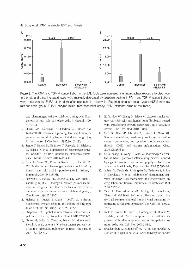
of tiplaxtinin (Figure 8). In the BAL fluid of rats, the
concentrations of PAI-1 and TGF-β were measured us-

Tuberculosis and Respiratory Diseases Vol. 70. No. 6, Jun. 2011
467
Figure 3. Western blot analysis of PAI-1, α-SMA and E-cadherin in rat ATII cells. Rat alveolar epithelial cells werestimulated with CSE (5%), TGF-β (5 ng/mL) for 72 hours and total cell lysates were taken for immunoblot. Both CSEand TGF-β induced EMT was again confirmed by increased α-SMA expression and decreased E-cadherin expression.CSE also increased the PAI-1 expression. Transfection of PAI-1 siRNA or addition of tiplaxtinin (50μg) to the rat ATIIcells reversed the CSE induced EMT and PAI-1 expression. Values given are the mean±SEM. Statistical analysis wasperformed using by Mann-Whitney U test. CSE: cigarette smoking extract; SEM: standard error of the mean.
ing the ELISA. The concentrations of PAI-1 and TGF-β,
both of which were increased by bleomycin, were de-
creased with the use of tiplaxtinin (Figure 9).
Discussion
The epithelial cells in the lung are the common target
sites for pulmonary damage. They play a key role in
the following clinical courses with no respect to wheth-
er a normal recovery is achieved or the fibrosis occurs.
In a phenomenon of the EMT where the epithelial cells
lose their own characteristics and are converted into the
mesenchymal cells, TGF-β is deeply involved. The re-
sulting activation of Smad and β-catenin is also asso-
ciated with the EMT12,13
. Idiopathic pulmonary fibrosis
(IPF) commonly occurs in smokers. But there are not
a great number of studies which have been conducted
to examine whether the smoking is associated with the
occurrence of pulmonary fibrosis, particularly, in rela-
tion to the EMT. It has been reported that the cigarette
smoking extract (CSE) triggers the occurrence of EMT
at a lower concentration in A549 cell lines and it is also
associated with the proliferation of myofibroblasts14.
These findings suggests that it can be involved in the
occurrence of idiopathic pulmonary fibrosis (IPF). In
addition, CSE increased the expression of PAI-1 in A549
epithelial cells and the increased PAI-1 increased the ex-
pression of IL-8 or LTB4. This might be associated with
the occurrence of inflammatory responses in patients
with chronic obstructive pulmonary disease15,16
. It is

JS Song et al: PAI-1 in alveolar EMT and fibrosis
468
Figure 5. Representative phase contrast images andimmunofluorescene stain-ing for PAI-1 (red) and α- SMA (green) expression with CSE, CSE plus PAI-1 siRNA transfection or ti-plaxtinin treatment in pri-mary rat type II epithelial cells. CSE induced α- SMA and PAI-1 expression in type II epithelial cells were inhibited either by PAI-1 siRNA transfection or PAI-1 specific inhibitor, tiplaxtinin (Original magnifi-cation, ×200). CSE: ciga-rette smoking extract.
Figure 4. Representative phase contrast images andimmunofluorescene stain-ing for E-cadherin (red) and α-SMA (green) expressionwith CSE, CSE plus PAI-1 siRNA transfection or ti-plaxtinin treatment in pri-mary rat type II epithelial cells; nuclei are stained with 4', 6-diamidino-2-phe-nylindole (blue). CSE in-duced EMT in type II epi-thelial cells were inhibited either by PAI-1 siRNA transfection or PAI-1 spe-cific inhibitor, tiplaxtinin (Ori-ginal magnification, ×200).CSE: cigarette smoking extract.
nowadays well established that the major functions of
PAI-1 are to suppress the fibrinolysis by plasmin and
thereby to contribute to the occurrence of pulmonary
and renal fibrosis. In the current experimental study,
however, when type II epithelial cells were isolated and
then cultured and they were exposed to the CSE, the
expression of PAI-1 was increased and this was directly
involved in the occurrence of EMT. It was also shown

Tuberculosis and Respiratory Diseases Vol. 70. No. 6, Jun. 2011
469
Figure 6. Effects of CSE and on TGF-β secretion of cul-tured rat ATII cells. Transfection of PAI-1 siRNA to the ATII cells or addition of tiplaxtinin (50μg) decreased the TGF-β concentrations in the supernatants of CSE-ex-posed rat ATII cells. CSE: cigarette smoking extract.
that the signal transduction pathway in this process acti-
vated the SNAIL via SMAD2 and ERK1/2 and type II epi-
thelial cells were converted into myofibroblasts accord-
ingly. In addition, with the use of tiplaxtinin that im-
pairs the fibrinolysis of PAI-1 due to the transfection
with PAI-1 siRNA or binding with PAI-1 in epithelial
cells17, the presence of this signal transduction pathway
was confirmed.
Of myriads of molecules that are involved in the EMT,
zinc-finger transcription factors such as SNAI1 (Snail)
and SNAI2 (Slug) play a role in regulating the EMT in
the diseases triggering the occurrence of cancer or fib-
rosis in the specific body organ18,19
. In many body or-
gans such as the lung, the most important cytokine that
is involved in the EMT, TGF-β1 plays a role in migrat-
ing Snail transcription factors into the nucleus and trig-
gering the occurrence of EMT in type II epithelial
cells20
. Also in the current experimental study, the CSE
increased the expression of Snail transcription factors in
type II epithelial cells. Following the transfection with
PAI-1 siRNA or pre-treatment with tiplaxtinin, the phos-
phorylation of SMAD-2 and the expression of Snail tran-
scription factor were suppressed. This was also accom-
panied by an EMT phenomenon that is characterized by
the decreased expression of α-SMA. A substantial num-
ber of studies have been conducted to examine whether
the suppressed effects of PAI-1 in type II epithelial cells
would also be found in an animal experimental model
of bleomycin-induced pulmonary fibrosis. According to
this, in rats which were orally given tiplaxtinin, as com-
pared with normal controls, the occurrence of pulmo-
nary fibrosis was significantly suppressed. Besides, the
concentration of TGF-β in the bronchoalveolar lavage
fluid was also decreased. When the occurrence of pul-
monary damage was induced by the treatment with
bleomycin in rats, the expression of such pro-coagulant
molecules such as PAI-1 or tissue factor was increased7.
In addition, the expression of PAI-1 gene arises from
reactive oxygen species that were increased by TGF-β
and TNF-α21
. Also in the lung of patients with idio-
pathic pulmonary fibrosis (IPF), as compared with nor-
mal healthy people, the concentration of PAI-1 was in-
creased22
. This implies that the fibrinolytic activity
would also be decreased due to the PAI-1-induced sup-
pression of tPA and uPA in the human body and this
would play a key role in the pathogenesis of pulmonary
fibrosis. As shown in the current results, however, PAI-1
promoted the EMT without suppressing PA. These re-
sults indicate that it might play a role in the pathophysi-
ology of pulmonary fibrosis.
Limitations of the current study are as follows:
1) It was demonstrated that the PAI-1 was directly in-
volved in the occurrence of CSE-induced EMT in type
II epithelial cells. But no studies have been conducted
to examine the PAI-1 receptors. There are no reports
about the PAI-1 receptors. In the current experimental
study, however, SMAD2 was phosphorylated by the CSE
and then suppressed by PAI-1 siRNA. This implies that
the TGF-β1 receptors might be shared.
2) It was not examined whether the EMT phenomen-
on would be suppressed by suppressing the expression
of Snail mRNA with the use of siRNA whose target was
Snail transcription factors in type II epithelial cells. In
A549 cells stimulated by TGF-β1 and type II alveolar
epithelial cells of mice, the expression of Snail mRNA
was increased20,23 and Snail transcription factors played
a key role in triggering the occurrence of TGF-β1-in-

JS Song et al: PAI-1 in alveolar EMT and fibrosis
470
Figure 7. CSE induced the ERK1/2, SMAD2 phosphorylation and Snail expression in cultured rat ATII cells. ATII cell lysates were subjected to western blotting using the pERK1/2, pSmad2 and Snail antibodies. Blots were analyzed bydensitometry. Transfection of PAI-1 siRNA to the ATII cells or addition of tiplaxtinin (50μg) decreased the CSE-inducedERK1/2, SMAD-2 phosphorylation and Snail expression. CSE: cigarette smoking extract.
duced EMT and the renal fibrosis24
. It can therefore be
inferred that the PAI-1-induced EMT, seen in the current
study, might undergo the signal transduction pathway
which is very similar to the TGF-β1-induced EMT.
3) The expression of SNAI2 (Slug) was not observed.
It has been known that SNAI2 as well as Snail plays
a key role in patients with TGF-β1-induced EMT or idi-
opathic pulmonary fibrosis (IPF) and an animal ex-
perimental model of bleomycin-induced pulmonary fib-
rosis20
. Also in the current study, SNAI2 might have
played a similar role to Snail transcription factor.
To summarize, CSE triggers the occurrence of EMT
where type II alveolar epithelial cells of rat origin are
converted into the myofibroblasts. This might be asso-
ciated with the phosphorylation of SMAD2 and ERK1/2
MAPK by the CSE-induced PAL-1 and the resulting intra-
cellular migration of Snail transcription factors. Besides,
there were such findings that the bleomycin-induced
pulmonary fibrosis was alleviated following the oral ad-
ministration of PAI-1 inhibitors in rats. This might also
due to the inhibitory effects of EMT to some extents.
Acknowledgement
The current work was financially supported by the
research funds from the Korean Society of Tuberculosis

Tuberculosis and Respiratory Diseases Vol. 70. No. 6, Jun. 2011
471
Figure 8. Representative photomicrographs of rat lung tissue stained with a hematoxylin-eosin in control (A), bleomycininjury (B) and tiplaxtinin plus bleomycin (C). Severe pulmonary inflammation and fibrosis occurred after bleomycin injuryon day 14 (B) and this histological change was markedly reduced by tiplaxtinin treatment (C). Lung fibrosis as evaluatedby Ashcroft score was markedly reduced by tiplaxtinin treatment after bleomycin injury. The Ashcroft score are mean±SEM from six rats for each group. SEM: standard error of the mean. All pictures were taken at ×100 magnifi-cation.
and the Korean Society of Pulmonology in 2010.
References
1. Selman M, King TE, Pardo A; American Thoracic
Society; European Respiratory Society; American
College of Chest Physicians. Idiopathic pulmonary fib-
rosis: prevailing and evolving hypotheses about its
pathogenesis and implications for therapy. Ann Intern
Med 2001;134:136-51.
2. Selman M, Pardo A. Role of epithelial cells in idiopathic
pulmonary fibrosis: from innocent targets to serial
killers. Proc Am Thorac Soc 2006;3:364-72.
3. Yao HW, Xie QM, Chen JQ, Deng YM, Tang HF.
TGF-beta1 induces alveolar epithelial to mesenchymal
transition in vitro. Life Sci 2004;76:29-37.
4. Loskutoff DJ, Quigley JP. PAI-1, fibrosis, and the elu-
sive provisional fibrin matrix. J Clin Invest 2000;106:
1441-3.
5. Fogo AB. Mesangial matrix modulation and glomer-
ulosclerosis. Exp Nephrol 1999;7:147-59.
6. Zhang LP, Takahara T, Yata Y, Furui K, Jin B, Kawada
N, et al. Increased expression of plasminogen activator
JS Song et al: PAI-1 in alveolar EMT and fibrosis
472
Figure 9. The PAI-1 and TGF-β concentration in the BAL fluids were increased after intra-tracheal exposure to bleomycinto the rats and these increased levels were markedly decreased by tiplaxtinin treatment. PAI-1 and TGF-β concentrationswere measured by ELISA at 14 days after exposure to bleomycin. Reported data are mean values±SEM from sixrats for each group. ELISA: enzyme-linked immunosorbent assay; SEM: standard error of the mean.
and plasminogen activator inhibitor during liver fibro-
genesis of rats: role of stellate cells. J Hepatol 1999;
31:703-11.
7. Olman MA, Mackman N, Gladson CL, Moser KM,
Loskutoff DJ. Changes in procoagulant and fibrinolytic
gene expression during bleomycin-induced lung injury
in the mouse. J Clin Invest 1995;96:1621-30.
8. Senoo T, Hattori N, Tanimoto T, Furonaka M, Ishikawa
N, Fujitaka K, et al. Suppression of plasminogen activa-
tor inhibitor-1 by RNA interference attenuates pulmo-
nary fibrosis. Thorax 2010;65:334-40.
9. Cho SH, Tam SW, Demissie-Sanders S, Filler SA, Oh
CK. Production of plasminogen activator inhibitor-1 by
human mast cells and its possible role in asthma. J
Immunol 2000;165:3154-61.
10. Eitzman DT, McCoy RD, Zheng X, Fay WP, Shen T,
Ginsburg D, et al. Bleomycin-induced pulmonary fib-
rosis in transgenic mice that either lack or overexpress
the murine plasminogen activator inhibitor-1 gene. J
Clin Invest 1996;97:232-7.
11. Richards RJ, Davies N, Atkins J, Oreffo VI. Isolation,
biochemical characterization, and culture of lung type
II cells of the rat. Lung 1987;165:143-58.
12. Chapman HA. Epithelial-mesenchymal interactions in
pulmonary fibrosis. Annu Rev Physiol 2011;73:413-35.
13. Chilosi M, Poletti V, Zamò A, Lestani M, Montagna L,
Piccoli P, et al. Aberrant Wnt/beta-catenin pathway ac-
tivation in idiopathic pulmonary fibrosis. Am J Pathol
2003;162:1495-502.
14. Liu Y, Gao W, Zhang D. Effects of cigarette smoke ex-
tract on A549 cells and human lung fibroblasts treated
with transforming growth factor-beta1 in a coculture
system. Clin Exp Med 2010;10:159-67.
15. Xiao W, Hsu YP, Ishizaka A, Kirikae T, Moss RB.
Sputum cathelicidin, urokinase plasminogen activation
system components, and cytokines discriminate cystic
fibrosis, COPD, and asthma inflammation. Chest
2005;128:2316-26.
16. Xu X, Wang H, Wang Z, Xiao W. Plasminogen activa-
tor inhibitor-1 promotes inflammatory process induced
by cigarette smoke extraction or lipopolysaccharides in
alveolar epithelial cells. Exp Lung Res 2009;35:795-805.
17. Izuhara Y, Takahashi S, Nangaku M, Takizawa S, Ishida
H, Kurokawa K, et al. Inhibition of plasminogen acti-
vator inhibitor-1: its mechanism and effectiveness on
coagulation and fibrosis. Arterioscler Thromb Vasc Biol
2008;28:672-7.
18. Cano A, Pérez-Moreno MA, Rodrigo I, Locascio A,
Blanco MJ, del Barrio MG, et al. The transcription fac-
tor snail controls epithelial-mesenchymal transitions by
repressing E-cadherin expression. Nat Cell Biol 2000;2:
76-83.
19. Batlle E, Sancho E, Francí C, Domínguez D, Monfar M,
Baulida J, et al. The transcription factor snail is a re-
pressor of E-cadherin gene expression in epithelial tu-
mour cells. Nat Cell Biol 2000;2:84-9.
20. Jayachandran A, Königshoff M, Yu H, Rupniewska E,
Hecker M, Klepetko W, et al. SNAI transcription factors

Tuberculosis and Respiratory Diseases Vol. 70. No. 6, Jun. 2011
473
mediate epithelial-mesenchymal transition in lung
fibrosis. Thorax 2009;64:1053-61.
21. Liu RM. Oxidative stress, plasminogen activator in-
hibitor 1, and lung fibrosis. Antioxid Redox Signal
2008;10:303-19.
22. Kotani I, Sato A, Hayakawa H, Urano T, Takada Y,
Takada A. Increased procoagulant and antifibrinolytic
activities in the lungs with idiopathic pulmonary
fibrosis. Thromb Res 1995;77:493-504.
23. Kim JH, Jang YS, Eom KS, Hwang YI, Kang HR, Jang
SH, et al. Transtorming growth factor beta1 induces
epithelial-to-mesenchymal transition of A549 cells. J
Korean Med Sci 2007;22:898-904.
24. Lange-Sperandio B, Trautmann A, Eickelberg O,
Jayachandran A, Oberle S, Schmidutz F, et al.
Leukocytes induce epithelial to mesenchymal transition
after unilateral ureteral obstruction in neonatal mice.
Am J Pathol 2007;171:861-71.