inhibition of class i histone deacetylases abrogates tumor...
TRANSCRIPT

1521-0111/94/2/793–801$35.00 https://doi.org/10.1124/mol.117.110924MOLECULAR PHARMACOLOGY Mol Pharmacol 94:793–801, August 2018Copyright ª 2018 by The American Society for Pharmacology and Experimental Therapeutics
Inhibition of Class I Histone Deacetylases Abrogates TumorGrowth Factor b Expression and Development of Fibrosis duringChronic Pancreatitis s
Marta Bombardo, Rong Chen, Ermanno Malagola, Enrica Saponara, Andrew P. Hills,Rolf Graf, and Sabrina Sonda1
Swiss Hepato-Pancreato-Biliary Center, Department of Visceral and Transplantation Surgery, University Hospital (M.B., R.C.,E.M., E.S., R.G., S.S.) and Center for Integrative Human Physiology (ZIHP), University of Zurich (R.G., S.S.), Zurich, Switzerland;and School of Health Sciences, College of Health and Medicine, University of Tasmania, Launceston, Australia (A.P.H., S.S.)
Received October 23, 2017; accepted May 31, 2018
ABSTRACTPancreatic fibrosis is the hallmark of chronic pancreatitis, ahighly debilitating disease for which there is currently no cure.The key event at the basis of pancreatic fibrosis is the depositionof extracellular matrix proteins by activated pancreatic stellatecells (PSCs). Transforming growth factor b (TGFb) is a potentprofibrotic factor in the pancreas as it promotes the activation ofPSC; thus, pharmacologic interventions that effectively re-duce TGFb expression harbor considerable therapeutic poten-tial in the treatment of chronic pancreatitis. In this study, weinvestigated whether TGFb expression is reduced by pharma-cologic inhibition of the epigenetic modifiers histone deacety-lases (HDACs). To address this aim, chronic pancreatitis wasinduced in C57BL/6 mice with serial injections of cerulein, and
the selective class 1 HDAC inhibitor MS-275 was administeredin vivo in a preventive and therapeutic manner. Both MS-275regimens potently reduced deposition of extracellular matrix anddevelopment of fibrosis in the pancreas after 4 weeks of chronicpancreatitis. Reduced pancreatic fibrosis was concomitant withlower expression of pancreatic TGFb and consequent reducedPSC activation. In search of the cell types targeted by theinhibitor, we found that MS-275 treatment abrogated theexpression of TGFb in acinar cells stimulated by ceruleintreatment. Our study demonstrates that MS-275 is an effec-tive antifibrotic agent in the context of experimental chronicpancreatitis and thus may constitute a valid therapeuticintervention for this severe disease.
IntroductionChronic pancreatitis is defined as a progressive inflamma-
tion of the pancreas, resulting in development of organfibrosis, which is at the core of the disease pathophysiol-ogy. This progressive condition is characterized by irregularsclerosis with focal, segmental, or diffuse destruction of theparenchyma. Consequently, gradual loss of exocrine andendocrine cellular components leads to pancreatic insuffi-ciency and eventually diabetes, which are associated withconsiderable morbidity, reduction of quality of life, and re-duction of life expectancy (reviewed inDiMagno andDiMagno,2013, 2016).Fibrosis is characterized by excessive production and de-
position of extracellular matrix (ECM) components in thepancreatic parenchyma, produced mainly by resident pancre-atic stellate cells (PSCs). In response to organ injury,
profibrogenic factors are released and activate PSCs, a processcharacterized by phenotypical cell alteration, proliferation, andECM protein synthesis.Despite advances in chronic pancreatitis research, to date,
the complex cellular and signaling mechanisms that drive thefibrotic process are not yet completely elucidated. This limitedknowledge explains why therapeutic approaches to counteractthe development of organ fibrosis are not currently availableand management of chronic pancreatitis remains a clinicalchallenge.In this study, we evaluated whether administration of
MS-275 (also known as entinostat), a selective inhibitor ofclass 1 histone deacetylases (HDACs), counteracts the devel-opment of pancreatic fibrosis using the widespread murinemodel of cerulein-induced chronic pancreatitis. The rationalefor this approach was 3-fold: 1) development of fibrosisactivates a substantial gene regulation, which is prominentlyorchestrated by epigenetic mechanisms (McDonnell et al.,2014; Weigel et al., 2015; Yang and Schwartz, 2015; Moran-Salvador and Mann, 2017); 2) HDACs are critical epigeneticregulators, and expression of class 1 HDACs is significantlyupregulated during the course of chronic pancreatitis(Bombardo et al., 2017); and 3) pharmacologic inhibitors of
This work received funding from the Swiss National Science Foundation (grantno. 310030 -146725) and the Amélie Waring Foundation.
1Current affiliation: School of Health Sciences, College of Health andMedicine, University of Tasmania, Newnham Campus, Launceston, Australia.
https://doi.org/10.1124/mol.117.110924.s This article has supplemental material available at molpharm.
aspetjournals.org.
ABBREVIATIONS: a-SMA, a-smooth muscle actin; ECM, extracellular matrix; HDAC, histone deacetylase; PCR, polymerase chain reaction; PSC,pancreatic stellate cell; TGFb, transforming growth factor b.
793
http://molpharm.aspetjournals.org/content/suppl/2018/06/07/mol.117.110924.DC1Supplemental material to this article can be found at:
at ASPE
T Journals on A
pril 1, 2020m
olpharm.aspetjournals.org
Dow
nloaded from

HDAC activity, originally developed as anticancer agents, arecurrently being investigated for their antifibrotic properties indifferent fibrotic diseases (recently reviewed in Pang andZhuang, 2010; Royce et al., 2014; Chen et al., 2015; Schuetzeet al., 2016).
Materials and MethodsAnimal Experiments. All animal treatments were performed in
accordancewith Swiss federal animal regulations and approved by thecantonal veterinary office of Zurich. All studies involving animalswere carried out in accordance with the Guide for the Care and Use ofLaboratory Animals as adopted and promulgated by theU.S. NationalInstitutes of Health. Mice used in this study were adult 8- to 10-week-old wild-type C57BL/6 mice in a weight range of 25–30 g (EnvigoLaboratories, Horst, The Netherlands). Animals were kept understandardized conditions under 12-hour light/dark cycles, with foodand water available ad libitum. Groups of 4 to 5 mice were kept instandard individually ventilated cages in a specific pathogen-freefacility. Food and water were provided ad libitum. Only male micewere used in this study.
Chronic pancreatitis was induced via six intraperitoneal injectionsof cerulein (50 mg/kg) administered hourly every 2nd day for up to6 weeks. Control animals received 0.9% NaCl injections. MS-275(Selleckchem,Houston, TX)was injected intraperitoneally at 20mg/kgevery 2nd day for 2 weeks, starting concomitantly (preventiveregimen) or 1 week after the beginning of cerulein injections (thera-peutic regimen). The concentration of MS-275 was chosen based onpreviously published in vivo studies using the inhibitor in mice(Dalgard et al., 2008; Nguyên et al., 2008; Murphy et al., 2014;Bombardo et al., 2017). Control animals received 10% dimethylsulf-oxide injections. Intraperitoneal injections were alternated dailybetween the left and right sides of the abdomen. Mice were examinedthroughout the development of pancreatitis, and their health statuswas recorded every 2nd day on a score sheet. Pancreatitis models usedin this study generated only a mild form of the disease. Animal weightloss did not exceed 10%, and no mortality was observed. After deepterminal anesthesia with isoflurane, micewere euthanized via cardiacpuncture exsanguination. Groups of five animals were tested for eachexperiment. Animals were assigned randomly to different experimen-tal groups for all in vivo studies. Data collection and evaluation of allin vivo and in vitro experiments were performed blinded to groupidentity.
Mammalian Cell Cultures. Cell culture reagents were fromGibco-BRL. Rat AR42J cells were maintained in Kaighn’s modifiedHam’s F-12 mediumwith 20% fetal bovine serum, supplemented with50 U/ml penicillin and 50 mg/ml streptomycin, and maintained atstable condition of 37°C in a 5% CO2 atmosphere. Cells were seeded insix-well plates, stimulated with 10 nM cerulein for 4 hours, and lysedin the plates for RNA extraction and real-time polymerase chainreaction (PCR) analysis.
Primary acini were isolated according to Algül et al. (2007) from6-week-old Wistar male rats from Charles River, Deutschland. Aciniwere preincubated with 1 mM MS-275 for 30 minutes and stimulatedwith 0.1 nM cerulein for 30 minutes in the presence of 1 mM MS-275.At the end of the treatment, cells were lysed in the plates for RNAextraction and real-time PCR analysis.
Immunohistochemistry. Pancreas specimens were embeddedin paraffin for histologic analyses, as previously described (Silvaet al., 2011). H&E and Masson’s trichrome staining were per-formed according to routine procedures. Microscopy analyseswere performed on a wide-field Nikon Eclipse Ti (Amsterdam,The Netherlands). Quantification of labeled cells was performedin at least 10 randomly selected high-power fields (�200) per slideusing the NIS Elements BR Analysis (Nikon, Amsterdam, TheNetherlands) and Cell^P analysis software (Olympus, Tokyo,Japan).
Western Blotting. Twenty milligrams of pancreatic tissue washomogenized in radioimmunopreciptation (RIPA) assay buffer con-taining a protease inhibitor cocktail (Roche Diagnostics, Mannheim,Germany). Protein concentrations were determined by a Bradfordprotein assay (BioRad, Hercules, CA). Twenty micrograms of proteinswas resolved by SDS-PAGE electrophoresis and blotted onto nitrocel-lulose membranes using a V3 Western Workflow system (BioRad)according to the manufacturer’s protocols.
Membranes were incubated with primary antibodies overnight at4°C. Primary antibodies used in this studywere: mouse anti-a-smoothmuscle actin (a-SMA, Dako, Glostrup, Denmark); rabbit anti a-tubulin(ab52894; Abcam, Cambridge, UK); rabbit anti-phospho-Smad3(Ser423/425) (Cell Signaling, Danvers, MA); and rabbit anti-GAPDH(Santa Cruz Biotechnology, Dallas, TX).
Nuclear Protein Extraction and HDAC Activity. Nuclearproteins were extracted from 20 mg of pancreatic tissue with theEpiQuik Nuclear Extraction Kit (Epigentek Group Inc, MountainView, CA), and HDAC activity was measured in the nuclear extractswith the fluorimetric EpiQuik HDAC activity/inhibition assay kit(Epigentek Group Inc.), according to the manufacturer’s instructions.
Transcript Analyses. Total RNA was extracted from pancre-atic tissue and acinar explants as described previously (Graf et al.,2002) and reverse-transcribed with qScript cDNA SuperMix(Quanta Biosciences, Beverly, MA). Gene expression wasmeasuredby real-time PCR on a 7500 Fast Real-Time PCR System (AppliedBiosystems, Carlsbad, CA) using Taqman probes (Applied Biosys-tems). Transcript levels were normalized using 18S RNA as areference and expressed as DDCt relative to the value of controlanimals or as DCt.
Statistical Analyses. Every group of mice compared in thedifferent experimental conditions was comprised 5 animals. Dataare expressed as means 6 standard deviation. Population character-isticswere compared among treatment groups using an unpaired, two-tailed Student’s t test when comparing two experimental conditionsor one-way analysis of variance, followed by Dunnett’s post-hoc testwhen comparing more than two experimental conditions. Holm-Bonferroni correction for multiple comparisons was used to keepthe family-wise error rate of dependent variables at 5%. Analyseswere performed using GraphPad Prism 4.0c (GraphPad Software,Inc., San Diego, CA).
ResultsDevelopment of Fibrotic Response during Chronic
Pancreatitis Correlates with Increased Levels ofHDAC Expression. To investigate the role of HDAC in thedevelopment of pancreatic fibrosis after induction of chronicpancreatitis, we first performed a time-course analysis afterinduction of the disease to determine the kinetics of thefibrotic response. Histologic evaluation of mice harvestedafter 2, 4, and 6 weeks of cerulein treatment revealed pro-gressive damage of pancreatic parenchyma and cell infiltra-tion (Fig. 1A) and pronounced ECM deposition (Fig. 1B).Quantification of fibrotic parameters showed increased ex-pression of collagen isoforms (Fig. 1C) and collagen depositionin the pancreas (Fig. 1D). Development of pancreatic fibrosis ismediated by activated pancreatic stellate cells (PSCs), whichconstitute the predominant source of ECM proteins, includingcollagens and fibronectin. Activation of PSCs, detected bya-smooth muscle actin (a-SMA) expression, reached a maxi-mum after 4 weeks of pancreatitis (Fig. 1E), thus mirroringthe kinetics of collagen expression. Expression of profibroticTGFb isoforms, the main activators of PSC, and TGFbreceptor II, critical for the development of pancreatic fibrosis(Yoo et al., 2005), also increased in a similar pattern during
794 Bombardo et al.
at ASPE
T Journals on A
pril 1, 2020m
olpharm.aspetjournals.org
Dow
nloaded from

the development of organ fibrosis (Fig. 1F). In addition,pancreatic expression of inflammatory components followedsimilar kinetics (Supplemental Fig. 1) (Bombardo et al., 2017).Based on these results showingmaximal levels of fibrosis after4 weeks of pancreatitis, we focused on this time point forfurther analyses.
Inhibition of Class 1 HDAC with MS-275 Reduces theDevelopment of Fibrosis after Induction of ChronicPancreatitis. We recently showed that gene expressionlevels of class 1 HDACs were upregulated during chronicpancreatitis (Bombardo et al., 2017). This finding was con-firmed by increased HDAC enzymatic activity (Fig. 2A) in
Fig. 1. Characterization of chronic pancreatitis development. (A) H&E and (B) Masson’s trichrome staining of pancreata at the indicated weeks ofpancreatitis induction revealed progressive destruction of parenchymal morphology, cell infiltration, and collagen deposition (green staining). (C)Quantitative PCR (qPCR) of collagen 1 and 3 expression in the pancreas at the indicated weeks of pancreatitis. (D) Quantification of collagen deposition,based on Masson’s trichrome staining, at the indicated weeks of pancreatitis. The amount of collagen is expressed as the percentage of total pancreaticarea. (E) qPCR of a-SMA expression in the pancreas at the indicated weeks of pancreatitis, indicative of pancreatic stellate cell activation. (F) qPCR ofTGFb isoforms and TGFb receptor 2 (TGFbR2) expression in the pancreas at the indicated weeks of pancreatitis. Results are average6 S.D. (n = 5), *P,0.05. Scale bar, 50 mm.
MS-275 Ameliorates Chronic Pancreatic Fibrosis 795
at ASPE
T Journals on A
pril 1, 2020m
olpharm.aspetjournals.org
Dow
nloaded from

Fig. 2. Preventive and therapeutic administration of MS-275 reduces the development of fibrosis during chronic pancreatitis. (A) Total HDAC activitydetected in pancreatic nuclear extract in control and mice treated with cerulein (Cer) for 4 weeks mice. (B) Schematic representation of 2 weeks ofpreventive (MS + Cer) and therapeutic (Cer + MS) MS-275 regimens during induction of chronic pancreatitis. Cer was administered on alternate daysover 4 weeks. MS-275 was administered on alternate days over 2 weeks. (C) H&E staining of pancreata after 4 weeks of chronic pancreatitis afterpreventive and therapeutic MS-275 regimens. (D) Masson’s trichrome staining of pancreata showing reduced collagen deposition (green) after 4 weeks of
796 Bombardo et al.
at ASPE
T Journals on A
pril 1, 2020m
olpharm.aspetjournals.org
Dow
nloaded from
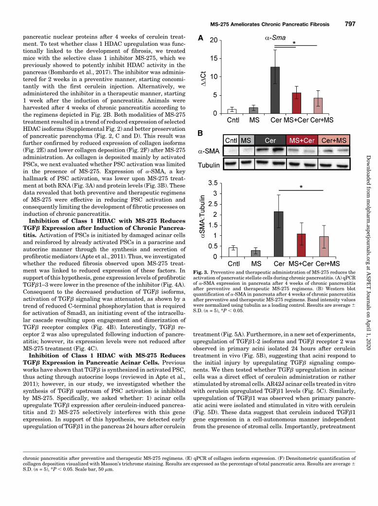
pancreatic nuclear proteins after 4 weeks of cerulein treat-ment. To test whether class 1 HDAC upregulation was func-tionally linked to the development of fibrosis, we treatedmice with the selective class 1 inhibitor MS-275, which wepreviously showed to potently inhibit HDAC activity in thepancreas (Bombardo et al., 2017). The inhibitor was adminis-tered for 2 weeks in a preventive manner, starting concomi-tantly with the first cerulein injection. Alternatively, weadministered the inhibitor in a therapeutic manner, starting1 week after the induction of pancreatitis. Animals wereharvested after 4 weeks of chronic pancreatitis according tothe regimens depicted in Fig. 2B. Both modalities of MS-275treatment resulted in a trend of reduced expression of selectedHDAC isoforms (Supplemental Fig. 2) and better preservationof pancreatic parenchyma (Fig. 2, C and D). This result wasfurther confirmed by reduced expression of collagen isoforms(Fig. 2E) and lower collagen deposition (Fig. 2F) after MS-275administration. As collagen is deposited mainly by activatedPSCs, we next evaluated whether PSC activation was limitedin the presence of MS-275. Expression of a-SMA, a keyhallmark of PSC activation, was lower upon MS-275 treat-ment at both RNA (Fig. 3A) and protein levels (Fig. 3B). Thesedata revealed that both preventive and therapeutic regimensof MS-275 were effective in reducing PSC activation andconsequently limiting the development of fibrotic processes oninduction of chronic pancreatitis.Inhibition of Class 1 HDAC with MS-275 Reduces
TGFb Expression after Induction of Chronic Pancrea-titis. Activation of PSCs is initiated by damaged acinar cellsand reinforced by already activated PSCs in a paracrine andautocrine manner through the synthesis and secretion ofprofibroticmediators (Apte et al., 2011). Thus, we investigatedwhether the reduced fibrosis observed upon MS-275 treat-ment was linked to reduced expression of these factors. Insupport of this hypothesis, gene expression levels of profibroticTGFb1–3 were lower in the presence of the inhibitor (Fig. 4A).Consequent to the decreased production of TGFb isoforms,activation of TGFb signaling was attenuated, as shown by atrend of reduced C-terminal phosphorylation that is requiredfor activation of Smad3, an initiating event of the intracellu-lar cascade resulting upon engagement and dimerization ofTGFb receptor complex (Fig. 4B). Interestingly, TGFb re-ceptor 2 was also upregulated following induction of pancre-atitis; however, its expression levels were not reduced afterMS-275 treatment (Fig. 4C).Inhibition of Class 1 HDAC with MS-275 Reduces
TGFb Expression in Pancreatic Acinar Cells. Previousworks have shown that TGFb is synthesized in activated PSC,thus acting through autocrine loops (reviewed in Apte et al.,2011); however, in our study, we investigated whether thesynthesis of TGFb upstream of PSC activation is inhibitedby MS-275. Specifically, we asked whether: 1) acinar cellsupregulate TGFb expression after cerulein-induced pancrea-titis and 2) MS-275 selectively interferes with this geneexpression. In support of this hypothesis, we detected earlyupregulation of TGFb1 in the pancreas 24 hours after cerulein
treatment (Fig. 5A). Furthermore, in a new set of experiments,upregulation of TGFb1-2 isoforms and TGFb receptor 2 wasobserved in primary acini isolated 24 hours after ceruleintreatment in vivo (Fig. 5B), suggesting that acini respond tothe initial injury by upregulating TGFb signaling compo-nents. We then tested whether TGFb upregulation in acinarcells was a direct effect of cerulein administration or ratherstimulated by stromal cells. AR42J acinar cells treated in vitrowith cerulein upregulated TGFb1 levels (Fig. 5C). Similarly,upregulation of TGFb1 was observed when primary pancre-atic acini were isolated and stimulated in vitro with cerulein(Fig. 5D). These data suggest that cerulein induced TGFb1gene expression in a cell-autonomous manner independentfrom the presence of stromal cells. Importantly, pretreatment
Fig. 3. Preventive and therapeutic administration of MS-275 reduces theactivation of pancreatic stellate cells during chronic pancreatitis. (A) qPCRof a-SMA expression in pancreata after 4 weeks of chronic pancreatitisafter preventive and therapeutic MS-275 regimens. (B) Western blotquantification of a-SMA in pancreata after 4 weeks of chronic pancreatitisafter preventive and therapeutic MS-275 regimens. Band intensity valueswere normalized using tubulin as a loading control. Results are average6S.D. (n = 5), *P , 0.05.
chronic pancreatitis after preventive and therapeutic MS-275 regimens. (E) qPCR of collagen isoform expression. (F) Densitometric quantification ofcollagen deposition visualized with Masson’s trichrome staining. Results are expressed as the percentage of total pancreatic area. Results are average6S.D. (n = 5), *P , 0.05. Scale bar, 50 mm.
MS-275 Ameliorates Chronic Pancreatic Fibrosis 797
at ASPE
T Journals on A
pril 1, 2020m
olpharm.aspetjournals.org
Dow
nloaded from

with the HDAC inhibitor MS-275 abrogated TGFb1 inductionin both AR42J cells and primary acini (Fig. 5, C and D).Furthermore, cerulein treatment increased expression of andHDAC1, but not HDAC2 and 3, in isolated acini (Fig. 5E),further suggesting that TGFb1 expression in acinar cells ispromoted by HDAC activity. Expression of TGFb2 was muchlower than TGFb1 and not regulated in this experimentalsetting in both cell types (Supplemental Fig. 3, A and B).
DiscussionTGFb is a potent fibrogenic factor that plays a pivotal role in
the development of fibrosis during chronic pancreatitis(Menke et al., 1997; Yoo et al., 2005; He et al., 2009; Li et al.,2016). One of the main effects exerted by TGFb is activation ofPSCs from a quiescent state to amyofibroblast-like phenotype(reviewed in Apte et al., 2011). In recent years, activated PSCshave attracted increasing attention as major mediators ofpancreatic fibrosis during chronic pancreatitis as they not onlymediate the development of fibrosis by producing ECMproteins, but they also amplify the fibrotic response in anautocrine and paracrine manner by secreting fibrogenicfactors, including TGFb (Kruse et al., 2000). In the presentstudy, we discovered that activity of HDACs in the pancreas isfunctionally linked to the development of fibrosis duringchronic pancreatitis, thus providing a potential therapeutictarget to counteract this disease. This hypothesis was further
tested in in vivo experiments where the selective inhibitor ofclass 1 HDACs MS-275 was administered in a preventive ortherapeutic manner during chronic pancreatitis. In bothregimen types, we observed a striking inhibition of pancreaticfibrosis and increased preservation of pancreatic parenchyma,suggesting that MS-275 exerts an antifibrotic effect, evenwhen administered after commencement of the disease.At the cellular level, reduced fibrosis detected upon MS-275
treatment was likely the result of reduced TGFb expression,leading to reduced activation of PSCs. An important questionarising from these data is the identity of the cells whose TGFbproduction is targeted by the inhibitor. Using in vitro exper-iments with isolated acinar cells, we found that short-termincubation with cerulein was sufficient to stimulate TGFbexpression in these cells. The fact that MS-275 treatmentpotently reduced cerulein-stimulated TGFb expression sug-gests that acinar cells are indeed a direct target of theinhibitor and contribute to the phenotype observed in vivo.In this regard, it would be important to explore further thetemporal regulation of TGFb isoform expression in acinar cellsto dissect the dynamic of their contribution to the developmentof pancreatic fibrosis.It is known that acinar cells are not the only source of TGFb
in the pancreas, as previous studies reported the presence ofTGFb1mRNA in stromal cells, including PSC, upon inductionof pancreatitis (Muller-Pillasch et al., 1999). In this regard, itis important to mention that TGFb synthesis in nonacinar
Fig. 4. Preventive and therapeutic administration of MS-275 reduces the expression of TGFb during chronic pancreatitis. (A) qPCR of TGFb isoformsexpression after 4 weeks of chronic pancreatitis after preventive and therapeutic MS-275 regimens. (B) Western blotting quantification of phospho-SMAD3 in pancreata after 4 weeks of chronic pancreatitis after preventive and therapeutic MS-275 regimens. Band intensity values were normalizedusing Gapdh as a loading control (Cntl). (C) qPCR of TGFb receptor 2 (TGFbR2) expression in pancreata after 4 weeks of chronic pancreatitis afterpreventive and therapeutic MS-275 regimens. Results are average 6 S.D. (n = 5), *P , 0.05.
798 Bombardo et al.
at ASPE
T Journals on A
pril 1, 2020m
olpharm.aspetjournals.org
Dow
nloaded from

cells may also depend on HDAC activity. In fact, treatmentof isolated PSCs with the pan-HDAC inhibitor sodiumvalproate inhibits TGFb expression and collagen synthesisin these cells (Bülow et al., 2007). Furthermore, anotherpossible source of TGFb production is inflammatory cells,which are recruited to the pancreas during the developmentof pancreatitis. This is particularly interesting as we re-cently demonstrated that MS-275 treatment effectivelyreduced the levels of inflammation during the course ofacute and chronic pancreatitis (Bombardo et al., 2017).Crosstalk between PSCs and distinct leukocyte popula-tions, including macrophages, promotes PSC activationand fibrosis during chronic pancreatitis (Xue et al., 2015).Future studies using coculture of acinar cells, PSC, andleukocytes are warranted to dissect the contribution of theindividual cell types in the production of TGFb upontreatment with MS-275 and the effect on PSC activation.
Although it is possible that reduced inflammation uponMS-275 administration leads to reduced fibrosis duringchronic pancreatitis, a recent study revealed that develop-ment of inflammation and fibrosis is two independent and,accordingly, not causal events in this disease. Specifically,using transgenic mice deficient in Cxcr2, the authors observedalmost complete ablation of inflammatory cell infiltrationupon chronic pancreatitis; however, this limited inflammatoryreaction did not prevent PSC activation; consequently, fibrosislevels were comparable in transgenic and wild-type controlmice (Steele et al., 2015). This striking example implies thatsignaling molecules derived from inflammatory cells may playa minor role in the development of pancreatic fibrosis duringchronic pancreatitis.Collectively, our results integrate with the current body of
evidence demonstrating the crucial role of HDACs in thedevelopment of fibrotic diseases. In this regard, compelling
Fig. 5. MS-275 administration inhibits TGFb expression in isolated acinar cells. (A) qPCR of TGFb isoforms and TGFb receptor 2 (TGFbR2) expressionin the whole pancreas 24 hours after cerulein treatment. (B) qPCR of TGFb isoforms and TGFb receptor 2 (TGFbR2) expression in primary acini isolated24 hours after in vivo cerulein treatment. Right panel: Micrograph of isolated pancreatic acini. (C) qPCR of TGFb1 expression in AR42J acinar cells uponcerulein stimulation. Right panel: Micrograph of AR42J cells. (D) qPCR of TGFb1 expression in isolated acinar cells upon in vitro treatment with ceruleinin the presence or absence of MS-275. (E) qPCR of class 1 HDAC expression in isolated acinar cells treated in vitro with cerulein. Results are average6S.D. (n = 5 to 6), *P , 0.05.
MS-275 Ameliorates Chronic Pancreatic Fibrosis 799
at ASPE
T Journals on A
pril 1, 2020m
olpharm.aspetjournals.org
Dow
nloaded from

evidence demonstrates that HDAC activity is necessary foractivation of hepatic stellate cells in vitro (reviewed in Chenet al., 2015). The requirement of HDAC activity in drivingmyofibroblastic differentiation and ECM protein synthesiswas also observed in different fibroblast populations presentin skin, lung, and kidney (Glenisson et al., 2007; Yoshikawaet al., 2007; Guo et al., 2009). Moreover, the use of differentHDAC inhibitors showed beneficial effects in the treatmentof hepatic, renal, cardiac, and pulmonary fibrosis in vivo(Kee et al., 2013; Liu et al., 2013; Van Beneden et al., 2013;Khan and Jena, 2014; Nural-Guvener et al., 2014; Chenet al., 2015; Choi et al., 2015; Korfei et al., 2015). Thisfinding suggests that epigenetic mechanisms controlled byHDACs may be conserved in the development of differentfibrotic diseases.Conclusion. Counteracting the development of pancre-
atic fibrosis is a major and elusive therapeutic goal in thecontext of chronic pancreatitis. Our data revealed a potentantifibrotic effect of MS-275 treatment, which is mediatedat least in part by suppression of TGFb expression in acinarcells; however, it is possible that downregulation of addi-tional factors contributes to the observed phenotype. In thiscontext, it is worth mentioning that pancreatic expressionof interleukins-1 and -6, interleukins known to promoteautocrine and paracrine activation of PSC (Bynigeri et al.,2017), was reduced upon MS-275 treatment (Bombardoet al., 2017).Collectively, our data suggest on one hand that class
1 HDAC activity is critical for the timely controlled epigeneticregulation of key signaling molecules driving the developmentof fibrosis in this organ. On the other hand, our data provide anew perspective on the cell types involved in regulating theprocess and highlights the possibility that acinar cells act asactive mediators of pancreatic fibrosis.These results harbor important implications to explore
further the therapeutic potential of MS-275 in the context ofchronic pancreatitis patients. Additional studies that includeexperimental models with increased severity of pancreatitisand autoimmune pancreatitis are warranted to define theeffect of MS-275 in a broader spectrum of disease manifesta-tions. Moreover, investigations using conditional knockoutmousemodels are needed to achieve a global understanding ofthe individual HDAC isoforms’ functions in the different celltypes that are involved in the development of this disease.
Acknowledgments
We thank Theresia Reding and Udo Ungetuem for invaluablesupport and technical assistance.
Authorship Contributions
Participated in research design: Bombardo, Graf, Sonda.Conducted experiments: Bombardo, Chen, Malagola, Saponara.Performed data analysis: Bombardo, Chen, Malagola, Saponara.Wrote or contributed to the writing of the manuscript: Bombardo,
Hills, Graf, Sonda.
References
Algül H, Wagner M, Lesina M, and Schmid RM (2007) Overexpression of ErbB2 inthe exocrine pancreas induces an inflammatory response but not increased pro-liferation. Int J Cancer 121:1410–1416.
Apte M, Pirola R, and Wilson J (2011) The fibrosis of chronic pancreatitis: newinsights into the role of pancreatic stellate cells. Antioxid Redox Signal 15:2711–2722.
Bombardo M, Saponara E, Malagola E, Chen R, Seleznik GM, Haumaitre C, Qui-lichini E, Zabel A, Reding T, Graf R, et al. (2017) Class I histone deacetylase
inhibition improves pancreatitis outcome by limiting leukocyte recruitment andacinar-to-ductal metaplasia. Br J Pharmacol 174:3865–3880.
Bülow R, Fitzner B, Sparmann G, Emmrich J, Liebe S, and Jaster R (2007) Anti-fibrogenic effects of histone deacetylase inhibitors on pancreatic stellate cells.Biochem Pharmacol 74:1747–1757.
Bynigeri RR, Jakkampudi A, Jangala R, Subramanyam C, Sasikala M, Rao GV,Reddy DN, and Talukdar R (2017) Pancreatic stellate cell: Pandora’s box forpancreatic disease biology. World J Gastroenterol 23:382–405.
Chen PJ, Huang C, Meng XM, and Li J (2015) Epigenetic modifications by histonedeacetylases: biological implications and therapeutic potential in liver fibrosis.Biochimie 116:61–69.
Choi SY, Ryu Y, Kee HJ, Cho SN, Kim GR, Cho JY, Kim HS, Kim IK, and JeongMH (2015) Tubastatin A suppresses renal fibrosis via regulation of epigenetichistone modification and Smad3-dependent fibrotic genes. Vascul Pharmacol72:130–140.
Dalgard CL, Van Quill KR, and O’Brien JM (2008) Evaluation of the in vitro andin vivo antitumor activity of histone deacetylase inhibitors for the therapy of ret-inoblastoma. Clin Cancer Res 14:3113–3123.
DiMagno EP and DiMagno MJ (2016) Chronic pancreatitis: landmark papers, man-agement decisions, and future. Pancreas 45:641–650.
DiMagno MJ and DiMagno EP (2013) Chronic pancreatitis. Curr Opin Gastroenterol29:531–536.
Glenisson W, Castronovo V, and Waltregny D (2007) Histone deacetylase 4 is re-quired for TGFbeta1-induced myofibroblastic differentiation. Biochim BiophysActa 1773:1572–1582.
Graf R, Schiesser M, Lüssi A, Went P, Scheele GA, and Bimmler D (2002) Coordinateregulation of secretory stress proteins (PSP/reg, PAP I, PAP II, and PAP III) in therat exocrine pancreas during experimental acute pancreatitis. J Surg Res 105:136–144.
Guo W, Shan B, Klingsberg RC, Qin X, and Lasky JA (2009) Abrogation of TGF-beta1-induced fibroblast-myofibroblast differentiation by histone deacetylase in-hibition. Am J Physiol Lung Cell Mol Physiol 297:L864–L870.
He J, Sun X, Qian KQ, Liu X, Wang Z, and Chen Y (2009) Protection of cerulein-induced pancreatic fibrosis by pancreas-specific expression of Smad7. BiochimBiophys Acta 1792:56–60.
Kee HJ, Bae EH, Park S, Lee KE, Suh SH, Kim SW, and Jeong MH (2013) HDACinhibition suppresses cardiac hypertrophy and fibrosis in DOCA-salt hypertensiverats via regulation of HDAC6/HDAC8 enzyme activity. Kidney Blood Press Res 37:229–239.
Khan S and Jena G (2014) Sodium butyrate, a HDAC inhibitor ameliorates eNOS,iNOS and TGF-b1-induced fibrogenesis, apoptosis and DNA damage in the kidneyof juvenile diabetic rats. Food Chem Toxicol 73:127–139.
Korfei M, Skwarna S, Henneke I, MacKenzie B, Klymenko O, Saito S, Ruppert C, vonder Beck D, Mahavadi P, Klepetko W, et al. (2015) Aberrant expression and ac-tivity of histone deacetylases in sporadic idiopathic pulmonary fibrosis. Thorax 70:1022–1032.
Kruse ML, Hildebrand PB, Timke C, Fölsch UR, and Schmidt WE (2000) TGFbeta1autocrine growth control in isolated pancreatic fibroblastoid cells/stellate cellsin vitro. Regul Pept 90:47–52.
Li X, Nania S, Fejzibegovic N, Moro CF, Klopp-Schulze L, Verbeke C, Löhr JM,and Heuchel RL (2016) Cerulein-induced pancreatic fibrosis is modulated bySmad7, the major negative regulator of transforming growth factor-b signaling.Biochim Biophys Acta 1862:1839–1846.
Liu N, He S, Ma L, Ponnusamy M, Tang J, Tolbert E, Bayliss G, Zhao TC, Yan H,and Zhuang S (2013) Blocking the class I histone deacetylase ameliorates renalfibrosis and inhibits renal fibroblast activation via modulating TGF-beta andEGFR signaling. PLoS One 8:e54001.
McDonnell F, O’Brien C, and Wallace D (2014) The role of epigenetics in the fibroticprocesses associated with glaucoma. J Ophthalmol 2014:750459.
Menke A, Yamaguchi H, Gress TM, and Adler G (1997) Extracellular matrix is re-duced by inhibition of transforming growth factor beta1 in pancreatitis in the rat.Gastroenterology 113:295–303.
Moran-Salvador E and Mann J (2017) Epigenetics and liver fibrosis. Cell Mol Gas-troenterol Hepatol 4:125–134.
Muller-Pillasch F, Menke A, Yamaguchi H, Elsasser HP, Bachem M, Adler G,and Gress TM (1999) TGFbeta and the extracellular matrix in pancreatitis. Hep-atogastroenterology 46:2751–2756.
Murphy SP, Lee RJ, McClean ME, Pemberton HE, Uo T, Morrison RS, BastianC, and Baltan S (2014) MS-275, a class I histone deacetylase inhibitor,protects the p53-deficient mouse against ischemic injury. J Neurochem 129:509–515.
Nguyên TL, Abdelbary H, Arguello M, Breitbach C, Leveille S, Diallo JS, Yasmeen A,Bismar TA, Kirn D, Falls T, et al. (2008) Chemical targeting of the innate antiviralresponse by histone deacetylase inhibitors renders refractory cancers sensitive toviral oncolysis. Proc Natl Acad Sci USA 105:14981–14986.
Nural-Guvener HF, Zakharova L, Nimlos J, Popovic S, Mastroeni D, and Gaballa MA(2014) HDAC class I inhibitor, Mocetinostat, reverses cardiac fibrosis in heartfailure and diminishes CD901 cardiac myofibroblast activation. Fibrogenesis Tis-sue Repair 7:10.
Pang M and Zhuang S (2010) Histone deacetylase: a potential therapeutic target forfibrotic disorders. J Pharmacol Exp Ther 335:266–272.
Royce SG, Moodley Y, and Samuel CS (2014) Novel therapeutic strategies for lungdisorders associated with airway remodelling and fibrosis. Pharmacol Ther 141:250–260.
Schuetze KB, Koch KA, and McKinsey TA (2016) The potential of targeting epige-netic regulators for the treatment of fibrotic cardiac diseases. Future Med Chem 8:1533–1536.
Silva A, Weber A, Bain M, Reding T, Heikenwalder M, Sonda S, and Graf R (2011)COX-2 is not required for the development of murine chronic pancreatitis. Am JPhysiol Gastrointest Liver Physiol 300:G968–G975.
800 Bombardo et al.
at ASPE
T Journals on A
pril 1, 2020m
olpharm.aspetjournals.org
Dow
nloaded from

Steele CW, Karim SA, Foth M, Rishi L, Leach JD, Porter RJ, Nixon C, Jeffry EvansTR, Carter CR, Nibbs RJ, et al. (2015) CXCR2 inhibition suppresses acute andchronic pancreatic inflammation. J Pathol 237:85–97.
Van Beneden K, Mannaerts I, Pauwels M, Van den Branden C, and van Grunsven LA(2013) HDAC inhibitors in experimental liver and kidney fibrosis. FibrogenesisTissue Repair 6:1.
Weigel C, Schmezer P, Plass C, and Popanda O (2015) Epigenetics in radiation-induced fibrosis. Oncogene 34:2145–2155.
Xue J, Sharma V, Hsieh MH, Chawla A, Murali R, Pandol SJ, and Habtezion A (2015)Alternatively activated macrophages promote pancreatic fibrosis in chronic pan-creatitis. Nat Commun 6:7158.
Yang IV and Schwartz DA (2015) Epigenetics of idiopathic pulmonary fibrosis. TranslRes 165:48–60.
Yoo BM, Yeo M, Oh TY, Choi JH, KimWW, Kim JH, Cho SW, Kim SJ, and Hahm KB(2005) Amelioration of pancreatic fibrosis in mice with defective TGF-beta sig-naling. Pancreas 30:e71–e79.
Yoshikawa M, Hishikawa K, Marumo T, and Fujita T (2007) Inhibition of histonedeacetylase activity suppresses epithelial-to-mesenchymal transition induced byTGF-beta1 in human renal epithelial cells. J Am Soc Nephrol 18:58–65.
Address correspondence to: Sabrina Sonda, Pancreatitis Research Labo-ratory, Department of Visceral and Transplantation Surgery, UniversityHospital Zurich, Rämistrasse 100, 8091 Zurich, Switzerland. E-mail: [email protected]
MS-275 Ameliorates Chronic Pancreatic Fibrosis 801
at ASPE
T Journals on A
pril 1, 2020m
olpharm.aspetjournals.org
Dow
nloaded from