information statement status of cardiovascular … and quality/radiation pet... · e gagnon...
TRANSCRIPT
INFORMATION STATEMENT
Status of cardiovascular PET radiation exposureand strategies for reduction: An InformationStatement from the Cardiovascular PET TaskForce
James A. Case, PhD,a Robert A. deKemp, PhD,b Piotr J. Slomka, PhD,c
Mark F. Smith, PhD,d Gary V. Heller, MD, PhD,e and Manuel D. Cerqueira, MDf
a Cardiovascular Imaging Technologies, L.L.C, Kansas City, MOb University of Ottawa Heart Institute, Ottawa, Canadac Cedars-Sinai Medical Center, Los Angeles, CAd University of Maryland, Baltimore, MDe Gagnon Cardiovascular Institute, Morristown Medical Center, Morristown, NJf Cleveland Clinic Foundation, Cleveland, OH
Received Mar 23, 2017; accepted Mar 23, 2017
doi:10.1007/s12350-017-0897-9
Cardiovascular positron emission tomography (PET) imaging provides high-quality visual andquantitative myocardial perfusion and function images. In addition, cardiovascular PET canassess myocardial viability, myocardial inflammatory disorders such as cardiac sarcoid, andinfections of implanted devices including pacemakers, ventricular assist devices, and prostheticheart valves. As with all nuclear cardiology procedures, the benefits need to be considered inrelation to the risks of exposure to radiation. When performed properly, these assessments canbe obtained while simultaneously minimizing radiation exposure. The purpose of this infor-mation statement is to present current concepts to minimize patient and staff radiationexposure while ensuring high image quality.
Key Words: PET imaging Æ radiation protection Æ myocardial perfusion imaging: PET
Cardiovascular PET Task Force
Brian G. Abbott, MD
Timothy M. Bateman, MD
Rob S.B. Beanlands, MD
Manuel D. Cerqueira, MD
E. Gordon DePuey, MD
Gary V. Heller, MD, PhD
Justin B. Lundbye, MD
Patrick White
David C. Wolinsky, MD
INTRODUCTION
The use of radionuclide imaging has improved the
diagnosis and management of patients with known or
suspected coronary artery disease and heart failure. The
benefits of single-photon emission computed tomogra-
phy (SPECT) and positron emission tomography (PET)
are widely recognized and clinical studies are performed
worldwide. The International Atomic Energy Agency,
while recognizing the risk of ionizing radiation, reaf-
firmed the importance of imaging, ‘‘…it is hoped that its
(radiological imaging) use in medicine will increase, as
the benefits for patients are enormous, far exceeding the
risks.’’1 It is essential that these benefits are achieved
using the procedures that minimize the patient’s risk
from radiation. The United States Food and Drug
Administration and the American Society of Nuclear
Cardiology (ASNC) recognize that image quality and
reducing radiation exposure are not mutually exclusive
Reprint requests: James A. Case, PhD, Cardiovascular Imaging
Technologies, L.L.C, Kansas City, MO; [email protected]
J Nucl Cardiol
1071-3581/$34.00
Copyright � 2017 American Society of Nuclear Cardiology.

goals; ‘‘Lowering the radiation while maintaining or
improving image quality should be considered an
improvement in the quality of care’’.2,3
For both SPECT and PET, a step-by-step approach
should be followed in developing flexible protocols that
maximize patient benefit, while minimizing risk. Facil-
ities should not sacrifice diagnostic accuracy for the sake
of reducing radiation. Recognized steps include the
following:
(1) Establish a clear definition of the clinical question
and the appropriateness of the nuclear study based
on appropriate use criteria.
(2) Consider individual risks for exposure to ionizing
radiation: age, risk profile, and gender, especially
women of child bearing potential.
(3) Optimize radiation dose to the patient; consider
patient body habitus, number of images acquired,
and machine settings.
(4) Consider all sources of radiation exposure, including
transmission scans.
(5) Consider radiation exposure to the staff and the
public.
Extensive literature on techniques to reduce radia-
tion exposure in the performance of SPECT studies has
been reported.4 However, less information is available
on exposure from current radiopharmaceutical dosages
and strategies for dose reduction with cardiovascular
PET. This paper will focus on current PET radiation
exposure to patients and how to reduce exposure while
maintaining diagnostic accuracy. Definitions of radia-
tion measures and their units are given in Appendix 1.
PET RADIATION SOURCES
PET Radiotracers
The most commonly used cardiac PET tracers for
assessment of qualitative myocardial perfusion and
quantitative measurement of absolute myocardial blood
flow (MBF) are rubidium-82 chloride (82Rb) and nitro-
gen-13 (13N) ammonia. Fluorine-18 (18F)-
fluorodeoxyglucose (18F-FDG) is used to identify hiber-
nating myocardium, to diagnose cardiac sarcoid, and to
identify sites of myocardial device infection and other
inflammatory conditions.
The injected tracer activity (sometimes referred to
as injected dose) is expressed in units of millicuries
(mCi) or megabecquerels (MBq). However, conversion
to effective dose, in units of millisieverts (mSv) using
a tracer-specific effective dose coefficient (EDC),
makes it possible to estimate the effective radiation
dose and relate it to other medical procedures such as
x-ray CT and diagnostic angiography. A comparison of
patient radiation dose for various SPECT and PET
protocols using current International Commission on
Radiological Protection (ICRP) conversions is shown
in Table 1.5,6
Table 1. Common PET and SPECT protocols and radiation effective doses (E)5,6,10
Study protocol Isotope ModalityActivity(mCi)a
E (mSv)b
Rest ? stress perfusion 82Rb-chloride 3D PET 25 ? 25 2
2D PET 50 ? 50 4
Rest ? stress perfusion 13N-ammonia 3D PET 10 ? 10 2
2D PET 20 ? 20 4
Rest viability, sarcoid, or inflammation
(? perfusion)
18F-FDG (?13NH3 or 82Rb) 3D PET 5 (?10 or 25) 3.5 (? 1)
2D PET 10 (?20 or 50) 7 (? 2)
Stress-only perfusion (ultra-low-dose) 99mTc-sestamibi CZT-SPECT 3.5 1
Stress-only perfusion (full-dose) 99mTc-sestamibi GC-SPECT 30 10
Rest ? stress perfusion one-day (half-dose) 99mTc-sestamibi GC-SPECT 5 ? 15 6.499mTc-tetrofosmin 5.6
Rest ? stress perfusion one-day (full-dose) 99mTc-sestamibi GC-SPECT 10 ? 30 1399mTc-tetrofosmin 11
CZT cadmium zinc telluride, GC gamma cameraRadiation effective dose (E): activity (mCi) 9 37 (MBq/mCi) 9 effective dose constant (mSv/MBq) = effective dose (mSv)Effective dose constants: 82Rb-chloride = 1.1 9 10-3 mSv/MBq; 13N-ammonia = 2.7 9 10-3 mSv/MBq; 18F-FDG = 1.9 9 10-2
mSv/MBq; 99mTc-sestamibi = 8.0 9 10-3 mSv/MBq (stress), 7.9 9 10-3 mSv/MBq (rest); 99mTc-tetrofosmin = 8.0 9 10-3 mSv/MBq (stress), 6.3 9 10-3 mSv/MBq (rest)aDoses listed are typical for cardiac PET and can vary for specific instrumentation and application. For specific dose recom-mendation, refer to the appropriate imaging guidelines15 and manufacturer prescribing informationbEffective doses listed are based on the ICRP Publication 12810 and may differ from prescribing information
Case et al. Journal of Nuclear Cardiology�Status of cardiovascular PET radiation exposure and strategies for reduction

Rubidium-82. 82Rb is a potassium analog with a
half-life of about 75 seconds and is actively transported
across the cell membrane by the sodium-potassium
ATPase transport system. Because the half-life of 82Rb
is short, it is ideal for sequential rest and pharmacologic
stress studies.82Rb is produced from a strontium-82 (82Sr) / 82Rb
generator and administered using an infusion pump
system. The eluate from the 82Sr/82Rb generator must be
monitored daily before first patient use to ensure that any
contaminationwith long-lived 82Sr and strontium-85 (85Sr)
is below the United States Pharmacopeia (USP) limits.
The monitoring for 82Sr and 85Sr breakthrough is
one of the most important quality assurance steps. As the82Rb generator is used, the amount of these contami-
nants in the eluate increases. For the Cardiogen�-8282Rb generator, the manufacturer requires increased 82Sr
and 85Sr monitoring after 14 L of eluate volume is drawn
from the system and discontinuation of use of the
generator after 17 L.7 For the RUBY-FILL� system, the
manufacturer requires increased 82Sr and 85Sr monitor-
ing after 20 L of eluate volume is drawn from the system
and discontinuation of use of the generator after 30 L.8
Radiation dosimetry estimates acquired on modern
PET/CT imaging equipment suggest an average patient
dose of 2-4 mSv for a 82Rb rest/stress protocol depend-
ing on the injected activities and imaging protocol used
(see Table 1). These estimates for 82Rb represent the
current expert consensus and product labeling on 82Rb
dosimetry, though some estimates of radiation dose may
be lower.9–11
13N ammonia. 13N ammonia is synthesized by
proton bombardment of water in a cyclotron to yield 13N
nitrates, followed by reduction reactions to produce 13N
ammonia.12 13N ammonia is taken up by the myocytes
and is trapped as 13N glutamine via the glutamic acid-
glutamine pathway.12,13 13N ammonia has demonstrated
excellent first-pass extraction and reproducible flow
characteristics,13 and can be administered for sequential
rest and stress studies due to the short 10 minute half-life
of 13N. Generally, 13N ammonia is administered as 10-
20 mCi (370-740 MBq) for each rest and stress injection.18F-Fluorodeoxyglucose. Myocardial viability,
inflammation, and infection can be assessed with 18F-
fluorodeoxyglucose (18F FDG). 18F is cyclotron-pro-
duced by proton irradiation of 18O water and has a 110-
minute half-life. Deoxyglucose is then labeled with 18F
to yield 18F FDG, an analog of glucose.14 Standardized
patient preparation protocols are employed to ensure
either adequate 18F FDG uptake in the case of viability
studies, or to inhibit normal myocardial uptake in the
case of cardiac inflammation studies.15,16
To evaluate myocardial viability, infection, or
inflammation using 18F FDG, a comparator rest or stress
perfusion study is required, and should be part of the
radiation exposure calculation for the study. 82Rb and13N ammonia are the best perfusion comparators in terms
of radiation dose and imaging comparability to 18F FDG,
but if a SPECT study is available, it may be used,
preferably with attenuation correction applied. Injected
activities of 5-15 mCi (185-555 MBq) 18F FDG are
acceptable and 10 mCi (370 MBq) is most commonly
used for 2D PET imaging. The estimated radiation dose
for a 10 mCi (370 MBq) injection of 18F FDG is 7 mSv.5
Radiation from Transmission Sources
All myocardial PET protocols utilize a patient-
specific transmission scan for correction of soft tissue
attenuation. Early PET systems relied on ring or rotating
isotope line or point sources, but 3D PET/CT hybrid
systems are most commonly used today with x-ray
transmission. Depending on the protocol selected, the
radiation dose from these external sources can vary from
a small fraction to an amount exceeding the injected
radiopharmaceutical dose.
Line Sources. In 1983, Carrol defined a method
for measuring a system of line sources to obtain a
patient-specific attenuation scan and this system is still
in use.17 This method can provide a reliable attenuation
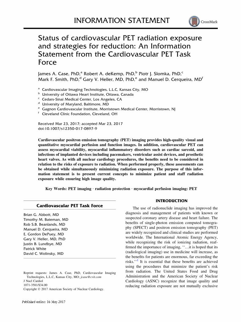
map at a very low patient dose (e.g., 0.04 mSv).15,18
Recent advances in iterative transmission reconstruction
algorithms significantly improve the quality of the
reconstructed transmission scans using as little as 60
seconds of acquisition times.19 See Figure 1. The major
limitations of line source systems are that transmission
imaging must be acquired in a 2D mode and septa must
be retracted before 3D emission imaging. Another
limitation is the limited availability of new line source
systems, as only one manufacturer produces a new PET
only system. Most systems are supported by refurbished
market suppliers. Despite the lack of new instrumenta-
tion, a large fraction of cardiac PET scanners in use
employ line source systems.
X-ray CT-Based Attenuation Correc-tion. PET/CT systems employ a conventional
multislice x-ray CT system capable of acquiring high-
resolution, diagnostic quality data and/or low-dose
attenuation correction scans.20 Radiation from CT-based
attenuation systems can be as little as 0.3 mSv21 to
upwards of 8.5 mSv,22 the latter dose being over twice
the radiation dose from a 82Rb perfusion examination.
Minimizing the radiation dose means using appropriate
protocols for cardiac PET/CT attenuation correction.
Several protocols have been introduced to ensure
high-quality transmission scanning. CT scans have
better signal-to-noise than line source attenuation scans.
However, CT-specific artifacts, such as breathing
Journal of Nuclear Cardiology� Case et al.
Status of cardiovascular PET radiation exposure and strategies for reduction
artifacts and metal artifacts associated with pacing and
ICD leads, can make PET/CT x-ray-based attenuation
correction more challenging than line source attenuation
correction.23
The CT portion of the cardiac PET/CT may provide
additive information to the perfusion study.24 Studies
that indicate the presence of coronary calcium in
patients with no known coronary disease can be helpful
in the evaluation of patient risk, and visual analysis of
coronary calcium detected with the low-dose CT scan
performed for attenuation correction may add useful
information without exposing the patient to a full
diagnostic CT.25
DOSE REDUCTION STRATEGIES
Cardiovascular PET myocardial perfusion imaging
with 82Rb, when implemented properly, is a low radiation
procedure, with total radiation doses of 4 mSv for typical
2D protocols (50 mCi for each infusion) and 2 mSv for
typical 3D protocols (25 mCi for each infusion).11
However, achieving these doses requires close adherence
to imaging standards and adapting protocols to meet the
needs of each patient. Laboratories should be aware that
recommended patient doses can vary based on PET
instrumentation.25 Major reductions can be achieved
through (1) the use of 3D imaging, (2) reducing the
number of attenuation scans, and (3) use of weight-
adjusted radioactivity administration.
Improving PET Image Quality WhileReducing Radiation with 3D Imaging
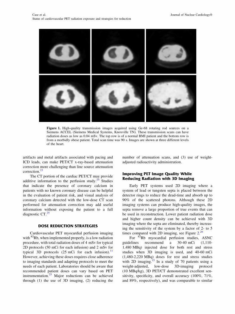
Early PET systems used 2D imaging where a
system of lead or tungsten septa is placed between the
detector rings to reduce the dead-time and absorb up to
90% of the scattered photons. Although these 2D
imaging systems can produce high-quality images, the
septa remove a large proportion of true events that can
be used in reconstruction. Lower patient radiation dose
and higher count density can be achieved with 3D
imaging where the septa are eliminated, thereby increas-
ing the sensitivity of the system by a factor of 2- to 5
times compared with 2D imaging, see Figure 2.26
For 82Rb myocardial perfusion studies, ASNC
guidelines recommend a 30-40 mCi (1,110-
1,480 MBq) injected dose for both rest and stress
studies when 3D imaging is used, and 40-60 mCi
(1,480-2,220 MBq) doses for rest and stress studies
with 2D imaging.15 In a study of 70 patients using a
weight-adjusted, low-dose 3D-imaging protocol
(10 MBq/kg), 3D PET/CT demonstrated excellent sen-
sitivity, specificity, and overall accuracy (100%, 71%,
and 89%, respectively), and was comparable to similar
Figure 1. High-quality transmission images acquired using Ge-68 rotating rod sources on aSiemens ACCEL (Siemens Medical Systems, Knoxville TN). These transmission scans can haveradiation doses as low as 0.04 mSv. The top row is of a normal BMI patient and the bottom row isfrom a morbidly obese patient. Total scan time was 90 s. Images are shown at three different levelsof the heart.
Case et al. Journal of Nuclear Cardiology�Status of cardiovascular PET radiation exposure and strategies for reduction

studies using 2D PET.27 In addition, absolute MBF
measurements appear to be equivalent using 2D and 3D
imaging, if the 3D activity is limited to avoid detector
saturation effects.28 When available, clinicians should
utilize 3D imaging so long as the hardware and software
have been validated for the specific cardiac application.
Reducing the injected dose from 50 mCi (1,850 MBq)
to 25 mCi (925 MBq) results in an average dose
reduction of 2 mSv for a typical rest-stress study.
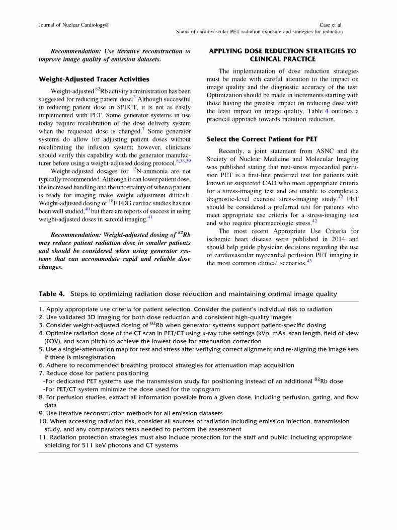
Sample imaging protocols for 2D and 3D imaging are
given in Figure 3. Current recommendations for injected
dose do not differ for 13N-ammonia (10-20 mCi) and 18F
FDG (5-15 mCi) when using 2D or 3D imaging.
Recommendation: validated 3D imaging should beused whenever possible for both dose reduction andhigh-quality images.
Optimizing Attenuation CorrectionTransmission Scan Settings
A common approach for attenuation correction of
PET studies is the use of x-ray CT. The CT image
quality required for attenuation correction is much lower
than for diagnostic quality images. Specifically, a CT
attenuation correction scan is acceptable for attenuation
correction if: the CT attenuation correction scan appears
uniform in the soft tissue regions when viewed using a
gray scale from -1,000 to 1,000 Hounsfield units; the
lung boundary is well defined and free of breathing
artifact; and the transmission scan is properly registered
with the emission data (See Figure 4).
Radiation dose optimization begins using x-ray tube
settings (kVp, mAs, scan length, field of view (FOV),
and scan pitch) to achieve the lowest dose. See Table 2.
For most applications, these settings can be kept to 100
kVp and 10 mAs.21 The lowest dose CT acquisition
mode uses a sequential ‘‘prospective ECG-triggered
axial scan.’’ This acquisition mode acquires a single slab
using a single rotation of the CT scanner followed by
moving the table to the next acquisition position.
Radiation dose can be kept to\1 mSv if the x-ray tube
is only on for data acquisition needed for attenuation
correction.21 When prospective triggering is not possi-
ble, ECG-based dose modulation is a critical
consideration for acquiring transmission data in the
retrospective ECG gated helical mode. Most systems
include a proprietary dose modulation algorithm and
should be used when helical scanning is used.21
Using iterative reconstruction of the CT scans can
reduce the need to use a high CT radiation dose to
acquire a high-quality CT scan for attenuation correc-
tion.29 Failure to follow a cardiac specific protocol can
result in as much as a tenfold increase in CT radiation.
The choice of a breathing protocol for acquiring the
transmission scan has a direct relationship on the
radiation dose received by the patient since different
scanning parameters are used for each protocol, see
Table 3.30–32 Three breathing approaches are commonly
used to minimize artifacts on the CT images: end-
expiration with or without breath holding, shallow free-
breathing, and cine-CT.30 Because each of these strate-
gies requires different scanning parameters, dose
reduction strategies for CT attenuation correction should
be customized to the breathing strategy employed. When
2D mode–only parallel
Accepted
3D mode–All photons accepted
All Accepted
photons accepted
Figure 2. In 2D PET imaging, contamination from non-coincidence and scattered photons is excluded using a systemof inter-plane septa. Although these septa are very effective inremoving these unwanted photons, it comes at the expense ofdecreasing the system sensitivity by a factor of 2–5 comparedto 3D PET systems.26
Journal of Nuclear Cardiology� Case et al.
Status of cardiovascular PET radiation exposure and strategies for reduction

dose strategies are optimized, the radiation from the
CT attenuation correction scan can be as little as
0.14 mSv using end-expiration or free-breathing
protocols.33
Recommendations: To achieve the lowest CT dose:
• Optimize kVp, mAs, typically 100 kVp or lessand 10 mAs or less.
5 min2D gated
scan
Txscan
5 min2D gated
scan
Txscan
(optional)
Vasodilator stress
50 mCi
3D, 7 minList mode
scan
CTscan
3D, 7 minList mode
scan
CTscan
(optional)
30 mCi 30 mCi
Vasodilator stress
50 mCi
2.5 min2D dynamic
scan
5 min3D scan
gated
Txscan
2.5 min2D dynamic
scan
5 min3D scan
gated
TxScan
(optional)
Vasodilator stress
40 mCi 40 mCi
A
B
C
Figure 3. Dynamic, gated, and 82Rb perfusion data can be acquired in PET using either frame orList-mode. A Standard 2D ECG gated PET protocol, B List-mode dynamic, gated, and perfusionprotocol, and C Frame-mode dynamic, gated, and perfusion protocol. It is important whendesigning an imaging protocol that diagnostic value is not sacrificed for reduced radiation dose.
Case et al. Journal of Nuclear Cardiology�Status of cardiovascular PET radiation exposure and strategies for reduction

• Utilize prospective ECG triggering when pos-sible. If helical scanning is required, dosemodulation should be employed.
• Confirm FOV settings for obtaining the attenu-ation correction scan are confined to the cardiacregion only without truncation of the body in-plane.
• Avoid cine-CT free-breathing protocols unlessthe PET/CT system is designed by the manu-facturer to perform this scan at a radiationdose comparable to either ECG triggering orhelical scanning with dose modulation.
Use of a Single-Transmission AttenuationScan
If the patient does not move substantially between
the rest and stress perfusion studies, acquiring separate
transmission scans may not be necessary. However, if
there is extreme patient movement, attenuation correc-
tion may not be properly applied without correcting for
the patient movement by re-aligning the emission and
transmission datasets.23,34 Failure to correctly re-align
these image sets will result in significant artifact and
lower diagnostic accuracy.
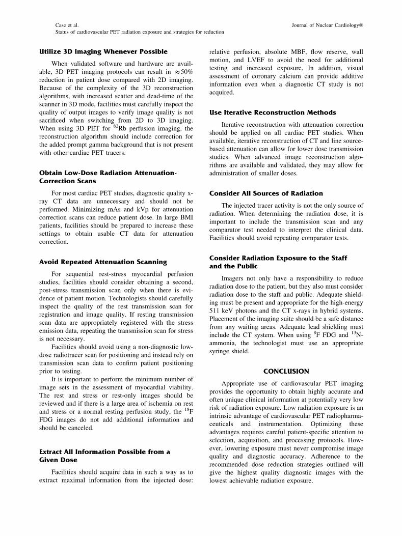
Figure 4. Examples of CT artifacts. A Breathing artifact froma free-breathing protocol. The liver can be seen in two differentplaces. B Metal artifact in a computed tomography attenuationcorrection image.
Table 2. Relationship between CT protocol settings radiation exposure and image quality
CT setting PurposeEffect on radiation
doseEffect on image
quality
kVp Increasing peak high voltage of
the X-ray tube, increases
energy of the electrons. This
increases the mean energy of
x-rays
Increasing kVp increases
radiation dose
Increasing kVp reduces
contrast, but improves
beam penetration
mAs Adjusts the number of electrons
being generated by the x-ray
tube (tube current)
Increasing mAs increases
radiation dose
Increasing mAs improves
signal-to-noise ratio
Pitch A measure of the amount of
overlap between adjacent
rotations of a helical CT scan
Increasing pitch decreases
radiation dose
Increasing pitch reduces
breath hold time
FOV Plane FOV is the size of the
in-plane region scanned in the
CT scan
Increasing axial and in-
plane FOV increases
radiation dose
Reducing FOV size to
smaller than the size of
the chest increases
truncation artifacts in
CT attenuation-
corrected images
Axial FOV is the size of the axial
length of a scan
Journal of Nuclear Cardiology� Case et al.
Status of cardiovascular PET radiation exposure and strategies for reduction

Recommendation: Use a single-attenuation cor-rection scan for rest and stress images after verifyingor correcting misregistration of the PET images.Technologists should only repeat transmission scan-ning when uncorrectable motion is present (examplesinclude patient rotation, breathing artifacts, or changesin arm position) or if the original transmission scanhas insufficient count density.
Patient Positioning with PET/CT Systems
Positioning of the heart in the center of the FOV can
be challenging with PET because of the small transverse
axis FOV of the scanner and the lack of tracer in the
patient prior to imaging. Optimal positioning is achieved
using a planar CT topogram.35
Recommendation: Minimize the region scannedfor the topogram to only cover the region of the heartfor perfusion imaging.
Patient Positioning with Dedicated PETSystems
One of the early approaches to positioning with
dedicated 2D PET systems was to inject a small amount
of 82Rb at rest prior to diagnostic scanning (5-10 mCi).
This approach, though effective in locating the heart,
exposes the patient to an additional 0.2-0.4 mSv and
requires an additional 10 minutes for the generator to
return to full strength. With the widespread utilization
of iterative reconstruction algorithms for line source
transmission scans, the acquisition time of transmission
scans for attenuation correction has been reduced to
less than 2 minutes. This has allowed more sites to use
transmission scans to confirm positioning instead of a
small dose of 82Rb. This approach introduces no
additional radiation exposure and does not impact
laboratory efficiency.
Recommendation: For dedicated PET systems, usethe transmission scan for positioning instead of a low-dose 82Rb scan.
Use Iterative Reconstruction on AllEmission Images
Iterative reconstruction with attenuation correction
should be applied to all cardiac PET studies (perfusion,
gated, and dynamic datasets). It can improve transmis-
sion scan quality and obviate the need for using higher
radiation doses. More recently time-of-flight and reso-
lution recovery reconstruction algorithms have been
investigated as techniques for improving image qual-
ity.36,37 Further study is needed before they can be used
routinely for dose reduction.
Table 3. Strategies for minimizing the dose from CT attenuation maps and imaging artifacts for thedifferent breathing protocols30-32
CT technique Description Techniques for minimizing dose
Shallow free breath For scanners that require more
than 10 seconds to cover
heart instruct the patient to
breathe using small breaths
through the study
Reduce current to\10 mAs
Reduce voltage to\100 kVp for lower BMI
patients
Use dose modulation
Use high-pitch acquisition (minimal slice
overlap)
End-expiration
breath hold
For scanners that require less
than 10 seconds to cover
heart instruct the patient to
breathe out slightly then hold
breath
Reduce current to\10 mAs
Reduce voltage to\100 kVp for lower BMI
patients
Use dose modulation
Use high-pitch acquisition (minimal slice
overlap)
Cine-CT For scanners with pre-installed
cine-CT protocol
Proprietary technique. Follow
manufacturers’ recommendations
Case et al. Journal of Nuclear Cardiology�Status of cardiovascular PET radiation exposure and strategies for reduction
Recommendation: Use iterative reconstruction toimprove image quality of emission datasets.
Weight-Adjusted Tracer Activities
Weight-adjusted 82Rb activity administration has been
suggested for reducing patient dose.3 Although successful
in reducing patient dose in SPECT, it is not as easily
implemented with PET. Some generator systems in use
today require recalibration of the dose delivery system
when the requested dose is changed.7 Some generator
systems do allow for adjusting patient doses without
recalibrating the infusion system; however, clinicians
should verify this capability with the generator manufac-
turer before using a weight-adjusted dosing protocol.8,38,39
Weight-adjusted dosages for 13N-ammonia are not
typically recommended.Although it can lower patient dose,
the increased handling and the uncertainty ofwhen a patient
is ready for imaging make weight adjustment difficult.
Weight-adjusted dosing of 18F FDG cardiac studies has not
beenwell studied,40 but there are reports of success in using
weight-adjusted doses in sarcoid imaging.41
Recommendation: Weight-adjusted dosing of 82Rbmay reduce patient radiation dose in smaller patientsand should be considered when using generator sys-tems that can accommodate rapid and reliable dosechanges.
APPLYING DOSE REDUCTION STRATEGIES TOCLINICAL PRACTICE
The implementation of dose reduction strategies
must be made with careful attention to the impact on
image quality and the diagnostic accuracy of the test.
Optimization should be made in increments starting with
those having the greatest impact on reducing dose with
the least impact on image quality. Table 4 outlines a
practical approach towards radiation reduction.
Select the Correct Patient for PET
Recently, a joint statement from ASNC and the
Society of Nuclear Medicine and Molecular Imaging
was published stating that rest-stress myocardial perfu-
sion PET is a first-line preferred test for patients with
known or suspected CAD who meet appropriate criteria
for a stress-imaging test and are unable to complete a
diagnostic-level exercise stress-imaging study.42 PET
should be considered a preferred test for patients who
meet appropriate use criteria for a stress-imaging test
and who require pharmacologic stress.42
The most recent Appropriate Use Criteria for
ischemic heart disease were published in 2014 and
should help guide physician decisions regarding the use
of cardiovascular myocardial perfusion PET imaging in
the most common clinical scenarios.43
Table 4. Steps to optimizing radiation dose reduction and maintaining optimal image quality
1. Apply appropriate use criteria for patient selection. Consider the patient’s individual risk to radiation
2. Use validated 3D imaging for both dose reduction and consistent high-quality images
3. Consider weight-adjusted dosing of 82Rb when generator systems support patient-specific dosing
4. Optimize radiation dose of the CT scan in PET/CT using x-ray tube settings (kVp, mAs, scan length, field of view
(FOV), and scan pitch) to achieve the lowest dose for attenuation correction
5. Use a single-attenuation map for rest and stress after verifying correct alignment and re-aligning the image sets
if there is misregistration
6. Adhere to recommended breathing protocol strategies for attenuation map acquisition
7. Reduce dose for patient positioning
–For dedicated PET systems use the transmission study for positioning instead of an additional 82Rb dose
–For PET/CT system minimize the dose used for the topogram
8. For perfusion studies, extract all information possible from a given dose, including perfusion, gating, and flow
data
9. Use iterative reconstruction methods for all emission datasets
10. When accessing radiation risk, consider all sources of radiation including emission injection, transmission
study, and any comparators tests needed to perform the assessment
11. Radiation protection strategies must also include protection for the staff and public, including appropriate
shielding for 511 keV photons and CT systems
Journal of Nuclear Cardiology� Case et al.
Status of cardiovascular PET radiation exposure and strategies for reduction
Utilize 3D Imaging Whenever Possible
When validated software and hardware are avail-
able, 3D PET imaging protocols can result in &50%
reduction in patient dose compared with 2D imaging.
Because of the complexity of the 3D reconstruction
algorithms, with increased scatter and dead-time of the
scanner in 3D mode, facilities must carefully inspect the
quality of output images to verify image quality is not
sacrificed when switching from 2D to 3D imaging.
When using 3D PET for 82Rb perfusion imaging, the
reconstruction algorithm should include correction for
the added prompt gamma background that is not present
with other cardiac PET tracers.
Obtain Low-Dose Radiation Attenuation-Correction Scans
For most cardiac PET studies, diagnostic quality x-
ray CT data are unnecessary and should not be
performed. Minimizing mAs and kVp for attenuation
correction scans can reduce patient dose. In large BMI
patients, facilities should be prepared to increase these
settings to obtain usable CT data for attenuation
correction.
Avoid Repeated Attenuation Scanning
For sequential rest-stress myocardial perfusion
studies, facilities should consider obtaining a second,
post-stress transmission scan only when there is evi-
dence of patient motion. Technologists should carefully
inspect the quality of the rest transmission scan for
registration and image quality. If resting transmission
scan data are appropriately registered with the stress
emission data, repeating the transmission scan for stress
is not necessary.
Facilities should avoid using a non-diagnostic low-
dose radiotracer scan for positioning and instead rely on
transmission scan data to confirm patient positioning
prior to testing.
It is important to perform the minimum number of
image sets in the assessment of myocardial viability.
The rest and stress or rest-only images should be
reviewed and if there is a large area of ischemia on rest
and stress or a normal resting perfusion study, the 18F
FDG images do not add additional information and
should be canceled.
Extract All Information Possible from aGiven Dose
Facilities should acquire data in such a way as to
extract maximal information from the injected dose:
relative perfusion, absolute MBF, flow reserve, wall
motion, and LVEF to avoid the need for additional
testing and increased exposure. In addition, visual
assessment of coronary calcium can provide additive
information even when a diagnostic CT study is not
acquired.
Use Iterative Reconstruction Methods
Iterative reconstruction with attenuation correction
should be applied on all cardiac PET studies. When
available, iterative reconstruction of CT and line source-
based attenuation can allow for lower dose transmission
studies. When advanced image reconstruction algo-
rithms are available and validated, they may allow for
administration of smaller doses.
Consider All Sources of Radiation
The injected tracer activity is not the only source of
radiation. When determining the radiation dose, it is
important to include the transmission scan and any
comparator test needed to interpret the clinical data.
Facilities should avoid repeating comparator tests.
Consider Radiation Exposure to the Staffand the Public
Imagers not only have a responsibility to reduce
radiation dose to the patient, but they also must consider
radiation dose to the staff and public. Adequate shield-
ing must be present and appropriate for the high-energy
511 keV photons and the CT x-rays in hybrid systems.
Placement of the imaging suite should be a safe distance
from any waiting areas. Adequate lead shielding must
include the CT system. When using 8F FDG and 13N-
ammonia, the technologist must use an appropriate
syringe shield.
CONCLUSION
Appropriate use of cardiovascular PET imaging
provides the opportunity to obtain highly accurate and
often unique clinical information at potentially very low
risk of radiation exposure. Low radiation exposure is an
intrinsic advantage of cardiovascular PET radiopharma-
ceuticals and instrumentation. Optimizing these
advantages requires careful patient-specific attention to
selection, acquisition, and processing protocols. How-
ever, lowering exposure must never compromise image
quality and diagnostic accuracy. Adherence to the
recommended dose reduction strategies outlined will
give the highest quality diagnostic images with the
lowest achievable radiation exposure.
Case et al. Journal of Nuclear Cardiology�Status of cardiovascular PET radiation exposure and strategies for reduction
Acknowledgement
The authors wish to thank Drs. Michael Desiderio,
Matthew Parker, and Raymond Russell for their independent
review and insightful comments on this manuscript.
Disclosures
R. deKemp is a consultant for, and receives unrestricted
grant funding and rubidium PET technology license revenues
from Jubilant DraxImage Inc. R. deKemp receives software
license revenues from INVIA Medical Imaging Solutions. J.
Case is an owner of Cardiovascular Imaging Technologies,
L.L.C., that produces PET processing software. P. Slomka
participates in software royalties at Cedars-Sinai Medical
Center for licensing of nuclear cardiology software and has
received research grant support from Siemens Medical
Systems. M. Smith receives research support from
Siemens Medical Solutions USA, Inc. G. Heller is a Medical
Advisor for Molecular Imaging Services and is a consultant to
Jubliant DraxImage Inc. M. Cerqueira is a consultant to
Astellas Pharma, USA and member of the Astellas Speakers
Bureau.
APPENDIX 1: USEFUL DEFINITIONS OFRADIATION
Activity (A)
The number of disintegrations per second (dps) and
the amount of administered radioactivity is usually
given in units of megabecquerels (MBq) or millicuries
(mCi). One Bq is equal to one decay per second and one
millicurie is 37 million decays per second.
Absorbed Dose (D)
The energy per unit mass deposited in tissue is
defined to be the absorbed dose, D, and is measured in
units of gray (Gy) or radiation absorbed dose (rad). One
Gy is equal to one joule/kg and equals 100 rad. This unit
is independent of the biological impact the radiation has
on tissue.
Equivalent Dose (HT)
The equivalent dose takes into consideration the
relative biological damage to tissue caused by radiation.
It is computed by multiplying the absorbed dose by a
weighting factor WR that is dependent on the radiation
type and particle energy, so that HT = D 9 WR. For
PET and PET/CT radiation, the weighting factor is one
for emitted positrons and photons. The units of equiv-
alent dose are sievert (Sv) or roentgen equivalent man
(rem). An absorbed dose in Gy or rad times a weighting
factor of one yields the same numerical value in Sv or
rem.
Whole Body Dose
The whole (or total) body dose is defined as the sum
of the energy deposited in each organ or body tissue,
with applied radiation weighting factors WR, divided by
the body mass. This is of limited value in nuclear
medicine due to non-uniform tracer distribution and has
been replaced by measures that reflect the radiation
sensitivity of different organs.
Effective Dose Equivalent (HE)
The effective dose equivalent, HE, was a weighted
average of the equivalent dose using tissue or organ
weights, WT, given by the International Commission on
Radiological Protection (ICRP) in 1979 [ICRP 1979].
The sum of the tissue weights is one. The ICRP has
updated this concept with the effective dose, E, with
new weighting factors (see below).
Effective Dose (E)
The effective dose is a tissue weighted average of
the equivalent dose in each individual organ. Specifi-
cally, effective dose (E) can be expressed as
E ¼X
T
WTHT ;
where HT is the equivalent dose absorbed by the Tth
organ and WT is the weighting factor for that organ,
reflecting the estimated cancer risk. The sum of the
weights is one. Adult organ and total body dosimetry
values are usually based on simplified kinetic models
and an idealized 70-kg person.
References
1. International Atomic Energy Agency. Radiological protection for
medical exposition to ionizing radiation. IAEA Safety Standard,
2002:RS-G-15.
2. US Food and Drug Administration. Initiative to reduce unneces-
sary radiation exposure from medical imaging, , February 2010,
http://www.fda.gov/downloads/Radiation-EmittingProducts/Radia
tionSafety/RadiationDoseReduction/UCM200087.pdf
3. Cerqueira MD, Allman KC, Ficaro EP, Hansen CL, Nichols KJ,
Thompson RC, et al. American Society for Nuclear Cardiology
Information Statement: Recommendations for reducing radiation
exposure in myocardial perfusion imaging. J Nucl Cardiol.
2010;17:709–18.
4. Piccinelli M, Garcia EV. Advances in software for faster proce-
dure and lower radiotracer dose myocardial perfusion imaging.
Prog Cardiovasc Dis. 2015;57:579–87.
Journal of Nuclear Cardiology� Case et al.
Status of cardiovascular PET radiation exposure and strategies for reduction

5. Hill KD, Einstein AJ. New approaches to radiation exposure.
Trends Cardiovasc Med. 2016;26:55–65.
6. Einstein AJ, Blankstein R, Andrews H, Fish M, Padgett R, Hayes
SW, et al. Comparison of image quality, myocardial perfusion, and
left ventricular function between standard imaging and single-in-
jection ultra-low-dose imaging using a high-efficiency SPECT
camera: the MILLISIEVERT study. J Nucl Med. 2014;55:1430–7.
7. Bracco Diagnostics. [Prescribing Information] CardioGen-82�
(Rubidium Rb 82 Generator), Bracco Diagnostics (NJ, USA)
revised April 2013. Available at: http://imaging.bracco.com/sites/
braccoimaging.com/files/technica_sheet_pdf/Cardiogen%20Full%
20Prescribing%20Information.pdf. Accessed June 30, 2016.
8. Jubilant DRAXIMAGE. [Prescribing Information] Ruby-Fill�
(rubidium Rb 82 generator), Jubilant DRAXIMAGE (Kirkland
Quebec). Available at: http://www.draximage.com/DATA/TEX
TEDOC/Approval-202153-Ruby-Fill-Labeling-30Sept-2016.pdf.
Accessed October 1, 2016.
9. Senthamizhchelvan S, Bravo PE, Esaias C, Lodge MA, Merrill J,
Hobbs RF, et al. Human biodistribution and radiation dosimetry of
82Rb. J Nucl Med. 2010;51:1592–9.
10. International Commission on Radiological Protection, 2015.
Radiation dose to patients from radiopharmaceuticals: A com-
pendium of current information related to frequently used
substances. ICRP Publication 128. Ann. ICRP 44(2S).
11. Hunter CR, Hill J, Ziadi MC, Beanlands RS, deKemp RA.
Biodistribution and radiation dosimetry of (82)Rb at rest and
during peak pharmacological stress in patients referred for
myocardial perfusion imaging. Eur J Nucl Med Mol Imaging.
2015;42:1032–42.
12. Saha GB. Fundamentals of nuclear pharmacy. 5th ed. New York:
Springer; 2004.
13. Schindler TH, Schelbert HR. Quantitation of myocardial perfu-
sion: absolute blood flow versus relative uptake. In: Dilsizian V,
Narula J, editors. Atlas of nuclear cardiology. 4th ed. New York:
Springer; 2013.
14. Taegtmeyer H, Dilsizian V. Imaging cardiac metabolism. In:
Dilsizian V, Narula J, editors. Atlas of nuclear cardiology. 4th ed.
New York: Springer; 2013.
15. Dilsizian V, Bacharach SL, Beanlands SR, Bergmann SR, Delbeke
D, Dorbala S, et al. ASNC imaging guidelines/SNMMI procedure
standard for positron emission tomography (PET) nuclear cardi-
ology procedures. J Nucl Cardiol. 2016;23:1187–226.
16. Dorbala S. Standard myocardial perfusion and cardiac FDG PET
protocols and associated patient radiation doses. http://www.ima
gewisely.org/*/media/ImageWisely%20Files/NucMed/Standard%
20Myocardial%20Perfusion.pdf. Accessed, May 10, 2016.
17. Carroll LR, Kertz P, Orcut O. The orbiting rod source: improving
performance in PET transmission correction scans. In: Esser PD.
Emission computed tomography: Current trends. New York: The
Society of Nuclear Medicine; 1983, p 235-247.
18. Slomka PJ, Pan T, Germano G. Recent advances and future pro-
gress in PET instrumentation. Semin Nucl Med. 2016;46:5–19.
19. Hsu BL, Case JA, Moser KW, Bateman TM, Cullom SJ. Recon-
struction of rapidly acquired germanium-68 transmission scans for
cardiac PET attenuation correction. J Nucl Cardiol. 2007;14:706–14.
20. Kinahan PE, Townsend DW, Beyer T, Sashin D. Attenuation
correction for a combined 3D PET/CT scanner. Med Phys.
1998;25:2046–53.
21. Dorbala S, Di Carli MF, Delbeke D, Abbara S, DePuey EG,
Dilsizian V, et al. SNMMI/ASNC/SCCT guideline for cardiac
SPECT/CT and PET/CT 1.0. J Nucl Med. 2013;54:1485–507.
22. Wu TH, Huang YH, Lee JJ, Wang SY, Wang SC, Su CT,
et al. Radiation exposure during transmission measurements:
Comparison between CT- and germanium-based techniques with a
current PET scanner. Eur J NuclMedMol Imaging. 2004;31:38–43.
23. Gould L, Pan T-S, Laghin C, Johnson NP, Guha A, Sdringola S.
Frequent diagnostic errors in cardiac PET/CT due to misregistration
of CT attenuation and emission PET images: A definite analysis of
causes, consequences and corrections. JNuclMed. 2007;48:1112–21.
24. Chang S, Nabi F, Xu J, Peterson LE, Achari A, Pratt CM, et al.
The coronary artery calcium score and stress myocardial perfusion
imaging provide independent and complementary prediction of
cardiac risk. J Am Coll Cardiol. 2009;54:1872–82.
25. Einstein AJ, Johnson LL, Bokhari S, Son J, Thompson RC,
Bateman TM, et al. Agreement of visual estimation of coronary
artery calcium from low-dose CT attenuation correction scans in
hybrid PET/CT and SPECT/CT with standard Agatston Score. J
Am Coll Cardiol. 2010;56:1914–21.
26. Cherry SR, Dahlborn M, Hoffman EJ. 3D PET using a conven-
tional multislice tomography without septa. J Comput Assist
Tomogr. 1991;15:655–68.
27. Knesaurek K, Machac J, Krynyckyi BR, Almeida OD. Compar-
ison of 2-dimensional and 3-dimensional 82Rb myocardial
perfusion PET imaging. J Nucl Med. 2003;4:1350–6.
28. Lekx KS, deKemp RA, Beanlands RS, Wisenberg G, Wells G,
Stodilka RZ, et al. 3D versus 2D dynamic 82Rb myocardial blood
flow imaging in a canine model of stunned and infarcted myo-
cardium. Nucl Med Commun. 2010;31:75–81.
29. Padole A, Ali Khawaja RD, Kalra M, Singh S. CT radiation dose
and iterative reconstruction techniques. AJR Am J Roentgenol.
2015;204:W384–92.
30. de Juan R, Seifert B, Berthold T, von Schulthess GK, Goerres GW.
Clinical evaluation of a breathing protocol for PET/CT. Eur
Radiol. 2004;14:1118–23.
31. Alessio AM, Kohlmyer S, Branch K, Chen G, Caldwell J, Kinahan
P. Cine CT for attenuation correction in cardiac PET/CT. J Nucl
Med. 2007;48:794–801.
32. Goerres GW, Kamel E, Heidelberg TN, Schwitter MR, Burger C,
von Schulthess GK. PET-CT image co-registration in the thorax:
Influence of respiration. Eur J Nucl Med Mol Imaging.
2002;29:351–60.
33. Xia T, Alessio AM, De Man B, Manjeshwar R, Asma E, Kinahan
PE. Ultra-low dose CT attenuation correction for PET/CT. Phys
Med Biol. 2012;57:309–28.
34. Case JA, Bateman TM. Taking the perfect nuclear image: Quality
control, acquisition, and processing techniques for cardiac SPECT,
PET, and hybrid imaging. J Nucl Cardiol. 2013;20:891–907.
35. O’Daniel JC, Stevens DM, Cody DD. Reducing radiation exposure
from survey CT scans. AJR Am J Roentgenol. 2005;185:509–15.
36. Akamatsu G, Ishikawa K, Mitsumoto K, Taniguchi T, Ohya N,
Baba S, et al. Improvement in PET/CT image quality with a
combination of point-spread function and time-of-flight in relation
to reconstruction parameters. J Nucl Med. 2012;53:1716–22.
37. Teoh EJ, McGowan DR, Macpherson RE, Bradley KM, Gleeson
FV. Phantom and clinical evaluation of the Bayesian penalized
likelihood reconstruction algorithm Q. Clear on an LYSO PET/CT
system. J Nucl Med. 2015;56:1447–52.
38. Kaster T, Mylonas I, Renaud JM, Wells GA, Beanlands RS,
deKemp RA. Accuracy of low dose rubidium-82 myocardial
perfusion imaging for detection of coronary artery disease using
3D PET and normal database interpretation. J Nucl Cardiol.
2012;19:1135–45.
39. Klein R, Adler A, Beanlands RS, de Kemp RA. Precision-con-
trolled elution of a 82Sr/82Rb generator for cardiac perfusion
imaging with positron emission tomography. Phys Med Biol.
2007;52:659–73.
Case et al. Journal of Nuclear Cardiology�Status of cardiovascular PET radiation exposure and strategies for reduction

40. Lindner O, Pascual TN, Mercuri M, Acampa W, Burchert W,
Flotats A, et al. Nuclear cardiology practice and associated radi-
ation doses in Europe: results of the IAEA Nuclear Cardiology
Protocols Study (INCAPS) for the 27 European countries. Eur J
Nucl Med Mol Imaging. 2016;43:718–28.
41. Skali H, Schulman AR, Dorbala S. 18F-FDG PET/CT for the
assessment of myocardial sarcoidosis. Curr Cardiol Rep.
2013;15:352.
42. Bateman TM, Dilsizian V, Beanlands RS, DePuey EG, Heller GV,
Wolinsky DA. American Society of Nuclear Cardiology and
Society of Nuclear Medicine and Molecular Imaging Joint Position
Statement on the Clinical Indications for Myocardial Perfusion
PET. J Nucl Cardiol. 2016;23:1227–31.
43. Ronan G, Wolk MJ, Bailey SR, Doherty JU, Douglas PS, Hendel
RC, et al. ACCF/AHA/ASE/ASNC/HFSA/HRS/SCAI/SCCT/
SCMR/STS 2013 multimodality appropriate use criteria for the
detection and risk assessment of stable ischemic heart disease: a
report of the American College of Cardiology Foundation
Appropriate Use Criteria Task Force, American Heart Association,
American Society of Echocardiography, American Society of
Nuclear Cardiology, Heart Failure Society of America, Heart
Rhythm Society, Society for Cardiovascular Angiography and
Interventions, Society of Cardiovascular Computed Tomography,
Society for Cardiovascular Magnetic Resonance, and Society of
Thoracic Surgeons. J Nucl Cardiol. 2014;21:192–220.
Journal of Nuclear Cardiology� Case et al.
Status of cardiovascular PET radiation exposure and strategies for reduction