influence of flanking homology and insert size on the
TRANSCRIPT

Environ. Biosafety Res. 6 (2007) 55–69 Available online at:c© ISBR, EDP Sciences, 2007 www.ebr-journal.orgDOI: 10.1051/ebr:2007027
Thematic Issue on Horizontal Gene Transfer
Influence of flanking homology and insert size on thetransformation frequency of Acinetobacter baylyi BD413
Deborah J. SIMPSON, Lisa F. DAWSON, John C. FRY*, Hilary J. ROGERS and Martin J. DAY
Cardiff School of Biosciences, Cardiff University, Main Building, Park Place, Cardiff, CF10 3TL, UK
RecA-mediated recombination requires regions of homology between donor and recipient DNA for successfulintegration. This paper investigates the effect of the relationship between the length of gene-sized inserts (434,733, 2228 and 2400 bp) and flanking sequence homology (100 – ca. 11 000 bp) on transformation frequencyin Acinetobacter baylyi strain BD413. Both insert size and size of the homologous region were varied, whichimproves on previous studies that kept insert size constant and varied only the homologous flank size. Transferfrequency of a non-homologous single small gene for gentamicin resistance (aac(3)I; 773 bp) was increased18-fold when flanking homology was changed from about 2000 bp to 8000 bp, but was reduced 234-fold whentwo genes were inserted (nptII-gfp; 2400 bp) between similar homologous regions. To investigate the effect ofsmaller regions of flanking homology (100 – 2000 bp), a partial nptII-gfp deletion (434 bp) was restored. Thisconfirmed that a minimum of 500 bp on each flank was required for transformation to be affected by flankinghomology. The data obtained allowed development of a multiple regression equation to predict transformationfrequency from homology, insert size and total fragment size for gene insertions. We also show that the ratio offlanking homology to insert size and not the total size of donor DNA is the most important variable determiningtransformation frequency. The equation developed was consistent with results previously reported by others,and so will be useful when using A. baylyi as a model for gene transfer by transformation in the laboratory,environment and for biosafety.
Keywords: Acinetobacter baylyi BD413 / natural transformation / predicting transfer frequency / gene transfer
INTRODUCTION
Natural transformation is the uptake and subsequentintegration of exogenous DNA by competent bacteria(Lorenz and Wackernagel, 1994; Thomas and Nielsen,2005). Recombination is mediated by RecA, and re-quires sequence homology between incoming donorDNA and sites in the bacterial genome (Dasgupta andRadding, 1982). Integration is essential for the expres-sion of DNA in transformed cells. The amount of ho-mology needed for recombination in well studied bacte-ria such as Escherichia coli (Watt et al., 1985), Ralstoniasolanacearum (Bertolla et al., 1997) and Bacillus subtilis(Khasanov et al., 1992) is about 20–70 bp, with increas-ing transformation frequency as homology increases.
Bacteria in the genus Acinetobacter are commonin many natural environments and Acinetobacter baylyishows particularly high natural transformation frequen-cies (ca. 10−3 transformants per cell; Juni, 1972; Lorenz
* Corresponding author: [email protected]
and Wackernagel, 1994; Vaneechoutte et al., 2006). Manystudies using this organism have shown that sequence ho-mology of regions flanking the DNA to be transferred af-fect transformation frequency. For example, recombina-tion was detected with 183 bp homology (de Vries andWackernagel, 2002) but there are no published data forsmaller regions. Gerischer and Ornston (2001) showedthat transfer frequencies of two point mutations fell asdistance increased from 2 base pairs to 10.5 kb. Trans-formation is more efficient with homology on both sidesof a sequence, but integration is also possible with one-sided homology, but at reduced frequency (de Vries andWackernagel, 2002). Sequence identity also affects trans-formation efficiency: for example, a decrease from 100%to 90% sequence similarity resulted in a 40-fold decrease(Majewski et al., 2000; Shen and Huang, 1986). In ad-dition to a requirement for sufficient homology, Palmenand Hellingwerf (1997) also highlight the importanceof intra-cellular nucleases affecting transformation. Theyhave shown that for A. baylyi approximately 500 bp ofthe incoming DNA is degraded during transfer of a point
Article published by EDP Sciences and available at http://www.ebr-journal.org or http://dx.doi.org/10.1051/ebr:2007027
D.J. Simpson et al.
mutation. Thus there is a body of literature indicating theimportance of the length of flanking homology in relationto transformation frequency, but no systematic study ofthis effect in Acinetobacter in relation to insert size.
Arber (2000) states that a major force in generatingdiversity is DNA acquisition. Thus the ability to exchangegenes horizontally, even at low frequencies, binds all or-ganisms together evolutionarily. Plant DNA is expectedto be present in large amounts and ubiquitously in the en-vironment, and Koonin et al. (2001) have proposed thatthe uptake of this DNA by bacteria would be a signifi-cant evolutionary force. Since A. baylyi is naturally com-petent and transformation proficient, it is a good modelorganism for studying gene transfer between plants andbacteria in nature. The size of the insert relative to theamount of homology required for the integration of non-homologous DNA is of relevance both to this evolution-ary process and also to the debate on genetically modifiedplants, as it is a likely limiting factor for horizontal geneflow of whole plant genes or transgenes into environmen-tal populations of bacteria. The aim of this work was todetermine the amount of flanking homology required forefficient integration of whole genes into A. baylyi, andto determine the relationship between the insert size, theamount of flanking sequence homology and transforma-tion frequency.
RESULTS AND DISCUSSION
Constructs used in experiments
Two types of genetic construct have been used in theseexperiments as donor DNA. The first type of constructused partial A. baylyi 16S rRNA gene flanks to act as ho-mologous regions around firstly a gentamicin resistancegene (Aac(3)I) and secondly two adjacent genes, encod-ing green fluorescent protein (gfp) and kanamycin re-sistance (nptII). The gentamicin construct (called BC3)acted as donor to provide a 773 bp insert for transferand the nptII-gfp donor insert (called PC1) was either2228 bp or 2400 bp depending on the promoter used(SP6 and psbA respectively). In both cases the 16S rRNAgene homology allowed transfer into one or more of theseven 16S rRNA genes in the wild type A. baylyi recipi-ent genome (Gralton et al., 1997). Using 16S rRNA genesin this way as sites for transformation is neither lethal(Strätz et al., 1996) nor does it reduce growth rate (Asaiet al., 1999). In experiments using lysates of A. baylyi car-rying these constructs as donor DNA, the genes were in-serted into the genome of the donors via the 16S flanks ofthe constructs. The second type of construct was a recipi-ent containing the nptII-gfp genes with a 434 bp deletionat the gene boundary, either when chromosomally inte-grated into the A. baylyi genome (BC1) or when carried
on a plasmid (BC2). These recipients were used in markerrescue experiments with various lengths of donor DNA,obtained from the complete nptII-gfp gene construct byappropriately designed PCR primers. These constructs al-lowed transformation experiments to be done with vari-ous lengths of flanking homology for three different genesized inserts, namely 2228–2400 bp, 773 bp and 434 bp.
Whilst making our constructs in the pGEM T easyplasmid, a pUC19 derived vector, we found that they weretaken up during natural transformation and stably main-tained by BD413, confirming a recent report (Graltonet al., 1997) showing that ColE1 based plasmids canreplicate autonomously in BD413. For this reason wehave used linear donor DNA in all the experiments re-ported here, either as PCR product, linearized plasmidor genomic DNA. This means that recombination alwaysinvolved an exchange of DNA. If we had used entireor intact plasmids, this would have made interpretationmore difficult, as these could become integrated by theCampbell-type integration process via their shared nptII-gfp homology.
Transformation experiments performedwith lysates
Transfer frequency of rifampicin resistance (a point mu-tation) using A. baylyi lysates as donor DNA and wild-type A. baylyi as recipient was 9.33± 1.2× 10⁻³ (Tab. 1).Transformation frequencies from lysates of our con-structs with insert sizes 773–2400 bp inserted into theA. baylyi genome as donor, with wild-type A. baylyi asrecipient ranged from 2.34−5.15 × 10⁻³ transformantsper recipient (Tab. 1) (N.B. these units are used for allsubsequent transformation frequencies given here). Thusthere was no significant difference in transformation fre-quency between the recombination of a point mutation ina coding gene and insertion of the 773–2400 bp gene con-structs used, when the DNA sequence was presented as acell lysate, which is when the homologous flanks werevery large (at least greater than 3000 bp each; Davidoff-Abelson and Dubnau, 1973). Although the chromosomeof A. baylyi BD413 has seven copies of the 16S rRNAgene (Gralton et al., 1997), the maximum recombinationfrequency obtained using the 16S rRNA sequence flanksin the donor construct was not significantly different fromthe maximum obtained for auxotrophic markers or the ri-fampicin resistance phenotype (around 10−2−10−3; Juniand Janik, 1969; Nielsen et al., 1997; Palmen et al.,1993).
Effect of total homology <2000 bp ontransformation frequency
Marker rescue experiments repaired the 434 bp deletionusing PCR products with flanking sequences around the
56 Environ. Biosafety Res. 6, 1 (2007)

Transformation of Acinetobacter baylyi with gene sized inserts
Table 1. Transformation of unmodified wild type Acinetobacter baylyi (strain BD413) with donor DNA of different flanking homol-ogy.
Donor DNA1 No. of Transformation Insert size Flanking Totaltransformants2 frequency2,3 (transformants/ (bp) sequence homology5
(mL−1) recipient cell) homology4 (bp) (bp)
BD413 lysate Rif resistant BD413 6.95± 1.21 × 10⁵ 9.33 ± 1.20 × 10⁻³ Point mutation * > 6000
PC1-SP6 in genome 7.16± 241 × 105 2.34 ± 0.60 × 10⁻³ 2228 * > 6000
PC1-psbA in genome 8.89± 2.63 × 10⁵ 3.25 ± 0.88 × 10⁻³ 2400 * > 6000
BC3 in genome 1.10± 0.15 × 10⁶ 5.15 ± 1.60 × 10⁻³ 773 * > 6000
PCR product PC1-SP6 12.5± 7.5 1.38 ± 0.01 × 10⁻⁸ 2228 246 & 196 442
PC1-psbA 8.3± 1.7 2.10 ± 0.74 × 10⁻⁸ 2400 246 & 196 442
BC3 6.43± 5.11 7.50 ± 2.59 × 10⁻⁹ 773 246 & 196 442
Linear plasmid PC1-SP6 4.0± 3.06 2.29 ± 1.23 × 10⁻⁸ 2228 246 & 196 442
1 All donor DNA except the Rif resistant lysate contained 16S rRNA homologous flanks, see Materials and Methods for details of constructs.2 Results are mean of 3 to 10 replicates ± SE of mean.3 MSD = 0.972 log10 units at P = 0.05 (and 1.169 at P = 0.01), this shows that the transformation frequencies for (i) lysates and (ii) lineardonor DNA (PCR product and linear plasmid) are significantly different from each other but not within these groups.4 Numbers indicate left and right lengths of flanking sequences; * indicates that the lengths of these sequences could not be determined asthe donor was lysate DNA (see footnote5).5 Maximum total donor fragment size for lysates taken up by BD413 was assumed to be >6000 bp (Davidoff-Abelson and Dubnau, 1973).
434 bp deletion site from intact nptII-gfp genes amplifiedfrom construct PC2. These showed the effect of the lengthof flanking homology, using 24 combinations of flank-ing sequence with flanks between 50 bp and 1100 bp.The results (Fig. 1A) showed a random cloud of trans-fer frequencies (geometric mean = 2.01 × 10⁻⁷) whenone or both regions of flanking homology were less than500 bp. These values were not significantly different fromeach other, were not linearly correlated, and the slope ofline of best fit was not significantly different from zero(r = −0.31; R2 = 9.5%; P = 0.119), which confirms therandomness of these values. This low frequency of trans-formation cannot be due to contamination of the PCRproduct with genomic DNA, because control experimentscarried out with genomic DNA diluted to the concentra-tions expected in the PCR products gave either no trans-formants or frequencies very much lower than those ob-served with >500 bp flanking homology. However, whenboth flanks were >500 bp in length, the transfer frequen-cies increased linearly (r = 0.986; P = 0.002), with thetotal homology on the semi-logarithmic plot presented(Fig. 1A). This indicates the dependence of transfer fre-quency on homology size for this 434 bp insert when ho-mology is above 500 bp on both flanks.
Note that in Figure 1A the recipients carried thedeleted genes both chromosomally (from construct BC1)and on a plasmid (BC2). These results are presented to-gether, as there was no significant difference between thetransfer frequencies for these constructs (BC1 = 4.4 ×
10−5; BC2 = 4.0 × 10⁻⁵; P = 0.856), despite a poten-tial 10-fold difference in target copy number, with sevencopies of the 16S rRNA gene in Acinetobacter sp. BD413(Gralton et al., 1997), and 10–50 copies of the plasmidper cell (West et al., 1994).
Notably, our data support the model proposed by Piferand Smith (1985) for Haemophilus influenzae, and con-firmed for A. baylyi BD413 (Palmen and Hellingwerf,1997), in which processing of donor DNA results in a re-quirement for a minimum sized flanking sequence for ef-ficient recombination, due to exonuclease degradation ofthe incoming single-stranded DNA before recombinationcan be started. Palmen and Hellingwerf (1997) showedthat this minimum flanking size was 500 bp for A. baylyiBD413 when repairing a point mutation, and our resultsshow that this minimum flank size remains 500 bp for amuch larger 434 bp fragment.
Our results differ from Palmen and Hellingwerf(1997) at low transformation frequencies. We have shownthat, within the limits of variability, transformation fre-quency was constant at about 2.0 × 10⁻⁷ (range = 2.5 ×10⁻⁸−1.0 × 10⁻⁶; n = 19), whereas Palmen and Helling-werf (1997) supposed that a semilogarithmic trend (n =3) was observed even at low frequencies. Thus ourdata suggest that transformation reaches a minimum fre-quency that is not decreased further by decreasing thesize of homologous flanks down to 50 bp upstreamand downstream of the insert DNA. This is the short-est amount of flanking sequence shown to be active in
Environ. Biosafety Res. 6, 1 (2007) 57

D.J. Simpson et al.
0 4000 8000 12000 16000Total homology (bp)
Insert = 773 bpInsert = 2400 bpUndigested, insert = 773 bpUndigested, insert = 2400 bp
0 500 1000 1500 2000
Total homology (bp)
>500 bp both flanks<500 bp both flanks>500 bp one flank only
10−3
10−4
10−5
10−6
10−7
10−8
Mea
n tra
nsfo
rmat
ion
frequ
ency
Flanking homology(A)
Mea
n tra
nsfo
rmat
ion
frequ
ency
(B)
MSD
MSD
Un
10−3
10−4
10−5
10−6
10−7
10−8
Figure 1. Effect of flanking homology on transformation frequency in A. baylyi BD413. Transformation frequency is expressed astransformants per recipient cell. (A) Experiments with PCR products (n = 5–12), using the marker rescue approach with flankinghomologies from 50–>850 bp on each side (see Tab. 5) of the donor DNA. The recipient contained deletion inactivated nptII and gfpgenes (434 bp deletion), in constructs BC1 (integrated into the A. baylyi genome) and BC2 (carried on plasmid). These strains weretransformed with donor DNA from PC2 PCR products, intact gfp and nptII genes or portions thereof. (B) Cell lysate experiments(n = 3–12). The wild type recipient was transformed with genomic donor DNA from two strains of A. baylyi carrying either constructBC3 (aac3(I); 773 bp) or PC1 (nptII-gfp; 2400 bp) inserted into a 16S rRNA gene. The genomic DNA was digested with restrictionenzymes to produce flanking homology sequences of 1974 to 11 195 bp and the recipient was wild type BD413. Un = control valuesusing undigested donor DNA, plotted separately on the right because the total homology is large but cannot be accurately estimated.MSD=minimum significant difference from the Tukey-Kramer test after analysis of variance (see Materials and Methods for details).
58 Environ. Biosafety Res. 6, 1 (2007)

Transformation of Acinetobacter baylyi with gene sized inserts
Acinetobacter to date, and is comparable to that seen inother species (Bertolla et al., 1997; de Vries and Wacker-nagel, 2002; Khasanov et al., 1992).
Transformation frequencies using PCR products ofthe 773–2400 bp gene inserts with small flanking se-quences (246 and 196 bp) as donor DNA were also low(7.5 × 10⁻⁹−2.1 × 10⁻⁸; Tab. 1). These values are sig-nificantly lower (P < 0.001) than those obtained withthe 434 bp insert discussed above. These results indicatethat there might be a minimum level of flanking homol-ogy beyond which transformation frequency does not de-cline further. So at saturating donor DNA concentrationssome molecules will always escape exonuclease degra-dation during DNA entry into A. baylyi BD413, but theproportion that escape depends on the length of the ho-mologous flanks around the insert.
It is interesting to note that ScaI cuts the plasmidbackbone at position 1876 of the pGEM plasmid back-bone thus providing 1820 bp and 1180 bp of non-homologous plasmid sequence on the two sides of theinsert in addition to the 246 bp and 196 bp of homol-ogous flanking sequence. The transformation frequencyinto wild type recipients of this construct was 2.29×10⁻⁸
(Tab. 1). So there was no significant increase in the trans-formation frequency by the addition of non-homologousflanking sequences exceeding the 500 bp which on aver-age is removed during DNA processing on entry into thecell (see Palmen and Hellingwerf, 1997; and above).
Effect of total homology >2000 bpon transformation frequency
To explore the effect of homology on transformation fre-quency of larger gene inserts further, the PCR products ofthe intact gentamycin (Aac(3)I) and nptII-gfp genes wereintegrated into the A. baylyi genome using the 16S rRNAflanking homologous sequences to generate donor strainscarrying PC1 and BC3 inserts. Genomic DNA of A. bay-lyi PC1-psbA (nptII-gfp; insert size 2400 bp) and BC3(aac3(I); insert size 773 bp) was digested with restrictionenzymes to produce fragments containing the insert withtotal flanking sequence homology ranging from 1974 to11 195 bp. The upstream and downstream flanking ho-mology varied between 641 bp and 6976 bp for thesefragments. These restriction digestion products were usedas donors to transform wild-type A. baylyi BD413. Undi-gested genomic DNA from A. baylyi PC1 and A. baylyiBC3 was used as controls to represent the maximal flank-ing sequence available. These data show that for bothinsert sizes transformation frequency increased with in-creasing total homology (Fig. 1B). Furthermore, the fre-quency for the smaller insert increased less than the largerinsert, and was over 1000-fold higher with 1974 bp of ho-mology. All the frequencies with the 2400 bp insert were
significantly different from the control frequencies usingundigested genomic DNA, apart from that obtained withlargest total homology (1974 bp, P < 0.001; 3991 bp,P < 0.001; 8296 bp, P = 0.006; 11 195 bp, P = 0.133).For the smaller construct, insert size 773 bp, transforma-tion frequency was not significantly different from thecontrol at any homology level.
These results show that for these larger inserts trans-formation frequency is dependent on total homologywhen flanking homology is greater than 500 bp on eachside of the insert. It is also clear that the larger insertshowed lower frequencies for similar lengths of homol-ogy than the smaller insert, and so transfer frequency isalso dependent on insert size. The curved shape of therelationships between frequency and total homology forboth insert sizes is similar to the shape predicted by Piferand Smith (1985) for their competition model for themechanism of transformation discussed earlier for largetransforming DNA fragments. Interestingly, the linear re-lationship noted (Fig. 1A) for the 434 bp insert with lessflanking homology is equivalent to the linear relationshippredicted for small DNA fragments from the Pifer andSmith (1985) model.
Relating transformation frequency to totalhomology and insert size
Multiple regression analysis was carried out on all dataobtained from the experiments with constructs carryinginsert sizes of 434 bp, 773 bp, 2228 bp and 2400 bp,and total flanking homology regions ranging from 100to 11 195 bp (n = 37). The aim of this analysis was todetermine which variables affected transformation fre-quency most, and to obtain a useful prediction equationto help other studies. The data for undigested DNA werenot included in these regression analyses, as the size ofthe homologous flanking sequence could only be esti-mated. The best regression equation used the ratio of to-tal homology to insert size, minimum homology and to-tal fragment size as variables to predict transformationfrequency. We did look at other variables (relative sizeand orientation of the flanking sequences, log10 transfor-mations of all the predictor variables) but these did notexplain any more variability in the data. Regression withthese variables showed that 60% of the variation in log10
transformation frequency observed could be explained bythe ratio of total homology to insert size and that 67% ofthe variation was accounted for by all three variables, butno more than 60% of the variation was explained by theratio and either of the other two variables. In all regres-sions with all combinations of variables, the ratio was al-ways the first variable selected by stepwise regression,and so can be considered the most important variable af-fecting transformation frequency. Furthermore, bivariate
Environ. Biosafety Res. 6, 1 (2007) 59

D.J. Simpson et al.
0 5 10 15 20Ratio of total homology/insert size (bp)
Insert = 434 bpInsert = 773 bpInsert = 2228−2400 bpUndigested, insert = 773 bpUndigested, insert = 2228 bpUndigested, insert = 2400 bpPublished data
Mea
n tra
nsfo
rmat
ion
frequ
ency
10−3
10−4
10−5
10−6
10−7
10−8
10−9
10−2
1
2
3
31 -
185 Un
MSD
Figure 2. Effect of the ratio of total homology to insert size on transformation frequency in A. baylyi BD413. The ratio of totalhomology to insert size was plotted for all data used in this study (overall means from all experiments, N = 39, n = 2–22). Recipientswere (i) wild type A. baylyi for 773 bp, 2228 bp and 2400 bp inserts flanked by 16S rRNA sequences and (ii) A. baylyi carryinginactivated gfp and nptII genes BC1 (chromosomally integrated) and BC2 (carried on plasmid). The circled numbers refer to data frompublished studies: 1 = Gebhard and Smalla (1998); 2 = de Vries and Wackernagel (2002); 3 = Strätz et al. (1996). Transformationefficiency is expressed as transformants per recipient cell. Un = control values using undigested donor DNA, plotted separately onthe right because the total homology is large but cannot be accurately estimated. MSD = minimum significant difference from theTukey-Kramer test after analysis of variance (see Materials and Methods for details).
regressions between transformation frequency and totalhomology, maximum and minimum homology and frag-ment size never accounted for more than 31% of the vari-ation in transformation frequency (range = 27–31%).
The best equation from this analysis is as follows:
Log10Tf = − 6.73 + 0.343 Rhi + 0.00196 Hmin
− 0.000567 Ftot
where: Tf = mean transformation frequency;Rhi = total homology divided by insert size;Hmin = size of minimum homologous flanking se-
quence;Ftot = total fragment size.This analysis shows that decreasing the amount of
flanking homology affected transformation frequency de-pending on insert size (Fig. 2). Also, this equation will
allow predictions of transformation frequency to be madefor a given construct with sizes of insert and flanking ho-mology falling within the range of our study, when trans-formed into A. baylyi BD413 under the conditions tested.This will be useful information for designing constructsfor studies involving recombination, and for predictingthe likely frequency of horizontal gene transfer.
Few studies reported in the literature give values forboth insert size and the length of flanking homologous re-gions. Despite this, when our equation was used to predicttransformation frequencies from the literature, data fromtwo studies (de Vries and Wackernagel, 2002; Gebhardand Smalla, 1998) fall well within the spread of our data(Fig. 2). The result from a third study (Strätz et al., 1996)is 104 fold higher than we predict from our equation. Inthis work Strätz et al. (1996) obtained a frequency of3 × 10−3 transformants per recipient of a single marker
60 Environ. Biosafety Res. 6, 1 (2007)

Transformation of Acinetobacter baylyi with gene sized inserts
gene, ApH3, into the A. baylyi BD413 chromosome.However, donor DNA was presented as purified plas-mid in the pDirect cloning vector (Clontech), which isa pUC19 derivative. Although Strätz et al. (1996) con-firmed chromosomal integration had occurred, no checkwas reported to establish the absence of the plasmid. So,as discussed earlier, it is possible that many of their trans-formants may also have contained plasmid-borne copiesof the construct, which would have enhanced the appar-ent transformation frequency. Thus their recombinationfrequencies using this vector may be artificially high.
Significance of this research
By deriving an equation that explains 67% of the variabil-ity in transformation frequency caused by changes in theratio of total homology to insert size, minimum homol-ogy and total fragment size, we have extended previouswork on the factors controlling transformation frequencyin bacteria (e.g., de Vries and Wackernagel, 2002; Palmenand Hellingwerf, 1997; Pifer and Smith, 1985). Further-more, the transformation model proposed by Pifer andSmith (1985) and Palmen and Hellingwerf (1997) is con-firmed for gene sized inserts by our results.
An issue we have not addressed so far is the presenceof hot spots in bacteria, where recombination frequenciesare higher (Stahl et al., 1975). However, given the similartransformation frequencies obtained with integration ofour constructs into the 16S rRNA genes and the restora-tion of rifampicin resistance, there was no evidence ofthe 16S rRNA genes acting as hot spots (or cold spots).In addition, we included published data in our work toensure its robustness, and found that our data are consis-tent with that of others (Fig. 2); thus we believe the equa-tion has general applicability for Acinetobacter BD413,within the limits of homology and insert size used here.
Finding a clear relationship between insert size andlength of flanking sequence with transformation fre-quency has clear implications for understanding hori-zontal gene transfer by natural transformation of bac-teria, both in the laboratory and more widely in fieldstudies. Recently, it was predicted that over the 70 mil-lion hectares planted with transgenic crops, we might ex-pect over 1018 bacteria to acquire plant transgenes basedon a transformation frequency of 10−17 (Heinemann andTraavik, 2004). Given that direct detection of transforma-tion in the field at such low frequencies is highly prob-lematic, incorporating all the variables into estimates ofbaseline transformation frequencies is important in mak-ing these kinds of global estimates. Our prediction equa-tion is based on use of saturating DNA concentrations,which are unlikely to occur in the field, so our equationis currently restricted to laboratory use. However, simi-lar approaches to ours could be used to take into account
insert size and length of homology in more comprehen-sive models, especially relevant to the transfer of planttransgenes to bacteria in the environment (Nielsen andTownsend, 2004; Pettersen et al., 2005).
Conclusion
This research shows that in A. baylyi a minimum of500 bp on each side of gene sized inserts was requiredfor transformation to be affected by flanking homologyand confirms that whole genes behave like point muta-tions in this respect. A multiple regression equation wasdeveloped from all the data, which predicted transfor-mation frequency from homology, insert size and totalfragment size for gene insertions. Our experiments alsoshow that the ratio of flanking homology to insert sizeand not the total size of donor DNA was the most impor-tant variable determining transformation frequency. Theequation developed will provide a useful base line whenusing A. baylyi as a model organism for gene transfer ofwhole genes by transformation in the laboratory, environ-ment and for biosafety research. Our work points the wayto the sort of experiments that should be considered forestablishing prediction models that can be used to guidefield studies and facilitate better estimation of the envi-ronmental transfer of transgenes from plants to bacteria.
MATERIALS AND METHODS
Bacterial strains
E. coli strains XL1 Blue (Stratagene, Cedar Creek, Texas,USA), Top10 (Invitrogen, Carlsbad, California, USA),and JM109 (Promega, Madison, Wisconsin, USA) wereused for cloning the constructs. These strains are recA−to prevent integration of the constructs into the E. coli 16SrRNA chromosomal genes, via homology provided by theAcinetobacter 16S rRNA gene flanking sequences. Thenon-methylating E. coli strain SSC-110 (Stratagene) wasused for preparing plasmids for ClaI digestion. A. baylyistrain BD413 (Vaneechoutte et al., 2006) was used as therecipient in transformation experiments. A strain of rif-resistant BD413 was obtained by spontaneous mutationon LB + rifampicin (100 µg.L−1) medium. All strainswere grown in LB medium (10 g.L−1 tryptone, 5 g.L−1
yeast extract, 10 g.L−1 NaCl), and on LB agar plates(1.5% agar). E. coli cultures were incubated at 37 ◦C.A. baylyi BD413 was grown at 30 ◦C. Other antibioticsused in media were as follows, kanamycin (50 µg.L−1),gentamicin (15 µg.L−1) and carbenicillin (50 µg.L−1).
Environ. Biosafety Res. 6, 1 (2007) 61

D.J. Simpson et al.
Table 2. PCR primers used for making constructs and transformant identification.
Primer Sequence 5’ to 3’ (restriction sites underlined) Restriction site
nptIIA ATGCGGATCCCTTTGACGTTGGAGTCCA BamHI
nptIIB CACGATCGATCTCAAGGATCTTACCGCT ClaI
nptIIC ATCAGTACGCGT GGATCCCTTTGACGTTGGA MluI; BamHI
nptIID CAGTGAATTCCTTTGACGTTGGAGTCC EcoRI
GFP1 GTGCGTCGACCTCTACTAGTGATCTCAATGAATATTGGTTGAC SalI
GFP2 CGCTCCGCTTATTTGTATAGTTCATCCATGC AciI
GFP3 ACTGGTCGACGCTATTTAGGTGACACTATA SalI
GFP4 AGCTGAATTCGATCTCAATGAATATTGGTTGA EcoRI
GFP5 ACTGTGAATTCGACTCCTACGGGAGGCAGCA EcoRI
16S1 ACGCTGAATTCGCTATTAGGTGACACTATAG EcoRI
16S2 CGATGTCGACGCGGTGTGTACAAGGC SalI
GENT1 ACGTGGATCCAGACTCGAATTGACATAA BamHI
GENT2 TGCAGGATCCCGAATTGTTAGGTGGC BamHI
GENT3 TCAGGCGCGCAGCTCGAATTGACATAAG BssHII
GENT4 ATCTGGCCCTAACGGCCTGAATTGTTAGGTGGC SfiI
Molecular methods
PCR was carried out using the MJ Research DNA engine(Genetic Research Instrumentation, Braintree, Essex,UK), using standard conditions. Pfu DNA polymerase(Promega) was used to amplify genes for cloning, andTaq DNA polymerase (Promega) was used for screeningby colony PCR. PCR amplification was carried out over30 cycles, with 30 s denaturation at 94 ◦C, 30 s anneal-ing at 55 ◦C, and 1 min extension per kb product withTaq DNA polymerase or 2 min per kb product with PfuDNA polymerase at 74 ◦C. PCR primers are listed in Ta-ble 2. PCR products were purified by using the QiagenPCR purification kit. Plasmid minipreps were carried outon 1.5 mL overnight cultures using the GenElute plas-mid miniprep kit (Sigma-Aldrich Company Ltd., Gilling-ham, Dorset, UK). Restriction digests were performedusing enzymes from Promega and New England Biolabs(Ipswich, Massachusetts, USA).
Production of donor DNA constructs
The nptII gene and its promoter (derived from theTn5 transposon) were PCR-amplified from the pCRIIcloning vector (Invitrogen). The gfp gene and psbA pro-moter were PCR-amplified from the pUTgfp-lux plasmid(Timms-Wilson and Bailey, 2001; Tab. 2), which containsa red-shifted gfp variant with maximum expression whenexcited with blue light (471 nm) (Heim et al., 1994). The
psbA promoter originates from the chloroplast of Ama-ranthus hybridus (Elhai, 1993). The acc(3)I gentamicinresistance gene was PCR-amplified from pUCP24 (Westet al., 1994; Tab. 2). Cloning was carried out using pGEMT Easy vector (Promega), and constructs for expression inA. baylyi BD413 were produced in the broad host rangeplasmid pUCP24 (West et al., 1994).
The constructs described in this paper were pro-duced to evaluate the movement of transgenes from trans-genic plants to soil bacteria. They were designed using100% homologous, two-sided, flanking sequences basedon amounts of homology required for efficient integra-tion (Bertolla et al., 1997; de Vries and Wackernagel,2002; Watt et al., 1985). Plasmid structures are illus-trated in Figure 3. Three plasmid constructs were pro-duced to provide donor sequences. Two of these pGEM-PC1 and pGEM-PC2 included either the psbA or SP6promoters. Only the psbA constructs are shown (Fig. 3)and are referred to as PC1-psbA (Fig. 3D) and PC2-psbA(Fig. 3B) respectively. Variant constructs with a SP6 pro-moter (replacing the psbA promoter), identical apart fromthis sequence, were also constructed. The third plasmidis pGEM-BC3, referred to as BC3 (Fig. 3E). Cloned se-quences of both BC3 and PC1 plasmid constructs wereflanked by the same conserved 442 bp 16S sequence.These plasmids were constructed as follows. The nptIIgene was PCR-amplified as a 1459 bp fragment, fromnucleotide 838–2299 of pCRII, using primers nptIIA andnptIIB (Tab. 2). After purification, the nptII fragment wasligated into pGEM T Easy vector, transformed into E. coli
62 Environ. Biosafety Res. 6, 1 (2007)
Transformation of Acinetobacter baylyi with gene sized inserts
AmpR
nptII
gfp
psbA16S
d)
nptII
AmpR
a)
RsrII
ClaI
SalI
AatII
SalI
gfp+psbApromoter
PCR-amplified from pGFPLUX
plasmid
AciI
pGEM-BC3
Aac(3)I PCR-amplified from
pUCP24
BssHII SfiI
AmpR
AmpR
16S
16S
16S
Aac(3)I
16S
PCR-amplify nptII, gfp, and promoter
Digest pGEM-16S with BssHII and SfiI
c)
AmpR
nptII
gfp
psbA
b)
e)
AmpR
nptII
gfp
psbA16S
d)
AmpR
nptII
gfp
psbA16S
d)
nptII
AmpR
a)
RsrII
ClaI
SalI
AatII
SalI
gfp+psbApromoter
PCR-amplified from pGFPLUX
plasmid
AciI
pGEM-BC3
Aac(3)I PCR-amplified from
pUCP24
BssHII SfiI
AmpR
AmpR
16S
16S
16S
Aac(3)I
16S
PCR-amplify nptII, gfp, and promoter
Digest pGEM-16S with BssHII and SfiI
c)
AmpR
nptII
gfp
psbA
b)
AmpR
nptII
gfp
psbA
b)
e)
PC2
PC1 BC3
BssHII
SfiI
(A)
(B) (C)
(D) (E)
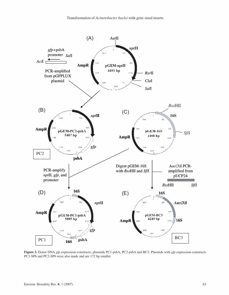
Figure 3. Donor DNA gfp expression-constructs; plasmids PC1-psbA, PC2-psbA and BC3. Plasmids with gfp expression-constructsPC1-SP6 and PC2-SP6 were also made and are 172 bp smaller.
Environ. Biosafety Res. 6, 1 (2007) 63
D.J. Simpson et al.
XL1 blue cells (Stratagene) and plated on LB platessupplemented with 50 µg.mL−1 kanamycin. The orien-tation of the nptII insert was determined by restrictiondigestion with AatII and RsrII. A plasmid clone, pGEM-nptII, (Fig. 3A) was selected with nptII oriented in the 5’to 3’ direction.
To produce plasmid construct PC2 (pGEM-PC2-psbA in Fig. 3B), gfp driven by the psbA promoter wasPCR-amplified from pUTgfp-lux plasmid using primersGFP1 and GFP2 (Tab. 2). The PCR product was purified,digested with AciI (to produce compatible sticky ends toClaI) and SalI, and ligated into the pGEM-nptII plasmiddigested with ClaI and SalI. The ligation was transformedinto E. coli JM109 (Promega) using kanamycin selec-tion. GFP fluorescence was monitored under blue light,using a GRI dark reader transilluminator (400–500 nm,maximum output at 450 nm). To confirm insertion of gfpinto the plasmid, fluorescent colonies were screened bycolony PCR using primers GFP1 and nptIIA (Tab. 2).Plasmids were sequenced to check the gfp gene orien-tation and the presence of all cassette components in theconstruct. In some clones, the psbA promoter was lostspontaneously and gfp expression was controlled by theSP6 promoter present in the pGEM vector. These plas-mids were termed pGEM-PC2-SP6.
Plasmid PC1 (pGEM-PC1-psbA in Fig. 3D) com-prised nptII and gfp genes flanked by A. baylyi BD41316S rRNA gene sequences. The Acinetobacter sequenceswere selected to have highly conserved regions at theouter edges (NCBI Accession number X89709, upstreamflank 305–546 and downstream flank 1176–1373 num-bered from the start of this sequence). PCR primers weredesigned to amplify the Acinetobacter 16S RNA genefrom the region 305–1373 (1069 bp). This region was am-plified from BD413 genomic DNA using primers 16S1and 16S2 (Tab. 2). Adenosine residues were added to theresulting fragment before ligation into the cloning siteof pGEM T Easy vector. Ligation mixtures were trans-formed into Top10 cells (Promega) and transformantswere selected on ampicillin. The presence of the 16Sfragment was determined by colony PCR using primers16S1 and 16S2 (Tab. 2), and the orientation was deter-mined by restriction digestion and sequencing. Cloneswith the 16S rRNA gene fragment running in the 5’ to 3’direction were selected. This plasmid was termed pGEM-16S (Fig. 3C).
Plasmid pGEM-16S (Fig. 3C) was digested with SfiI,and the ends were filled using T4 DNA polymerase be-fore digestion with BssHII to remove the central sec-tion of the 16S rRNA gene, and purification by gelextraction. To produce plasmid PC1 (pGEM-PC1-psbAin Fig. 3D), the nptII-gfp cassette was PCR-amplifiedfrom plasmids pGEM-PC2-SP6 and pGEM-PC2-psbA,using primers nptIIC and GFP1, and nptIIC and GFP3
respectively (Tab. 2). The PCR products were purifiedand digested with MluI, which creates compatible endsto BssHII, before ligation with the pre-digested pGEM-16S plasmid (Fig. 3C), and transformation into E. coliJM109. Transformants were selected for kanamycin re-sistance (50 µg.mL−1) and GFP fluorescence, and con-firmed by PCR, and restriction digestion. The resultingplasmids were termed pGEM-PC1-SP6 and pGEM-PC1-psbA (Fig. 3D). To produce plasmid BC3, the acc(3)Igene with promoter was amplified from pUCP24 usingprimers GENT3 and GENT4 (Tab. 2). The PCR prod-uct was digested with BssHII and SfiI, and ligated withpGEM-16S (Fig. 3C) pre-digested with the same en-zymes, before transformation into E. coli JM109. Trans-formants were selected on gentamicin (15 µg.mL−1), andtermed plasmid construct BC3 (pGEM-BC3 in Fig. 3E).Inserts from PC1 and BC3 were subsequently trans-formed into the A. baylyi genome using the 16S rDNAflanking sequences for recombination into one or more ofseven 16S rRNA genes in the A. baylyi genome.
Production of recipient DNA constructs
Two plasmid constructs were produced containing in-activated nptII and gfp genes; sequences from theseconstructs were subsequently transformed into A. baylyiBD413. These plasmid constructs were pGEM-BC2(Fig. 4C) and pGEM-BC1 (Fig. 4E). Note that the donorplasmid constructs PC1-psbA and PC2-psbA, and recip-ient plasmid constructs BC1-psbA and BC2-psbA werealso produced with the SP6 promoters (i.e., PC1-SP6,etc.) but only the psbA versions are shown in Figure 4.
The GFP protein is inactivated by the removal of nineamino acids from the C-terminal end (Li et al., 1997),and the nptII protein can be inactivated by the removalof 22 amino acids from the C-terminal end (Beck et al.,1982). As the coding portion of the C-terminal ends wereadjacent in plasmid construct PC2, both genes were in-activated by digestion with RsrII and BstBI (Fig. 4A),which removes 150 bp from nptII and 100 bp from gfp.After digestion, the sticky ends were filled in using T4DNA polymerase (Promega), the plasmid was religated,and used to transform E. coli JM109, to produce plas-mid BC2 (pGEM-BC2-psbA in Fig. 4C). TransformedE. coli colonies containing plasmid BC2 were no longerkanamycin resistant or fluorescent and were thereforeselected using carbenicillin (AmpR) at 50 µg.mL−1. Totransfer the BC2 insert into A. baylyi BD413 the nptII-gfpregion of plasmid BC2 was PCR-amplified using primersnptIIA and either GFP4 (for BC2-psbA) or GFP5 (forBC2-SP6), and ligated into the broad host range plasmidpUCP24, digested with BamHI and EcoRI. The ligationmixture was used to transform E. coli JM109 cells andtransformants were selected on gentamicin. Subsequently
64 Environ. Biosafety Res. 6, 1 (2007)

Transformation of Acinetobacter baylyi with gene sized inserts
Figure 4. Recipient DNA plasmid constructs BC1 and BC2 with deletion inactivated nptII and gfp genes. Plasmids pGEM-BC1and pGEM-BC2 were also produced with gfp expression controlled by the SP6 promoter, which results in plasmids that are 172 bpsmaller.
Environ. Biosafety Res. 6, 1 (2007) 65

D.J. Simpson et al.
Table 3. Restriction enzymes used for genomic DNA digestion.
Restriction enzyme Sequence homology (bp)
Left flank Right flank Total
BspHI 1333 641 1974
NsiI 2551 1440 3991
MluI 5067 3229 8296
EcoRV1 6976 4219 11 195
1 EcoRV used for A. baylyi BD413 carrying construct PC1 ingenome digestion only.
the pUCP24 plasmid carrying the BC2 construct wastransformed into A. baylyi.
To produce plasmid BC1 the acc(3)I gene (confer-ring resistance to gentamicin; PCR product size 773 bp)was PCR-amplified from pUCP24 using primers GENT1and GENT2 (Tab. 2) and digested with BamHI. ThePCR product was ligated with plasmid PC1 (Fig. 4B)pre-digested with BamHI, to produce pGEM-PC1-psbA-GENT (Fig. 4D). This was digested with RsrII and BstBIas described for plasmid BC2, to produce plasmid con-struct BC1 (insert size 3170 bp; pGEM-BC1-psbA inFig. 4E).
ScaI linearised plasmid and PCR product (PCR prod-uct size 3670 bp) amplified from plasmid construct BC1using primers 16S1 and 16S2 (Tab. 2) were used to trans-fer the BC1 insert into the A. baylyi BD413 genome, andtransformants were selected for gentamicin resistance.
Preparation of donor DNA
Genomic DNA was extracted from A. baylyi BD413 con-taining inserts PC1 and BC3 by CTAB extraction (Juni,1972). Overnight culture (1.5 mL) was centrifuged at topspeed in an Eppendorf 5415C centrifuge for 5 min. Thesupernatant was removed and the bacteria were resus-pended in 500 µL of lysis solution (100 µg.mL−1 pro-teinase K, in 0.5% SDS) and incubated at 55 ◦C for60 min, followed by addition of 100 µL 5M NaCl, and80 µL CTAB solution (10% w/v CTAB in 0.7M NaCl).After mixing by inversion the samples were incubated for15 min at 65 ◦C, before 680 µL chloroform:isoamyl al-cohol (1:25) was added and the tubes shaken gently toform an emulsion, and centrifuged for 5 min. The up-per aqueous layer was transferred to a clean tube andthe DNA was collected by isopropanol precipitation. Ge-nomic DNA was digested with the restriction enzymeslisted in Table 3, which were selected on the basis oftheir presence in A. baylyi BD413 16S rRNA gene, andabsence from the inserted marker genes. BC3 genomicDNA was not digested with EcoRV due to the presence
Table 4. PCR primers used to produce donor DNA for transfor-mations.
Primer Sequence 5’ to 3’
50BPF ATGGCCGCTTTTCTGGATT
50BPR CTGTCCTTTTACCAGACAACC
101BPF ACTCGTCGTGATCCATG
100BPR CGTTCAACTAGCAGACCAT
200BPF CCGGTCTTGTCGATCAGGATGATCTGGACGAAG
228BPR GATGGAAACATTCTTGGACAC
400BPF TGTCACTGAAGCGGGAAG
405BPR TACCCAGATCATATGAAACAGC
600PBF GAGAGGCTATTCGGCTATGA
P600BPR ACTGGAGTTGTCCCAATTC
S600BPR CTGGAGTTGTCCCAATTCTT
800BPF GGACAGCAAGCGAACC
986BPF CAAATTCAGGGCGCAAG
GFP1 GTGCGTCGACCTCTACTAGTGATCTCAATGAATA
TTGGTTGAC
GFP-3 ACTGGTCGACGCTATTTAGGTGACACTATA
NPTIID CAGTGAATTCCTTTGACGTTGGAGTCC
16S1 ACGCTGAATTCGCTATTAGGTGACACTATAG
16S2 CGATGTCGACGCGGTGTGTACAAGGC
of an EcoRV site within the gentamicin resistance gene.Linear plasmids of pGEM-PC1 and BC3 were preparedby digestion with ScaI. PCR products for transformationof inactivated nptII-gfp construct with varying regionsof homology were produced using the primers shownin Table 4, which gave the PCR product sizes given inTable 5. All PCR products were checked for purity by gelelectrophoresis. All products were pure, except in caseswhere product was from constructs with 16S rRNA geneinserts. In these cases two bands were seen, one fromthe native 16S rRNA gene and one from the construct.Fragments from the constructs were purified by gel ex-traction and purity confirmed by further gel electrophore-sis. Bacterial lysates were produced by resuspending thecells from 1 mL of overnight culture in 500 µL sterilesaline citrate (0.15 M NaCl, 0.015 M sodium citrate and0.05% SDS), and incubating at 60 ◦C for 1 h with occa-sional shaking. The resulting lysates were stored at 4 ◦Cuntil used.
Transformation experiments
Transformation experiments were carried out as de-scribed by Williams et al. (1996). A 25 mm diameter,0.45 µm pore size nitrocellulose filter was placed on a nu-trient LB agar plate with appropriate antibiotic selection:50 µg.mL−1 kanamycin in transformations using donor
66 Environ. Biosafety Res. 6, 1 (2007)

Transformation of Acinetobacter baylyi with gene sized inserts
Table 5. PCR products used as donors for transformation exper-iments with 434 bp insert constructs.
Left flank Right flank Total homology PCR product
(bp) (bp) (bp) (bp)
50 50 100 540
50 101 151 591
50 228 278 718
50 405 455 895
50 600 650 1090
101 100 201 641
101 50 150 590
200 228 428 868
200 50 250 690
400 405 805 1245
400 50 450 890
600 600 1200 1640
600 50 650 1090
600 405 1005 1445
800 600 1400 1840
800 50 850 1290
800 100 900 1340
800 228 1028 1468
800 405 1205 1645
989 600 1589 2029
989 50 1039 1479
989 228 1217 1657
1100 650 1750 2190
1100 850 1950 2390
PCR products were amplified from plasmid construct PC2 whichhas no 16S flanking sequences, and contains the intact nptII and gfpgenes. PCR products used as donors span a 434 bp deleted region inthe recipients which inactivates both gfp anf nptII genes and, thusthey are able to rescue the two markers in the recipients carrying theinactivated gfp nptII constructs (pUCP24-BC2 and BC1). Donorshave varying lengths of homology flanking this 434 bp region asshown. Total homology = left homology plus right homology.
DNA PC1 and carrying construct PC2 and 15 µg.mL−1
gentamicin for donor DNA carrying construct BC3. Typ-ically, an excess of donor DNA (1 µg in 100 µL) wasadded to the filter and allowed to dry before 100 µL ofrecipient culture was applied (108 cells). Recipient cul-tures were grown overnight in LB broth with shaking(120 rpm) at 30 ◦C; cell numbers were determined byOD670 and diluted in Ringer’s solution (Fisher Scientific,Loughborough, UK) to 109 per mL. Bacterial lysate (pre-
pared as described earlier) or genomic DNA of donorA. baylyi strains, PCR product and circular or linearisedplasmid DNA of donor constructs were used as donorDNA. As a control, 100 µL of recipient culture was addedto a nitrocellulose filter without addition of donor DNA,and the sterility of the donor DNA lysate was confirmedby plating. The plates were incubated at 30 ◦C for 24 h.Each filter was aseptically removed, placed in 3 mL ofsterile quarter strength Ringers Solution (Fisher), andvortex mixed for 1 min. Serial, 10-fold dilutions to 10−7
were plated as triplicate 20 µL drops. For lower trans-formation frequencies 200 µL to 3 mL (concentrated to100 µL) of mixture was spread on a selective plate. Thisconcentration procedure is effective and yields appropri-ately increased proportions of transformants (Rochelleet al., 1988; Williams et al., 1996). Plates were incubatedat 30 ◦C for 48 h before colonies were counted and flu-orescence was monitored. For low frequency transforma-tions (<10 colonies per plate) all putative transformantswere confirmed by colony PCR using appropriate primersfrom Table 2, followed by gel electrophoresis to confirmproduct size. Where more than ten putative transformantswere obtained in the experiment, at least five colonieswere verified in the same way.
To establish that the transformation protocol wasworking effectively, chromosomal integration transfor-mation was carried out using a lysate of a rifampicin re-sistant A. baylyi BD413 as a positive control. Transfor-mation occurred at a frequency of 9.3 ± 1.2 × 10⁻³ (n =3). Transformation frequencies using uncut chromosomalDNA from A. baylyi carrying constructs BC2 (Km-r) andBC3 (Gm-r) were not significantly different, confirmingthat there was no difference in transformation frequencydue to the antibiotic selection used. We also confirmedthat DNA concentration was saturating in all experimentsby regularly calibrating transformation frequency againstDNA concentration.
Statistics and analysis
Transfer frequencies, all expressed as transformants perrecipient, obtained using different constructs were com-pared using one-way and two-way analysis of variance.Where necessary, log10 x + 1 transformations were car-ried out on the data to achieve normality of residuals andhomogeneity of variance. Means were compared with theTukey-Kramer minimum significant difference (MSD)and sum of squares simultaneous test procedure (Fry,1993). Significant differences are cited at P ≤ 0.05 unlessotherwise stated. Multiple regression was used to predicttransformation frequency using the following predictorvariables (n = 37): total fragment size, insert size, leftand right homologous flanking region sizes, total homol-ogy (calculated as the sum of both flank sizes) and ratios
Environ. Biosafety Res. 6, 1 (2007) 67
D.J. Simpson et al.
of total homology and insert size. The log10 of transfor-mation frequency was used as the dependent variable toensure evenness and because the errors with this transfor-mation were not related to the magnitude of the variable.Various transformations of these variables were used toexplore improving the regression equation and evennessof the data. The best regression equation was selected bycomparing results from the stepwise, forward, backwardelimination, and best subsets regression procedures (Fry,1993). The best equation was selected on the basis of theR2 and Mallow’s Cp statistics, coupled with the degree ofvariability in the predicted values and the equation’s abil-ity to predict data not used to construct it. Statistical anal-ysis was carried out using Minitab version 14.2 (MinitabInc, State College, Pennsylvania, USA).
ACKNOWLEDGEMENTS
We wish to thank the Molecular Biology Support Unitat Cardiff University for sequencing, and Mark Baileyand Tracy Timms-Wilson at CEH-Oxford for supply-ing the pUTgfplux plasmid. This research was fundedby a joint BBSRC and NERC research grant (number72GM114168).
Received September 29, 2006; accepted May 7, 2007.
REFERENCES
Arber W (2000) Genetic variation: molecular mechanisms andimpact on microbial evolution. FEMS Microbiol. Rev. 24: 1–7
Asai T, Condon CN, Voulgaris J, Zaporojets D, Shen B,Al-Omar M, Squires C, Squires CI (1999) Construction andinitial characterization of Escherichia coli strains with fewor no intact chromosomal rRNA operons. J. Bacteriol. 181:3803–3809
Beck E, Ludwig G, Auerswald EA, Reiss B, Schaller H(1982) Nucleotide sequence and exact localization of theneomycin phosphotransferase gene from transposon Tn5.Gene 19: 327–336
Bertolla F, Van Gijsegem F, Nesme X, Simonet P(1997) Conditions for natural transformation of Ralstoniasolanacearum. Appl. Environ. Microbiol. 63: 4965–4968
Dasgupta C, Radding CM (1982) Polar branch migration pro-moted by RecA-protein - effect of mismatched base-pairs.Proc. Natl. Acad. Sci. USA 79: 762–766
Davidoff-Abelson R, Dubnau D (1973) Conditions affectingthe isolation from transformed cells of Bacillus subtilis ofhigh molecular weight single-stranded deoxyribonucleic acidof donor origin. J. Bacteriol. 116: 146–53
de Vries J, Wackernagel WM (2002) Integration of foreignDNA during natural transformation of Acinetobacter sp. byhomology-facilitated illegitimate recombination. Proc. Natl.Acad. Sci. USA 99: 2094–2099
Elhai J (1993) Strong and regulated promoters in the cyanobac-terium Anabaena PCC 7120. FEMS Microbiol. Lett. 114:179–184
Fry JC (1993) Biological Data Analysis: A Practical Approach,IRL Press, Oxford.
Gebhard F, Smalla K (1998) Transformation of Acinetobactersp. strain BD413 by transgenic sugar beet DNA. Appl.Environ. Microbiol. 64: 1550–1554
Gerischer U, Ornston LN (2001) Dependence of linkage ofalleles on their physical distance in natural transformation ofAcinetobacter sp. strain ADP1. Arch. Microbiol. 176: 465–469
Gralton EM, Campbell AL, Neidle EL (1997) Directed intro-duction of DNA cleavage sites to produce a high-resolutiongenetic and physical map of the Acinetobacter sp. strainADP1 (BD413UE) chromosome. Microbiology 143: 1345–1357
Heim R, Prasher DC, Tsien RY (1994) Wavelength mutationsand posttranslational autoxidation of green fluorescent pro-tein. Proc. Natl. Acad. Sci. USA 91: 12501–12504
Heinemann JA, Traavik T (2004) Problems in monitoring hor-izontal gene transfer in field trials of transgenic plants. Nat.Biotechnol. 22: 1105–1109
Juni E (1972) Interspecies transformation of Acinetobacter: ge-netic evidence for a ubiquitous genus. J. Bacteriol. 112: 917–931
Juni E, Janik A (1969) Transformation of Acinetobacter cal-coaceticus (Bacterium anitratum). J. Bacteriol. 98: 281–288
Khasanov FK, Zhvingila DJ, Zailhudlin AA, Prozorov AA,Bashkirov VI (1992) Homologous recombination betweenplasmid and chromosomal DNA in Bacillus subtilis requiresapproximately 70 bp of homology. Mol. Gen. Genet. 234:494–497
Koonin EV, Makarova KS, Aravind L (2001) Horizontal genetransfer in prokaryotes: quantification and classification. Ann.Rev. Microbiol. 55: 709–742
Li X, Zhang G, Ngo N, Zhao X, Kain SR (1997) Deletionsof the Aequorea victoria green fluorescent protein define theminimal domain required for fluorescence. J. Biol. Chem.272: 28545–28549
Lorenz MG, Wackernagel WM (1994) Bacterial gene-transfer by natural genetic-transformation in the environment.Microbiol. Rev. 58: 563–602
Majewski J, Zawadzki P, Pickerell P, Cohan FM, DowsonCG (2000) Barriers to genetic exchange between bacte-rial species: Streptococcus pneumoniae transformation. J.Bacteriol. 182: 1016–1023
Nielsen KM, Townsend JP (2004) Monitoring and modellinghorizontal gene transfer. Nat. Biotechnol. 22: 1110–1114
Nielsen KM, van Weerelt MDM, Berg TN, Bones AM,Hagler AN, Van Elsas JD (1997) Natural transforma-tion and availability of transforming DNA to Acinetobactercalcoaceticus in soil microcosms. Appl. Environ. Microbiol.63: 1945–1963
68 Environ. Biosafety Res. 6, 1 (2007)

Transformation of Acinetobacter baylyi with gene sized inserts
Palmen R, Hellingwerf KJ (1997) Uptake and processing ofDNA by Acinetobacter calcoaceticus – a review. Gene 192:179–190
Palmen R, Vosman B, Buijsman P, Breek CKD, HellingwerfKJ (1993) Physiological characterisation of natural transfor-mation in Acinetobacter calcoaceticus. J. Gen. Microbiol.139: 295–305
Pettersen A-K, Bøhn T, Primicero R, Shorten P, Soboleva T,Nielsen KM (2005) Modelling suggests frequency estimatesare not informative for predicting the long-term effects of hor-izontal gene transfer in bacteria. Environ. Biosafety Res. 4:223–233
Pifer ML, Smith HO (1985) Processing of donor DNA dur-ing Haemophilus influenzae transformation: analysis using amodel plasmid system. Proc. Natl. Acad. Sci. USA 82: 3731–3735
Rochelle PA, Day MJ, Fry JC (1988) Occurrence, trans-fer and mobilisation in epilithic strains of Acinetobacter ofmercury-resistance plasmids capable of transformation. J.Gen. Microbiol. 134: 2933–2941
Shen P, Huang HV (1986) Homologous recombination inEscherichia coli: Dependence on substrate length and homol-ogy. Genetics 112: 441–457
Stahl FW, Crasemann JM, Stahl MM (1975) Rec-mediatedrecombinational hot spot activity in bacteriophage lambda.III. Chi mutations are site-mutations stimulating rec-mediatedrecombination. J. Mol. Biol. 94: 203–212
Strätz M, Mau M, Timmis KN (1996) System to study hor-izontal gene exchange among microorganisms without culti-vation of recipients. Mol. Microbiol. 22: 207–215
Thomas CM, Nielsen KM (2005) Mechanisms of, and barriersto, horizontal gene transfer between bacteria. Nat. Microbiol.Rev. 3: 711–721
Timms-Wilson TM, Bailey MJ (2001) Reliable use ofgreen fluorescent protein in fluorescent pseudomonads. J.Microbiol. Meth. 46: 77–80
Vaneechoutte M, Young DM, Ornston LN, De BaereT, Tanny AN, van Der Reijden EC, Tjernberg I,Dijkshoorn L (2006) Naturally transformable Acinetobactersp. strain ADP1 belongs to the newly described speciesAcinetobacter baylyi. Appl. Environ. Microbiol. 72: 932–936
Watt VM, Ingles JC, Urdea MS, Rutter WJ (1985)Homology requirements for recombination in Escherichiacoli. Proc. Natl. Acad. Sci. USA 82: 4768–4772
West SHE, Schweizer HP, Dall C, Sample AK, Runyen-Janecky LJ (1994) Construction of improved Escherichia-Pseudomonas shuttle vectors derived from pUC18/19 andsequence of the region required for their replication inPseudomonas aeruginosa. Gene 148: 81–86
Williams HG, Day MJ, Fry JC, Stewart GJ (1996) Naturaltransformation in river epilithon. Appl. Environ. Microbiol.62: 2994–2998
Environ. Biosafety Res. 6, 1 (2007) 69