sequences flanking the transmembrane segments facilitate ... · sequences flanking the...
TRANSCRIPT

Sequences flanking the transmembrane segmentsfacilitate mitochondrial localization and membranefusion by mitofusinXiaofang Huanga,b, Xin Zhoua,b, Xiaoyu Hua,b,1, Amit S. Joshic, Xiangyang Guoa,b, Yushan Zhua,b, Quan Chena,b,d,William A. Prinzc, and Junjie Hua,b,e,f,2
aDepartment of Genetics and Cell Biology, College of Life Sciences, Nankai University, Tianjin 300071, China; bTianjin Key Laboratory of Protein Sciences,Nankai University, Tianjin 300071, China; cLaboratory of Cell and Molecular Biology, National Institute of Diabetes and Digestive and Kidney Diseases,National Institutes of Health, Bethesda, MD 20892; dState Key Laboratory of Membrane Biology, Institute of Zoology, Chinese Academy of Sciences, Beijing100101, China; eNational Laboratory of Biomacromolecules, Center for Excellence in Biomacromolecules, Institute of Biophysics, Chinese Academy ofScience, Beijing 100101, China; and fUniversity of Chinese Academy of Sciences, Beijing 100101, China
Edited by Peter J. Novick, University of California, San Diego, La Jolla, CA, and approved October 5, 2017 (received for review May 26, 2017)
Mitochondria constantly divide and fuse. Homotypic fusion of theouter mitochondrial membranes requires the mitofusin (MFN)proteins, a family of dynamin-like GTPases. MFNs are anchoredin the membrane by transmembrane (TM) segments, exposingboth the N-terminal GTPase domain and the C-terminal tail (CT) tothe cytosol. This arrangement is very similar to that of the atlastin(ATL) GTPases, which mediate fusion of endoplasmic reticulum(ER) membranes. We engineered various MFN-ATL chimeras togain mechanistic insight into MFN-mediated fusion. When MFN1 islocalized to the ER by TM swapping with ATL1, it functions inthe maintenance of ER morphology and fusion. In addition, anamphipathic helix in the CT of MFN1 is exchangeable with thatof ATL1 and critical for mitochondrial localization of MFN1.Furthermore, hydrophobic residues N-terminal to the TM seg-ments of MFN1 play a role in membrane targeting but not fusion.Our findings provide important insight into MFN-mediatedmembrane fusion.
membrane fusion | membrane targeting | mitochondria | endoplasmicreticulum | mitofusin
Mitochondria are double membrane-bound organelles thatgovern ATP production and many other important cellular
processes. Mitochondrial membranes undergo frequent fusionto maintain a ribbon-like morphology and actively divide forclearance of damaged portions or redistribution during cell di-vision (1–3). These membrane dynamics are tightly regulated toensure proper mitochondrial functioning. Mitochondrial fusionand fission are mediated primarily by dynamin-like proteins(DLPs) (4). In mammals, mitofusin (MFN) (Fzo1p in yeast) fusesouter mitochondrial membranes (5–7), and OPA1 (Mgm1p inyeast) merges inner mitochondrial membranes (8, 9), whereasDrp1 (Dnm1p in yeast) controls mitochondrial fission (10).Among the DLPs, the MFN family was the first to be identi-
fied and suggested as membrane remodeling factors (11). MFN/Fuzzy Onion/Fzo1p proteins localize to the outer membrane ofmitochondria, and their deletion causes fragmentation (12), in-dicating a lack of membrane fusion. Two MFN isoforms havebeen identified in mammalian cells (5). Both MFN1 and MFN2are ubiquitously expressed and essential during embryonic de-velopment (13), but they exhibit distinct activities in mediatingfusion (14). Mutations in MFN2 cause the neurodegenerativedisease Charcot–Marie–Tooth Type 2A (CMT2A) (15), and themitochondrial morphology regulated by MFN proteins has beenlinked to several critical physiological functions, including car-diomyocyte differentiation (16, 17), hematopoietic stem cellmaintenance (18), spermatogenesis (19), and neuron-controlledenergy metabolism (20, 21). MFN-mediated mitochondrialfusion is thought to be regulated by membrane potential (22),phosphorylation (23, 24), ubiquitination (25–27), acetylation
(28), and numerous interacting proteins, such as MIB, MARCH-V,Gβ, Bax, and GPAT (29–33). Given the difficulties purifying andreconstituting MFN proteins, their sufficiency for membranefusion is unclear.MFNs comprise an N-terminal cytosolic GTPase followed by a
helical bundle (HB) domain, a transmembrane (TM) domain,and a cytosolic C-terminal tail (CT). Two heptad repeats (HRs)have been predicted to reside in the HB and CT. MFNs arethought to localize in the mitochondrial membranes via the TMdomain. However, deletion of the HB or CT of MFN2 results inthe diffusion of some proteins into the cytosol (22), suggestingthat the mitochondrial targeting of MFN is determined by mul-tiple sequence elements. How MFN mediates mitochondrialfusion is also poorly understood. GTPase activity is knownto be required (14, 22). The crystal structure of the CT ofMFN1 revealed homotypic interactions in the form of an anti-parallel coiled coil (34), suggesting that this region of MFNs (thesecond predicted heptad repeat, HR2) promotes the tetheringof apposing mitochondrial membranes before fusion. Recentstructural analysis of the minimal GTPase domain (MGD) ofMFN1 revealed that membrane tethering is likely achieved
Significance
Mitochondria constantly connect through membrane fusion.The merging of the outer mitochondrial membrane requiresmitofusin (MFN) proteins. MFN is a membrane-anchoredGTPase, but whether it is sufficient to achieve fusion, and ifso how, is largely unknown. We have taken advantage of asimilar GTPase named atlastin (ATL), which mediates fusionof the endoplasmic reticulum (ER), as its mechanism is bet-ter understood. Domain swapping experiments show thatMFN is capable of fusing membranes, even on the ER. TheC-terminal tail of MFN contains an amphipathic helix thatpromotes fusion. MFN is properly inserted into the mitochon-drial membrane with the help of the helix and neighboringhydrophobic residues. These findings provide insight into howmitochondria fuse.
Author contributions: X. Huang, X.Z., Q.C., W.A.P., and J.H. designed research; X. Huang,X.Z., X. Hu, A.S.J., and X.G. performed research; X. Huang, X.Z., X. Hu, A.S.J., Y.Z., and J.H.analyzed data; and J.H. wrote the paper.
The authors declare no conflict of interest.
This article is a PNAS Direct Submission.
This open access article is distributed under Creative Commons Attribution-NonCommercial-NoDerivatives License 4.0 (CC BY-NC-ND).1Present address: Department of Pharmacology, University of Texas Southwestern Med-ical Center, Dallas, TX 75390.
2To whom correspondence should be addressed. Email: [email protected].
This article contains supporting information online at www.pnas.org/lookup/suppl/doi:10.1073/pnas.1708782114/-/DCSupplemental.
www.pnas.org/cgi/doi/10.1073/pnas.1708782114 PNAS | Published online November 1, 2017 | E9863–E9872
CELL
BIOLO
GY
PNASPL
US
Dow
nloa
ded
by g
uest
on
Janu
ary
11, 2
020

through its GTP-dependent dimerization (35, 36). Cryo-EM to-mography studies using isolated mitochondria confirmed thattethering/docking is GTP-dependent (37). Whether other re-gions of MFN contribute to the fusion activity remains to beinvestigated.Another class of dynamin-like GTPases that mediates fusion
of endoplasmic reticulum (ER) membranes has been identified:atlastin (ATL) in mammals, Sey1p in yeast, and RHD3 in plants(38–41). ATL and MFN are functionally analogous and share thesame domain structure and membrane topology. The fusionmechanisms of ATL and Sey1p are better understood than thoseof MFN proteins. The crystal structure of the cytosolic domain ofhuman ATL1 has been determined in three different confor-mations (42–44). In all structures, the GTPase domain of ATLforms a dimer, but the following three-helix bundle (3HB) docksto GTPase in either the same or the paired ATL molecule,orienting different directions relative to the GTPase dimer. Afusion model has been proposed in which GTP binding inducesinteractions between ATL molecules across the apposing mem-branes, and subsequent GTP hydrolysis causes conformationalchanges to force the HBs of engaging ATL molecules to comevery close, pulling the two membranes together. In addition, theCT of ATL forms an amphipathic helix that binds and perturbsthe membrane bilayer, facilitating the fusion process, and theTM segments are required for efficient fusion, probably via me-diation of the nucleotide-independent oligomerization of ATLmolecules (45). Structural and biochemical studies of Sey1p haverevealed a conserved mechanism with some unique features (46).Here, we elucidate how MFN mediates homotypic membrane
fusion based on our knowledge of ATL. We show that the role ofthe MFN-CT appears to be different from that previously proposed.An amphipathic helix in the CT is critical for MFN-mediated fusion.Furthermore, the same helix and a loop region in the HB play a rolein localizing MFN onto the outer mitochondrial membrane.
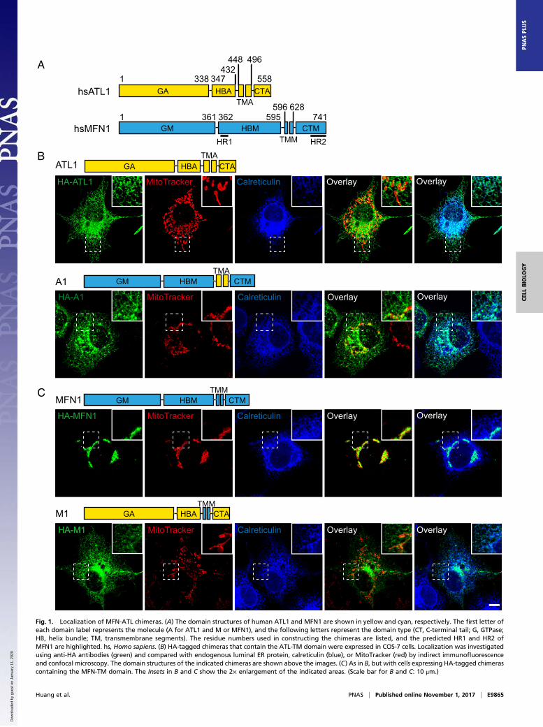
ResultsConstruction and Localization of MFN-ATL Chimeras. We generatedMFN-ATL chimeras to determine whether MFN or ATL retainmembrane fusion activity when they are relocated to a differentorganelle and whether MFN domains function similarly totheir ATL counterparts. To ensure surgical precision in con-struction of the chimera, we performed a detailed sequenceanalysis of human MFN1. Overall, the secondary structures ofMFN1 predicted by the “PredictProtein” program (47) exhibiteda pattern very similar to that observed experimentally for humanATL1 and MFN1 (Fig. 1A and Fig. S1). The N-terminal MGD ofMFN1 is composed of a classical GTPase and HB that is com-plemented by α11b from the CT. Three helices, numbered α7 toα9, were predicted between the MGD and TM segments ofMFN1. We named this region the HBM domain (Fig. 1A, M forMFN1), and it is likely the second HB of MFN1, given thatMGD already possesses one 4HB (35, 36). The previously de-fined HR1 (residues 373 to 400) is within α7. The TM segmentsof MFN1 (TMM) appear to be 33 residues in length, which wasconfirmed by online software TMHMM (www.cbs.dtu.dk/services/TMHMM/) and is considerably shorter than a conventional two-pass TM region, even if it forms a hairpin. The CT of MFN1(CTM) was predicted to be extensively α-helical (including α10,α11a, and α11b). The latter two helices were previously known asHR2 and have appeared as one long helix in the crystal structure(34). Collectively, these analyses suggest that MFN1 can be di-vided into four parts: GM (1–361), HBM (362–595), TMM (596–628), and CTM (629–741) (Fig. 1A). Similarly, the correspondingparts of ATL1 have been defined as the following: GA (1–338),HBA (339–447), TMA (448–496), and CTA (497–558) (Fig. 1A,A for ATL1).To relocate MFN or ATL, we switched their TM segments, the
key targeting element for an integral membrane protein. For
clarity, we named all of the chimeras used here according to theidentity of their TM domain; chimeras containing TMA aredesignated as A-type and chimeras containing TMM are desig-nated as M-type.To move MFN1 to the ER, we created chimera A1 (GTPa-
seMFN1-HBMFN1-TMATL1-CTMFN1 or GM-HBM-TMA-CTM).Immunofluorescence and the MitoTracker signal showed thatA1 properly localized to the ER, but not the mitochondria, eventhough the mitochondrial morphology is slightly altered (Fig.1B), possibly due to interactions between A1 and MFN1 (Fig.S2). In addition, A1 was found mostly in the tubular ER and verylittle protein was seen in peripheral ER sheets or the nuclearenvelope, a feature reminiscent of ATL1 (Fig. 1B). To rule outthe possibility that some of A1 remains on the mitochondria, weexpressed A1 in MFN1-deleted mouse embryonic fibroblast (MEF)cells and monitored the mitochondrial morphology. A1 appearedas an ER pattern and, similar to untransfected cells, A1-expressingcells exhibited fragmented mitochondria (Fig. S3), suggesting alack of A1 on mitochondria. Finally, deletion of the HBM (A2,GM-TMA-CTM) or CTM (A3, GM-HBM-TMA) from A1 didnot alter the ER localization (Fig. S4A). These results indicatethat the TMA is sufficient for localization of MFN1 to the ER.When the TM domain of ATL1 was replaced with that of
MFN1, the resulting mutant M1 (GA-HBA-TMM-CTA) did notlocalize to the mitochondria as expected; instead, it remained onthe ER and exhibited dispersed distribution in the cytosol insome cells (Fig. 1C and Fig. S4C). Consistently, expression ofM1 resulted in little rescue of defective mitochondrial mor-phology in MFN1-deleted MEF cells (Fig. S3). Truncation of theHR1- or HR2-containing regions in MFN2 has been shown tocause protein redistribution into the cytosol (48). We confirmedthese findings using MFN1 protein lacking the newly defined HB(including HR1) or CT (including HR2) (Fig. S4B). Thus, theTMM is likely necessary, but not sufficient, for outer mito-chondrial membrane targeting.
Functional Tests for MFN-ATL Chimeras Localized in the ER. We thentested the function of relocated MFNs. As the cytosolic domainsof these MFN-containing chimeras could potentially interactwith endogenous MFNs, we used a Saccharomyces cerevisiaesystem in which such cross-talk is less likely due to sequencedivergence between ectopically expressed MFN mutants andendogenous Fzo1p (the yeast ortholog of MFN). To determinethe activity of MFN localized in the ER, we analyzed whetherMFN-ATL chimeras can replace Sey1p, a functional ortholog ofATL. Yeast cells lacking Sey1p and either Yop1p or Rtn1p,proteins that shape the ER tubules (49, 50), exhibited abnormalcortical ER morphology; in particular, the tubular ER networklargely disappeared, and many areas of the cortex were void ofER, indicating a lack of ER fusion (39). These defects can berestored by the expression of wild-type (WT) Sey1p or humanATL1 (38, 39). When HA-tagged A1 was transformed intosey1Δyop1Δ cells using a CEN vector with the endogenous SEY1orFZO1 promoter, no expression was detected (Fig. S5A). Thelevels of these chimeras became detectable only when using ahigh copy 2μ vector with a GAL promoter (Fig. S5A) and wereequivalent to that of human ATL1 expressed under the endog-enous SEY1 promoter in a CEN vector (Fig. S5A). To confirmthe localization of these chimeras in yeast cells, GFP-taggedchimeras were expressed in sey1Δ cells and compared with thelocalization of ER-targeted red fluorescent protein (RFP) (ss-RFP-HDEL). All A-type chimeras localized to the ER (Fig. S5B)as in mammalian cells.To test whether some chimeras also localize to mitochondria
and influence their morphology, we expressed A1 in fzo1Δ cells,in which mitochondria are fragmented. As observed with MFN-deleted MEF cells, A1 expression in yeast cells did not affectthe mitochondrial morphology (Fig. S5C). Finally, we also
E9864 | www.pnas.org/cgi/doi/10.1073/pnas.1708782114 Huang et al.
Dow
nloa
ded
by g
uest
on
Janu
ary
11, 2
020
B
C
HA-ATL1 CalreticulinMitoTracker Overlay Overlay
ATL1 GA HBATMA
CTA
HA-A1 CalreticulinMitoTracker Overlay OverlayA1 GM HBM CTM
TMM
HA-MFN1 CalreticulinMitoTracker Overlay Overlay
MFN1 GM HBM CTM
TMA
hsATL11 338 347
432448 496
558GA HBA
TMACTA
hsMFN1
A
1 361 362 741595596 628
GM HBMTMM
CTM
HR2HR1
TMMM1 GA HBA CTA
HA-M1 MitoTracker Calreticulin OverlayOverlay
Fig. 1. Localization of MFN-ATL chimeras. (A) The domain structures of human ATL1 and MFN1 are shown in yellow and cyan, respectively. The first letter ofeach domain label represents the molecule (A for ATL1 and M or MFN1), and the following letters represent the domain type (CT, C-terminal tail; G, GTPase;HB, helix bundle; TM, transmembrane segments). The residue numbers used in constructing the chimeras are listed, and the predicted HR1 and HR2 ofMFN1 are highlighted. hs, Homo sapiens. (B) HA-tagged chimeras that contain the ATL-TM domain were expressed in COS-7 cells. Localization was investigatedusing anti-HA antibodies (green) and compared with endogenous luminal ER protein, calreticulin (blue), or MitoTracker (red) by indirect immunofluorescenceand confocal microscopy. The domain structures of the indicated chimeras are shown above the images. (C) As in B, but with cells expressing HA-tagged chimerascontaining the MFN-TM domain. The Insets in B and C show the 2× enlargement of the indicated areas. (Scale bar for B and C: 10 μm.)
Huang et al. PNAS | Published online November 1, 2017 | E9865
CELL
BIOLO
GY
PNASPL
US
Dow
nloa
ded
by g
uest
on
Janu
ary
11, 2
020

expressed A1 in sey1Δyop1Δ cells and observed no changesin mitochondrial morphology (Fig. S5D). These results con-firmed that ATL1-TM efficiently redirects MFN1 to the ER,even in yeast cells.The cortical ER of sey1Δyop1Δ cells expressing the chimeras
was visualized by Sec63p-GFP. To better evaluate the ER mor-phology, we added a category of partially normal ER in whichsome fenestrated ER was observed, but areas of the cortexlacked ER (Fig. 2A). We found that, when A1 (GM-HBM-TMA-CTM) was expressed, a substantial number of cells had normalER morphology (Fig. S5 E and F). In contrast, when the GTP-binding mutant A1 K88A was expressed, the ER morphologydefects were not rescued (Fig. S5E). The ER defects were alsoretained with the expression of A2 (GM-TMA-CTM) and A3(Fig. S5E). In addition, when MFN1 lacking HB or CT wastransfected into MFN1-deleted MEF cells, mitochondria frag-mentation was not restored (Fig. S3). Taken together, these re-sults suggest that MFN1 is functional on the ER, with the HBMand CTM playing roles in fusion.
The CT of ATL1 facilitates fusion using an amphipathic helix(45). The tailless mutant of ATL1, when expressed at endoge-nous levels using SEY1 promoter, failed to rescue the ER mor-phology defects in sey1Δyop1Δ cells (Fig. 2B). Thus, we tested thefunction of MFN-CT by tail swapping. As expected, the additionof MFN1-CT (tailless-CTM) partially rescued the defectiveATL1 tailless mutant and maintained proper ER morphology(Fig. 2B; see Fig. S5A for expression levels). Notably, neither WTATL1 nor ATL tailless containing mutants was able to restoremitochondria morphology in MFN1-deleted MEF cells (Fig. S3),consistent with their localization on the ER. These results in-dicate that the CTM is exchangeable with the CTA.To further test whether chimeras containing MFN1 mediate
functional ER fusion, we monitored their activities in a cellproliferation assay. Previously, it was reported that cells eitherlacking Sey1p or carrying ufe1-1 mutation, a temperature-sensitiveallele of the essential ER SNARE Ufe1p, grow normally, but thecombination of the two mutants causes very severe growth defects(38). As expected, the expression of human ATL1, but not the
A periphery center
norm
alab
norm
alpa
rtial
lyno
rmal
B
ATL1ATL1 taillessTailless-CTM
GA HBATMA
CTA
α10CTM
Tailless-CTMα10
ufe1-1 + vector
vector
ufe1
-1 s
ey1Δ
+
0
0.2
0.4
0.6
0.8
1.0
1.2
1.4
1000 2000 3000 4000 5000 6000
Cel
lDen
sity
(O.D
600
nm)
Time [min]
ufe1-1 + vectorvectorATL1ATL1 tailless
ufe1
- 1s e
y 1Δ
+C D
0
0
20
40
60
80
100
% o
f cel
ls w
ith in
dica
ted
ER
mor
phol
ogy
vecto
rATL1
Tailles
s
-CTMATL1
taille
ss
Tailles
s
-CTMα1
0
normalpartially normalabnormal
ATL1 tailless
Tailless-CTM
Tailless-CTMα10
ATL1
Tailless-CTMTailless-CTMα10
Fig. 2. Functional analysis of MFN-ATL chimeras in yeast cells. (A) A GFP fusion protein containing the ER protein Sec63p was expressed in yeast cells lackingSey1p and Yop1p (sey1Δyop1Δ cells). The ER morphology represented by the Sec63p-GFP signal was visualized by confocal microscopy, with the microscopefocused on either the center or the periphery of the cell. ER morphology was categorized into three classes. Note that the partially abnormal class exhibitssome fenestrated ER but is void of ER in some areas at the periphery of the cell. (Scale bar: 1 μm.) (B) Empty vector or indicated chimeras were expressed with aCEN plasmid under the control of the endogenous SEY1 promoter in sey1Δyop1Δ cells. See Fig. S5 for expression levels and localization of the chimeras. The ERmorphology was determined as in A, and each category was colored as in A. A total of 80 to 150 cells were categorized for each sample. The experiments,performed in a blinded manner, are averages of three repetitions. (C) The same chimeras as in B were transformed into ufe1-1sey1Δ, and empty vector wastransformed into ufe1-1 or ufe1-1sey1Δ. Serial 10-fold dilutions of cells were spotted onto an SC medium and incubated at 30 °C for 3 to 4 d. (D) Growth ratesof the same cells as in C were determined by measuring OD600.
E9866 | www.pnas.org/cgi/doi/10.1073/pnas.1708782114 Huang et al.
Dow
nloa
ded
by g
uest
on
Janu
ary
11, 2
020

B
T
NQ
Q
R
K KF
F
YM
I
E
LVV
N
C
A
A
C641
646650643 653
6421.0
1.2
1.4 5-doxyl12-doxyl
α10A650W
α11aF677WI702W
α11bI708WF729W
dmATLCTH
Trp
F0/F
α10F646W
657
DA
Fabnormalnormal
0
20
40
60
80
100
% o
f cel
ls w
ith in
dica
ted
mito
chon
dria
l mor
phol
ogy
vecto
rW
TA64
1DF64
6DA65
0D
G
H
02468
101214
ATL1
Tailless
Tailless-CTMα10
0 5 10 15 20 25 30 35
Tailless-CTMα11
% T
otal
Flu
ores
cenc
e
Time [min]
E
MFN
1 K
OM
FN1
MFN
1 A
650D
MFN
1 F6
46D
MitoDsRed
MFN
1 A
641D
HA Merge Zoom
0
2
4
6
8
10
12
Tailless
Tailless-CTMα10Tailless-CTMα11Tailless-CTM
0 10 15 20 255
%
Tot
al F
luor
esce
nce
1:2000
-30000-22500-15000
-75000
7500
200 220 240 260wavelength (nm)
α10+PSPC
peptide+liposomepeptide
wavelength (nm)
α10+EPL
200 220 240 260
A641D+EPL
wavelength (nm)200 220 240 260
wavelength (nm)
F646D+EPL
200 220 240 260
1:1000
Time [min]
M.R
.E.
(deg
cm
2 dm
ol-1)
7000
-1000
-9000
-17000
-25000
M.R
.E.
(deg
cm
2 dm
ol-1)
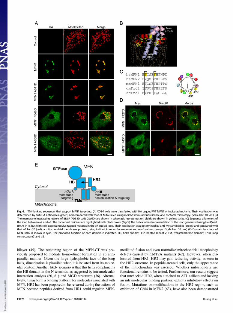
Fig. 3. An amphipathic helix in the MFN1-CT. (A) Sequence alignment of the CTs from various MFNs. The predicted and observed α-helices of MFN1 arelabeled with cylinders. The sequences of three synthetic peptides used in this figure are underlined in red. Mutated residues are numbered, with critical onesin cyan, moderately critical ones in green, and noncritical ones in yellow. The conservation of the important residues is highlighted in black boxes. (B) Helicalwheel representation of α10 was generated using program HeliQuest (heliquest.ipmc.cnrs.fr/). Hydrophobic, negatively charged, and positively chargedresidues are shown in yellow, red, and blue, respectively. Mutated residues are numbered. (C) Indicated peptides were added to liposomes with or without PCcontaining doxyl groups at position 5 or 12 of the hydrocarbon. The quenching of the fluorescence of Trp in the peptide was measured and expressed as F0/F(maximal fluorescence with doxyl-free liposomes divided by maximal fluorescence with doxyl-containing ones). DmATL CTH is a positive control, and aminoacid Trp is a negative control. Data shown are the mean and SE of three experiments. (D) Circular dichroism spectra of indicated peptides were recorded in theabsence (black lines) or presence (red lines) of liposomes. EPL, liposomes made of E. coli polar lipids; M.R.E., mean residue ellipticity; PCPS, liposomes made of PC:PS(mole percent 85:15). (E) HA-taggedWTMFN1 or indicated mutants were transfected into MFN1-deleted MEF cells. Their localization was determined by anti-HAantibodies (green) and compared with that of MitoDsRed, a mitochondrial targeted marker protein, using indirect immunofluorescence and confocal microscopy.The right panels show the 4.7× enlargement of the indicated areas. (Scale bars: 10 μm.) (F) The mitochondrial morphology of indicated samples was categorized as“normal” or “abnormal”. A total of 100 to 120 cells were counted for each sample. All graphs are representative of at least three repetitions. (G) Full-lengthdmATL, ATL tailless, tailless-CTMα10, and tailless-CTMα11 were purified and reconstituted into donor and acceptor vesicles at a 1:2,000 protein-to-lipid ratio. GTP-dependent fusion of donor and acceptor vesicles was monitored by the dequenching of an 7-nitro-2-1,3-benzoxadiazol-4-yl (NBD)-labeled lipid present in thedonor vesicles. All reactions were initiated by addition of GTP. (H) As in G, with indicated constructs tested at a 1:1,000 protein-to-lipid ratio.
Huang et al. PNAS | Published online November 1, 2017 | E9867
CELL
BIOLO
GY
PNASPL
US
Dow
nloa
ded
by g
uest
on
Janu
ary
11, 2
020
tailless mutant, efficiently rescued the poor growth of the ufe1-1sey1Δ cells (Fig. 2C). When ATL tailless-CTM was tested, thegrowth was slightly restored (Fig. 2C), which is consistent with theresults of the ER morphology assay. The same results wereobtained when cell proliferation was measured using an automatedgrowth curve system (Fig. 2D). These results support the notionthat the MFN-CT is functionally analogous to, although less effi-cient than, ATL1-CT in facilitating membrane fusion.
An Amphipathic Helix in the MFN-CT Facilitates Fusion and Localization.The CT of ATL forms an amphipathic helix that inserts intothe membrane and perturbs the lipid bilayer without causingsignificant lysis. Because the CT of MFN1 can partially replacethe CT of ATL, we investigated whether a similar amphipathichelix is present in the MFN1-CT. Helical wheel analysis revealedthat the first helix in the CT (α10) exhibited an amphipathicpattern (Fig. 3 A and B). The second helix (α11) can form acoiled coil structure (34), which requires hydrophobic resi-dues to point in the same direction, and as such is naturallyamphipathic.We synthesized three peptides corresponding to sequences of
the helical region in the MFN1-CT (α10 peptide, residues 629 to659; α11a peptide, residues 676 to 705; and α11b peptide, resi-dues 706 to 735) and tested whether they insert into the lipidbilayer as reported with other amphipathic helices (45). If thehydrophobic surface of an amphipathic helix contains a trypto-phan (Trp), upon membrane insertion, the Trp residue wouldcome into contact with the hydrophobic tails of lipids. Thefluorescence of Trp can be quenched by doxyl groups conjugatedto the hydrocarbon chains. Not all of the peptides contain Trp;thus, we replaced residues in the predicted hydrophobic faceswith Trp (α10 peptide, F646W or A650W; α11a peptide, F677W/I702W; α11b peptide, I708W/F729W). To determine whethersubstitution with Trp affects the function of MFN1, we trans-fected COS-7 cells with F646W or A650W. Similar to WTMFN1, both mutants aggregated mitochondria in the perinuclearregion (Fig. S6A) and were able to restore mitochondrial mor-phology in MFN1-deleted MEF cells (Fig. S6B), suggesting thatthe Trp mutants are functional. In a doxyl-quenching assay, weobserved direct contact of Trp residues with membranes onlywhen the α10 peptide was used (Fig. 3C). The quenching wasmore prominent with 5-doxyl than 12-doxyl, suggesting shallowinsertion (Fig. 3C). Similar quenching was observed with apeptide from the equivalent amphipathic helix in the ATL-CTfrom Drosophila, but not with free Trp (Fig. 3C). Taken together,the results indicate that the α10 of MFN1, which forms an am-phipathic helix that inserts into the membrane, may be func-tionally analogous to the CT of ATL.Circular dichroism (CD) measurements provided further evi-
dence of an interaction between the amphipathic helix and lipids.The α10 peptide corresponding to the WT sequence becamemore helical in the presence of liposomes (Fig. 3D). Helicalstructures appeared when Escherichia coli polar lipids (EPLs), alipid mixture that is partly similar to the membrane compositionof mitochondria, were used to generate liposomes, but not whenphosphatidyl-choline (PC)/phosphatidyl-serine (PS) was used(Fig. 3D). F646D and A650D, mutations in the hydrophobic faceof the first half helix, abolished the interactions and exhibitedno helical folding, even in the presence of liposomes, whereasI657D, a later mutation, caused no defects (Fig. 3D and Fig.S6C). The hydrophilic face of the helix appears to be less im-portant as mutation of A641 had little effect (Fig. 3D). Consis-tently, none of the α11 peptides exhibited lipid-induced helicalformation (Fig. S6C). To measure the helical formation of thepeptides in a more physiologically relevant environment, lipo-somes containing a lipid mixture of outer mitochondrial mem-branes (51) were used. Similar to EPL-containing liposomes,α10 peptide exhibited a helical propensity in the presence of
these liposomes (Fig. S6D). Individual lipid types were thenremoved one by one from the liposomes, and the CD spectrumwas determined with the α10 peptide. Cardiolipin (CL) andphosphatidyl-inositol (PI) were more important than PC andphosphatidyl-ethanolamine (PE) (Fig. S6D), confirming thepreference of more mitochondria-specific lipids by the peptide.These results show that folding of the α10 amphipathic helix isinduced upon interaction with the lipid bilayer, with the first halfof the helix being more important.To directly test the function of MFN1-α10, the α10 region was
attached to the ATL tailless mutant (tailless-CTMα10) instead ofthe entire MFN1-CT (tailless-CTM). The chimeric protein wasable to rescue the ER morphology defects in sey1Δyop1Δ cells(Fig. 2B) and growth defects in ufe1-1sey1Δ cells (Fig. 2 C andD), equivalent to that of WT ATL1 and even better than tailless-CTM. In addition, when MFN1-α10 was linked to the DrosophilaATL tailless mutant, the purified reconstituted chimera proteinwas able to restore ∼30% of the fusion defect of the taillessmutant at a protein-to-lipid ratio of 1:2,000 (Fig. 3G, see Fig. S7A and B for reconstitution controls). Conversely, fusion bytailless ATL was not promoted by α11. Consistent with ERmorphology and cell proliferation assays, the presence of α11appeared to partly inhibit the activity of α10 as tailless-CTM wasless competent in mediating fusion than tailless-CTMα10 (Fig.3H). These results suggest that the α10 amphipathic helix ofMFN1 is capable of facilitating ATL-mediated fusion.To further probe the function of the amphipathic helix, we
tested the effect of point mutations on MFN-mediated mito-chondrial fusion using MFN1-deleted MEF cells. As shownpreviously, expression of WT MFN1 rescued the mitochondriafragmentation in these cells, but expression of F642D, F646D,and A650D, the hydrophobic face mutants, failed to do so (Fig.3 E and F and Fig. S7D). Consistent with CD measurements,mutation A641D did not affect the function of MFN1 (Fig. 3 Eand F). Based on the helical wheel prediction, both K643 andK653 flank the hydrophobic face. Mutation of K643D, the formerlysine, but not K653D, reduced the ability of MFN1 to maintainmitochondrial fusion (Fig. S7D). Similarly, I657D, a mutant thatexhibited unaltered lipid association (Fig. S6C), restored the mi-tochondrial morphology to that of WT protein (Fig. S7D). Theseresults confirm that the α10 amphipathic helix is critical for MFN1function in maintaining mitochondrial morphology in cells.We noticed that some F646D and A650D proteins did not
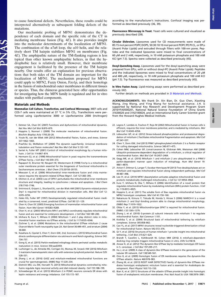
localize to the mitochondria (Fig. 3E). To test whether the am-phipathic helix plays a role in MFN1 localization, we expressedHA-tagged WT or mutant MFN1 in COS-7 cells. Consistent withprevious observations, overexpressed MFN1 localized to themitochondria, causing condensation of mitochondria around thenucleus, which is likely indicative of excessive fusion (Fig. 4A).When the hydrophobic face of the amphipathic helix was dis-rupted (F642D, F646D, and A650D), some of the mutant pro-teins diffused into the cytosol (Fig. 4A and Fig. S7E). Thesemutants also failed to cause hyperfusion of the mitochondria. Incontrast, the hydrophilic surface mutant A641D behaved like theWT protein; it localized to the mitochondrial membranes and itsoverexpression resulted in perinuclear condensation of the mi-tochondria (Fig. 4A). The localization of additional mutants (K643D,K653D, and I657D) correlated with their ability to maintain mi-tochondrial morphology (Fig. S7E). These results indicate that theα10 amphipathic helix plays a dual role facilitating mitochondrialtargeting and fusion by MFN1.
α7α8 Loop Facilitates Mitochondrial Targeting of MFN. The role ofthe α10 amphipathic helix in MFN1 localization explains thedeletion of MFN1-CT causing mistargeting. Next, we investi-gated how the HB region is involved in mitochondrial targetingas the mutant lacking the HB domain exhibited the same defects(Fig. S4B). Bacterial dynamin-like protein (BDLP) has been reported
E9868 | www.pnas.org/cgi/doi/10.1073/pnas.1708782114 Huang et al.
Dow
nloa
ded
by g
uest
on
Janu
ary
11, 2
020

to use a paddle domain and a loop in the trunk region to insert intomembranes (52) (Fig. 4B). The paddle of BDLP is reminiscent ofthe predicted transmembrane hairpin of MFN1, and the loop re-gion consists of three short amphipathic helices. We hypothesizethat a corresponding loop in the HB domain (equivalent to thetrunk domain of BDLP) that connects α7 and α8, and as such is inproximity to the lipid bilayer, plays a role in mitochondrial mem-brane targeting of MFN1. Helical wheel analysis of the looprevealed a less typical amphipathic character (Fig. 4C). Never-theless, two conserved Phe residues were found on one side of thehelical wheel. We replaced F427 and F431 with Asp and tested themutants in COS cells. As predicted, the mutated MFN proteinspartly localized to the cytosol (Fig. 4D). Interestingly, unlike α10mutants, the properly targeted F427D and F431D proteins wereable to aggregate mitochondria around the nucleus. These resultssuggest that the α7α8 loop affects localization of MFN1 viamembrane insertion of bulky hydrophobic residues but is less likelyto be involved in regulating fusion activity.
TM-Flanking Elements Are Conserved in Fzo1p. To test if the TM-flanking elements identified in MFN are conserved in eukary-otes, we analyzed the sequence of Fzo1p, the yeast homolog ofMFN. The overall predicted secondary structure was conserved be-tween Fzo1p and MFNs (Fig. S1). In the CT of Fzo1p, a predictedhelix (755 to 775) that is equivalent to α10 in MFN1 is present im-mediately following the TM regions, and in the predicted α7α8 loopof Fzo1p, Y552, and Y555 are weakly equivalent to F427 andF431 in human MFN1 (Fig. 4C and Fig. S1). The structures ofthe α10 regions in MFN1 and Fzo1p were modeled using Rap-torX (raptorx.uchicago.edu/StructurePrediction/predict/). Residues631 to 657 of MFN1 were modeled as α-helical (Fig. S7C), which isconsistent with the second half of the α10 peptide being lessimportant. In addition, the model revealed that F642, F646, andA650 form a hydrophobic patch that faces one direction (Fig.S7C). Similarly, residues 763 to 775 of Fzo1p were modeled as ahelix, with L765, Y769, and L733 being the hydrophobic face(Fig. S8A). These analyses suggest that the elements used byMFN1 for membrane targeting and fusion are likely conservedin Fzo1p.To test whether residues found in Fzo1p are critical for
maintaining proper mitochondrial morphology, we expressedWT and mutant Fzo1p under control of the endogenous FZO1promoter (Fig. S8B) in fzo1Δ cells and monitored mitochondrialmorphology using a mitochondria-targeted GFP marker (mito-GFP). As previously reported (6), deletion of Fzo1p caused thefragmentation of mitochondria, and reintroduction of WT Fzo1prestored the ribbon-like morphology (Fig. S8C). Mutations in theα7α8 loop (Y552D and Y555D) and the hydrophilic face of α10(N764D) did not compromise mitochondrial morphology, butmutations in the hydrophobic face of α10 (L765D, Y769D, andL773D) did (Fig. S8D). Notably, previous studies confirmed therole of α10 residues in supporting cell proliferation (53); Y769Pand L773P (within the predicted α10) are inactive, but mutationsof later leucines cause no defects. To further investigate themembrane targeting of Fzo1p mutants, we performed a mem-brane sedimentation assay. Mutations in hydrophobic residues inboth the α7α8 loop and α10 helix affected the membrane tar-geting of Fzo1p (Fig. S8E). These results suggest that Fzo1putilizes similar hydrophobic residues as MFN for proper locali-zation and function.
DiscussionOur results provide important insights into the mechanism un-derlying MFN-mediated homotypic fusion of the outer mito-chondrial membranes. We show that this dynamin-like GTPaseis capable of mediating membrane fusion outside of mitochon-dria, suggesting that it is a bona fide fusogen without the re-quirement of mitochondria-specific cofactors. The domains of
MFN protein, especially the CT, play similar roles as those ofATL.The GTPase domain of MFN1 appears to be longer than
previously estimated (48), based on a comparison with cytATL1,which is consistent with MFN having an extra HB associatingwith its GTPase (35, 36). Secondary structure prediction andstructural analogy with BDLP suggest that the region betweenthe GTPase and TM domain of MFN is likely an HB, similar toATL. Finally, MFN is predicted to have one hydrophobic seg-ment that may be a TM domain. Given that both the N and Ctermini are cytosolic (48), the TM segments of MFN1 likely forma hairpin without traversing the bilayer completely. The short TMdomain compared with ATL implicates an additional mechanismfor membrane targeting.The fusion activities of many other membrane fusogens, in-
cluding SNAREs and ATL proteins, have been demonstrated invitro using reconstituted vesicles containing purified proteins(54). Such direct evidence has not been reported for MFN. Invitro fusion assays using isolated mitochondria have shownthat the MFN family is necessary for fusion of the outermitochondrial membranes (55). However, several importantmitochondria-related factors, including inner membrane poten-tials and mitochondria-resident proteins, such as outer membraneprotein Ugo1p (56), can drastically affect MFN- or Fzo1p-mediatedfusion, which prevents the conclusion that MFN may act alonefor fusion. Here, we showed that, when MFN1 is relocated to theER using the ATL1-TM domain (chimera A1), it can replace ERfusogen Sey1p in maintaining ER morphology and mediate ERfusion in yeast cells. The partial restoration of ER morphology waslikely caused by low and uneven levels of A1 expression among thetested cells. Alternatively, the previously reported regulatory pro-teins for MFN family members are needed to boost the activity ofthese fusogenic proteins to a necessary level. Nevertheless, ourevidence favors the notion that MFN is intrinsically capable ofmediating membrane fusion.In addition to the GTPase, we showed that the HB of MFN1 is
indispensable. Based on recent structural studies (35, 36), HBMis likely complemented by the first half of HR2 (α11a), mim-icking the configuration of the trunk domain of BDLP. In ATL,the stalk-like HB relays the conformational changes initiatedin the GTPase domain to the TM domain and embeddedmembrane. Whether HBM moves similarly remains to beinvestigated.The TM domains of both ATL and MFN likely form hairpin
structures. In the case of ATL, the two TMs are in very closeproximity, and the MFN-TM domain may not even cross themembrane completely, representing an extreme case of hairpinstructure. The short TMM is consistent with the mitochondrialmembranes being thinner than other cellular membranes (57),and the TM helices of mitochondrial membrane proteins arerelatively short (58). TM hairpins are commonly found in pro-teins that prefer high curvature of the lipid bilayer (59, 60).Because the outer surface of a mitochondrion is not as bent asthat of ER tubules, it is possible that the hairpin configurationsof the TM domains may be useful in other aspects, probablyrelated to the fusion process. We previously demonstrated thatthe ATL-TM domain is not exchangeable with other TM do-mains (45). Similarly, we would predict that the MFN-TM domain issequence-specific. However, when the ATL1-TM domain is used byMFN1 (chimera A1), it appears to be active. In addition, both TMsmediate oligomerization independently of nucleotide (45), suggest-ing the existence of some unknown features of the TM domainsshared by these proteins.The most intriguing results for MFN fusion are due to its CT,
which includes a previously characterized HR2 fragment. Ourresults indicate that the MFN-CT, particularly the α10 helix, isfunctionally equivalent to the ATL-CT, which is known to forman amphipathic helix that binds to, and destabilizes, the lipid
Huang et al. PNAS | Published online November 1, 2017 | E9869
CELL
BIOLO
GY
PNASPL
US
Dow
nloa
ded
by g
uest
on
Janu
ary
11, 2
020
bilayer (45). The remaining region of the MFN-CT was pre-viously proposed to mediate homo-dimer formation in an anti-parallel manner. Given the large hydrophobic face of the longhelix, dimerization is plausible when it is isolated from its molec-ular context. Another likely scenario is that this helix complimentsthe HB domain in the N terminus, as suggested by intramolecularinteraction analysis (60, 61) and MGD structures (36). Alterna-tively, it may form a binding platform for molecules associated withMFN. HR2 has been proposed to be released during the actions ofMFN because peptides derived from HR1 could regulate MFN-
mediated fusion and even normalize mitochondrial morphologydefects caused by CMT2A mutants (62). However, when dis-located from HR1, HR2 may gain tethering activity, as seen inthe HR2 structure. In peptide-treated cells, only the appearanceof the mitochondria was assessed. Whether mitochondria arefunctional remains to be tested. Furthermore, our results suggestthat unchecked HR2, when attached to ATL tailless and lackingan intramolecular binding partner, exhibits inhibitory effects onfusion. Mutations or modifications in the HR2 region, such asoxidation of C684 in MFN2 (63), have also been demonstrated
HA MitoDsRed Merge
Con
trol
MFN
1M
FN1
A64
1D
hsMFN1 EFCSEFHPNPDhsMFN2 DYQMDFHPSPVmmMFN1 EFCSEFHPTPSdmFzo1 SFSQPFHPEFPscFzo1
P E
E
H
DS N
C
NF
FPC
Myc Tom20 Merge
MFN
1 F4
27D
MFN
1 F4
31D
A B
C
Paddle(TMs)
Trunk( 7, 8)
D
427431
MFN
1 F6
46D
MFN
1A
650D
EYPK-YQGLGQ
EGTPase
HBCytosol
MitochondriaTMs
α10
HR2
membranedestabilization & targeting
7 8membrane
targeting
MFN
Fig. 4. TM-flanking sequences that support MFN1 targeting. (A) COS-7 cells were transfected with HA taggedWTMFN1 or indicated mutants. Their localization wasdetermined by anti-HA antibodies (green) and compared with that of MitoDsRed using indirect immunofluorescence and confocal microscopy. (Scale bar: 10 μm.) (B)The membrane interacting regions of BDLP (PDB ID code 2W6D) are shown in schematic representation. Lipids are shown in yellow sticks. (C) Sequence alignment ofthe loop between α7 and α8. The conserved residues are highlighted with black boxes. (Right) The helical wheel representation of the loop generated using HeliQuest.(D) As in A, but with cells expressing Myc-taggedmutants in the α7 and α8 loop. Their localization was determined by anti-Myc antibodies (green) and compared withthat of Tom20 (red), a mitochondrial membrane protein, using indirect immunofluorescence and confocal microscopy. (Scale bar: 10 μm.) (E) Domain functions ofMFN. MFN is shown in cyan. The proposed function of each domain is indicated. HB, helix bundle; HR2, heptad repeat 2; TM, transmembrane domain; α7α8, loopconnecting α7 and α8.
E9870 | www.pnas.org/cgi/doi/10.1073/pnas.1708782114 Huang et al.
Dow
nloa
ded
by g
uest
on
Janu
ary
11, 2
020
to cause functional defects. Nevertheless, these results could beinterpreted alternatively as subsequent folding defects of themutations.Our mechanistic probing of MFN1 demonstrates the de-
pendence of each domain and the specific role of the CT inmediating membrane fusion (Fig. 4E). It also provides insightinto the molecular determinants of the localization of MFN1.The combination of the α7α8 loop, the α10 helix, and the rela-tively short TM hairpin stabilizes MFN1 on membranes (Fig.4E). The amphipathic nature of the TM-flanking regions is lesstypical than other known amphipathic helices, in that the hy-drophobic face is relatively small. However, their membraneengagement is facilitated by the presence of the TM hairpinnearby. Our results offer an explanation for previous observa-tions that both sides of the TM domain are important for thelocalization of MFN1. The mechanism proposed for MFN1could apply to MFN2, Fuzzy Onion, Fzo1p, and their homologsin the fusion of mitochondrial outer membranes in different tissuesor species. Thus, the chimeras generated here offer opportunitiesfor investigating how the MFN family is regulated and for in vitrostudies with purified components.
Materials and MethodsMammalian Cell Culture, Transfection, and Confocal Microscopy. MEF cells andCOS-7 cells were maintained at 37 °C in 5% CO2. Transfections were per-formed using Lipofectamine 3000 or Lipofectamine 2000 (Invitrogen)
according to the manufacturer’s instructions. Confocal imaging was per-formed as described previously (36, 39).
Fluorescence Microscopy in Yeast. Yeast cells were cultured and visualized aspreviously described (39).
Circular Dichroism. Liposomes used for CD measurements were made of85:15mol percent POPC:DOPS, 50:30:10:10mol percent POPC:PE:PI:CL, or EPLs(Avanti Polar Lipids) and extruded through filters with 100-nm pores. Pep-tides and the indicated liposomes were mixed to final concentrations of60 μM and 2 mM, respectively, in 10 mM potassium phosphate and 100 mMKCl (pH 7.5). Spectra were collected as described previously (45).
Doxyl-Quenching Assay. Liposomes used for the doxyl-quenching assay wereprepared from EPLs, with or without doxyl-PC (Avanti Polar Lipids). Peptidesand the indicated liposomes were mixed to final concentrations of 20 μMand 400 μM, respectively, in 10 mM potassium phosphate and 100 mM KCl(pH 7.5). Trp fluorescence was measured as described previously (45).
In Vitro Fusion Assay. Lipid-mixing assays were performed as described pre-viously (42).
Further details on methods are provided in SI Materials and Methods.
ACKNOWLEDGMENTS. We thank Dr. Xin Bian, Dr. Guo Chen, XiaotingWang, Jing Wang, and Ying Wang for technical assistance. J.H. issupported by National Key Research and Development Program Grant2016YFA0500201, National Natural Science Foundation of China Grants31630020 and 3142100024, and an International Early Career Scientist grantfrom the Howard Hughes Medical Institute.
1. Detmer SA, Chan DC (2007) Functions and dysfunctions of mitochondrial dynamics.
Nat Rev Mol Cell Biol 8:870–879.2. Hoppins S, Nunnari J (2009) The molecular mechanism of mitochondrial fusion.
Biochim Biophys Acta 1793:20–26.3. Youle RJ, van der Bliek AM (2012) Mitochondrial fission, fusion, and stress. Science
337:1062–1065.4. Praefcke GJ, McMahon HT (2004) The dynamin superfamily: Universal membrane
tubulation and fission molecules? Nat Rev Mol Cell Biol 5:133–147.5. Santel A, Fuller MT (2001) Control of mitochondrial morphology by a human mito-
fusin. J Cell Sci 114:867–874.6. Hermann GJ, et al. (1998) Mitochondrial fusion in yeast requires the transmembrane
GTPase Fzo1p. J Cell Biol 143:359–373.7. Rapaport D, Brunner M, Neupert W, Westermann B (1998) Fzo1p is a mitochondrial
outer membrane protein essential for the biogenesis of functional mitochondria in
Saccharomyces cerevisiae. J Biol Chem 273:20150–20155.8. Meeusen S, et al. (2006) Mitochondrial inner-membrane fusion and crista mainte-
nance requires the dynamin-related GTPase Mgm1. Cell 127:383–395.9. Olichon A, et al. (2003) Loss of OPA1 perturbates the mitochondrial inner membrane
structure and integrity, leading to cytochrome c release and apoptosis. J Biol Chem
278:7743–7746.10. Smirnova E, Griparic L, Shurland DL, van der Bliek AM (2001) Dynamin-related protein
Drp1 is required for mitochondrial division in mammalian cells. Mol Biol Cell 12:
2245–2256.11. Hales KG, Fuller MT (1997) Developmentally regulated mitochondrial fusion medi-
ated by a conserved, novel, predicted GTPase. Cell 90:121–129.12. Chen H, Chan DC (2005) Emerging functions of mammalian mitochondrial fusion and
fission. Hum Mol Genet 14:R283–R289.13. Chen H, et al. (2003) Mitofusins Mfn1 and Mfn2 coordinately regulate mitochondrial
fusion and are essential for embryonic development. J Cell Biol 160:189–200.14. Ishihara N, Eura Y, Mihara K (2004) Mitofusin 1 and 2 play distinct roles in mito-
chondrial fusion reactions via GTPase activity. J Cell Sci 117:6535–6546.15. Züchner S, et al. (2004) Mutations in the mitochondrial GTPase mitofusin 2 cause
Charcot-Marie-Tooth neuropathy type 2A. Nat Genet 36:449–451, and erratum (2004)
36:660.16. Kasahara A, Cipolat S, Chen Y, Dorn GW, 2nd, Scorrano L (2013) Mitochondrial fusion
directs cardiomyocyte differentiation via calcineurin and Notch signaling. Science 342:
734–737.17. Gong G, et al. (2015) Parkin-mediated mitophagy directs perinatal cardiac metabolic
maturation in mice. Science 350:aad2459.18. Luchsinger LL, de Almeida MJ, Corrigan DJ, Mumau M, Snoeck HW (2016) Mitofusin
2 maintains haematopoietic stem cells with extensive lymphoid potential. Nature 529:
528–531.19. Zhang J, et al. (2016) GASZ and mitofusin-mediated mitochondrial functions are
crucial for spermatogenesis. EMBO Rep 17:220–234.20. Dietrich MO, Liu ZW, Horvath TL (2013) Mitochondrial dynamics controlled by mito-
fusins regulate Agrp neuronal activity and diet-induced obesity. Cell 155:188–199.21. Schneeberger M, et al. (2013) Mitofusin 2 in POMC neurons connects ER stress with
leptin resistance and energy imbalance. Cell 155:172–187.
22. Legros F, Lombès A, Frachon P, Rojo M (2002) Mitochondrial fusion in human cells isefficient, requires the inner membrane potential, and is mediated by mitofusins. MolBiol Cell 13:4343–4354.
23. Leboucher GP, et al. (2012) Stress-induced phosphorylation and proteasomal degra-dation of mitofusin 2 facilitates mitochondrial fragmentation and apoptosis. Mol Cell47:547–557.
24. Chen Y, Dorn GW, 2nd (2013) PINK1-phosphorylated mitofusin 2 is a Parkin receptorfor culling damaged mitochondria. Science 340:471–475.
25. Cohen MM, Leboucher GP, Livnat-Levanon N, Glickman MH, Weissman AM (2008)Ubiquitin-proteasome-dependent degradation of a mitofusin, a critical regulator ofmitochondrial fusion. Mol Biol Cell 19:2457–2464.
26. Gegg ME, et al. (2010) Mitofusin 1 and mitofusin 2 are ubiquitinated in a PINK1/parkin-dependent manner upon induction of mitophagy. Hum Mol Genet 19:4861–4870.
27. Anton F, Dittmar G, Langer T, Escobar-Henriques M (2013) Two deubiquitylases act onmitofusin and regulate mitochondrial fusion along independent pathways. Mol Cell49:487–498.
28. Lee JY, et al. (2014) MFN1 deacetylation activates adaptive mitochondrial fusion andprotects metabolically challenged mitochondria. J Cell Sci 127:4954–4963.
29. Eura Y, Ishihara N, Oka T, Mihara K (2006) Identification of a novel protein thatregulates mitochondrial fusion by modulating mitofusin (Mfn) protein function. J CellSci 119:4913–4925.
30. Hoppins S, et al. (2011) The soluble form of Bax regulates mitochondrial fusion viaMFN2 homotypic complexes. Mol Cell 41:150–160.
31. Nakamura N, Kimura Y, Tokuda M, Honda S, Hirose S (2006) MARCH-V is a novelmitofusin 2- and Drp1-binding protein able to change mitochondrial morphology.EMBO Rep 7:1019–1022.
32. Ohba Y, et al. (2013) Mitochondria-type GPAT is required for mitochondrial fusion.EMBO J 32:1265–1279.
33. Zhang J, et al. (2010) G-protein β2 subunit interacts with mitofusin 1 to regulatemitochondrial fusion. Nat Commun 1:101.
34. Koshiba T, et al. (2004) Structural basis of mitochondrial tethering by mitofusincomplexes. Science 305:858–862.
35. Cao YL, et al. (2017) MFN1 structures reveal nucleotide-triggered dimerization criticalfor mitochondrial fusion. Nature 542:372–376.
36. Qi Y, et al. (2016) Structures of human mitofusin 1 provide insight into mitochondrialtethering. J Cell Biol 215:621–629.
37. Brandt T, Cavellini L, Kühlbrandt W, Cohen MM (2016) A mitofusin-dependentdocking ring complex triggers mitochondrial fusion in vitro. Elife 5:e14618.
38. Anwar K, et al. (2012) The dynamin-like GTPase Sey1p mediates homotypic ER fusionin S. cerevisiae. J Cell Biol 197:209–217.
39. Hu J, et al. (2009) A class of dynamin-like GTPases involved in the generation of thetubular ER network. Cell 138:549–561.
40. Orso G, et al. (2009) Homotypic fusion of ER membranes requires the dynamin-likeGTPase atlastin. Nature 460:978–983.
41. Zhang M, et al. (2013) ROOT HAIR DEFECTIVE3 family of dynamin-like GTPases me-diates homotypic endoplasmic reticulum fusion and is essential for Arabidopsis de-velopment. Plant Physiol 163:713–720.
42. Bian X, et al. (2011) Structures of the atlastin GTPase provide insight into homotypicfusion of endoplasmic reticulum membranes. Proc Natl Acad Sci USA 108:3976–3981.
Huang et al. PNAS | Published online November 1, 2017 | E9871
CELL
BIOLO
GY
PNASPL
US
Dow
nloa
ded
by g
uest
on
Janu
ary
11, 2
020
43. Byrnes LJ, Sondermann H (2011) Structural basis for the nucleotide-dependent di-merization of the large G protein atlastin-1/SPG3A. Proc Natl Acad Sci USA 108:2216–2221.
44. Byrnes LJ, et al. (2013) Structural basis for conformational switching and GTP loadingof the large G protein atlastin. EMBO J 32:369–384.
45. Liu TY, et al. (2012) Lipid interaction of the C terminus and association of thetransmembrane segments facilitate atlastin-mediated homotypic endoplasmic re-ticulum fusion. Proc Natl Acad Sci USA 109:E2146–E2154.
46. Yan L, et al. (2015) Structures of the yeast dynamin-like GTPase Sey1p provide insightinto homotypic ER fusion. J Cell Biol 210:961–972.
47. Rost B, Yachdav G, Liu J (2004) The PredictProtein server. Nucleic Acids Res 32:W321–W326.
48. Rojo M, Legros F, Chateau D, Lombès A (2002) Membrane topology and mitochon-drial targeting of mitofusins, ubiquitous mammalian homologs of the trans-membrane GTPase Fzo. J Cell Sci 115:1663–1674.
49. Voeltz GK, Prinz WA, Shibata Y, Rist JM, Rapoport TA (2006) A class of membraneproteins shaping the tubular endoplasmic reticulum. Cell 124:573–586.
50. Hu J, et al. (2008) Membrane proteins of the endoplasmic reticulum induce high-curvature tubules. Science 319:1247–1250.
51. van Meer G, Voelker DR, Feigenson GW (2008) Membrane lipids: Where they are andhow they behave. Nat Rev Mol Cell Biol 9:112–124.
52. Low HH, Löwe J (2006) A bacterial dynamin-like protein. Nature 444:766–769.53. Griffin EE, Chan DC (2006) Domain interactions within Fzo1 oligomers are essential
for mitochondrial fusion. J Biol Chem 281:16599–16606.
54. Weber T, et al. (1998) SNAREpins: Minimal machinery for membrane fusion. Cell 92:
759–772.55. Meeusen S, McCaffery JM, Nunnari J (2004) Mitochondrial fusion intermediates re-
vealed in vitro. Science 305:1747–1752.56. Hoppins S, Horner J, Song C, McCaffery JM, Nunnari J (2009) Mitochondrial outer and
inner membrane fusion requires a modified carrier protein. J Cell Biol 184:569–581.57. Pelttari A, Helminen HJ (1979) The relative thickness of intracellular membranes in
epithelial cells of the ventral lobe of the rat prostate. Histochem J 11:613–624.58. Pogozheva ID, Tristram-Nagle S, Mosberg HI, Lomize AL (2013) Structural adapta-
tions of proteins to different biological membranes. Biochim Biophys Acta 1828:
2592–2608.59. Park SH, Zhu PP, Parker RL, Blackstone C (2010) Hereditary spastic paraplegia proteins
REEP1, spastin, and atlastin-1 coordinate microtubule interactions with the tubular ER
network. J Clin Invest 120:1097–1110.60. Shnyrova A, Frolov VA, Zimmerberg J (2008) ER biogenesis: Self-assembly of tubular
topology by protein hairpins. Curr Biol 18:R474–R476.61. Huang P, Galloway CA, Yoon Y (2011) Control of mitochondrial morphology through
differential interactions of mitochondrial fusion and fission proteins. PLoS One 6:
e20655.62. Franco A, et al. (2016) Correcting mitochondrial fusion by manipulating mitofusin
conformations. Nature 540:74–79.63. Shutt T, Geoffrion M, Milne R, McBride HM (2012) The intracellular redox state is a
core determinant of mitochondrial fusion. EMBO Rep 13:909–915.
E9872 | www.pnas.org/cgi/doi/10.1073/pnas.1708782114 Huang et al.
Dow
nloa
ded
by g
uest
on
Janu
ary
11, 2
020