inflammation(3)
TRANSCRIPT

Surgical Infections of Thorax
Infection----It is the invasion of the body by pathogenic micro-organisms and the reaction of the tissues(inflammation) to their presence and to the toxins produced by them

PATHOLOGY
• Once the Infection has occurred ,it may: 1.Resolve
• 2.Cause Local Tissue Necrosis(Damage)• 3. Spread in to the Body---• a)Direct spread.• b)Lymphatic spread. • c)Haematogenous spread• 4. Or become Chronic

GENERAL INVESTIGATIONS
• BLOOD---Acute infections are usually characterised by POLYMORPHONUCLEAR LEUCOCYTOSIS, while Chronic Infections by LYMPHOCYTOSIS
• BLOOD CULTURE is usually positive in Bacteraemia, Septicaemia and Pyaemia
• Discharge if present can be subjected to C/S Exam• Other investigations depend upon site of
infection,e.g. radiography in bone osteomyelitis,CT scan in Mediastinal Abscess& USG in Lung or Chest Wall Abscess

GENERAL TREATMENT
• Antibiotics and conservative Rx--- Resolutionmay be there
• When Abscess ---• 1.Aspiration • 2. Inscision and Drainage of Pus, • 3. Slough is excised • C/S report ----Specific Antibiotics• Later Repair of the defects may be required

CLASSIFICATION OF INFLAMMATORY DISEASES OF THORAX
A.INFECTIONS OF THE CONTAINERB. INFECTIONS OF THE CONTENTSA. CONTAINER:
1. SKIN2.S/C TISSUE3. I/C MUSCLES4.I/C NERVES AND VESSELS5. PARIETAL PLEURA6.DIAPHRAGM

CONTENTS
• 1.LUNGS• 2.OESOPHAGUS• 3.TRACHEO-BRONCHEAL TREE• 4.MEDIASTINUM• 5.HEART• 6.GREAT VESSELS• THORACIC DUCT

CONTAINER INFECTION• ACUTE INFLAMMATIONS• 1. CELLULITIS• 2. ABCESS• a) Pyogenic• b)Pyaemic• c) Cold Abscess
• 3.NECROTISING FASCITIS• 4.Erysipelas• 5.Carbuncle• 6.Fruncle or Boil• 7.Bacteraemia and Septicaemia

CHRONIC INFLAMMATION
• CH. NON SPECIFIC INFL. • 1.CH. OSTEOCHONDRITIS• 2.CH OSTEOMYELITIS RIBS• 3.CH PLEURISY• CH.SPECIFIC INFLAMMATION• 1. TUBERCULOSIS OF RIBS• 2.ACTINOMYCOSIS OF CHEST WALL

CONTENTS INFLAMMATION
• 1.EMPYEMA THORACIC• 2.LUNG ABSCESS• 3.PUL .TUB. WITH CAVITATION• 4.CONSTRICTIVE PERICARDITIS• 5.SYPHILITIC AORTA• 6.MEDIASTINITIS

TUBERCULOSIS OF RIBS
• Tuberculosis is the most common inflammatory lesion of the ribs, second only to metastatic neoplasm as a destructive cause of a rib lesion.
• IN retrospectively analyzed CT findings of 13 lesions in eight patients with pathologically proven rib tuberculosis. The presenting symptoms were:

SYMPTOMS
• painful mass in five, • chest pain in two, • and non tender mass in one.• Five patients had concomitant
pulmonary tuberculosis.

INVESTIGATIONS
• On CT, all showed a juxta costal soft tissue mass with central low attenuation and peripheral rim enhancement (a so called «cold abscess»).
• Only 4 of 13 lesions demonstrated bone destruction: two were osteolytic expansile lesions with cortical disruption and two were mild cortical irregularities

SPREAD
• HAEMATOGENOUS SPREAD• LOCAL SPREAD

TREATMENT OF TUB RIBS
• ATT• DEPENDING ON THE RESPONSE • FURTHER SURGICAL TREATMENT WILL
DEPEND .

ACTINOMYCOSIS THORAX
• There are two peak age periods of actinomycotic infection: 11-20 year oldand 30-50 year old.
• Men are affected three times as often as women.
• Only 27% of actinomycotic infections occur in people under 20 years old

ACTINOMYCOSIS
• There are three major clinical forms of• actinomycosis-• the cervicofacial, • thoracic and• abdominopelvic. • The thoracic form accounts for about 15% of cases, • the cervicofacial form for about 55% and abdominal form for
about 20%.• Infection of other organs including skin, brain,
pericardium and extremities accounts for 10% ofcases

• Pulmonary actinomycosis is most common inpatients with alcoholism and chronic obstructivelung disease.
• The primary location involves theperibronchial tissue, bronchioles and alveola,
• its organisms may spread from lung to pleura,ribs, spine, heart, pericardium and chest wall
• without regards for tissue plane and boundaries The reason may relate to the proteolytic activity of the bacteria.

ACTINOMYCOSIS
• The thorax is involved in approximately 13-15per cent of all cases of actinomycosis
• Thoracic disease may occur by means of direct extension from the cervicofacial and abdominopelvic regions

• The presence of a chronic pleural effusion with underlying lung changesand periosteal rib involvement is a well recognized mode of presentation and is usually accepted as a diagnostic triad.

• bronchial brush biopsy may prove more helpful where there is doubt as to the diagnosis.

Treatment
• PENICILLIN IS DRUG OF CHOICE• 10-20 million units I/M for 2-3 weeks and
then Oral Penicillin V 500 mg 4 times a day• OR• SULPHONAMIDES e.g. Sulfamethoxazole 2-4
gm daily in divided doses• Continued for many months till the S/S
disappear• SURGERY---I/D or LOCAL DEBRIDEMENT

EMPYEMA
There are no universally accepted guidelines for management of empyema thoracic .

Definition
• presence of pus or microorganism in the pleural fluid. Microorganisms may be seen on smear examination or on culture.
• In the absence of microorganism,– The pH of pleural fluid is less than 7.0– Lactic dehydrogenase (LDH) is more than 1000 IU/L– glucose is less than 40 mg/dl– lactate is more than 5 mol/L or 45 mg/ml

Organisms• Staphylococcus aureus, Streptococcus pneumoniae and
Streptococcus pyogenes • Pneumococcal pneumonia presents with effusion in 40%
patients, empyema occurs only in 5% • Anaerobes and enterobacter are common in mixed
infections. Anaerobes are more common after 6 years of age. For anaerobes, aspiration pneumonia is the most common cause followed by lung abscess, sub diaphrag-matic abscess and spreading infection from adjacent sites, e.g. periodontal, retropharyn-geal, peritonsillar and neck abscesses.
• Tuberculous

Stages of Empyema
• Exudative stage (1-3 days )
• Fibrino purulent stage (4 to 14 days)
• Organizing stage (after 14 days)
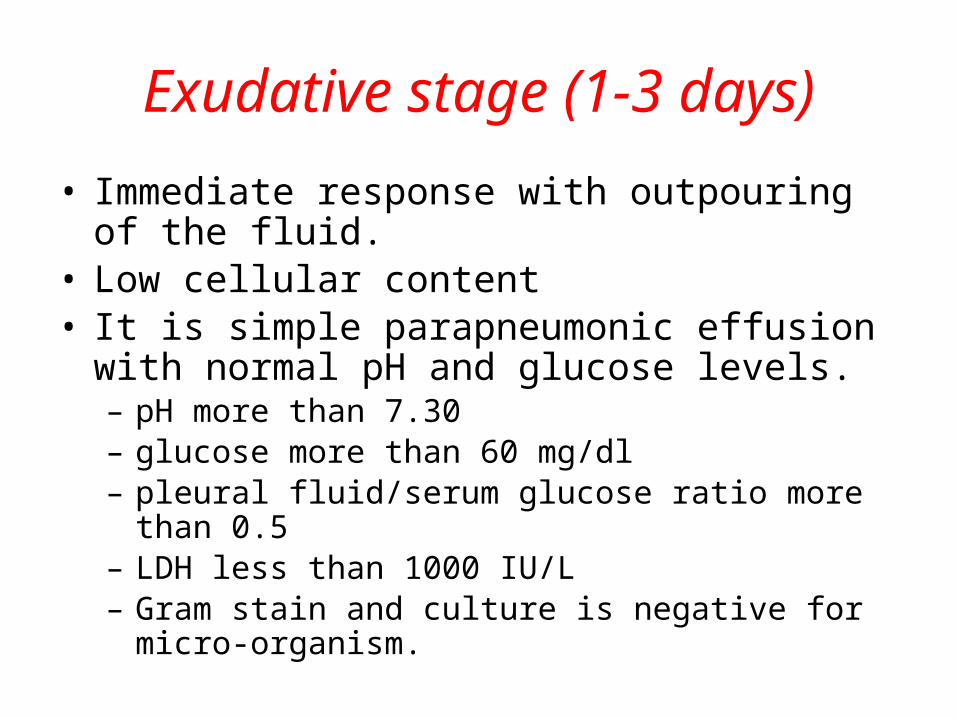
Exudative stage (1-3 days)
• Immediate response with outpouring of the fluid. • Low cellular content • It is simple parapneumonic effusion with normal
pH and glucose levels. – pH more than 7.30– glucose more than 60 mg/dl– pleural fluid/serum glucose ratio more than 0.5– LDH less than 1000 IU/L – Gram stain and culture is negative for micro-
organism.

Fibrino purulent stage (4 to 14 days)
• Large number of poly-morphonuclear leukocytes and fibrin accumulates
• Fluid pH and glucose level fall while LDH rises. • Acumulation of neutro-phils and fibrin, effusion becomes
purulent and viscous leading to development of empyema.
• There is progressive tendency towards loculations and formation of a limiting membranes.
• Pleural fluid analysis– Purulent fluid or pH less than 7.10, glucose less than 40 mg/dl
and LDH more than 1000 IU/L. Gram stain and culture reports show microorganism.

Organizing stage (after 14 days)
• Fibro-blasts grow into exudates on both the visceral and parietal pleural surfaces
• Development of an inelastic membrane "the peel". • Thickened pleural peel may prevent the entry of
anti-microbial drugs in the pleural space and in some cases can lead to drug resistance.
• Most common in S. aureus infection. • Thickened pleural peel can restrict lung movement
and it is commonly termed as trapped lung

CXR
• Large pleural effusion can be diagnosed in posteroanterior view
• Lateral decubitus view with affected side inferior facilitates recognition of smaller volumes of fluid.
• X-ray in different positions helps to recognize the extent of parenchymal infection and may reveal loculated fluid

USG
• Very useful tool for diagnosis, guidance of thoraco-centesis, or pleural catheter placement.
• Sonography can distinugish solid from liquid pleural abnormalities with 92% accuracy compared to 68% accuracy with chest X-ray. When both are combined, accuracy rises to 98%
• USG shows limiting membranes suggesting the presence of loculated collections even when they are invisible by CT scan.

Thoracocentesis and Pleural Fluid Analysis
• If effusion is free flowing and greater than one centimeter from inside of the chest wall to the pleural fluid line on the lateral decubitus view, immediate diagnostic thoracocentesis should be done.
• If loculated, thoracocentesis should be done under ultrasound guidance. The site for thoracocentesis is 1 cm below upper level of dullness

Thoracocentesis and Pleural Fluid Analysis
• Two third of the cases of anaerobic infection have malodorous empyema
• Protein level and specific gravity is rarely helpful in differentiating stages of empyema
• In some cases with frank pus, organisms are neither seen on Gram stain nor grown in culture. Such cases must raise a suspicion of chylous effusion
• cell fragments will sediment where a chylous effusion will remain opaque after centrifugation
• Tuberculous empyema can be confirmed by stains for acid fast bacilli in fewer than 25% cases but pleural biopsy and culture can diagnose more than 90% cases
• ADA more than 70 U/L supports the diagnosis of tuberculous pleural empyema
• PCR

Goal of treatment
1. Control of infection
2. Drainage of pus
3. Expansion of lungs

Treatment Options
• Non-Operative– Antibiotics– Thoracocentesis– ICTD– Fibrinolysis
• Operative– VATS– Thoracotomy
• Primary non-operative Salvage operative
• Primary operative

Emperical antibiotics
• Anti Staph antibiotic + Cephalosporin + Aminoglycoside
• Suspectedanaerobic infection Clindamycin should be added

Antibiotics
• Paren-teral therapy should be continued for 48-72 hours after abatement of fever and then oral therapy can be used to complete the course.
• Antibiotic should be continued until patient is afebrile, WBC count is normal, radiograph show consider-able clearing
• Duration of therapy– H. influnezae, S. pneumonia: 10-14 days– Staph aureus: 3-4 weeks

TUBE THORACOSTOMY
• Tube thoracostomy is usually the first step in the treatment of acute empyema. The success rate for tube thoracostomy is 70-85%

TUBE THORACOSTOMY IN LATE CASES
• Despite the expected low success rate for tube thoracostomy in the treatment of late empyema, it remains a first line therapy, if for no other reason than to attempt to decrease the severity of pleural sepsis until further therapy can be instituted.

FIBRINOLYTIC THERAPY
• The use of fibrinolytic therapy is associated with resolution of empyema thoracis in many cases

VAT
• VEDIO ASSISTED THORACOSCOPIC DEBRIDEMENT (VAT) debridement has achieved satisfactory results in the management of empyema in the literature.

RIB RESECTION
• Rib resection and insertion of large bore drain is another successful method
• This can only be achieved when a large bore tube is placed accurately in the most dependent part of the collection for a sufficient duration before organization occurred.
DECORTICATION
• Decortication represents the most invasive treatment for organized empyema cavities. Decortication allows a more rapid recovery with a decreased number of chest tube days, and decreased length of hospital stay.

THORACOPLASTY
• Thoracoplasty was a common procedure in the pre chemotherapeutic era of pulmonary tuberculosis. It plays an important but less prominent role in the treatment of tuberculosis and has relevance in non-tuberculous empyemas

MORTALITY
• For all stages, mortality rate may be as high as 10% in healthy patients and 50% in elderly or debilitated patients

Bronchopleural fistula
• ICTD• Decortication and fistula closure: after 2-3 weeks
of ICTD• Gradual tube withdrawal• Thoracoplasty or resectional surgery• Response depends on
– Size of fistula, – state of underlying lung and contralateral lung, – presence or absence of systemic illness, – nutritional rehabilitation

LUNG ABSCESS
• Lung abscesses are considered acute or chronic depending on the duration of symptoms at the time of patient presentation.
• The arbitrary dividing time is 4-6 weeks. • Primary lung abscess are commonly
observed in patients who are predisposed to aspiration or in otherwise healthy individuals,

• whereas secondary lung abscesses represent complications of a pre existing local lesion such as a bronchogenic carcinoma or a systemic disease (eg, HIV infection) that compromises immune function

ETIOLOGY
• Lung abscesses have numerous infectious causes. • Anaerobic bacteria continue to be accountable for
most cases. These bacteria predominate in the upper respiratory tract and are heavily concentrated in areas of oral-gingival disease.
• Other bacteria involved in lung abscesses are gram-positive and gram-negative organisms. However, lung cavities may not always be due to an underlying infection.

FACTORS CONTRIBUTING TO LUNG ABSCESS
• Oral cavity disease – Periodontal disease – Gingivitis
• Altered consciousness – Alcoholism – Coma

–Drug abuse –Anesthesia –Seizures
• Immunocompromised host –Steroid therapy –Chemotherapy –Malnutrition –Multiple trauma

• Esophageal disease –Achalasia –Reflux disease –Depressed cough and gag reflex –Esophageal obstruction
• Bronchial obstruction –Tumor –Foreign body –Stricture
• Generalized sepsis

PATHOGENESIS
• Aspiration of infectious material is the most frequent etiologic mechanism in the development of pyogenic lung abscess.
• Aspiration due to dysphagia (eg, achalasia) or to compromised consciousness (eg, alcoholism, seizure, cerebrovascular accident, head trauma) appears to be a predisposing factor.
• Poor oral hygiene, • dental infections, • and gingival disease are also common in these
patients.

• Although lung abscesses can occur in edentulous patients, an occult carcinoma should be considered. Edentulous patients very seldom, if ever, develop a putrefied abscess because they lack periodontal flora.
• Patients with alcoholism and those with chronic illnesses frequently have oropharyngeal colonization with gram-negative bacteria, especially when they undergo prolonged endotracheal intubation and are administered agents that neutralize gastric acidity.
• A pyogenic lung abscess can also develop from aspiration of infectious material from the oropharynx into the lung when the cough reflex is suppressed in a patient with gingivodental disease.

PATHOLOGY
• Abscesses generally develop in the right lung and involve the posterior segment of the right upper lobe, the superior segment of the lower lobe, or both.
• This is due to gravitation of the infectious material from the oropharynx into these dependent areas.
• Initially, the aspirated material settles in the distal bronchial system and develops into a localized pneumonitis.
• Within 24-48 hours, a large area of inflammation results, consisting of exudate, blood, and necrotic lung tissue. The abscess frequently connects with a bronchus and partially empties.

• After pyogenic pneumonitis develops in response to the aspirated infected material,
• liquefactive necrosis can occur secondary to bacterial proliferation and an inflammatory reaction to produce an acute abscess.
• As the liquefied necrotic material empties through the draining bronchus, a necrotic cavity containing an air-fluid level is created.
• The infection may extend into the pleural space and produce an empyema without rupture of the abscess cavity.
• The infectious process can also extend to the hilar and mediastinal lymph nodes, and these too may become purulent

Bacteriology of lung abscess
• Gram-negative organisms –Bacteroides species –Fusobacterium species –Proteus species –Aerobacter species –Escherichia coli

• Gram-positive organisms – Peptostreptococcus species – Microaerophilic streptococcus – Clostridium species – Staphylococcus species – Actinomyces species
• Opportunistic organisms – Candida species – Legionella species – Mycobacterium species

Clinical Features
• have had symptoms for at least 2 weeks• intermittent febrile course, productive cough,
weight loss, general malaise, and night sweats. Initially.
• foul sputum is not observed in the course of the infection; however, after cavitation occurs, putrid expectorations are quite prevalent.

• The odor of the breath and sputum of a patient with an anaerobic lung abscess is often quite pronounced and noxious and may provide a clue to the diagnosis.
• Hemoptysis may occasionally follow the expectoration of putrid sputum.

• Primary lung abscesses that occur following staphylococcal suppurative pneumonia in infants and children lack the typical indolent recurrent course of the more common postaspiration infections.
• Their onset tends to be abrupt and more threatening, producing :
• chills, fever, tachycardia, tachypnea, and unremitting production of putrid sputum.
• Auscultation may reveal coarse rhonchi and absent breath sounds.
• Clubbing of the fingers is sometimes noted.

Clinical Types
• Anaerobic necrotizing pneumonia• Usually, anaerobic necrotizing pneumonia is chiefly
restricted to one pulmonary segment or lobe, although it may progress to encompass an entire lung or both lungs.
• This type of anaerobic lung infection is the most serious. • The inflammatory process often spreads quickly and
causes destruction characterized by greenish staining of the lung and a huge amount of putrid tissue, resulting in pulmonary gangrene.
• These patients are gravely ill with a progressive septic course.
• Leukocytosis is obvious, and the sputum is putrid.

• Secondary lung abscess• In cases of secondary lung abscess, the
fundamental process (eg, bacteremia, endocarditis, septic thrombophlebitis, subphrenic infection) is generally apparent along with the pulmonary pathology.
• Infections below the diaphragm may extend to the lung or pleural space by way of the lymphatics, either directly through the diaphragm or via defects in it.
• The most typical hematogenous lung abscesses are observed in persons with staphylococcal bacteremia, especially in children.

• These abscesses are multiple and are located in the periphery of the lung.
• Infections may arise in or posterior to an obstruction (eg, an enlarged mediastinal lymph node) and migrate to the lungs.
• Septic emboli from bacterial endocarditis or emboli from deep pelvic veins may result in metastatic lung abscess.
• Septic emboli are suggested when multiple lesions appear over an extended period.
• Fewer than 5% of bland pulmonary infarcts become secondarily infected.
• Secondary infection of infarcts is suggested if fever and leukocytosis are present. Abscess formation may also occur within a necrotic pulmonary tumor.

Amoebic lung abscess
• Patients who develop an amoebic lung abscess often have symptoms associated with a liver abscess.
• These may include right upper quadrant pain and fever.
• After perforation of the liver abscess into the lung, the individual may develop a cough and expectorate a chocolate or anchovy paste–like sputum that has no odour.
• The patient may give a history of diarrhoea and travel outside the country

Chest radiographs
• The distinctive characteristic of lung abscess, the air-fluid level, (2weeks)can only be observed on a chest x-ray film taken with the patient upright or in the lateral decubitus position. In the presence of associated pleural thickening, atelectasis, or pneumothorax, the air-fluid level may be obscured. When better anatomic interpretation is required, CT scans have proven very useful.
CT SCAN
• Chest CT scan images are valuable for demonstrating cavitation within an area of consolidation,
• for evaluating the thickness and regularity of the abscess wall,
• and for determining the exact position of the abscess with regard to the chest wall and bronchus.
• CT scan images can also aid in evaluating the extent of bronchial involvement proximal or distal to the abscess.

Invasive diagnostic procedures
• transtracheal aspirates, • transthoracic aspirates, • and fiberoptic bronchoscopy• TO BE DONE prior to the institution of
antibiotic therapy • benefits of such procedures are controversial • Not Routinely but only for Atypical cases

Differential diagnosis
• Differential diagnoses of a cavitary lung lesion
• Anaerobic infection – Gram-negative bacteria – Pseudomonas species – Legionella species – Haemophilus influenzae species
• Gram-positive bacteria – Staphylococcus species – Streptococcus species – Mycobacterium species – Fungi

• Parasitic – E histolytica – Paragonimus westermani
• Septic – Embolism – Cavitary infarction – Bland infarction – Wegener vasculitis – Neoplasms – Bronchogenic carcinoma – Metastatic carcinoma – Lymphoma
• Sequestration – Bulla with fluid – Empyema with air fluid levels

Medical Treatment
• Antibiotics in lung abscess
• Anaerobic organisms–First choice - Clindamycin (Cleocin ) –Alternative - Penicillin –Oral therapy - Clindamycin,
metronidazole (Flagyl), amoxicillin (Amoxil)

• Gram-negative organisms • First choices – Cephalosporins,
aminoglycosides, quinolones • Alternatives – Penicillines and cephalexin
(Bio cef) • Oral therapy - Trimethoprim/sulfa methoxa
zole (Septran)• Pseudo monal organisms: First choices
include aminoglycosides, quinolones, and cephalosporin

• Gram-positive organisms –First choices - Oxacillin (Bactocill),
clindamycin, cephalexin, nafcillin (Nafcil), and amoxicillin
–Alternatives - Cefuroxime (Ceftin) and clindamycin
–Oral therapy - Vancomycin (Lyphocin)• Nocardial organisms: First choices
include trimethoprim/sulfamethoxazole and tetracycline (Sumycin).

DRAINAGE
• Most lung abscesses communicate with the tracheo bronchial tree early in the course of the infection and drain spontaneously during the course of therapy.
• Dependent drainage (with appropriate positions based on the pulmonary segment) is commonly advocated using chest physical therapy and sometimes broncho scopy.
• .

• Bronchoscopy can also facilitate abscess drainage by aspiration of the appropriate bronchus through the bronchoscope.
• Transbronchial drainage by catheterization of the appropriate bronchus under fluoroscopy has been successful

• Generally, augmenting this passive drainage with invasive procedures is unnecessary.
• In fact, attempts at therapeutic bronchoscopy may sometimes produce adverse consequences.
• Reports have been received of bronchoscopy-induced release of large amounts of purulent material from the involved lung segment into other parts of the lung,
• occasionally inducing acute respiratory failure, acute respiratory distress syndrome (ARDS), or both.

• Course of treatment• If treatment is started in the acute stage of the disease and is
continued for 4-6 weeks, • approximately 85-95% of patients with anaerobic lung abscesses
respond to medical management alone. Successful medical therapy resolves symptoms with no radiographic evidence or only a residual thin-walled cystic cavity (<2 cm after 4-6 wk of antibiotic therapy).
• The success of medical therapy is dependent on the duration of symptoms and the size of the cavity before the initiation of therapy.
• Antibiotic therapy is rarely successful if symptoms are present for longer than 12 weeks before the initiation of antibiotic therapy or if the original diameter of the cavity is more than 4 cm.
• When patients with lung abscesses do not respond to proper medical therapy, consider the probability of an underlying malignancy.

SURGICAL TREATMENT
• Contraindications to surgery • Several important factors must be considered prior
to undertaking surgery. • Because of the high risk of spillage of the abscess
into the contra lateral lung, it is almost essential that a double-lumen tube be used to protect the airway.
• If this is not available, surgery poses a very high risk of abscess in the other lung and a risk of ARDS.
.

• In such cases, postponing the surgery is a wise decision.
• Another, less-satisfactory method to deal with this problem includes positioning the patient in the prone position.
• The surgeon must be skilled in resecting the abscess and in rapid clamping of the bronchus to prevent spillage into the trachea.
• These factors are extremely important when dealing with the surgical aspects of treating a lung abscess. If doubt persists, postponing the surgery is best.

• Surgical treatment is now rarely necessary and is almost never the initial choice in the treatment of lung abscesses.
• In current practice, fewer than 15% of patients need surgical intervention for the unchecked disease and for complications that occur in both the acute and chronic stages of the disease.

• Surgical management is reserved for specific indications such as little or no response to medical treatment, inability to eliminate a carcinoma as a cause, critical hemoptysis, and complications of lung abscess (eg, empyema, bronchopleural fistula).
• In addition, if after 4-6 weeks of medical treatment a notable residual cavity remains and the patient is symptomatic, surgical resection is advocated

INDICATIONS OF SURGERY
• Probable carcinoma • Significant hemoptysis• Percutaneous drainage• Percutaneous drainage of a complicated abscess
(ie, one associated with fever and signs of sepsis) is beneficial in selected patients who do not respond to adequate medical therapy. These are ventilator-dependent patients who are not candidates for extensive thoracic procedures.

• Other indications for drainage include ongoing sepsis despite adequate antimicrobial therapy,
• progressively enlarging lung abscess in imminent danger of rupture,
• failure to wean from mechanical ventilation, and contamination of the opposite lung.
• In current practice, most of these lung abscesses are drained under CT guidance.
• Results achieved with percutaneous drainage show it to be safe and effective compared to surgery.
• Percutaneous drainage is rarely complicated by empyema, hemorrhage, or bronchopleural fistula.
• Although a few patients who undergo percutaneous drainage develop bronchopleural fistulas, most of these fistulas close spontaneously with resolution of the abscess cavity.
• Percutaneous drainage may be used to stabilize and prepare critically ill patients for surgery

• Abscess from gram-negative and opportunistic bacteria
• Hospital-acquired gram-negative infections are usually due to nosocomial organisms (eg, Pseudomonas, Enterobacter, Proteus). Patients with these infections are often elderly, debilitated with numerous major medical disorders, or have sustained multiple trauma. These patients are typically treated in a critical care unit.

• The infection is usually with a resistant organism originating from a single source. The lung abscess appears rapidly as an area of pneumonitis with associated pleural involvement. These patients often require per cutaneous drainage as an emergency procedure. Unfortunately, the infection is systemic and often out of control, and the pulmonary pathology represents only one aspect of a multi organ involvement with a rapidly deteriorating course.

• Among fungal infections, Candida albicans has become a major organism in lung abscesses. Fungal infections are difficult to treat, and amphotericin/fluconazole and surgical drainage remain the only modalities of treatment; however, at best, they have had only limited success.

COMPLICATIONS
• Approximately one third of lung abscesses are complicated by empyema. This may be observed with or without broncho- pleural fistulas. Haemoptysis is a common complication of a lung abscess and can be treated with bronchial artery embolization. Occasionally, the haemoptysis can be massive, thus requiring urgent surgery. Brain abscess may also be a complication in patients who receive inadequate treatment.

• Occasionally, the haemoptysis can be massive, thus requiring urgent surgery. Brain abscess may also be a complication in patients who receive inadequate treatment

PROGNOSIS
• The prognosis of patients with lung abscesses depends on the underlying or predisposing pathologic event and the speed with which appropriate therapy is established.
• Negative prognostic factors include a large cavity (>6 cm), necrotizing pneumonia, multiple abscesses, immuno compromise, age extremes, associated bronchial obstruction, and aerobic bacterial pneumonia.

• The mortality rate associated with an anaerobic lung abscess is less than 15%, although it is slightly higher in patients with necrotizing anaerobic pneumonia and pneumonia caused by gram-negative bacteria.
• The prognosis associated with amoebic lung abscess is good when treatment is prompt