infection in the newborn infant: the importance of
TRANSCRIPT

Henry Ford Hospital Medical Journal
Volume 13 | Number 2 Article 3
6-1965
Infection In The Newborn Infant: The ImportanceOf Prematurity And Obstetric ComplicationsPhilip J. Howard
Follow this and additional works at: https://scholarlycommons.henryford.com/hfhmedjournal
Part of the Life Sciences Commons, Medical Specialties Commons, and the Public HealthCommons
This Article is brought to you for free and open access by Henry Ford Health System Scholarly Commons. It has been accepted for inclusion in HenryFord Hospital Medical Journal by an authorized editor of Henry Ford Health System Scholarly Commons. For more information, please [email protected].
Recommended CitationHoward, Philip J. (1965) "Infection In The Newborn Infant: The Importance Of Prematurity And Obstetric Complications," HenryFord Hospital Medical Bulletin : Vol. 13 : No. 2 , 145-156.Available at: https://scholarlycommons.henryford.com/hfhmedjournal/vol13/iss2/3

Henry Ford Hosp. Med. Bull. Vol. 13, June, 1965
INFECTION IN THE NEWBORN INFANT
The Importance of Prematurity and Obstetric Complications.
PHILIP J. HOWARD, M.D.*
T H E OBJECT OF THIS paper is to emphasize the importance of prematurity in increasing the hazard of infection, to summarize the large place of infection in causing illness and death in the newborn group, and to show the relation of certain complications of pregnancy to mortality. Premature rupture of the amniotic membranes is, in many cases, also related to infandle infecdon. Suggestions are given for the recognition of septic states, and illustradons of infection in sample cases.
Perinatal mortality has progressively declined with the advent of improved nutrition, earlier recognidon of di.sease states such as toxemia, and better obstetrical technique. At present, a perinatal mortality rate of approximately 30 per 1000 dehveries is attained by hospitals, by many states, and by some countries. This total figure of 30 is composed of stillbirths and hebdomadal infant deaths in about equal parts. This mortality figure is directly affected by the incidence and degree of prematurity in the population sample being studied, and must be considered. Prematurity varies in incidence from 4.9 percent to 13 percent (Table I ) . Its known causes are social, economic, and nutritional, and among its suspected causes are endocrine diseases.
Table I Incidence of Prematurity'
Identity of Group Total Births Premature Infants (Under 2500 gms.)
Ford Hospital 1,213 8.9% Detroit-1962 44,021 (live) 7.0 Boston Lying-In 30,273 7.8 University Minnesota 845 5.7 University Oregon 917 10.0 N . Y. Med. Col. 976 13.0 Rotunda, Dublin 4,881 4.9
The premature infant has a physiologic predisposition toward infection because of ineffective lung expansion, anemia, hypo gamma-globulinemia, and other factors.
'•'Department of Pediatrics Presented in part at the Fourth Annual Michigan Conference on Maternal and Perinatal Welfare March 11, 1965, Flint, Michigan.
145

HOWARD
Extra precautions are regularly used in premature care by incubation, isolation and controlled oxygenation. Thus a continued attempt is made to protect them from infection.
Infections are directly responsible for a definite fraction of neonatal and perinatal
deaths. The detailed studies of the pathology of these groups carried on by Potter,^
by Nesbitt,^ and by Bound," are extremely valuable because these causes, in minor
forms, cause the morbidity in nursery groups, in many samples of which proper
treatment leads to a good result. Potter's report showed the results of 8905 autopsies
in the newborn, and she reported 1197 or 13.4 percent due to infection. The
following tables I I and I I I show the reports of Nesbitt^ and Bound."
Table I I Relative Importance of Causes of Fetal and Neonatal Deaths Infants 1000 grains and Over.=
Disease Fetal Neonatal Total
Anoxia-visible petechial hemorrhage 25% 9% 34'% No Cause Found 23 9 32 Birth Injury 2 8 10 Malformation 4 5 9 Abnormal Pulmonary Ventilation 0 7 7 Infection 1 4,5 5.5 Erythroblastosis .6 1.1 1.7 Other .7 .1 .8
Total 56.3 43.7 100.
Table I I I Classification of Perinatal Mortality. Causes of Death in 337 Consecutive autopsies.^
Causes Stillbirths Premature Term Total Percent
Antepartum with maceration 61 — — 61 18.0% Intrapartum Asphyxia 44 5 6 55 16.3% Malformed 24 11 17 52 15.5% Birth Trauma 8 14 15 37 11.0% R.D.S. — 30 6 36 10.7% Antepartum Asphyxia 36 — 36 10.7% Pneumonia — 8 1 1 IV 5.6% Erythroblastosis 8 3 4 15 4.4% Intraventricular Hemorrhage 10 1 1 1 3.3% Miscellaneous 4 I 3 8 2.4% Previable — 7 7 2 .1%
Total 337 100. %
These analyses of causes show that infection plays an important part in mortality.
It is often not clearly evident whether abnormal pulmonary ventilation and respiratory
disease syndrome are types of infection. It is known, however, that any interference
with alveolar exchange promotes local congestion and infection. These studies show
146

INFECTION IN THE NEWBORN INFANT
at least 5.6% and possibly as high as 16.3% of perinatal deaths in which infection plays a role as the primary cause of damage, or as a secondary invader. Infection is a tangible reality subject to study, culture, isolation and hopefully successful treatment.
Table IV
Mortahty Rate According to Diagnosis in High-Risk Pregnancies.'
Incidence/ Mortality Diagnosis 1000 Total Stillbirths Live Births Totals
Births % % % Eclampsia 0.8 28.0 12.0 40.0 Hydramnios 4.7 17.2 22.1 39.3 Premature separation 12.6 21.2 12.7 33.9 Prolapsed cord 4.8 19.7 6.1 25.8 Pre-eclampsia (Grade 2) 6,2 14.7 6.8 21.5 Placenta previa 5.7 7.5 13.8 21.3 Breech (single) 36.5 9.2 8.5 17.7 Diabetes 6.0 6.0 9.2 15.2 Twin birth 22.7 4.3 6.3 10.6 Phlebitis 5.7 4.5 2,3 6.8 Pyelonephritis 6.3 3.1 3.6 6.7 Hypertension and proteinuria 13.9 5.4 11.7 6.1 Pre-eclampsia (Grade 1) 34.8 4.2 1.5 5.7 Pyelitis 32.7 2.1 2.1 4.2 A l l ho.spitalized patients. 30,765 1.6 1.5 3.1
Table V
Individual Contribution to Total Hospital Perinatal MortalityS
Cause of Death Incidence/ 1000 Total
Births*
Stillbirths/
Live-born Deaths/
Perinatal Mortal i ty/
Incidence/ 1000 Total
Births* 1000 Total 1000 Total 1000 Total
Incidence/ 1000 Total
Births* Births Births Births
Breech (Single) 36.5 3.4 3.1 6.5 Premature separation 12.6 2.7 1,6 4.3 Twin birth 22.7 0.8 1.4 2.2 Pre-eclampsia (Grade 1) 34.8 1.5 0.5 2.0 Hydramnios 4.7 0.8 1,0 I.S Pyelitis 32.7 0,7 0.7 1.4 Pre-eclampsia (Grade 2) 6.2 0.9 0.4 1.3 Placenta previa 5.7 11,4 0.8 1.2 Prolapsed cord 4.8 0.9 0.3 1.2 Diabetes 6.0 0.4 (1,6 1.0 Hypertension and proteinuria 13.9 (1.7 0.1 II.X
Pyelonephritis 6.3 0,2 0.2 0.4 Phlebitis 5,7 ().? 0,1 0.4 Eclampsia l),S 0.2 11,1 0.3 Total hospital perinatal mortality 16.1 14.9 31.0
HOWARD
There are certain complications of pregnancy associated not only with death
of the infant but also with disease states such as anoxia and brain damage. The
cause of death in one infant will be the cause of slight or moderate trouble in
another. This has been clearly stated by Clifford' and realized by all dealing with
newborn problems. Tables IV and V hst comphcations of pregnancy with their
individual risk to the infant. Eclampsia is shown to be the most dangerous compli
cations being associated with 40 percent perinatal mortality or 400 deaths per 1000
eclamptic deliveries. Other complications are listed in descending order of risk to
pyelitis carrying a risk of 4.2 percent. The first item in Table V shows breech
presentation to be the complication which produces the most perinatal deaths in the
total infant population. Breech delivery is not only dangerous, but common and
accounts for 6.5 deaths of the total 31 perinatal deaths per 1000 deliveries. Other
complications are listed in descending order of total importance in producing perinatal
mortality, eclampsia being the last because it is so rare.
The infants of these complex pregnancies then are the most threatened, and
among them are many with minor anoxia, slight trauma,, and especially beginning
infection. These conditions must bs recognized and treated early to obtain the best
results.
The place of premature rupture of the amniotic membranes is simply that of
an added complication. In the extensive computer analysis of 30,765 deliveries de
tailed in Tables IV and V, this complication was not separately studied. It is, however,
at times an important help in alerting the physician to possible infection. During
one year May 1963 to May 1964 this complication was studied at the Henry Ford
Hospital. Among 1213 deliveries there were 46 with premature amniotic membrane
rupture of 20 hours or more before delivery. Seven known infections were observed,
and 18 infants were suspected of infection and treated. The seven infected infants
had either positive culture or X-Ray evidence of pneumonia. Details of this group
are shown on Table V I .
Infection must be suspected in the newborn infant on very slight signs, such as
anoxia, cyanosis, irregular breathing, weak cry and poor sucking reflex. Jaundice
or increased bilirubin if unexplained by erythroblastosis or obstruction suggests infection.
Unusual exposure to infection by early rupture of membranes, prolonged labor or
maternal prepartum fever, always .suggest germ or virus invasion of the fetus. Four
conditions, respiratory distress syndrome, birth trauma, pneumonia, and intraven
tricular hemorrhage cause two-thirds of infant deaths, and are often indistinguishable
at birth on clinical grounds. These conditions should have cuhure studies, constant
observation, and in most cases prophylactic treatment for three days only unless
the diagnosis is definite.
148

INFECTION IN THE NEWBORN INFANT
Table V I Seven Infants born after Prolonged Rupture Time who showed infection or
pneumonia. Maternal complications, and infant treatment shown.
Case Sex Birth Weight (gms)
Rupt. Time*
Obstetric Complications
Infant Condition and Rx
1 M 2700 59 hrs Age 20. Sickle cell anemia. Gray. I I Para O. Failed medical induction Caesarian. Unmarried. Psychotic Hbg 8.7 gms
Weak Cultures-Throat Staph Albus; cord, cornybacteria Achro 60 mg. d. x 3 days
2 M 2690 34 hrs Age 27. Gray. V Para I I I . Miscarriage 1. Amniotic Ft. culture; E. coli and peptococcus. T. 102 F. Chlor. 2. gm d Pen. 625 mg d. Labor induced.
Weakness Pen. 150 mg. d x 3d Strep 60 mg. d. x 3d Cultures and X-Ray neg.
3 F 1710 5 days Age 34. Grav. IV Para I I . One Tubal Preg. Pen & Strep Id.
Weakness. Throat Cult. Staph Albus Pen 500 mg. d. X 4d. Strep 60 mg. d. x 4d.
4 F 3500 24 hrs Age 31. Labor 55 hrs. Grav. I I . Obese. Edema, Para O. Wgt 197 lbs. Pen 625. mg I . V. Strep 500 mg 1. M.
Weakness. Bl Culture E. Coli and enterococci Pen 350 mg. d. x 3d Strep 60 mg. d. x 3d
5 M 4380 26 hrs Age 38. Grav. V Para I I . Obese Wgt 177. Labor 8 hrs. Serology positive. Abortions 11 B. P. increased
Weakness. Throat culture Coliform. Bl cult cornybact. Pen 350 mg. d. x 3d Strep 120 mg. d. x 3d
6 M 1230 66 hrs Age 30 Caesarian section
Pneumonia by X-Ray Pen 125 mg. d. x 3d Strep 60 mg. d. x 3d
7 F 3120 5 days Age 40. Grav. X I Para V. Hypertension Induced
Weakness. Cord Bl Coliform. Pen 375 mg. d. x 3d Strep 100 mg. d. x 3d
Pen — Penicillin. 62.5 mg. equals 100,000 units. Strep — Streptomycin.
*Rupt Time equals hours before dehvery of rupture of amniotic membranes.
The following case reports of newborn infants illustrate some of the situations
associated with infection.
CASE 1. Baby M . V. was bom Jan. I I , 1960 and lived 14 days. Her birth weight
was 1300 gms (2 lbs. 13 oz). She was the second of twins. The mother was 35
years old. This was her second pregnancy and it was terminated by Caesarian section
for placenta previa. The infant was a normal premature with an Apgar score of 8
given regular care of gavage feedings, oxygen and temperature control. She gained
daily from the 9th day on and the 13th day showed spells of apnoea, an increased
respiratory rate to 70 per minute, a cardiac rate of 180 per minute. There were also
abdominal distension, and bowel patterning. Rapid increase in weakness in the next
149

HOWARD
Figure 1 Brain damage following early meningitis, a. Front view. b. Side view. Case 2.
/
w
WW
Figure 2 Pseudomonas sepsis, abscesses, and pneumonia associated with congenital lamellar ichthyosis, a. Front view. b. Back view. Case 6.
150

INFECTION IN THE NEWBORN INFANT
few hours and death followed. Clinical diagnosis was intestinal obstruction and heart failure. The autopsy showed E. coli meningitis.
Early blood and spinal f l u id cultures followed by chlormycetin treatment are at present thought best in E. coli sepsis or meningitis.
CASE 2. Baby G. DiV. was born Aug. 16, 1964, and survived. His birth weight was 2724 gms (6 lbs) and he was born after a 12 hour labor, and was the mother's third child He was discharged home the third day of life in apparently good condition. The fourth day of life, the parents undertook an auto trip to St. Louis. During the trip the infant became febrile with temperature of 106°, and also was non-responsive. They sought treatment in Indianapolis where an injection of penicillian was given. On arrival in St. Louis, infant was placed in the C.G.M. Hospital and remained there 9 days. The findings were temperature 102 6F respiratory rate 80 per minute, pulse 140 per minute, a molded skin, minimum movement facial weakness, dry right cornea, skull sutures separated from dehydration, and decreased muscle tone. The clinical diagnosis was sepsis with meningitis, or brain abnormality secondary to birth injury, convulsive disorder secondary to this plus possible hypoglycemia or hypocalcemia Subdural taps were negative as well as spinal fluid cultures. The treatment was rehydration and antibiotics. On returning to Detroit at the age of 2 weeks, the infant had bulging fontanelles. separated sutures, and poor nutrition. Subdural hematomas were found and drained. The subsequent history has been one of surgical treatment and Pudenz valve insertion. Final diagnosis possible sepsis, possible meningitis, brain injury, hydrocephalus permanent brain damage. (Fig. 1).
Exposure of newborn infants should be avoided. I f travel becomes imperative, the infant should be returned to the hospital for temporary care.
CASE 3. Baby D. L. was born Nov. 23, 1964 and her birth weight was 3240 gms. (7 lbs 1 oz). The mother was 21 years of age and this was her first pregnancy. The amniotic membranes ruptured 2 days before delivery and an induction was attempted with intravenous pituitrin. A Caesarian section was then done. The infant appeared normal, having an Apgar score of 10. The fourth day baby became jaundiced and failed to gain. Mother and infant both were Group A Rh positive. The bilirubin rose to 21.2 mg percent, and an exchange transfusion was given. The blood culture showed staphlococcus albus coagulase negative and alpha streptocci moderately sensitive to penicilhn. Treatment was achromycin 100 mg per day for 6 days and penicillin 500 mg per day for 6 days. Infant showed immediate recovery.
CASE 4. Baby R. K. was born Jan. 6, 1960 and died of infection the third day. The birth weight was 3000 gms (6 lbs). The mother was 22 years of age, was gravida 2, had one living child, and had diabetes. The labor lasted 12 hours 37 minutes, was at the 32nd week of gestadon. was a breech presentation rotated to occiput anterior, and delivered with mid forceps. Infant seemed normal and had an apgar score of 9. However, by 6 hours of age had become cyanotic and weak. Oxygen was given constantiy. Icterus developed and by second day reached 16.5 mg percent. The mother was blood group A, Rh positive, the infant was group B, Rh positive. Cyanosis increased and resuscitadon was needed. The Bilirubin reached 23.8 mg percent and exchange transfusion was tried. After exchanging 400 cc. infant expired. The blood culture showed staphylococcus coagulase negative, and streptococcus The post mortem showed atelectasis, and hver defect.
Diabedc offspring are especially subject to the many possible comphcations. This infant was too weak to survive respiratory distress, erythroblastosis, sepsis and exchange transfusion. The exchange was undertaken here with fu l l knowledge of the many complications.
CASE 5. Baby Dal. was born Sept. 15, 1960. Her birth weight was 4260 gms (9 lbs 6 oz). The mother was gravida 4, and had previous Caesarian section for placenta previa, so section was repeated. The infant at once showed respiratory distress, with respiratory rate of 125 per minute. There developed the second day a lung infiltrate. The trachea and bronchi were suctioned by bronchoscopy and respiratory rate dropped to 85 per minute. Jaundice developed the 3rd day, bilirubin rose to 8.1 mg and blood cultures showed staphylococcus. Treatment was carried on with achromycin 400 mg daily for 3 days, Chloromycetin 160 mg daily for 3 days, and erythromycetin 160 mg daily for 3 days. X-Rays of the lungs the 4th day showed nodules, consolidation, and hyperlucency. The X-Ray cleared by the 13th day and infant steadily improved and was discharged. The staphylococcus was very sensitive to erythromycin.
This was considered a staphylococcus septicaemia and pneumonia which responded to treatment.
151

HOWARD
CASE 6. Baby K. K. was born at term July 10, 1964 and lived three and a half months. She was the second baby, pregnancy and delivery were normal. The first infant died of congenital skin disease and septicaemia. Present infant showed epidermolysis and respiratory difficulty at birth, followed by pseudomonas sepsis, pneumonia, convulsions, progressive skin disease, and failure to grow. Antibiotics, transfusion of whole blood, and of packed cells, bronchoscopy and careful nursing all failed and infant steadily failed and expired. The hemoglobin was 10 gms or above most of the time. The gamma globulin was 0.36 gms percent. Final diagnosis was Bronchopneumonia, local abscesses, sepsis due to pseudomonas and congenital ichthyosis lamellar type. The newborn is markedly deficient in bactericidins to gram negative enteric bacilli.' Commercial gamma globulin is low in antibodies to gram negative organisms. In spite of whole blood and plasma, colistin and other support, this infant became progressively worse. (Fig. 2) .
CASE 7. Baby B. A. was born prematurely Apri l 21, 1963. His birth weight was 1255 gms. (2 lbs. 12 oz) and he lived 36 hours. The mother, 37 years of age, was gravida 4, and had lost one former premature weighing 4 lbs. 13 oz. The rupture time was 18 hrs. 46 min. Labor time 4 hrs. 48 minutes. The presentation was vertex, and she was said to be in 24th week. The baby was weak and had an Apgar score of 5. The respiratory rate was 40, but increased to 70, retraction of chest was minimal but increased, cyanosis became more marked, and baby became progressively weaker and expired. Chnical diagnosis was respiratory disease syndrome, meaning hyaline membrane disease. X-Ray of chest showed extensive redcular formation of both lungs, nodular densities, probable lung hemorrhage,, and slight air bronchogram. Autopsy showed congenital heart disease, atelectasis, and pulmonary hemorrhage. This baby illustrates difticulty of diagnosis in this group. (Fig. 3) .
Figure 3
Respiratory distress syndrome and pulmonary hemorrhage. Autopsy showed also, congenital heart disease and atelectasis. Illustrates difficulty in diagnosis. Case 7.
152

INFECTION IN THE NEWBORN INFANT
CASE 8. Baby E. D. was born prematurely weighing 2000 gms (4 lbs. 9 oz) and lived 55 hrs and 17 min. She was the second living child of mother, gravida V, who was a diabetic A Caesarian section was done at the 36th week. The infant's "Apgar score was 9 and by the 4th hour she showed mild retraction and cyanosis. These signs increased along with respiratory rate and grunting respiration. Treatment consisted of mist, oxygen, achromycin 100 mg daily for 2 days, solu-cortef i.m. 20 mg twice daily for 2 days, but infant became progressively weaker and expired. X-Ray showed bilateral, peribronchial intiltra'ion and air bronchogram. The autopsy showed 70% of all alveoh affected with hyaline alveolar membrane. Lhis demonstrates the rapid general development of hyahne membrane in some infants. (Fig. 4) .
CASE 9. Baby K. W. was born prematurely weighing 1230 gms (2 lbs 11 oz). The mother 30 years old, was gravida I X , had history of three miscarriages, and six hving children' AmnioUc niembranes ruptured 3 days before delivery. Pituitrin inducdon was unsuccessful and Caesarian section was needed. The infant had slight retraction but an Apgar score of 9 At 6 hours of age. slight retraction, cyanosis and increased respiratory rate were present Ireatment consisted of oxygen, mist, temperature control, penicillin 125 mg day for 3 days streptomycin 60 mg per day for 3 days. X-Ray of chest showed "hyaline membrane disease"' 1 he nose and throat cultures were negative. Infant gained steadily from 5th day and went home weighing SVi lbs. (Fig. 5) .
CASE 10 Baby T. H . was born at term September 30, 1964, weighing 3700 gms. (8 lbs 2 oz). The mother, 20 years old, was primiparous, the amniotic membranes ruptured 36 hours before delivery. Induction was aUempted with pituitrin intravenously, but as this was unsuccessful, a Caesarean was done. The infant was in fair condidon and given an Apgar score of 8. There was moderate respiratory difficulty for a few hours. Nose and throat cultures and gastric content cultures were negative, but blood culture was positive for pepto-streptococcus, a fastidious organism that prefers anaerobiosis initially for growth. Treatment
Figure 4
Hyaline membrane disease. Case 8.
153

HOWARD
BHII^Ii
Figure 5
Respiratory distress syndrome in premature infant weighing 1230 gms. Progressive complete recovery. Case 9.
was penicillin 187.5 mg per day for 3 days, and streptomycin 75 mg per day for 3 days. Infant recovered well. This child illustrates the necessity of blood culture on all infants with respiratory difficulty.
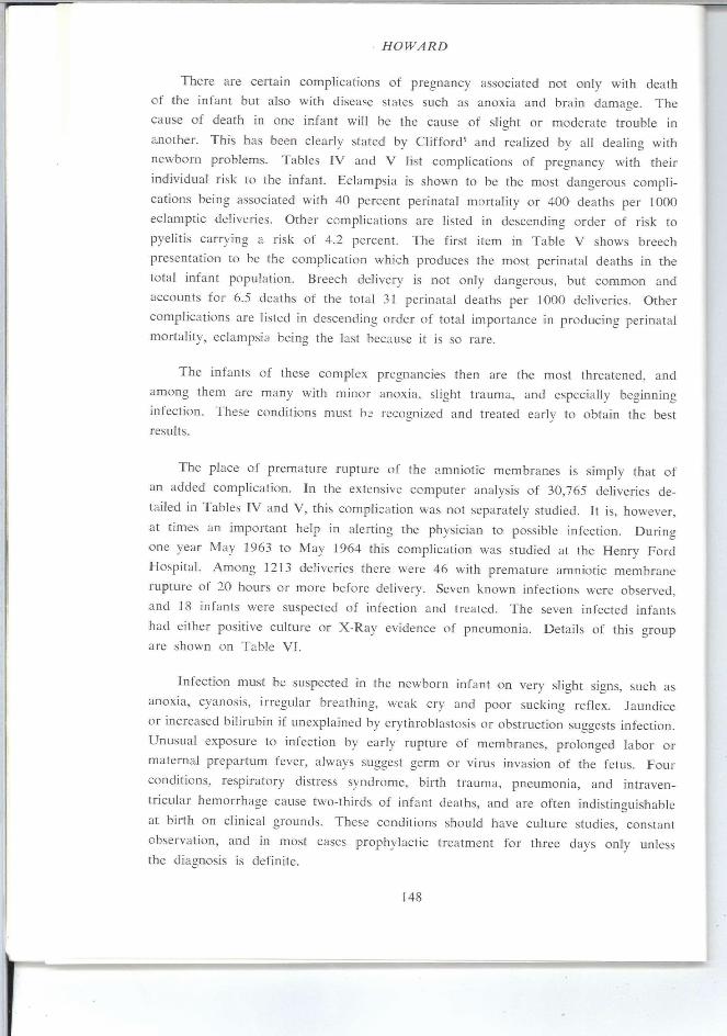
CASE 11. Baby D. W. was born prematurely, September 17th, 1964 weighing 2410 gms. (5 lbs. 5 oz). The mother, 37 years old, was gravida V with 3 living children. The amniodc membranes ruptured 3 days before delivery, labor was induced with intravenous pituitrin, and delivery aided with forceps. The mother was given penicillin and streptomycin antepartum. The infant was slightly weak and cyanotic but given an Apgar of 8, and treated with penicillin 125 mg per day for 4 days, and streptomycin 50 mg daily for 11 days. A slight systolic murmur developed into a patent ductus murmur and jundice developed with a bilirubin of 23.9 mg percent, requiring an exchange transfusion. Respiration was a little irregular, and the 9th day of Kfe seemed worse. X-Rays of lungs now showed a progressing pneumonia. Infant kept on streptomycin, and chlormycetin started 50 mg per day for 2 days, and then 30 mg per day for 3 days. The lungs cleared and infant recovered completely and was discharged. Typical course of late gram-neg bacillus infection. Blood cultures were negative. (Fig. 6) .
CASE 12. Baby D. was born weighing 1820 gms. (4 lbs.) Dec. 27, 1962. The mother, age 38, was gravida V I . with four living children, and she had formerly worked in a Tuberculosis Sanatorium. Her chest X-Rays during this pregnancy were completely negative. Infant's only home contact with mother was Jan. 15 to Jan. 22, 19th to 26th days of life. Infant was born with good strength and activity, and seemed normal the first 19 days of life, gaining 13 ounces. At age of 26 days infant was returned to the hospital with fever and respiratory difficulty and X-Ray taken Jan. 22nd showed extensive tuberculosis, tuberculin test was positive Jan. 31st and gastric washings showed tubercle bacilli. Treatment was started with streptomycin isoniazid and para amino salicylic acid. Steroids and tube feedings were used. At 8 months of age infant weighed 11 lbs. Mother's uterus was curetted and tubercle bacilli demonstrated. Twenty-one days of infection are necessary to develop a positive tuberculin test. Such an exposure as this infant had is quite rare.^
154
INFECTION IN THE NEWBORN INFANT
Figure 6
Delayed pneumonia showing 9th day of life probably due to gram-negative organism invasion. Case 11.
SUMMARY
Prematurity increases the risk of infection during the neonatal period. Extensive
autopsy studies of perinatal deaths suggest that 5.6 percent to 16.3 percent of them
are caused or complicated by infection. Severe pregnancies disturbed by conditions
such as eclampsia or breech presentation show an increased perinatal mortality a.nd
morbidity. Prominent among the causes of disease in the newborn are infections
secondary to prematurity, atelectasis, and anoxia. Premature rupture of the amniotic
membranes opens a channel for direct infection of the infant lung and intestinal
tract. The recognition of infection in the newborn infant depends largely on minute
physical signs found in situations of high risk.
155
HOWARD
REFERENCES
1. Michigan Department of Health: Michigan Health Statistics, 1962, Lansing, Michigan; Cl i f ford , S. N . : High-risk pregnancy. I . New Eng. J. Med. 271:243, 1964; Jacobsen, H . N . , and Reid, D. E.: High-risk pregnancy. I I . New Eng. J. Med. 271:302, 1964: Rotunda Hospital, Dublin, Ireland: Clinical Report, 1963.
2. Bundesen, H . N . , Potter, E. L., Fishbein, W. I . , Bauer, F. C , and Platzke, G. V . : Progress in the^^prevemion of needless neonatal deaths, Chicago Health Department: Annual Report,
3. Nesbitt, R. E. L. , and Anderson, G. W.: Perinatal mortality, Obst. Gynec. 8:50, 1956.
4. Bound, J. P., Buder, N . R., and Spector, W. G.: Classification and causes of perinatal mortahty
Brit. Med. J. 2:1191, 1956.
5. Cl i f fo rd , S. H . : High-risk pregnancy. I . New Eng. J. Med. 271:243, 1964.
6. Gitlin, D., Rosen, F. S., and Michael, J. G.: Transient 19S gammaglobulin deficiency in the newborn infant, and its significance. Pediatrics 31:197, 1963.
7. Eickhoff, T. C , Klein, J. O., Daly, A . K., Ingall, D., and Finland, M . : Neonatal sepsis and others infections due to group B beta-hemolytic streptococci. New Eng. J. Med. 271-1221 1964.
8. Kempton, G. B., and Howard, P. J.: Congenital tuberculosis, Henry Ford Hosp. Med. Bull 1:6, 1953. y f •
156