indications for coronary angiography - …cdn.intechweb.org/pdfs/23195.pdf · indications for...
TRANSCRIPT

3
Indications for Coronary Angiography
Karl Poon and Darren Walters The Prince Charles Hospital
Australia
1. Introduction
Since the first serendipitous selective coronary angiography was performed by pediatric
cardiologist Dr. Mason Sones in 1958 during ventriculography and aortography (Sones et
al., 1959), invasive coronary angiography has become well established as the diagnostic gold
standard for coronary artery disease.
Coronary angiography provides definitive information of “luminology” (radiographic
outline using a contrast agent) of the coronary arteries. It is the standard with which other
diagnostic modalities are compared. Millions are performed annually worldwide. Whilst the
risk of coronary angiography of complications is very low in experienced hands the
procedure is invasive and, requires dedicated infrastructure and expense. There are certain
specific indications for coronary angiography. The general indication for a coronary
angiography is to define the coronary artery anatomy. More detailed guidelines for
coronary angiography date back to 1999 from the American College of Cardiology (Scanlon
et al., 1999). This of course predated the enormous technological advancement over the past
decade, the development in computed tomography coronary angiography, the widespread
adoption of coronary stenting, the improvement afforded by drug eluting stents, and the
availability of new antiplatelet therapy that enhanced the safety of all coronary
interventions.
This chapter examines the appropriate indications for invasive coronary angiography in the
current era, assesses the role of non invasive CT coronary angiography, and incorporates the
emerging adjunctive role in the management of patients undergoing percutaneous cardiac
structural interventions. Case vignettes are included in this chapter to illustrate the
indications and use of coronary angiography in the current era.
It must be stressed whilst guidelines do provide recommendations regarding the indications
for invasive coronary angiography, many more complex clinical and non clinical factors
exist for each individual patient and the clinician must exercise clinical acumen to proceed
to perform an invasive diagnostic and/or therapeutic procedure.
2. Indications for coronary angiography
Coronary angiography involves the opacification of the lumen in coronary arteries and
acquisition of this luminogram under fluoroscopy. Consequently the indication for coronary
angiography is predominantly focused on the diagnosis of any conditions that can lead to
luminal compromise. This anatomical study defines the origin, course and pattern of
www.intechopen.com
Advances in the Diagnosis of Coronary Atherosclerosis
34
epicardial arteries, with a spatial resolution that is still superior to other non-invasive
imaging modalities. There are however limitations to coronary angiography. Aside from the
obvious invasive nature and attendant risks, it provides only a projection of the lumen
outlined by contrast and hence no information on the extent of atheroma contained within
the vessel wall or the functional significance of the stenosis which can only be inferred. Both
of these limitations can be overcome by the use of intravascular modalities such as
intravascular ultrasound, optical coherence tomography and fractional flow reserve
assessment. When combined with these adjuncts, coronary angiography serves as a
formidable tool in guiding revascularization strategies, with a body of scientific data that
has been well validated to provide long term prognostic benefit.
The guiding principle in determining the indication behind the decision to undertake
coronary angiography in a given patients is similar to that underpinning the decision to
undertake any invasive investigations in medicine. The decision to proceed is based on that
the knowledge gained from the investigation outweighs the perceived risk, that the result
will benefit the patient, in that it will likely change management and the available treatment
option will improve symptoms and/or prognosis. The risks and complications of coronary
angiography are covered elsewhere in this book and this chapter will focus on the
indications of coronary angiography. Relative and absolute contraindications are also
covered briefly.
The indications for coronary angiography have been divided into the follow clinical
categories:
1. Coronary artery disease 1. Stable pattern
i. suspected coronary artery disease ii. known coronary artery disease
2. Unstable pattern i. Acute coronary syndrome with unstable haemodynamics or rhythm ii. Unstable angina and Non-ST elevation MI iii. ST elevation MI iv. Out of hospital cardiac arrest
3. Special considerations i. Congestive cardiac failure ii. Preoperative assessment for non cardiac surgery iii. The value of non invasive computed tomography coronary angiography
4. Relative and absolute contraindications 2. Prelude to structural cardiac disease/valvular heart disease intervention 3. Case vignettes of the use of coronary angiography in the contemporary era
3. Coronary artery disease
Coronary angiography is pivotal in the diagnostic algorithm as the gold standard for the
confirmation of coronary artery stenosis. A number of well defined clinical scenarios
often lead to the suspicion of coronary artery disease. This section is separated into
patients who present in stable pattern, symptomatic or asymptomatic; and patients who
present in an acute manner highly suggestive of coronary artery disease, i.e. acute
coronary syndromes. Specifics scenarios, such as the role of coronary angiography in the
www.intechopen.com
Indications for Coronary Angiography
35
investigation of congestive cardiac failure, pre-valvular surgery and the emerging role of
computed tomography coronary angiography will be covered too.
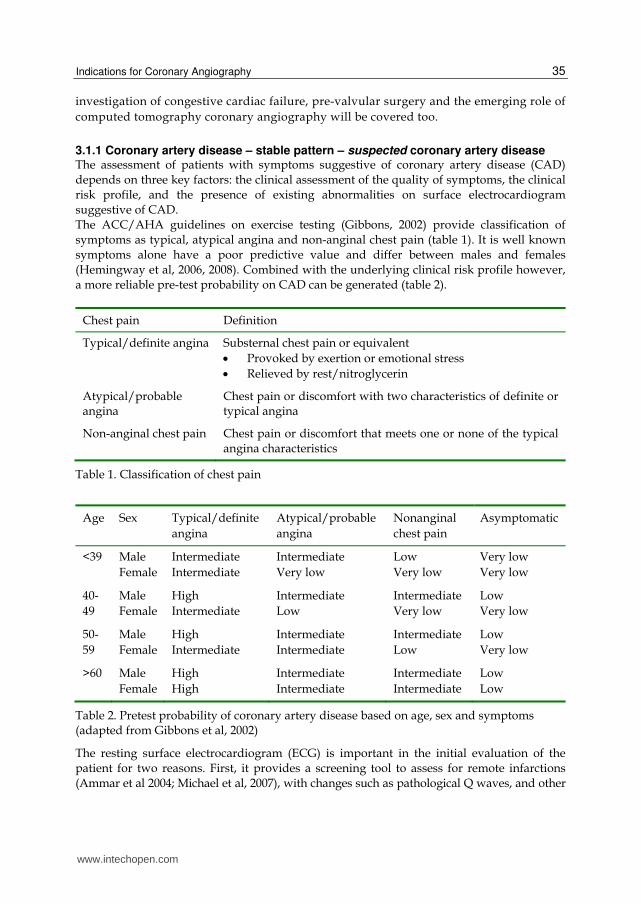
3.1.1 Coronary artery disease – stable pattern – suspected coronary artery disease The assessment of patients with symptoms suggestive of coronary artery disease (CAD) depends on three key factors: the clinical assessment of the quality of symptoms, the clinical risk profile, and the presence of existing abnormalities on surface electrocardiogram suggestive of CAD. The ACC/AHA guidelines on exercise testing (Gibbons, 2002) provide classification of symptoms as typical, atypical angina and non-anginal chest pain (table 1). It is well known symptoms alone have a poor predictive value and differ between males and females (Hemingway et al, 2006, 2008). Combined with the underlying clinical risk profile however, a more reliable pre-test probability on CAD can be generated (table 2).
Chest pain Definition
Typical/definite angina Substernal chest pain or equivalent
Provoked by exertion or emotional stress
Relieved by rest/nitroglycerin
Atypical/probable angina
Chest pain or discomfort with two characteristics of definite or typical angina
Non-anginal chest pain Chest pain or discomfort that meets one or none of the typical angina characteristics
Table 1. Classification of chest pain
Age Sex Typical/definite
angina
Atypical/probable
angina
Nonanginal
chest pain
Asymptomatic
<39 Male
Female
Intermediate
Intermediate
Intermediate
Very low
Low
Very low
Very low
Very low
40-
49
Male
Female
High
Intermediate
Intermediate
Low
Intermediate
Very low
Low
Very low
50-
59
Male
Female
High
Intermediate
Intermediate
Intermediate
Intermediate
Low
Low
Very low
>60 Male
Female
High
High
Intermediate
Intermediate
Intermediate
Intermediate
Low
Low
Table 2. Pretest probability of coronary artery disease based on age, sex and symptoms (adapted from Gibbons et al, 2002)
The resting surface electrocardiogram (ECG) is important in the initial evaluation of the patient for two reasons. First, it provides a screening tool to assess for remote infarctions (Ammar et al 2004; Michael et al, 2007), with changes such as pathological Q waves, and other
www.intechopen.com

Advances in the Diagnosis of Coronary Atherosclerosis
36
non-specific ST segment and T wave changes. Second, any such changes may guide the choice of the appropriate functional study. The presence of a left bundle branch block, for example, essentially renders exercise stress electrocardiography uninterpretable and confounds assessment of stress echocardiography or perfusion studies (La Canna et al, 1992). Non-invasive tests such as exercise or pharmacological stress studies form an important
pathway in the diagnostic algorithm. The value of a functional study cannot be
overemphasized in patients with atypical or noncardiac chest pains. A negative functional
study, such a negative treadmill test to 12 minutes on the Bruce protocol, provides sufficient
prognostic information to negate any further investigations (Myers et al 2002; Marshall et al,
2010). The ischaemic burden as determined from imaging tests also provides pertinent
guidance as to the appropriateness of revascularization. A substudy (Shaw et al 2008) of the
COURAGE trial (Boden et al, 2007), which showed no benefit of PCI over optimized medical
therapy, did show mortality and MI benefit with revascularizing patients with >10%
ischaemic myocardial burden. A well executed functional study may also assist in
localizing the appropriate territory for revascularization, especially in the setting of multi-
vessel intermediate lesions.
Patients with positive stress tests, especially with high risk features (table three), suggestive
of significant ischaemic burden, should proceed to coronary angiography for risk
stratification with a view to possible percutaneous or surgical revascularization. Indeed, of
the indications of coronary angiography outlined in the ACC guideline for stable angina, a
positive stress test with high risk features carries the highest level of evidence (Level A)
compared to all other indications (level B to C).
Noninvasive test results predicting high risk for adverse outcome (> 3% annual mortality rate)
Severe resting left ventricular dysfunction (LVEF <35%)
High-risk treadmill score (score -11)
Severe exercise induced left ventricular dysfunction (exercise LVEF <35%)
Stress-induced large perfusion defect (particularly if anterior)
Stress-induced moderate-size multiple perfusion defects
Large, fixed perfusion defect with left ventricular dilatation or increased lung uptake
Stress-induced moderate-size perfusion defect with left-ventricular dilatation or increased
lung uptake
Echocardiographic wall motion abnormality (involving >2 segments) developing at low
dose of dobutamine ( 10mg/kg/min) or at a low heart rate (<120bpm)
Stress echocardiographic evidence of extensive ischaemia
Table 3. High risk features in noninvasive tests
In some patients, however, it may be appropriate to consider coronary angiography as the initial investigation to confirm coronary artery disease. Patients who are symptomatic despite adequate or maximal antianginal therapy should proceed to coronary angiography
www.intechopen.com
Indications for Coronary Angiography
37
with a view to revascularization for symptomatic benefit. As outlined in the ESC guideline on myocardial revascularization (Wijns et al, 2010), patients deemed to have a high pretest probability of coronary disease are advised against (IIIA and IIIB recommendations) non-invasive tests but to proceed to coronary angiography (IA recommendation). Patients intolerant of non-invasive testings, or with left ventricular dysfunction, could be considered for coronary angiography. Cardiac transplant patients continue to form an important group of the patients in whom coronary angiography is routinely performed for follow up for transplant vasculopathy. It continues to be a class II recommendation for patients awaiting organ transplant aged 40 years or older.
3.1.2 Coronary artery disease – stable pattern – known coronary artery disease There is no role for surveillance routine coronary angiography in stable asymptomatic patients
with known CAD, previous coronary interventions or coronary bypass surgery, unless as
part of a research protocol. Patients who have documented coronary artery disease and
previous interventions or CABG, who have developed recurred or worsening symptoms,
coronary angiography is indicated and may be combined with percutaneous
revascularization. It is reasonable to proceed to coronary angiography directly in these
patients. Whilst functional non-invasive tests will still provide localizing data for inducible
ischaemia, intravascular testing modalities such as intravascular ultrasound, optical
coherence tomography and fractional flow reserve testing allow a robust evidence based
approach to guide any revascularization (Tonino et al 2009; Pijls et al 2007).
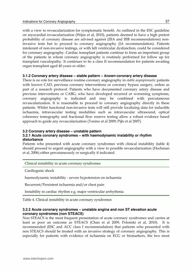
3.2 Coronary artery disease – unstable pattern 3.2.1 Acute coronary syndromes – with haemodynamic instability or rhythm disturbance Patients who presented with acute coronary syndromes with clinical instability (table 4) should proceed to urgent angiography with a view to possible revascularization (Hochman et al, 2006) either percutaneously or surgically if indicated.
Clinical instability in acute coronary syndromes
Cardiogenic shock
haemodynamic instability - severe hypotension on ischaemia
Recurrent/Persistent ischaemia and/or chest pain
Instability in cardiac rhythm e.g. major ventricular arrhythmia
Table 4. Clinical instability in acute coronary syndromes
3.2.2 Acute coronary syndromes – unstable angina and non ST elevation acute coronary syndromes (non STEACS) Non STEACS is the most frequent presentation of acute coronary syndromes and carries at least as poor an outcome as STEACS (Chan et al 2009, Polonski et al, 2010). It is recommended (ESC and ACC class I recommendation) that patients who presented with non STEACS should be treated with an invasive strategy of coronary angiography. This is especially for patients with evidence of ischaemia on ECG or biomarkers, the two most
www.intechopen.com
Advances in the Diagnosis of Coronary Atherosclerosis
38
powerful predictors of adverse events, as a prelude to definitive treatment for unstable coronary culprit lesions. Several meta-analyses including a Cochrane review (Hoenig et al, 2006; Fox et al, 2010; Mehta et al, 2005) of randomized controlled trials have concluded a routine invasive strategy improved ischaemic endpoints - reducing recurrent ischaemia, rehospitalisation and revascularization; and a more recent meta-analysis of three RCTs (FRISC-II, ICTUS, RITA-3) in 2010 (Fox et al, 2010) showed the benefit of a routine invasive strategy in cardiovascular death or non fatal MI was maintained at five years. Whilst the meta-analysis was conclusive, one must note the heterogeneity in the trials included. The most controversial trial, published in 2005, comparing a routine invasive vs. conservative strategy, ICTUS (de Winter et al, 2005), was the only trial to show no difference in the ischaemic end points between the two strategies. Five year follow up data from this study continued to show no long-term benefit of an early invasive strategy in reducing death or MI (Damman et al, 2010). Notably, 54% of the initially conservatively treated patients received revascularization within one year. It is likely the ability of the ICTUS trial to demonstrate difference in clinical outcomes has been significantly compromised by the cross over rate (Cannon, 2004) from medical therapy to invasive treatment. As a result of the heterogeneity between trials (inclusion criteria, definitions of MI, intensity
of pharmacological treatment and frequency of revascularization) and the confounding
results, this issue of routine invasive vs. conservative treatment strategy continues to spurn
passionate debate (Bittl & Maron, 2010; Thompson, 2010). The 2011 ACC guidelines
(Anderson et al, 2011) acknowledge that in selective patients without high-risk features who
have been stabilized, an initially conservative strategy may not be inappropriate, especially
those with serious co morbidities or contraindications to angiography and intervention who
have an elevated risk for clinical events, for example, patients with advanced malignancy
and a history of contrast allergy who declines invasive procedures. The decision to avoid
angiography in these patients may be made by considering the clinical evaluation of the
physician and patient preference. It is those patients enjoying satisfactory quality of life who
demonstrate the highest clinical risk profile that derive the most benefit and should receive
an early invasive strategy.
When an early invasive approach has been decided for a patient, the exact timing to
invasive coronary angiography is not very well established and subject to ongoing research.
This of course differs from the management in STEACS where urgent reperfusion is vital.
Various trials have attempted to answer the question of timing of coronary angiography in
NSTEACS. The risk of adverse cardiovascular events is time dependant. These events in the
unstable patient group are more common in the early phase after initial clinical
presentation. An early invasive treatment may prevent impending reinfarction or ischaemia,
but the counterargument is that an adequate pre procedural period of pharmacotherapy
may “prepare” the lesion better to minimize complications such as no reflow. The data is
not definitive. Small trials (Neumann et al, 2003), non-clinical primary endpoints
(Montalescot et al, 2009), possibly underpowered studies (Mehta et al, 2009) are all factors
that make definitive evidence based recommendations difficult.
Whilst the evidence differs between various trials, it is reasonable the low to intermediate
risk NSTEACS patients undergo coronary angiography within 72 hours. This is reflected in
the conservative arm of recent studies in whom a delayed approach is now considered a
median of 24 hours after the onset of symptoms compared to 72 hours 10 years ago. Based
on subgroup analysis on the TIMACS study in which patients with a GRACE score >140
www.intechopen.com
Indications for Coronary Angiography
39
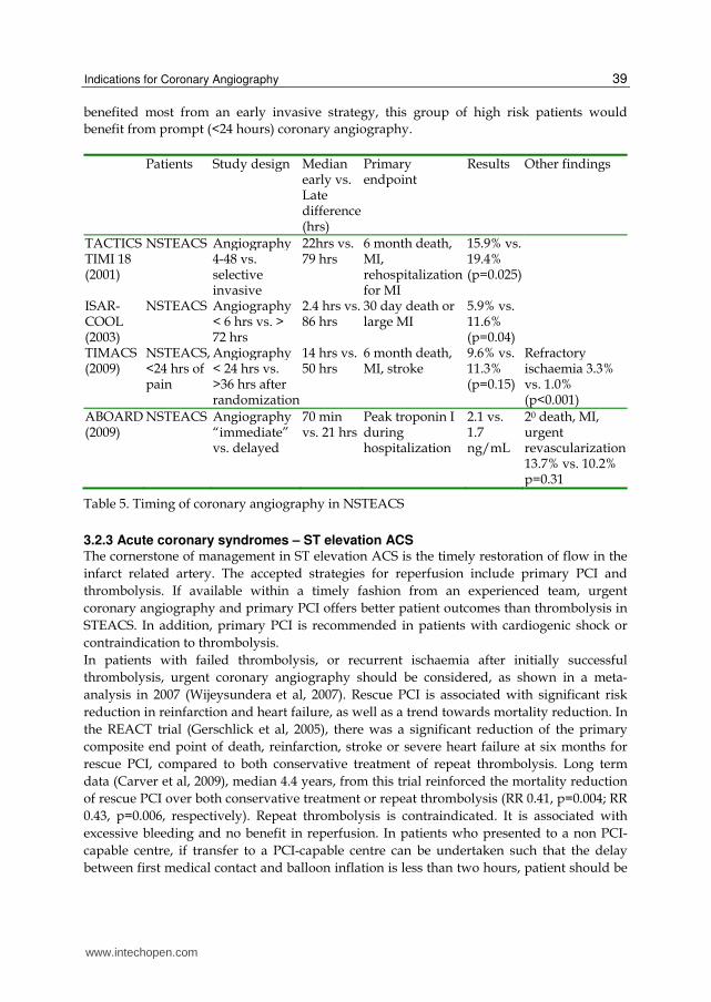
benefited most from an early invasive strategy, this group of high risk patients would
benefit from prompt (<24 hours) coronary angiography.
Patients Study design Median early vs. Late difference (hrs)
Primary endpoint
Results Other findings
TACTICS TIMI 18 (2001)
NSTEACS Angiography 4-48 vs. selective invasive
22hrs vs. 79 hrs
6 month death, MI, rehospitalization for MI
15.9% vs. 19.4% (p=0.025)
ISAR-COOL (2003)
NSTEACS Angiography < 6 hrs vs. > 72 hrs
2.4 hrs vs. 86 hrs
30 day death or large MI
5.9% vs. 11.6% (p=0.04)
TIMACS (2009)
NSTEACS, <24 hrs of pain
Angiography < 24 hrs vs. >36 hrs after randomization
14 hrs vs. 50 hrs
6 month death, MI, stroke
9.6% vs. 11.3% (p=0.15)
Refractory ischaemia 3.3% vs. 1.0% (p<0.001)
ABOARD (2009)
NSTEACS Angiography “immediate” vs. delayed
70 min vs. 21 hrs
Peak troponin I during hospitalization
2.1 vs. 1.7 ng/mL
20 death, MI, urgent revascularization 13.7% vs. 10.2% p=0.31
Table 5. Timing of coronary angiography in NSTEACS
3.2.3 Acute coronary syndromes – ST elevation ACS The cornerstone of management in ST elevation ACS is the timely restoration of flow in the
infarct related artery. The accepted strategies for reperfusion include primary PCI and
thrombolysis. If available within a timely fashion from an experienced team, urgent
coronary angiography and primary PCI offers better patient outcomes than thrombolysis in
STEACS. In addition, primary PCI is recommended in patients with cardiogenic shock or
contraindication to thrombolysis.
In patients with failed thrombolysis, or recurrent ischaemia after initially successful
thrombolysis, urgent coronary angiography should be considered, as shown in a meta-
analysis in 2007 (Wijeysundera et al, 2007). Rescue PCI is associated with significant risk
reduction in reinfarction and heart failure, as well as a trend towards mortality reduction. In
the REACT trial (Gerschlick et al, 2005), there was a significant reduction of the primary
composite end point of death, reinfarction, stroke or severe heart failure at six months for
rescue PCI, compared to both conservative treatment of repeat thrombolysis. Long term
data (Carver et al, 2009), median 4.4 years, from this trial reinforced the mortality reduction
of rescue PCI over both conservative treatment or repeat thrombolysis (RR 0.41, p=0.004; RR
0.43, p=0.006, respectively). Repeat thrombolysis is contraindicated. It is associated with
excessive bleeding and no benefit in reperfusion. In patients who presented to a non PCI-
capable centre, if transfer to a PCI-capable centre can be undertaken such that the delay
between first medical contact and balloon inflation is less than two hours, patient should be
www.intechopen.com
Advances in the Diagnosis of Coronary Atherosclerosis
40
transferred to the PCI-capable centre. Established logistics and personnel arrangement must
be in place for this to take place.
In patients with successful thrombolysis, the timing of transfer to a PCI-capable facility and
the timing of angiography have been addressed in several contemporary trials and meta-
analyses. Several recent meta-analyses (Borgia et al, 2010; Desch et al, 2008; Savio et al, 2010)
which included studies such as CARESS-AMI (Di Mario et al, 2009), TRANSFER-AMI
(Cantor et al, 2010) and NORDISTEMI (Bøhmer et al, 2009), demonstrated that early transfer
to a PCI capable facility is associated with a significant reduction in the combined endpoint
of 30 day mortality, re-infarction, ischaemia. The benefit appears to be driven most by a
reduction of re-infarction and ischaemia. Current ESC and ACC guidelines recommend this
pharmacoinvasive strategy, or “drip and ship” strategy, the prompt transfer of
thrombolyzed patients to a PCI capable hospital within 24 hours.
The timing of coronary angiography deserves special mention. In stable patients
successfully thrombolyzed, having been transferred to a PCI capable facility, coronary
angiography and PCI should be deferred until 3 to 24 hours after thrombolysis. Whilst
immediate coronary angiography appears attractive a proposition, evidence from
“facilitated PCI” suggests otherwise. Facilitated PCI is a strategy of immediate angiography
and PCI after pharmacotherapy such as upstream full or half dose thrombolytic therapy,
with or without glycoprotein IIb/IIIa inhibitor. Several trials have addressed this question.
ASSENT-4 (ASSENT-4 investigators, 2006), assessing tenecteplase then PCI compared to
primary PCI, showed increased rates of ischaemic and bleeding complications with
facilitated PCI, and FINESSE (Ellis et al, 2008, reduced reteplace and abciximab then PCI vs.
abciximab then PCI vs. PCI) showed no difference in the mortality, ventricular function or
cardiogenic shock but excess bleeding. A meta-analysis (Keeley et al, 2006) of 18 trials over 3
decades showed no benefit of facilitated PCI over primary PCI. There is currently no data to
recommend the use of “facilitated” PCI. Pharmacoinvasive strategy is encouraged but
facilitated PCI is not.
Coronary angiography should be performed prior to definitive surgical treatment for
mechanical complications of any myocardial infarction (e.g. ventricular septal defect, acute
mitral regurgitation or ventricular aneurysm). There are some situation however where the
delays associated with angiography may outweigh the benefits of defining the coronary
anatomy. For example angiography should not delay emergent surgical treatment of VSD
and shock or aortic dissection.
3.2.4 Out of hospital cardiac arrest (OOHCA) Patients who have survived an out of hospital cardiac arrest with return of spontaneous
circulation (ROSC) may or may not present with diagnostic ischaemic ECG changes. In
patients with definitive changes such as ST elevation, they should be treated as outlined
above with an immediate invasive strategy. In haemodynamically and rhythmically stable
patients after cardiac arrest, the appropriate timing (immediate vs. deferred) of coronary
angiography is the subject of particular interest. Up to 80% of OOHCA patients have had an
acute coronary syndrome as the precipitant (Pell et al, 2003) and ECG changes are not
necessarily predictive (Aurore et al, 2011). Given this finding, it was not surprising some
studies (Dumas et al, 2010; Reynolds et al, 2009; Spaulding et al, 1997; Sunde et al, 2007; Stub
et al 2010) have shown an improved outcome, including cardiac ejection fraction,
www.intechopen.com

Indications for Coronary Angiography
41
Patients Study design Primary
endpoints
Results Hrs to PCI
in transfer
group
Other
findings
CARESS-in-
AMI (2008)
75 y.o. or
younger
STEACS, ½
reteplace
with
abciximab,
heparin,
aspirin
within 12
hours of
pain
Immediate
transfer for
angiography
±PCI vs.
standard
treatment
(with transfer
for rescue if
needed)
30 day
death,
reinfarction,
refractory
ischaemia
RR 0.40
(p=0.004)
<3
(median 2.2)
TRANSFER-
AMI (2009)
STEACS,
tenecteplase
within 12
hours of
pain
Immediate
transfer for
angiography±
PCI within 6
hours vs.
standard-
treatment
(deferred >
24 hrs)
30 day
death,
reinfarction,
recurrent
ischaemia,
new CCF,
cardiogenic
shock
RR 0.64
(p=0.004)
<6
(median 3.9)
NORDISTEMI
(2010)
75 y.o. or
younger,
STEACS,
teneteplase
<6 hr of
pain,
tenecteplase
Immediate
angiography
±PCI vs.
Deferred
12 month
death,
reinfarction,
stroke, new
ischaemia
RR 0.72
(NS)
As soon as
possible
(median 2.7)
30 day
death,
reinfarction,
stroke or
new
ischaemia
(RR 0.49
p=0.03)
Table 6. Timing of transfer of STEACS patients to a PCI capable facility
neurological recovery and survival, with this strategy. The largest study of this patient
group is the PROCAT registry (Dumas et al, 2010) from Paris suggested that immediate
angiography and successful PCI improved survival (odds ratio 2.06, p=0.013). Given the
evidence leading centers have adoped a strategy of immediate coronary angiography and
revascularisation as indicated after an OOHCA with ROSC. Indeed, this is the
recommendation endorsed by the International Liaison Committee on Resuscitation
(ILCOR, 2010) but the level of evidence is relatively low. Coma or significant neurological
compromise are common in this patient group and these should not preclude consideration
of angiography with full neurological recovery in initially comatose STEACS patients found
in various registries (Garot et al, 2007; Hosmane et al, 2009). Further, the induction of
www.intechopen.com
Advances in the Diagnosis of Coronary Atherosclerosis
42
hypothermia (Bernard et al, 2002; Wolfrum et al, 2008) should be incorporated as part of the
treatment strategy for those undergoing immediate angiography.
3.3 Special considerations 3.3.1 Congestive cardiac failure Invasive coronary angiography is useful in the investigation of left ventricular systolic dysfunction for an ischemic aetiology, especially in the presence of suggestive features such as angina, remote history of severe angina or even silent infarction, ischaemia on surface electrocardiogram, or regional wall motion abnormalities on cardiac imaging. Since the 1999 ACC guideline, the advance of non-invasive computed tomography coronary angiography (Andreini et al, 2009), with its high sensitivity and negative predictive value, may justify its use for this particular indication rather than invasive coronary angiography. The negative predictive value of a normal scan has been shown to approach 100%.
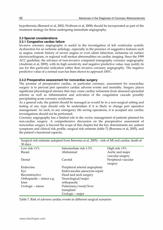
3.3.2 Preoperative assessment for noncardiac surgery The premise of preoperative cardiac, in particular coronary, assessment for noncardiac surgery is to prevent peri operative cardiac adverse events and mortality. Surgery places significant physiological stresses that may cause cardiac ischaemia from stenosed epicardial arteries as well as inflammation and activation of the coagulation cascade possibly precipitating acute coronary syndromes. As a general rule, the patient should be managed as would be in a non-surgical setting and testing of any type should only be undertaken if it is likely to change peri operative management. As such, in any emergency life saving operations, it is accepted any cardiac investigations should not be performed. Coronary angiography has a limited role in the routine management of patients planned for non-cardiac surgery. A comprehensive discussion on the preoperative assessment of noncardiac surgery is beyond the scope of this chapter but the key determinants are: patient symptoms and clinical risk profile, surgical risk estimate (table 7) (Boersma et al, 2005), and the patient’s functional capacity.
Surgical risk estimate (adapted from Boersma et al, 2005) – risk of MI and cardiac death at 30 days
Low risk <1% Intermediate risk 1-5% High risk >5% Breast Abdominal Aortic and major
vascular surgery Dental Carotid Peripheral vascular
surgery Endocrine Peripheral arterial angioplasty Eye Endovascular aneurysm repair Reconstructive Head and neck surgery Orthopaedic – minor e.g. knee
Neurological/major orthopaedic
Urologic – minor Pulmonary/renal/liver transplant
Urologic – major
Table 7. Risk of adverse cardiac events in different surgical scenarios
www.intechopen.com
Indications for Coronary Angiography
43
Patients who have stable angina or are asymptomatic can generally proceed with surgery.
Patients who have worsening symptoms of ischaemia probably deserve further assessment,
namely in the form of functional studies, prior to elective surgery. The same indications for
coronary angiography and/or intervention should apply here as would be in the non-
surgical setting. The cardiac risks of the particular operation should be assessed. It is a class
I recommendation (Poldermans et al, 2009) that pre-operative coronary angiography be
performed in acute coronary syndromes as well as in patients with angina refractory to
medical therapy. Pre-operative angiography is not recommended in cardiac-stable patients
undergoing low-risk and even intermediate risk surgery.
Evidence of improved outcome with revascularization prior to surgery however is lacking.
There is little data pertaining to the benefit of revascularization in patients undergoing non
cardiac vascular surgery, a subset of surgical patients known for high cardiac co-morbidity.
One study (McFalls et al, 2004 - Coronary Artery Revascularization Prophylaxis CARP
study) on stable CAD patients with preserved LV failed to demonstrate improved outcome
with prophylactic revascularization compared to optimized medical therapy. Another study
(Poldermans et al, 2007 - DECREASE-V pilot study), assessed the same concept on complex
CAD with impaired LV systolic function, and reached the same conclusion. This was despite
75% of these cases having three vessel or left main coronary artery disease. On the currently
limited available data, prophylactic revascularization cannot be recommended.
3.3.3 Non invasive computed tomography coronary angiography (CTCA) Over the past decade the technological advances in CTCA have meant that the technology is
emerging as an important investigation in a range of clinical scenarios. Aside from its non-
invasive nature, CTCA provides information about both vessel lumen and important
information on the composition of plaque and the vessel wall as well as other cardiac
structures. However, it is important to note the spatial and temporal resolutions remain
inferior to invasive coronary angiography with the current technology. Various multicentre
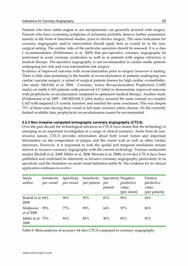
studies (Budoff et al, 2008; Miller et al, 2008; Mowatt et al, 2008) on 64 slice CTCA have been
published and confirmed its inferiority to invasive coronary angiography particularly in its
specificity and the limitation on small vessel definition (table 8). The evidence for its clinical
application continues to evolve.
Study
author
Sensitivity
per vessel
Specificity
per vessel
Sensitivity
per patient
Specificity
per
patient
Negative
predictive
value
(per atient)
Positive
predictive
value
(per patient)
Budoff et al
2008
84% 90% 95% 83% 99% 64%
Meijboom
et al 2008
95% 77% 99% 64% 97% 86%
Miller et al
2008
75% 93% 85% 90% 83% 91%
Table 8. Meta-analyses of accuracy 64 slice CTCA compared to coronary angiography
www.intechopen.com
Advances in the Diagnosis of Coronary Atherosclerosis
44
Guidelines on the appropriateness of CTCA (Taylor et al, 2010) published in 2010 attempted
to address the use of this rapidly evolving technology. The indications chosen mirrored that
of invasive coronary angiography. In view of its high sensitivity, CTCA is particularly
suited to low to intermediate risk patients in the setting of stable symptomatic patients, low
risk acute coronary syndromes and heart failure patients. Patients with normal CTCA
findings convey a very low risk of future cardiac events and cardiac mortality (Hulten et al,
2011). Patients at high risk of coronary artery disease are advised against CTCA as this
represents hindrance to timely diagnosis and management of CAD with the additional risk
of contrast and radiation. It has no role in the preoperative assessment of non-cardiac
surgery. It is deemed appropriate in the assessment of graft patency for symptomatic
patients with previous CABG. To reflect the emerging role of left main stenting, CTCA has
been deemed appropriate as a means for routine follow up screening of left main stent(s) in
asymptomatic patients.
Unless the limitations of CTCA can be sufficiently improved upon, it is likely CTCA will
continue to be utilized in a complementary role to invasive coronary angiography in the
assessment of CAD. Of course the major advantage of angiography remains the ability to
proceed on to coronary intervention as part of the imaging procedure.
3.4 Relative and absolute contraindications The only absolute contraindication is if a patient refuses the test fully understanding the risk
and benefit of his/her decision, refuses any treatment for CAD regardless of the result of the
coronary angiography, or if other medical co-morbidities rendering imaging of the
coronaries and revascularization inappropriate or impossible. Subjecting the patient to even
the small risks of a coronary angiogram with no foreseeable gain cannot be justified.
Relative contraindications include situations which increase the likelihood of complications,
e.g. bleeding diathesis, coagulopathy (iatrogenic i.e. warfarin or pathological), renal
impairment, anaemia, contrast allergy, active infection and aortic valve vegetation. If these
relative contraindications are reversible the patient should be appropriately prepared and
coronary angiography deferred.
4. Valvular heart disease and structural heart disease intervention
The assessment of the coronary anatomy prior to cardiac valvular surgery is routine
although not necessarily evidence driven. Whilst surgery for valvular heart disease has been
performed for many years, percutaneous intervention for structural or valvular heart
disease has been generating significant interest over the recent years. Transcatheter aortic
valve implants (TAVI) have already demonstrated significant mortality benefit in patients
deemed unsuitable for surgery compared to medical therapy, in the first RCT on TAVI
(PARTNERS trial) (Leon et al, 2010). Several investigational percutaneous treatment options
also exist for mitral regurgitation. Percutaneous coronary revascularization has been a
prerequisite for the TAVI procedures and as such all patients underwent coronary
angiography. The incidence of coronary disease in this patient group is high, up to 70% the
PARTNERS trial. Coronary angiography is a pivotal part in the assessment for TAVI
suitability. The increasing percutaneous treatment options will likely generate in the future
specific college endorsed guideline to incorporate the role of coronary angiography in pre
structural heart intervention..
www.intechopen.com

Indications for Coronary Angiography
45
4.1 Pre valvular surgery It is routine to perform coronary angiography prior to valve surgery although there is no evidence to guide such practice (Bonow et al, 2006; Vahanian et al, 2007). This is especially logical in older patients with cardiac risk factors, or suggestion of CAD from symptoms, ECG, cardiac imaging or non-invasive function studies. The indication is less clear in younger patients, and special caution must be exercised in aortic valve surgery for infective endocarditis given the risk for vegetation embolization. No definitive data are available to define the clinical benefit of revascularization in the setting of corrective valve surgery. Whilst it is reasonable to expect benefit in prognostic disease, the same generalization may not apply to single vessel non prognostic lesions which would otherwise not necessarily be revascularized in a non-surgical setting. The role of CTCA is yet to be defined but its application here may reasonably mimic that outlined previously, in a low to intermediate risk group of patients undergoing valvular surgery (Meijboom et al, 2006).
4.1.1 Pre percutaneous valve implant This is a developing area in which the role of coronary angiography appears crucial. Coronary angiography and if necessary percutaneous revascularization were a requisite for all TAVI trials so as to allow for direct comparison with the CABG + AVR subgroup. It seems nonetheless reasonable for this group of high risk patients, often with symptoms indistinguishable from ischaemia, to undergo coronary angiography. In addition, the left heart catheterization is pivotal for necessary measurement of aortic dimensions, angulation, coronary ostia position and origin, and assessment of vascular access. It should be noted the indication of revascularization in patients undergoing TAVI stems from its equivalent in surgical treatment, the exact timing and completeness of revascularization are yet to be defined. The more difficult question relates to the need for coronary angiography in patients who may not otherwise meet the indications for coronary angiography, e.g. pulmonary valve implant in a young congenital heart disease patient, mitral valve clip in severe functional mitral regurgitation. These patients are managed the patient in a similar fashion to the conventional indications for coronary angiography.
4.1.2 Other structural cardiac interventions There is limited evidence to support routine coronary angiography in structural cardiac interventions such as patent foramen ovale and atrial septal defect closure, coarctation stenting, patent ductus arteriosus interventions. It is often performed however based on the individual patient’s clinical risk profile. The discovery of prognostically significant coronary artery disease not amenable to percutaneous coronary interventions may sway treatment towards a complete surgical treatment option. Coronary angiography does not increase the invasive nature of these procedures and add little incremental risk.
5. Case vignettes of the use of coronary angiography in the contemporary era
Case One – atrial septal defect with multi-vessel coronary artery disease
A 73 year old female presented with NYHA (New York Heart Association) class II
exertional dyspnoea. Her cardiac risk factors included hypertension and hyperlipidaemia. A
www.intechopen.com
Advances in the Diagnosis of Coronary Atherosclerosis
46
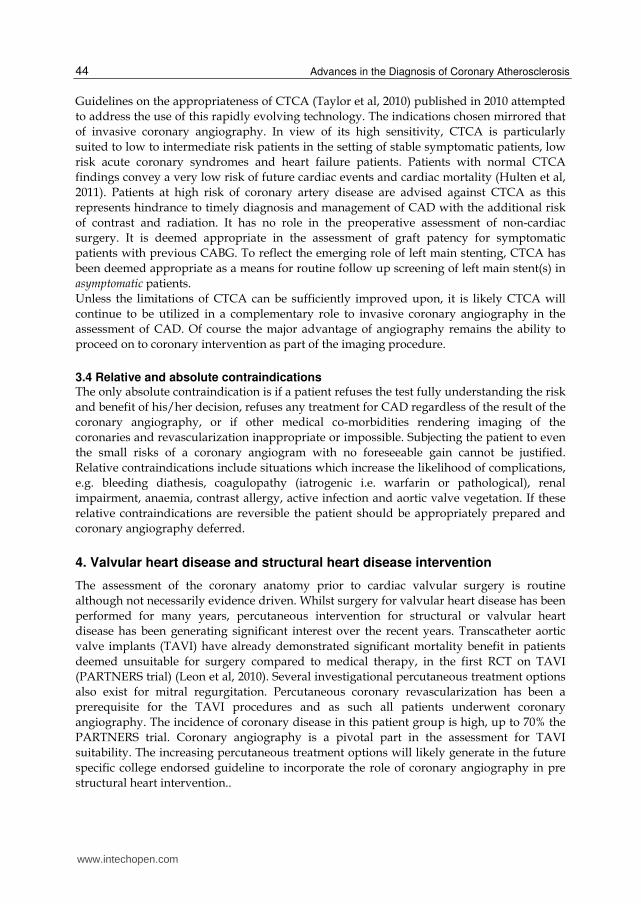
transthoracic echocardiogram demonstrated two small secundum atrial septal defects
(ASD), with moderate right ventricular dilatation and normal systolic function.
Percutaneous ASD closure was attempted. Coronary angiography was performed,
especially given the uncharacteristically late presentation of her ASD, as well as her cardiac
risk factors. This demonstrated prognostically significant triple vessel disease (Figure 1a, 1b,
2c), with an osital 90% left circumflex artery stenosis, 90% mid left anterior descending
artery bifurcation stenosis and a 90% distal right coronary artery stenosis. Given the
unfavourable percutaneous prospect for the coronary lesions, she was referred for surgical
complete revascularization and ASD closure. She was discharged uneventfully one week
after surgery. Coronary angiography should be considered prior to structural cardiac
interventions in patients with cardiac risk factors and may alter management.
Fig. 1a. Left anterior artery stenoses
www.intechopen.com

Indications for Coronary Angiography
47
Fig. 1b. Ostial left circumflex stenosis
Fig. 1c. Posterolateral branch stenosis
www.intechopen.com

Advances in the Diagnosis of Coronary Atherosclerosis
48
Case Two – post TAVI angiogram
An 84 year old male represented with angina two years after a successful transcatheter
aortic valve implant (TAVI) with a bioprosthetic CoreValve®. His NYHA symptomatology
improved from Class IV to Class I. His coronary angiogram and graft study prior to his
TAVI showed patent grafts (LIMA-LADD1-LAD; SVG-OM1; SVG-PDA) but severe native
disease including a severely diseased grafted PDA with no prospect for surgical or
percutaneous revascularization. A coronary angiogram and graft study was performed
through his CoreValve® to assess if his underlying disease had progressed (Figure 2a).
Fig. 2a. Angiography with a 5 French Judkins Left 5 catheter through a CoreValve ® bioprosthesis
www.intechopen.com

Indications for Coronary Angiography
49
This demonstrated patent grafts as previously noted but progression in his PDA disease (Figure 2b). Percutaneous, or surgical, treatment remained an unfavourable prospect and he was successfully treated with intensified medical therapy with CCS (Canadian Cardiovascular Society) I angina. Coronary angiography can be safely performed through CoreValve struts but care must be exercised with the choice and manipulation of catheters.
Fig. 2b. Severe native posterior descending artery disease not amenable to percutaneous treatment
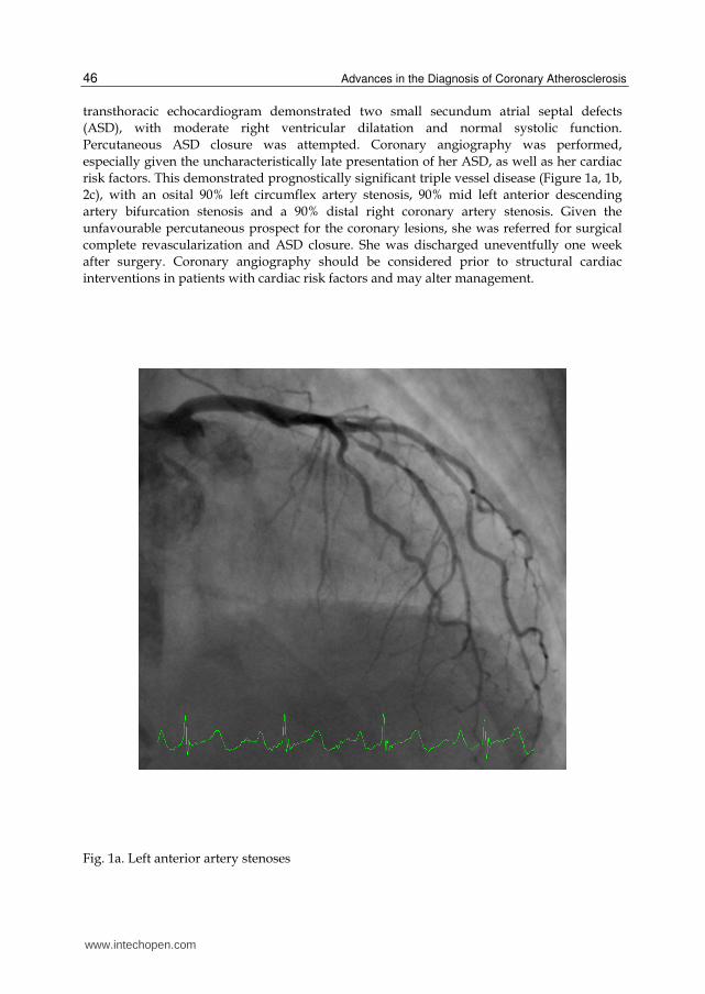
Case Three – percutaneous treatment of left main stenosis and aortic stenosis
A 76 year old male with severe aortic stenosis was referred for possible percutaneous treatment due to his high surgical risk. Assessment for TAVI entailed a coronary angiogram, aortogram and an iliofemoral angiogram (Figure 3a). Coronary angiography revealed a significant left main stenosis (Figure 3b) which on intravascular ultrasound measured 5.8 mm2. A multidisciplinary heart team assessed the data and concluded surgical treatment to be prohibitively high risk. The patient then underwent left main stenting with a drug eluting stent under IVUS guidance. Satisfactory stent expansion was attained with a final diameter of 4.5mm (Figure 3c). The patient then proceeded to a successful TAVI (CoreValve®) implant and continued to enjoy excellent symptom relief at six months.
www.intechopen.com
Advances in the Diagnosis of Coronary Atherosclerosis
50
Coronary angiography, and arterial (aortic, iliac, and femoral) assessment, form an integral part of TAVI assessment. Elderly patients with severe aortic stenosis often have severe co-existing coronary artery disease as well as peripheral vascular disease as demonstrated by this case. Optimizations of the coronary status, as well as an appreciation of the peripheral vasculature, are absolutely paramount to a successful TAVI implant and to avoid vascular complications.
Fig. 3a. Tortuous aorta and iliac arteries, important information for TAVI transfemoral access.
www.intechopen.com

Indications for Coronary Angiography
51
Fig. 3b. Mid shaft left main stenosis
www.intechopen.com
Advances in the Diagnosis of Coronary Atherosclerosis
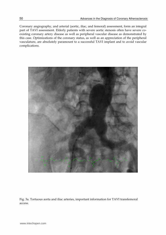
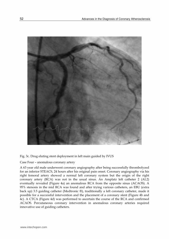
52
Fig. 3c. Drug eluting stent deployment in left main guided by IVUS
Case Four – anomalous coronary artery
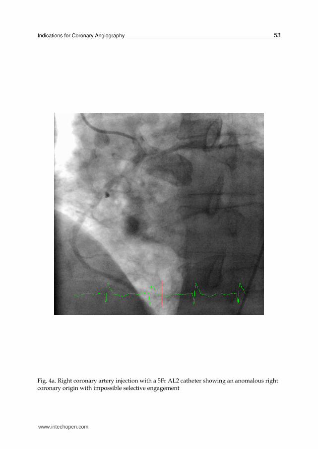
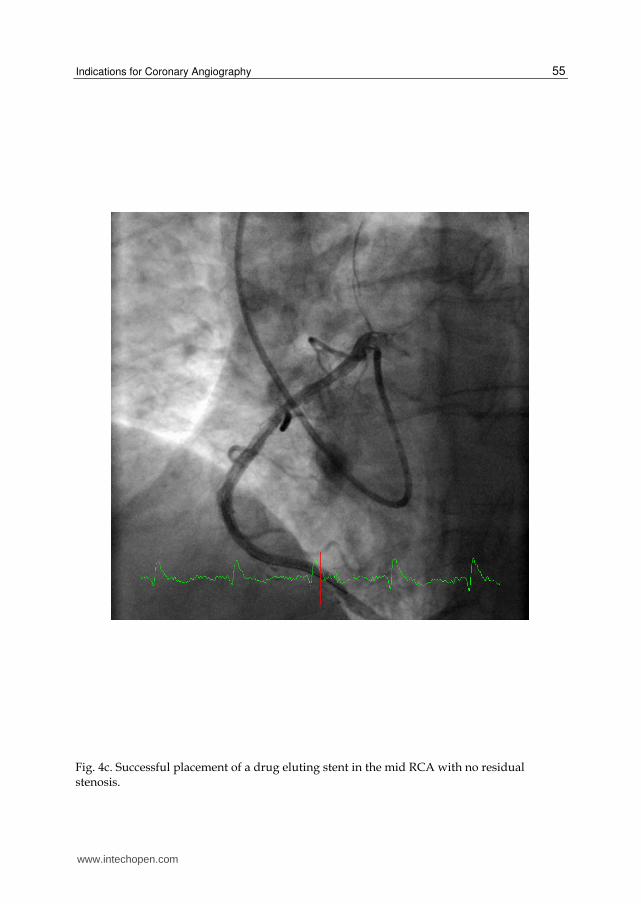
A 63 year old male underwent coronary angiography after being successfully thrombolyzed for an inferior STEACS, 24 hours after his original pain onset. Coronary angiography via his right femoral artery showed a normal left coronary system but the origin of the right coronary artery (RCA) was not in the usual sinus. An Amplatz left catheter 2 (AL2) eventually revealed (Figure 4a) an anomalous RCA from the opposite sinus (ACAOS). A 95% stenosis in the mid RCA was found and after trying various catheters, an EBU (extra back up) 3.5 guiding catheter (Medtronic ®), traditionally a left coronary catheter, made it possible for a successful intervention and the placement of a coronary stent (Figure 4b and 4c). A CTCA (Figure 4d) was performed to ascertain the course of the RCA and confirmed ACAOS. Percutaneous coronary intervention in anomalous coronary arteries required innovative use of guiding catheters.
www.intechopen.com
Indications for Coronary Angiography
53
Fig. 4a. Right coronary artery injection with a 5Fr AL2 catheter showing an anomalous right coronary origin with impossible selective engagement
www.intechopen.com

Advances in the Diagnosis of Coronary Atherosclerosis
54
Fig. 4b. A 6Fr EBU catheter used to engage the anomalous origin. Traditional right coronary catheters would not have been useful.
www.intechopen.com
Indications for Coronary Angiography
55
Fig. 4c. Successful placement of a drug eluting stent in the mid RCA with no residual stenosis.
www.intechopen.com

Advances in the Diagnosis of Coronary Atherosclerosis
56
Fig. 4d. CTCA 3D reconstruction shows the origin and course of the anomalous right coronary artery clearly and defines its ante-aortic course.
Case Five – CTCA in graft assessment
A 69 year old male developed atypical chest pain 12 months after his coronary bypass graft (LIMA-LAD; RA-ramus; SVG-OM1). During the initial angiographic assessment he underwent IVUS of his left main and left anterior descending artery indeterminate lesions. IVUS showed his LM to be 5.44mm2 and LAD 3.96mm2. These were thought be suggest significant stenoses. To investigate his current atypical chest pain an MPS (Figure 5a) was performed which showed no inducible ischaemia or symptoms. A CTCA (Figure 5b) was performed which however suggested possible occluded LIMA graft. A coronary angiogram and graft study (Figure 5c and 5d) confirmed this finding that the LIMA graft had become atretic and normal coronary flow seen in the LAD. His other grafts were patent as was shown on the CTCA. This case demonstrated the value of CTCA in identifying graft patency and position, often facilitating the location of the graft anastomosis at the aorta. The atypical symptoms, patent coronary arteries and grafts, correlated well with his negative functional study. The LIMA-LAD graft probably underwent atresia as a result of no physiological implication to his intermediate LM and LAD lesions even though the diameters measured fulfilled conventional criteria for significant lesions.
www.intechopen.com

Indications for Coronary Angiography
57
Fig. 5a. Myoperfusion study with no significant inducible ischaemia.
www.intechopen.com

Advances in the Diagnosis of Coronary Atherosclerosis
58
Fig. 5b. Position of two aortic grafts demonstrated clearly on CTCA. No LIMA flow demonstrated.
www.intechopen.com

Indications for Coronary Angiography
59
Fig. 5c. LAD artery with no obvious proximal stenosis and normal coronary flow.
www.intechopen.com

Advances in the Diagnosis of Coronary Atherosclerosis
60
Fig. 5d. Small atretic LIMA.
Case Six – balanced ischaemia on MPS in a high risk patient
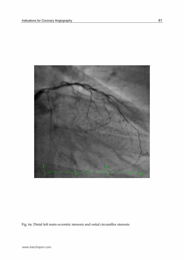
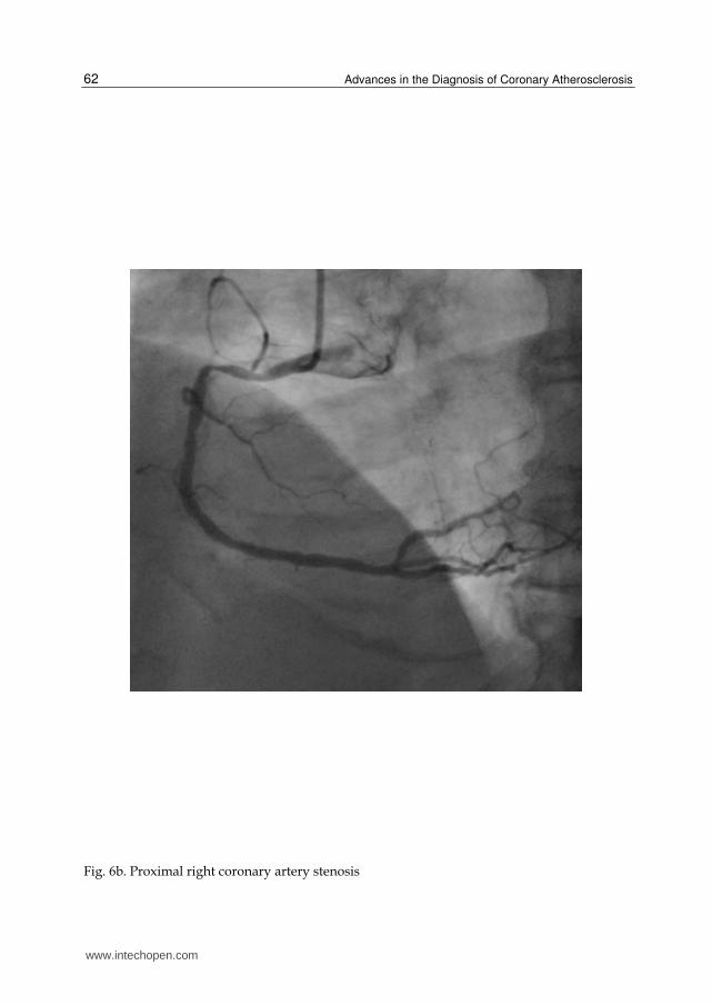
A 67 year old male with 20 years of diabetes mellitus II and 40 pack year history of smoking presented with six months of worsening exertional dyspnoea and chest pain. A myoperfusion study performed at a satisfactory level of workload demonstrated a small area of mildly reversible ischaemia in the inferolateral area. Given the abnormal findings, the patient proceeded to coronary angiography. This revealed severe distal left main artery stenosis, with a subtotally occluded left circumflex and a proximal severe RCA stenosis. Left ventricular function was normal. The extent and severity of disease was of prognostic significance. The patient underwent uneventful coronary bypass grafting and was discharged from the hospital one week after. This patient exhibited a high pretest probability based on the clinical profile. Whilst the myoperfusion study was suggestive, it underestimated the extent of disease likely due to the balanced ischaemia from the RCA and LM stenoses. Complex distal left main stenosis, especially bifurcation stenosis, is best treated with surgical revascularization.
www.intechopen.com
Indications for Coronary Angiography
61
Fig. 6a. Distal left main eccentric stenosis and ostial circumflex stenosis.
www.intechopen.com
Advances in the Diagnosis of Coronary Atherosclerosis
62
Fig. 6b. Proximal right coronary artery stenosis
www.intechopen.com
Indications for Coronary Angiography
63
6. Conclusion
Coronary angiography remains the most accurate diagnostic modality for coronary artery
disease. It provides a definitive assessment of coronary artery lumen and provides an
avenue to percutaneous intervention. It is pivotal in the treatment on unstable or acute
coronary syndromes and life saving in cardiogenic shock and STEACSs. Indications for
invasive coronary angiography have changed incrementally over the decade since
ACC/AHA guidelines on the topic were published. The advent of noninvasive CTCA has
changed the landscape for the diagnosis of CAD but CTCAs seem destined only for the low
to intermediate risk patients. Coronary angiography also serves a pivotal role in the
burgeoning field of percutaneous structural cardiac interventions, especially percutaneous
valve implants.
7. References
Andreini D, Pontone G, Bartorelli AL, Agostoni P, Mushtaq S, Bertella E, Trabattoni D,
Cattadori G, Cortinovis S, Annoni A, et al. (2009). Sixty-Four-Slice Multidetector
Computed Tomography: An Accurate Imaging Modality for the Evaluation of
Coronary Arteries in Dilated Cardiomyopathy of Unknown Etiology Circ
Cardiovasc Imaging 2(3): 199 - 205.
Anderson JL, Adams CD, et al. (2011). 2011 ACCF/AHA Focused Update Incorporated into
the ACC/AHA 2077 Guidelines for the management of patients with unstable
angina/Non-ST Elevation myocardial infarction, A report of the American College
of Cardiology Foundation/American Heart Association Task Force on Practice
Guidelines, Circulation. 2011; 123 [Epub ahead of print]
ASSENT-4 investigators, (2006). Primary versus tenecteplase-facilitated percutaneous
coronary intervention in patients with ST-segment elevation acute myocardial
infarction (ASSENT-4 PCI): randomised trial. Lancet 367:569–578.
Aurore A, Jabre P, Liot P, Margenet A, Lecarpentier E, Combes X. (2011). Predictive factors
for positive coronary angiography in out-of-hospital cardiac arrest patients. Eur J
Emerg Med. 18(2):73-6.
Bernard SA, Gray TW, Buist MD, Jones BM, Silvester W, Gutteridge G, et al. (2002).
Treatment of comatose survivors of out-of-hospital cardiac arrest with induced
hypothermia. N Engl J Med 346: 557 – 563
Bittl JA, Maron DJ. (2010). Having It Both Ways J. Am. Coll. Cardiol., 55(9): 865 - 866.
Boden WE, O'Rourke RA, Teo KK, et al. (2007). Optimal medical therapy with or without
PCI for stable coronary disease. N Engl J Med 356:1503-1516
Boersma E, Kertai MD, Schouten O, Bax JJ, Noordzij P, Steyerberg EW, Schinkel AF, van
Santen M, Simoons ML, Thomson IR, Klein J, van Urk H, Poldermans D. (2005).
Perioperative cardiovascular mortality in noncardiac surgery: validation of the Lee
cardiac risk index. Am J Med 118:1134–1141.
Bøhmer E, Hoffmann P, Abdelnoor M, Arnesen H, Halvorsen S. (2010). Efficacy and safety
of immediate angioplasty versus ischemia-guided management after thrombolysis
in acute myocardial infarction in areas with very long transfer distances: results of
www.intechopen.com

Advances in the Diagnosis of Coronary Atherosclerosis
64
the NORDISTEMI (NORwegian study on DIstrict treatment of ST-Elevation
Myocardial Infarction) J Am Coll Cardiol 55:102-110.
Bonow RO, Carabello BA, Kanu C, de Leon AC Jr, Faxon DP, Freed MD, Gaasch WH, Lytle
BW, Nishimura RA, O'Gara PT, O'Rourke RA, Otto CM, Shah PM, Shanewise JS,
Smith SC Jr, Jacobs AK, Adams CD, Anderson JL, Antman EM, Faxon DP, Fuster V,
Halperin JL, Hiratzka LF, Hunt SA, Lytle BW, Nishimura R, Page RL, Riegel B.
(2006). ACC/AHA 2006 guidelines for the management of patients with valvular
heart disease: a report of the American College of Cardiology/American Heart
Association Task Force on Practice Guidelines (writing Committee to Revise the
1998 guidelines for the management of patients with valvular heart disease)
developed in collaboration with the Society of Cardiovascular Anesthesiologists
endorsed by the Society for Cardiovascular Angiography and Interventions and the
Society of Thoracic Surgeons. J Am Coll Cardiol. 48: e1–e148.
Borgia, F, Goodman SC, Halvorsen S, Cantor WJ, Piscione F, Le May MR, Fernandez-Aviles
F, Sanchez PL, Dimopoulos K, Scheller B, Armstrong PW and Di Mario C. (2010).
Early routine Percutaneous coronary intervention after fibrinolysis vs. standard
therapy in ST-segment elevation myocardial infarction: a meta-analysis, Eur Heart J
31 (17):2156-2169
Budoff MJ, Dowe D, Jollis JG, et al. (2008). Diagnostic performance of 64-multidetector-row
coronary computed tomographic angiography for evaluation of coronary artery
stenosis in individuals without known coronary artery disease. J Am Coll Cardiol
52:1724–32.
Cannon CP. (2004). Revascularisation for everyone? Eur Heart J 25:1471–1472
Cantor WJ, Fitchett D, Borgundvaag B, Ducas J, Heffernan M, Cohen EA, Morrison LJ,
Langer A, Dzavik V, Mehta SR, Lazzam C, Schwartz B, Casanova A, Goodman SG.
(2009). Routine early angioplasty after fibrinolysis for acute myocardial infarction.
N Engl J Med 360:2705–2718.
Carver A, Rafelt S, Gershlick AH, et al. REACT Investigators. (2009). Longer-term follow-up
of patients recruited to the REACT (Rescue Angioplasty Versus Conservative
Treatment or Repeat Thrombolysis) trial J Am Coll Cardiol 54:118-126.
Chan MY, Sun JL, Newby LK, Shaw LK, Lin M, Peterson ED, Califf RM, Kong DF, Roe MT.
(2009). Long-term mortality of patients undergoing cardiac catheterization for ST-
elevation and non-ST elevation myocardial infarction, Circulation. 19(24):3110-7
Damman P, Hirsch A, Windhausen F, Tijssen JGP, de Winter RJ, ICTUS Investigators.
(2010). 5-year clinical outcomes in the ICTUS (Invasive versus Conservative
Treatment in Unstable coronary Syndromes) trial: a randomized comparison of an
early invasive versus selective invasive management in patients with non–ST-
segment elevation acute coronary syndrome J Am Coll Cardiol 55:858-864.
de Winter RJ, Windhausen F, Cornel JH, et al (2005). Invasive versus Conservative
Treatment in Unstable Coronary Syndromes (ICTUS) Investigators. Early invasive
versus selectively invasive management for acute coronary syndromes. N Engl J
Med 353: 1095-1104.
Desch S, Eitel I, Rahimi K, de Waha S, Schuler G, Thiele H, (2010). Timing of invasive
treatment after fibrinolysis in ST elevation myocardial infarction – a meta-analysis
www.intechopen.com

Indications for Coronary Angiography
65
of immediate or early routine versus deferred or ischaemia-guided randomized
controlled trials, Heart, 96(21):1695-1702
Di Mario C, Dudek D, Piscione F, Mielecki W, Savonitto S, Murena E, Dimopoulos K,
Manari A, Gaspardone A, Ochala A, Zmudka K, Bolognese L, Steg PG, Flather M.
(2008). Immediate angioplasty versus standard therapy with rescue angioplasty
after thrombolysis in the Combined Abciximab Reteplase Stent Study in Acute
Myocardial Infarction (CARESS-in-AMI): an open, prospective, randomised,
multicentre trial. Lancet 371:559–568
D'Souza SP, Mamas MA, Fraser DG, Fath-Ordoubadi F. (2010). Routine early coronary
angioplasty versus ischaemia-guided angioplasty after thrombolysis in acute ST-
elevation myocardial infarction: a meta-analysis Eur Heart J first published online
October 28, 2010
Dumas F, Cariou A, Manzo-Silberman S, et al. (2010) Immediate percutaneous coronary
intervention is associated with better survival after out-of-hospital cardiac arrest:
insights from the PROCAT (Parisian Region Out of hospital Cardiac ArresT)
registry. Circ Cardiovasc Interv 3:200–7
Ellis SG, Tendera M, de Belder MA, van Boven AJ, Widimsky P, Janssens L, Andersen HR,
Betriu A, Savonitto S, Adamus J, Peruga JZ, Kosmider M, Katz O, Neunteufl T,
Jorgova J, Dorobantu M, Grinfeld L, Armstrong P, Brodie BR, Herrmann HC,
Montalescot G, Neumann FJ, Effron MB, Barnathan ES, Topol EJ. (2008). Facilitated
PCI in patients with ST-elevation myocardial infarction. N Engl J Med 358:2205–
2217.
Fernandez-Aviles F, Alonso JJ, Castro-Beiras A, Vazquez N, Blanco J, Alonso-Briales J,
Lopez-Mesa J, Fernandez-Vazquez F, Calvo I, Martinez-Elbal L, San Roman JA,
Ramos B. (2004). Routine invasive strategy within 24 hours of thrombolysis
versus ischaemia-guided conservative approach for acute myocardial infarction
with ST-segment elevation (GRACIA-1): a randomised controlled trial. Lancet
364:1045–1053
Fox KA, Clayton TC, Damman P, Pocock SJ, de Winter RJ, Tijssen JG, Lagerqvist B,Wallentin
L. (2010). Long-term outcome of a routine versus selective invasive strategy in
patients with non-ST-segment elevation acute coronary syndrome a meta-analysis
of individual patient data. J Am Coll Cardiol 55:2435–2445.
Garot P, Lefevre T, Eltchaninoff H, Morice MC, Tamion F, Abry B, Lesault PF, Le Tarnec JY,
Pouges C, Margenet A, Monchi M, Laurent I, Dumas P, Garot J, Louvard Y. (2007).
Six-month outcome of emergency percutaneous coronary intervention in
resuscitated patients after cardiac arrest complicating ST-elevation myocardial
infarction. Circulation. 115: 1354–1362.
Gershlick AH, Stephens-Lloyd A, Hughes S, et al. (2005). Rescue angioplasty after failed
thrombolytic therapy for acute myocardial infarction. N Engl J Med. 353:2758–
68
Hoenig MR, Doust JA, Aroney CN, Scott IA. (2006). Early invasive versus conservative
strategies for unstable angina & non-ST-elevation myocardial infarction in the stent
era. Cochrane Database Syst Rev 3: CD004815.
www.intechopen.com

Advances in the Diagnosis of Coronary Atherosclerosis
66
Hosmane VR, Mustafa NG, Reddy VK, et al. (2009). Survival and neurologic recovery in
patients with ST-segment elevation myocardial infarction resuscitated from cardiac
arrest. J Am Coll Cardiol 53:409–15.
Hulten EA, Carbonaro S, Petrillo SP, Mitchell JD, Villines TC. (2011). Prognostic value of
cardiac computed tomography, A systematic review and meta-analysis, J. Am. Coll.
Cardiol 57:1237-1247.
ILCOR, Part 9: Acute coronary syndromes 2010 International Consensus on
Cardiopulmonary Resuscitation and Emergency Cardiovascular Care Science with
Treatment Recommendations_Nolan, JP, Hazinski MF, et al, Part 1: Executive
summary 2010 International Consensus on Cardiopulmonary Resuscitation and
Emergency Cardiovascular Care Science With Treatment Recommendations
Resuscitation 81S (2010) e175–e212Resuscitation 81S (2010) e1–e25
Keeley EC, Boura JA, Grines CL. (2006). Comparison of primary and facilitated
percutaneous coronary interventions for ST-elevation myocardial infarction:
quantitative review of randomised trials Lancet 367:579-588
Leon MB, Smith CR, Mack M, et al. (2010). Transcatheter aortic-valve implantation for aortic
stenosis in patients who cannot undergo surgery. N Engl J Med 2010;
DOI:10.1056/NEJMoa1008232
McFalls EO, Ward HB, Moritz TE, Goldman S, Krupski WC, Littooy F, Pierpont G, Santilli S,
Rapp J, Hattler B, Shunk K, Jaenicke C, Thottapurathu L, Ellis N, Reda DJ,
Henderson WG. (2004). Coronary-artery revascularization before elective major
vascular surgery. N Engl J Med 351:2795 – 2804.
Mehta SR, Cannon CP, Fox KA, Wallentin L, Boden WE, Spacek R, Widimsky P,
McCullough PA, Hunt D, Braunwald E, Yusuf S. (2005). Routine vs selective
invasive strategies in patients with acute coronary syndromes: a collaborative
meta-analysis of randomized trials. JAMA 293:2908–2917.
Mehta SR, Granger CB, Boden WE, et al. (2009). Early versus delayed invasive intervention
in acute coronary syndromes. N Engl J Med. 360: 2165–75.
Meijboom WB, Meijs MF, Schuijf JD, et al. (2008) Diagnostic accuracy of 64-slice computed
tomography coronary angiography: a prospective, multicenter, multivendor study.
J Am Coll Cardiol 52:2135-44
Meijboom WB, Mollet NR, Van Mieghem CAG, Kluin J, Weustink AC, Pugliese F,
Vourvouri E, Cademartiri F, Bogers AJJC, Krestin GP, et al. (2006). Pre-Operative
Computed Tomography Coronary Angiography to Detect Significant Coronary
Artery Disease in Patients Referred for Cardiac Valve Surgery J. Am. Coll. Cardiol.,
48(8): 1658 - 1665.
Miller JM, Rochitte CE, Dewey M, et al. (2008). Diagnostic performance of coronary
angiography by 64-row CT. N Engl J Med 359:2324–36.
Montalescot G, Cayla G, Collet JP, et al. (2009). Immediate vs delayed intervention for acute
coronary syndromes: a randomized clinical trial. JAMA. 302:947–54.
Mowatt G, Cook JA, Hillis GS, et al. (2008). 64-Slice computed tomography angiography in
the diagnosis and assessment of coronary artery disease: systematic review and
meta-analysis. Heart 94:1386–93.
www.intechopen.com
Indications for Coronary Angiography
67
Neumann FJ, Kastrati A, Pogatsa-Murray G, et al. (2003). Evaluation of prolonged
antithrombotic pretreatment (“cooling-off” strategy) before intervention in patients
with unstable coronary syndromes: a randomized controlled trial. JAMA. 290:1593–
9.
Pell JP, Sirel JM, Marsden AK, Ford I, Walker NL, Cobbe SM. (2003). Presentation,
management, and outcome of out of hospital cardiopulmonary arrest: comparison
by underlying aetiology. Heart 89:839—42
Poldermans D, Bax JJ, Boersma E, De Hert S, Eeckhout E, Fowkes G, Gorenek B, Hennerici
MG, Iung B, Kelm M, Kjeldsen KP, Kristensen SD, Lopez-Sendon J, Pelosi P,
Philippe F, Pierard L, Ponikowski P, Schmid JP, Sellevold OF, Sicari R, Van den
Berghe G, Vermassen F, Hoeks SE, Vanhorebeek I. (2009). Guidelines for
preoperative cardiac risk assessment and perioperative cardiac management in
noncardiac surgery: the Task Force for Preoperative Cardiac Risk Assessment and
Perioperative Cardiac Management in Non-cardiac Surgery of the European
Society of Cardiology (ESC) and endorsed by the European Society of
Anaesthesiology (ESA). Eur Heart J 30:2769–2812.
Reynolds JC, Callaway CW, El Khoudary SR, Moore CG, Alvarez RJ, Rittenberger JC. (2009).
Coronary angiography predicts improved outcome following cardiac arrest:
propensity-adjusted analysis. J Intensive Care Med 24:179–86.
Spaulding CM, Joly LM, Rosenberg A, et al. (1997). Immediate coronary angiography in
survivors of out-of-hospital cardiac arrest. N Engl J Med 336:1629–33.
Stub D, Hengel C, Chan W, Jackson D, Sanders K, Dart AM, Hilton A, Pellegrino V, Shaw
JA, Duffy SJ, Bernard S, Kaye DM. (2011). Usefulness of cooling and coronary
catheterization to improve survival in out-of-hospital cardiac arrest. Am J Cardiol.
107(4):522-7. Epub 2010 Dec 22.
Sunde K, Pytte M, Jacobsen D, et al. (2007) Implementation of a standardised treatment
protocol for post resuscitation care after out-of-hospital cardiac arrest. Resuscitation
73:29— 39.
Taylor AJ, Cerqueira M, Hodgson JM, et al. (2010).
ACCF/SCCT/ACR/AHA/ASE/ASNC/NASCI/SCAI/SCMR 2010 appropriate
use criteria for cardiac computed tomography: a report of the American College of
Cardiology Foundation Appropriate Use Criteria Task Force, the Society of
Cardiovascular Computed Tomography, the American College of Radiology, the
American Heart Association, the American Society of Echocardiography, the
American Society of Nuclear Cardiology, the North American Society for
Cardiovascular Imaging, the Society for Cardiovascular Angiography and
Interventions, and the Society for Cardiovascular Magnetic Resonance J Am Coll
Cardiol 56:1864-1894.
Thompson PL. (2010). The invasive approach to acute coronary syndrome: true promise or
false premise? Med J Aust; 192 (12): 694-695.
Vahanian A, Baumgartner H, Bax J, Butchart E, Dion R, Filippatos G, Flachskampf F, Hall R,
Iung B, Kasprzak J, Nataf P, Tornos P, Torracca L, Wenink A. (2007). Grupo de
Trabajo sobre el Tratamiento de las Valvulopatías de la Sociedad Europea de
www.intechopen.com

Advances in the Diagnosis of Coronary Atherosclerosis
68
Cardiología: guidelines on the management of valvular heart disease. Eur Heart J.
28: 230–268.
Wijeysundera HC, Vijayaraghavan R, Nallamothu BK, et al. (2007). Rescue angioplasty or
repeat fibrinolysis after failed fibrinolytic therapy for ST-segment myocardial
infarction: a meta-analysis of randomized trials. J Am Coll Cardiol. 49:422–30
Wolfrum S, Pierau C, Radke PW, Schunkert H, Kurowski V. (2008). Mild therapeutic
hypothermia in patients after out-of-hospital cardiac arrest due to acute STsegment
elevation myocardial infarction undergoing immediate percutaneous coronary
intervention. Crit Care Med 36:1780–6
www.intechopen.com

Advances in the Diagnosis of Coronary AtherosclerosisEdited by Prof. Suna Kirac
ISBN 978-953-307-286-9Hard cover, 378 pagesPublisher InTechPublished online 09, November, 2011Published in print edition November, 2011
InTech EuropeUniversity Campus STeP Ri Slavka Krautzeka 83/A 51000 Rijeka, Croatia Phone: +385 (51) 770 447 Fax: +385 (51) 686 166www.intechopen.com
InTech ChinaUnit 405, Office Block, Hotel Equatorial Shanghai No.65, Yan An Road (West), Shanghai, 200040, China
Phone: +86-21-62489820 Fax: +86-21-62489821
Coronary artery disease (CAD) and its consequences are most important morbidity and mortality reasons inthe developed and developing countries. To prevent hard end-points, early definitive diagnosis and optimumtherapy play significant role. Novel advanced diagnostic tests which are biomarkers of inflammation, celladhesion, cell activation and imaging techniques provide to get the best result in the detection andcharacterization of calcified or uncalcified atherosclerotic plaques. In spite of last developments in the imagingmethods, coronary catheterization is still frequently performed. Following the first cardiac catheterizationperformed in 1844, date by date historical developments and the mechanics of cardiac catheterizationtechniques, risks associated with coronary angiography, and also, preventions and treatments of possiblecomplications have been presented in this book. Other important issue is radiation exposure of patients andstaff during coronary angiography and scintigraphy. Radiation dose reduction techniques, general radiationprotection principles have been discussed in related chapters.
How to referenceIn order to correctly reference this scholarly work, feel free to copy and paste the following:
Karl Poon and Darren Walters (2011). Indications for Coronary Angiography, Advances in the Diagnosis ofCoronary Atherosclerosis, Prof. Suna Kirac (Ed.), ISBN: 978-953-307-286-9, InTech, Available from:http://www.intechopen.com/books/advances-in-the-diagnosis-of-coronary-atherosclerosis/indications-for-coronary-angiography