coronary angiography - universiti sains malaysiacoronary angiography indication & limitation...
TRANSCRIPT

5/8/2017
1
Coronary Angiography
Indication & Limitation
Introduction
• Coronary angiogram is part of general group of procedures known as cardiac catheterizations.
• Cardiac catheterization can both diagnose and treat heart and blood vessel conditions.
• Cardiac catheterization is a minimally invasive procedure to access the coronary circulation (coronary angiogram) and heart chambers (ventriculogram).
• Cardiac catheterization requires the use of fluoroscopy to visualize the path of the catheter as it enter the coronary arteries.
• During coronary angiogram, a type of dye that is visible by an X-ray machine is injected into the blood vessels. The x-ray machine rapidly takes a series of images offering a look at coronary vessels.
Case
• 55 years old gentleman, businessman, chronic smoker, hypercholesterolemia and diabetes mellitus.
• Developed acute central chest pain during meeting. Associated with palpitation, shortness of breath, lower jaw numbness.

5/8/2017
2
Normal ECG

5/8/2017
3
Indication for Coronary Angiogram
• Acute ST elevation MI
• Non- ST elevation acute coronary syndrome
• Unstable angina
• Stable angina
• Abnormal stress test
• Unexplained heart failure
• Dangerous cardiac arrhythmia
• Suspected Prinzmetal angina (coronary vasospasm)
Indication for Cardiac Catheterization
• Valvular heart disease
• Aortic dissection
• Congenital heart diseases
• Initial and follow up assessment for heart transplant
Contraindications There is no absolute contraindication
Relative contraindications include:
• Coagulopathy
• Uncontrolled hypertension
• Fever from infection
• Decompensated congestive heart failure
• Pregnancy
• Active infection
• Renal failure
• Contrast medium allergy
• Inability for patient to cooperate
Complication & Risk
• Death
• Myocardial infarction
• Stroke
• Arrhythmia
• Coronary artery perforation, hemorrhage
• Contrast induced nephropathy
Overall risk is less than 1%
Pre-catheterization Care
• Informed consent obtained
• History taking
• Physical examination
• CXR
• Blood investigation
• ECG
• Echocardiogram
• Exercise stress test
• Cardiac perfusion studies
Pre-catheterization Care
• Branula
• Nil by mouth 4-6 hours before procedure

5/8/2017
4
Cardiac Cath Lab Ancillary Equipment
• Emergency trolley
• Oxygen & suction pump
• Defibrillator
• Temporary pacemaker
• Pulse oximetry
• Blood pressure cuff
• Activated clotting time (ACT) equipment
Patient Participation
• Patient is usually awake during catheterization, only local anesthesia and minimal general sedation given.
• Performing procedure with patient awake is safer as patient can immediately report any discomfort and facilitate rapid correction.
Catheter Introduction
• Prepare catheter introduction site with aseptic technique – shaved and cleaned
• Can be at femoral (most common), radial, brachial, jugular and subclavian areas.
• Seldinger technique used.
Seldinger Technique

5/8/2017
5
Transradial Approach
Transfemoral Approach

5/8/2017
6
LAD Stenosis Data Collection
• Hemodynamic parameter- blood pressure
• ECG
• Oximetry readings
• Cardiac output
• Blood samples to measure oxygen saturation level
Interventional Procedures
Balloon angioplasty
• Also known as Plain old balloon angioplasty (POBA)
• Employs balloon to dilate the coronary artery stenosis
• The placement of the catheter is placed much in the same way as standard coronary angiography
Balloon Angioplasty

5/8/2017
7
• Special steerable guide wire is used
• Guide wire is advanced to stenotic area using the balloon catheter
• Balloon is pushes through to the stenotic area
• Balloon is inflated and compress fatty deposits
• Followed by arteriography to make sure it blood is flowing
• This may be done repeated times to assure maximum dilatation
• Restenosis occurs in 30-50%
PTCA with Stent Placement
• Stent (tube-shape device) placed in the coronary arteries to keep the arteries open.
• Restenosis is lower.
PTCA - Percutaneous transluminal coronary angioplasty
Atherectomy
• Atherectomy devices remove the fatty deposit or thrombus material within artery
• Directional coronary atherectomy devices having a specialized cutting device to shave out the plaque
• There is a special nose cone that collect the free floating particles
• The tip is a football shape and is embedded with diamond particles
• Special torque guide wire between 160,000-200,000 rpm
• The plaque is pulverized into particles (the size of RBC) and removed by the reticulo-endothelial system
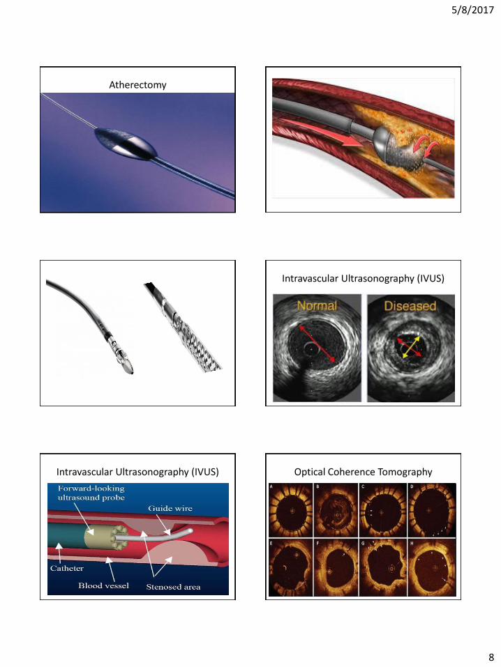
5/8/2017
8
Atherectomy
Intravascular Ultrasonography (IVUS)
Intravascular Ultrasonography (IVUS) Optical Coherence Tomography

5/8/2017
9
Post-catheterization Care
• Firm pressure is applied to puncture site for 15 -30minutes
• Wound sites are cleaned and dressed • The patient will be observed in recovery for 4-8
hours • The insertion site will be checked frequently for
signs of bleeding • Medications and discharged instruction are given • Lots of fluid should be taken • Vital signs should be monitored for 24 hours
PCI Success
1. Angiographic success
Residual stenosis < 10%, TIMI 3 flow, no occlusion of a significant side-branch, flow-limiting dissection, distal embolization or angiographic thrombus
2. Procedural success
Angiographic success without in-hospital major complications (eg death, MI, stroke, emergent CABG)
3. Clinical success
Procedural success with relief of signs and symptoms of myocardial ischemia
Challenges
• Restenosis is the body’s response to injury of the vessel wall from angioplasty and stent (foreign body)
• 50% of POBA, suffered significant restenosis
• Stent provide a mechanical framework to hold the artery wall open.
• When stent is used and restenosis occurs, this is called in-stent restenosis (ISR)
• Bare-metal stent
• Drug-eluting stent (sirolimus, everolimus, paclitaxel)
• Absorbable/ biodegradable stent

5/8/2017
10
Duration of Antiplatelet
• Double antiplatelet (DAPT) is combination of (aspirin+prasugrel) or (aspirin+ticagrelor) is recommended
DAPT must be continued for up to 12 months after STEMI, with strict minimum of:
• 1 month for patients receiving BMS
• 6months for patients receiving DES
Limitation of Coronary Intervention
• Left main stem disease
• 3- vessel disease
Coronary Artery Bypass Graft (CABG) is better option
Transradial vs Transfemoral
• The actual gold-standard for percutaneous remain femoral access, mainly due to its easy feasibility and short learning curve
• Since its introduction in 1989 for coronary angiography, radial approach has gained progressive widespread in worldwide
1. The radial approach has lower incidence of local complication
2. Avoidance of post-procedural bed-rest
3. Improve quality of life
Transradial vs Transfemoral
• The transradial and transfemoral approach are equivalent in terms of major safety with similar rate of MACE
• However, the transradial approach is more technically demanding with global procedural failure of around 7%
MACE – Major Adverse Cardiac Event

5/8/2017
11
Allen’s test – performed ± Oxymetry test
In the presence of an abnormal Allen’s test, the radial artery should not be used for catheterization
Radial Access – Proximal to Styloid Process
Learning Curve
• Getting access
• Radial artery spasm
• Anatomical variation
• Transversing the subclavian
• Catheter shape selection for cannulation
• Catheter control
• Patent hemostasis after pulling out the sheath
Radial Loop and Radial Recurrent Artery

5/8/2017
12
Verapamil Eliminate Spasm Problem Complication of Transradial Access
• Radial artery occlusion
• Forearm hematoma and pain
• Radial artery pseudoaneurysm
• Radial or brachial artery perforation
• Uncontrolled bleeding with compartment syndrome
• Need for femoral conversion
Femoral Approach
• Puncture site 1-2 cm below inguinal ligament
• Locate inguinal ligament running from anterior superior iliac spine to pubic tubercle
• Use skin crease to mark skin entry
• Fluoroscopy of inferior border of femoral head
Contraindication for Transfemoral Access
• Local skin infection
• Obesity
• Abdominal aortic aneurysm
• Femoral peripheral vascular disease
Complication of Transfemoral Access
• Distal embolization
• Dissection
• Pseudoaneurysm
• Retroperitoneal hematoma

5/8/2017
13
History
In 1929, Werner Forssmann demonstrated that a simple Rubber catheter could be passed to the pulmonary artery through the antecubital vein.