increased expression of claudin-17 promotes a malignant
TRANSCRIPT

RESEARCH Open Access
Increased expression of claudin-17promotes a malignant phenotype inhepatocyte via Tyk2/Stat3 signaling and isassociated with poor prognosis in patientswith hepatocellular carcinomaLemeng Sun1, Liangshu Feng2 and Jiuwei Cui1*
Abstract
Background: Hepatocellular carcinoma (HCC) is the second leading cause of cancer death in Asia; however, themolecular mechanism in its tumorigenesis remains unclear. Abnormal expression of claudins (CLDNs), a family oftight junction (TJ) proteins, plays an important role in the metastatic phenotype of epithelial-derived tumors byaffecting tight junction structure, function and related cellular signaling pathways. In a previous study, we used atissue chip assay to identify CLDN17 as an upregulated gene in HCC. Here we aimed to use molecular biologytechnology to explore the effect of CLDN17 on the malignant phenotype of HCC and the underlying molecularmechanism, with the objective of identifying a new target for HCC treatment and the control of HCC metastasis.
Method: The expression levels of CLDN17 in HCC tissues and histologically non-neoplastic hepatic tissues wereexplored by immunohistochemistry. Stable transfection of the hepatocyte line HL7702 with CLDN17 was detectedby real-time polymerase chain reaction (PCR), western blotting and immunofluorescence. The impact of CLDN17on the malignant phenotype of HL7702 cells in vitro was assessed by a Cell Counting Kit-8 (CCK8) assay, a Transwellassay and a wound-healing experiment. Western blotting was utilized to detect the activation state of Tyrosinekinase 2 (Tyk2) / signal transducer and activator of transcription3 (Stat3) pathway. A Tyk2 RNA interference (RNAi)was utilized to determine the impact of the Tyk2/Stat3 signaling pathway on the malignant phenotype of hepatocytes.
Results: In this work, our research group first found that CLDN17 was highly expressed in HCC tissues and was associatedwith poor prognosis. In addition, we demonstrated that CLDN17 affected the Stat3 signaling pathway via Tyk2 andultimately enhanced the migration ability of hepatocytes.
Conclusion: In conclusion, we confirmed that the upregulated expression of CLDN17 significantly enhancesthe migration ability of hepatocytes in vitro and we found that the activation of the Stat3 pathway by Tyk2may an important mechanism by which CLDN17 promotes aggressiveness in hepatocytes.
Keywords: Hepatocytes, Tight junction, Migration, CLDN17, Tyrosine kinase 2, Signal transducer and activatorof transcription 3
* Correspondence: [email protected] Cell and Cancer Center, The First Bethune Hospital, Jilin University,Changchun, Jilin 130021, People’s Republic of ChinaFull list of author information is available at the end of the article
© The Author(s). 2018 Open Access This article is distributed under the terms of the Creative Commons Attribution 4.0International License (http://creativecommons.org/licenses/by/4.0/), which permits unrestricted use, distribution, andreproduction in any medium, provided you give appropriate credit to the original author(s) and the source, provide a link tothe Creative Commons license, and indicate if changes were made. The Creative Commons Public Domain Dedication waiver(http://creativecommons.org/publicdomain/zero/1.0/) applies to the data made available in this article, unless otherwise stated.
Sun et al. Diagnostic Pathology (2018) 13:72 https://doi.org/10.1186/s13000-018-0749-1

BackgroundPrevious studies have shown that the metastasis ofepithelial-derived tumors is accompanied by abnormalitiesin tight junction (TJ) structure and function [1, 2]. Clau-dins (CLDNs) are the key proteins that form TJ, and accu-mulating evidence suggests that tumor cells frequentlyexhibit changes in the expression and localization ofCLDNs [3]. For example, CLDN1 demonstrated to beoverexpressed in colorectal cancer (CRC) compared withthe level in the normal mucosa, and CLDN1 targetingwith an anti-CLDN1 monoclonal antibody (mAb) resultedin decreased growth and survival of colorectal cancer(CRC) cells, suggesting that CLDN1 could be a new po-tential therapeutic target for CRC [4]. In addition, adatabase-augmented, exosome-based mass spectrometryapproach identified circulating CLDN3 as a biomarker inpatients with prostate cancer [5]. Moreover, high-levelcytoplasmic CLDN3 expression is an independent pre-dictor of poor survival in patients with breast cancer [6].The TJ protein CLDN4 has been reported to be overex-pressed in advanced ovarian cancer (OC) and Kaplan-Meier survival analyses and the log-rank test suggest thathigh expression of CLDN4 may have prognostic value inOC [7]. These observations revealed that the alterations inCLDNs expression may be related to tumorigenesis andcancer progression in various types of human carcinoma.Additionally, CLDNs have been shown to participate
in the transduction of intracellular/extracellular sig-nals and may be related to tumorigenesis and cancerprogression in human various carcinomas [8, 9]. Forinstance, genetic and pharmacological studies con-firmed that the expression of CLDN3 was downregu-lated in colon cancer and that the loss of CLDN3induced Wnt/β-catenin activation in a transducer andactivator of transcription 3 (Stat3)-dependent mannerto promote colon cancer malignancy [10]. Besides, arecent study revealed that enhanced CLDN18 expres-sion activated ERK1/2 to contributed to the malignantpotentials of bile duct cancer [11]. Our preliminarywork showed that CLDN17 was strongly expressed inHCC tissues and cell lines and weakly expressed innon-neoplastic tissues and hepatocyte lines, whichrevealed that upregulated CLDN17 expression mayplay a role in the development of HCC. Furthermore,gene chip screening revealed that CLDN17 overex-pression activated the tyrosine kinase 2 (Tyk2)/Stat3pathway signaling pathway. To date, there has beenno report on the impact of CLDN17 on the malig-nant phenotype of hepatocytes. In this study, we uti-lized molecular biology and other techniques tostudy the role and mechanisms of CLDN17 in malig-nant phenotype of hepatocytes and to identify noveltargets for HCC treatment and the control of earlymetastasis.
MethodsAntibodiesRabbit polyclonal antibodies against CLDN17 (cat. no.ab233333) and mouse anti-human β-actin (cat. no.ab8226) were purchased from Abcam (Massachusetts,US). Rabbit anti-human phospho-Stat1 (cat. no. #7649),rabbit anti-human phospho-Stat3 (cat. no. #9145, rabbitanti-human phospho-Tyk2 (cat. no. #68790), rabbitanti-human Stat1 (cat. no.#14,994), rabbit anti-humanStat3 (cat. no. #9139) and rabbit anti-human Tyk2 (cat.no. #13531) were purchased from Cell Signaling Tech-nology (Boston, USA).
Cell cultureHuman hepatocyte line (HL7702) and HCC cell lines(HepG2, Hep3B and Huh1) utilized in this study werepurchased from Shanghai Cell Bank of the ChineseAcademy of Sciences. These cell lines were cultured inDulbecco’s modified Eagle’s medium supplemented with10% fetal bovine serum (FBS) at 37 °C in a humidifiedincubator containing 5% CO2.
Plasmid construction and transfectionThe plasmid p-EGFP-C1/CLDN17 (NM_012131) wasconstructed and amplified by KeyGen BioTech Com-pany. Two micrograms of plasmid DNA was transfectedinto cells using the SuperFect Transfection Reagent(TaKaRa, Japan) according to the manufacturer’s proto-col. A cell line stably expressing CLDN17 was selectedin medium containing G418 (Thermo Fisher Scientific,Waltham, MA).
Real-time polymerase chain reaction (PCR)Total RNA was extracted using a Perfect Pure RNA Cul-tured Cell Kit (Thermo Fisher Scientific, Waltham, MA) ac-cording to the manufacturer’s protocol. Real-time PCRreactions was carried out as previously described [12]. Theprimer pairs used for CLDN17 and glyceraldehyde phos-phate dehydrogenase (GAPDH) were as follows: CLDN17forward (5′-ACCCAGCCATCCACATAG-3′) and reverse(5′- CCCTTGCTTCTTTCTGTTG-3′); and GAPDHforward (5’-AACGTGTCAGTCGTGGACCTG-3′) and re-verse (5’-AGTGGGTGTCGCTGTFGAAGT-3′). The rela-tive expression was based on the expression ratio of atarget gene compared with that of GAPDH.
Western blottingA bicinchoninic acid (BCA) Protein Assay Kit (PierceChemical Co., Rockford, Illinois, USA) was utilized todetect protein concentrations. Total protein (30 micro-grams) was separated via 10% sodium dodecyl sulfate-polyacrylamide gel electrophoresis (SDS-PAGE) gel andthen transferred onto a nitrocellulose membrane (Millipore,Temecula, California, USA). Next, the membrane was
Sun et al. Diagnostic Pathology (2018) 13:72 Page 2 of 10
blocked and investigated with the following primary anti-bodies: rabbit anti-human phospho-Stat1, rabbit anti-hu-man Stat1, rabbit anti-human phospho-Stat3, rabbitanti-human Stat3, rabbit anti-human phospho-Tyk2, rabbitanti-human Tyk2, rabbit anti-human CLDN17 and mouseanti-human β-actin. After 3 washes with phosphate-buff-ered saline (PBS), the membrane was incubated with horse-radish peroxidase (HRP)-conjugated secondary antibody(Santa Cruz Biotechnologies, California, USA) at a 1:1000dilution at 4 °C. Immunoreactive bands were detected usingECL western blot reagents (GE, Fairfield, Connecticut,USA) and analyzed with Image Lab 6.0.1 Software fromBio-Rad Laboratories.
Immunofluorescence methodThe cells were fixed with 4% paraformaldehyde for10 min at room temperature (RT) and then perme-abilized with 0.1% Triton X-100 (Sigma-Aldrich, cat. no.9002-93-1). Then, after blocking with 2% bovine serumalbumin (Bote Biotechnological Corporation, Jilin,China) diluted in PBS for 30 min, the cells were probedwith a primary rabbit anti-human CLDN17 antibody,which was diluted in blocking solution (1:1000 dilution)for 30 min at RT. The cells were incubated with AlexaFluor®647-conjugated anti-rabbit IgG antibody (ab150093,Santa Cruz Biotechnologies, California, USA) at a 1:1000dilution.
Cell counting Kit-8 assayCell proliferation curve generated by the colorimetricwater-soluble tetrazolium salt assay using a Cell Count-ing Kit-8 (Dojindo, Kumamoto, Japan) as the protocol.The cells were seeded into 96-well plates in triplicate,and cell proliferation was recorded per 12 h for 4 days.
Wound-healing assayThe cells were maintained in a monolayer at 70% conflu-ence on 24-well plastic dishes and the monolayer wasscratched with a 100-μl pipette tip. The wounds werephotographed light microscope (E100, Nikon InstrumentsInc., Japan) (magnification × 200) at the same location at0, 12 and 24 h.
Transwell chamber methodThe cells were grown in a monolayer at 90% conver-gence and were maintained in FBS-free medium for12 h. Matrigel (BD Biosciences, cat. no. 356234) wasadded to the upper Boyden chamber (Millipore, Bedford,MA) in 24-well plates and the plates were maintained ina cell incubator at 37 °C for 15 min. Then, medium con-taining chemotactic factors, which had been collectedfrom the cell culture, was added to the 24-well plate.The cells were supplemented with Matrigel and culturedin a cell incubator at 37 °C for 6 h.
RNA interference (RNAi) methodFrozen glycerol bacterial stocks containing pGCSIL-scramble and pGCSIL-Tyk2-RNAi were purchased fromNanjing KeyGen Biotech Co., Ltd. The target wasTyk2-RNAi (29473), and the control insert sequencewas pGCSIL-scramble. HEK 293 T cells (0.2 × 10⁷) wereseeded and maintained for 24 h to achieve 70–80% con-fluence in 6-well dishes (Costar, Cam- bridge, MA).Three plasmids, including of pGCSIL-Tyk2-RNAi orpGCSIL-scramble, 5 μg of the packaging vector pHelper1.0 and 5 μg of a vesicular stomatitis virus glycoprotein(VSVG) expression plasmid vector, were added toOpti-MEM, with a final volume of 1.0 ml. Then, 50 μl ofLipofectamine was added to 950 μl of FBS-free medium.These two solutions were mixed and added to the cells.Lentiviral particles were harvested 48 h after transfec-tion, and the viral titer was determined by countinggreen fluorescent protein (GFP)-expressing cells under afluorescence microscopy (Nikon Diaphot 300®) withfilters 96 h after transfection.
Patients and tissue samplesBiopsies were collected from 52 patients with pathologic-ally confirmed the diagnoses of HCC who received treat-ment at The First Bethune Hospital of Jilin Universitybetween June 2007 and May 2012. The patients were care-fully chosen based on the following criteria: no history ofradiotherapy or chemotherapy and no prior malignantdisease. The grade and classification of the HCC patientswere based on the American Joint Committee on Cancer(AJCC) tumor node metastasis (TNM) staging system.Thirty cases of histologically non-neoplastic hepatic tis-sues and 10 cases of cirrhosis tissues were also obtainedfrom hepatitis B virus infected patients who were treatedat The First Bethune Hospital, Jilin University during theperiod between October 2006 and September 2011 thatwere identified to be histologically non-neoplastic. Therewere 16 men and 14 women with an average age of 49 years.The medical records of the patients were reviewed to deter-mine the clinical and pathological characteristics.
ImmunohistochemistryAn immunohistochemistry was utilized to explore theexpression patterns of CLDN17 in 52 HCC tissues, 10cirrhosis tissues and 30 non-neoplastic hepatic tissues.Of the 52 cases, 42 cases exhibited HBsAg infection, 17cases exhibited occurrence and metastasis, and 34 caseswere coupled with cirrhosis. The experimental methodwas described previously [13], and the antibody utilizedwas a rabbit anti-human CLDN17 antibody. The evalu-ation of protein expression levels was based on the per-centage of positively stained tumor cells in combinationwith the staining intensity as previously described [14].
Sun et al. Diagnostic Pathology (2018) 13:72 Page 3 of 10

Follow-upThe patients with a pathologically confirmed diagnosisof HCC were followed-up for 60 months after diagnosisto assess occurrence and metastasis and to determinesurvival. The survival status of the patients was deter-mined through a telephone interview or an outpatientvisit before December 2017.
Statistical methodsAll of the experiments were repeated 3 times, and all ofthe data are based on the mean ± SD of at least 3 experi-mental results. The experimental results were analyzedusing Student’s t-test, and the prognostic significanceand value of CLDN17 expression was determined by the
Chi-square test/Chi-square goodness-of-fit test. P < 0.05was considered statistical significance.
ResultsThe expression of CLDN17 was upregulated in HCC cell linesand tissuesReal-time PCR and western blotting were utilized todetect the expression of CLDN17 in the human hepato-cyte line and the HCC cell lines (Huh1, HepG2 andHep3B). We found that the mRNA and protein expres-sion levels of CLDN17 were low or absent in the humanhepatocyte line HL7702 but high in HCC cell linesHuh1, HepG2 and Hep3B (Fig. 1a-c).
Fig. 1 The expression levels of CLDN17 in HCC cell lines and tissues. a The relative mRNA level of CLDN17 in the hepatocyte line and HCC celllines; (b) The relative protein expression of CLDN17 in the hepatocyte line and HCC cell lines; (c) The corresponding statistical analysis of proteinexpression in the hepatocyte line and HCC cell lines; (d) The correlation between the expression of CLDN17 and survival in HCC patients; (e) Theprotein expression of CLDN17 in hepatocyte tissues, cirrhosis tissues, tissues adjacent to the tumors and HCC tissues. Note: * represents P < 0.05,** represents P < 0.01, compared with the empty vector groups
Sun et al. Diagnostic Pathology (2018) 13:72 Page 4 of 10

CLDN17 expression was explored in 52 HCC tissues,10 histologically non-neoplastic cirrhosis tissues, 10 his-tologically non-neoplastic tissues adjacent to the tumorand 30 histologically non-neoplastic hepatic tissues. Asshown in Fig. 1e, the expression of CLDN17 in hepatictissues and HCC tissues is located mainly in thecytoplasm and membrane. High expression of CLDN17was observed in 53.8% (28/52) of HCC tissues 30.0% (3/10)of non-neoplastic cirrhosis tissues, 40.0% (4/10) of histolog-ically non-neoplastic tissues adjacent to the tumor and in40.0% (12/30) of hepatic tissues (P = 0.0001 < 0.001)(Table 1).The log-rank test was utilized to analyze correlations
between CLDN17 and clinical survival. As shown in Fig.1d, patients with positive expression of the CLDN17protein in tumors (median survival, 46.15 months) had anotably shorter survival than those with negativeexpression of the CLDN17 protein (median survival,57.79 months).
The relationships between CLDN17 and clinicalpathological indicators were also analyzed, and theexpression of CLDN17 was not associated with age(P = 1.000), HBsAg absent status (P = 1.000), cirrhosis(P = 1.000), serum AFP level (P = 0.637) of HCC pa-tients or clinical staging (P = 1.000) of HCC patients.However, CLDN17 expression was associated with
HCC occurrence and metastasis (P = 0.001 < 0.01),histological grade (P = 0.001 < 0.01) and TNM stage(P = 0.001 < 0.01) (Table 1).
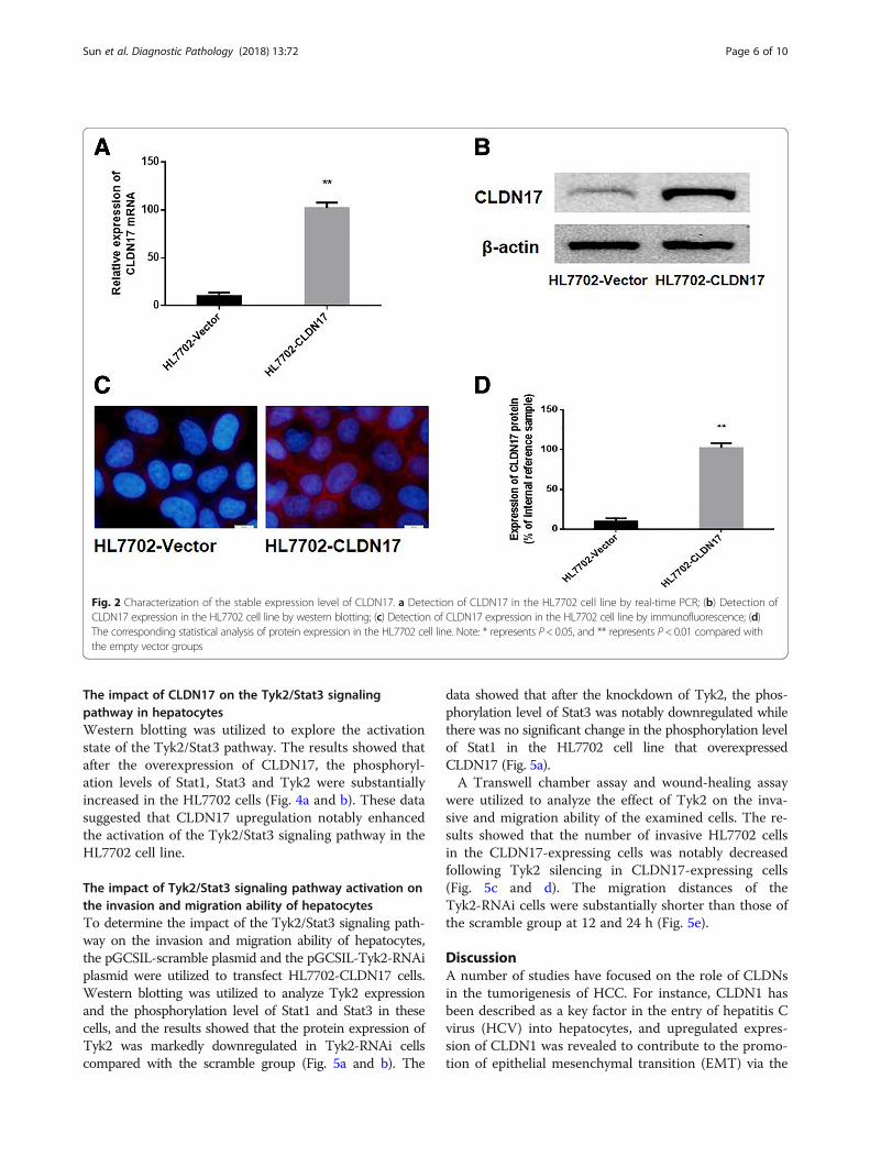
Stable transfection of a hepatocyte line with CLDN17The p-EGFP-C1/CLDN17 plasmid was utilized totransfect HL7702 cells. After G418 screening, amonoclonal strain of HL7702 cells was obtained,which was termed HL7702-CLDN17. Real-time PCRand western blotting were also utilized to detect theexpression of CLDN17 in cultured cells. The resultsshowed that the mRNA and protein expression levelsof CLDN17 in the HL7702-CLDN17 group were not-ably higher than those in the empty vector groups (P= 0.0001 < 0.01; P = 0.0001 < 0.01, respectively) (Fig. 2a, band d). Immunofluorescence was utilized to detect thelocalization of CLDN17 in HL7702-CLDN17 cells. The re-sults showed that the expression of CLDN17 was primar-ily localized to the cell cytoplasm and membrane (Fig. 2c).These results demonstrated that clonal HL7702 cell linethat stably expressed CLDN17 had been successfullyestablished.
The impact of CLDN17 on the proliferation and migrationof hepatocyte linesThe growth curve for the HL7702 cell line was gener-ated by the CCK-8 method. The data revealed thatthe proliferation rates of HL7702-CLDN17 cells at 48and 72 h were notably higher than those of theempty vector groups (Fig. 3a). A wound-healing ex-periment was utilized to detect the impact ofCLDN17 on the migration ability of hepatocytes (Fig.3b). The results showed that the migration distancesof HL7702-CLDN17 cells were substantially greaterthan those of the empty vector groups at 12 and 24 h(P = 0.0022, < 0.01). The Transwell chamber methodwas also utilized to detect the migration ability of he-patocytes. Twelve hours after the cells were seeded,the cells that invaded through the membrane of thechamber were observed. The results showed that thenumber of invasive cells in the HL7702-CLDN17group was notably higher than in the empty vectorgroups (Fig. 3c and d). These results suggested thatCLDN17 clearly promoted the proliferation and mi-gration ability of hepatocytes in vitro.
Table 1 Expression of CLDN17 and the clinicopathologicalcharacteristics in HCC patients
Item n CLDN17(+)
CLDN17(−)
P
HCC tissues 52 28 24 < 0.01*
Hepatic tissues 30 12 18
Age (years)
≤ 60 21 11 10 1.000
> 60 31 17 14
HbsAg
+ 42 23 19 1.000
- 10 5 5
Cirrhosis
+ 34 18 16 1.000
- 18 10 8
Occurrence andmetastasis
+ 17 13 4 < 0.01*
- 35 15 20
Serum AFP (ng/ml)
< 400 22 13 9 0.637
> 400 30 15 15
TNM stage (AJCC)
I~II 27 17 10 < 0.01*
III~IV 25 11 14
Histological grade
Well-differentiated 24 15 9 < 0.01*
Moderately and poorly differentiated 28 13 15*Statistical significance was found with the Chi-square test/Chi-SquareGoodness-of-Fit Test
Sun et al. Diagnostic Pathology (2018) 13:72 Page 5 of 10
The impact of CLDN17 on the Tyk2/Stat3 signalingpathway in hepatocytesWestern blotting was utilized to explore the activationstate of the Tyk2/Stat3 pathway. The results showed thatafter the overexpression of CLDN17, the phosphoryl-ation levels of Stat1, Stat3 and Tyk2 were substantiallyincreased in the HL7702 cells (Fig. 4a and b). These datasuggested that CLDN17 upregulation notably enhancedthe activation of the Tyk2/Stat3 signaling pathway in theHL7702 cell line.
The impact of Tyk2/Stat3 signaling pathway activation onthe invasion and migration ability of hepatocytesTo determine the impact of the Tyk2/Stat3 signaling path-way on the invasion and migration ability of hepatocytes,the pGCSIL-scramble plasmid and the pGCSIL-Tyk2-RNAiplasmid were utilized to transfect HL7702-CLDN17 cells.Western blotting was utilized to analyze Tyk2 expressionand the phosphorylation level of Stat1 and Stat3 in thesecells, and the results showed that the protein expression ofTyk2 was markedly downregulated in Tyk2-RNAi cellscompared with the scramble group (Fig. 5a and b). The
data showed that after the knockdown of Tyk2, the phos-phorylation level of Stat3 was notably downregulated whilethere was no significant change in the phosphorylation levelof Stat1 in the HL7702 cell line that overexpressedCLDN17 (Fig. 5a).A Transwell chamber assay and wound-healing assay
were utilized to analyze the effect of Tyk2 on the inva-sive and migration ability of the examined cells. The re-sults showed that the number of invasive HL7702 cellsin the CLDN17-expressing cells was notably decreasedfollowing Tyk2 silencing in CLDN17-expressing cells(Fig. 5c and d). The migration distances of theTyk2-RNAi cells were substantially shorter than those ofthe scramble group at 12 and 24 h (Fig. 5e).
DiscussionA number of studies have focused on the role of CLDNsin the tumorigenesis of HCC. For instance, CLDN1 hasbeen described as a key factor in the entry of hepatitis Cvirus (HCV) into hepatocytes, and upregulated expres-sion of CLDN1 was revealed to contribute to the promo-tion of epithelial mesenchymal transition (EMT) via the
Fig. 2 Characterization of the stable expression level of CLDN17. a Detection of CLDN17 in the HL7702 cell line by real-time PCR; (b) Detection ofCLDN17 expression in the HL7702 cell line by western blotting; (c) Detection of CLDN17 expression in the HL7702 cell line by immunofluorescence; (d)The corresponding statistical analysis of protein expression in the HL7702 cell line. Note: * represents P< 0.05, and ** represents P< 0.01 compared withthe empty vector groups
Sun et al. Diagnostic Pathology (2018) 13:72 Page 6 of 10

c-Abl/Raf/Ras/ERK signaling pathway [15, 16]. Further-more, it was demonstrated that CLDN3 is an epigeneti-cally silenced tumor suppressor gene in HCC, and itsoverexpression notably inhibits metastasis by suppress-ing the EMT via the Wnt/β-catenin signaling pathway inHCC cells [13]. Besides, CLDN14 was epigenetically
silenced via the trimethylation of lysine 27 on histoneH3 (H3K27ME3) and was a novel prognostic biomarkerin HCC [17]. Given the correlation between the expres-sion levels of these CLDNs and the tumorigenesis ofHCC, CLDNs represent potential novel therapeutic tar-gets in patients with HCC.
Fig. 3 The impact of CLDN17 on the proliferation and migration ability of cells in vitro. a A growth curve for the HL7702 cell line was generatedby the CCK-8 method; (b) A wound healing assay was utilized to detect the migration ability of the HL7702 cell line in vitro; (c) The Transwellchamber method was utilized to detect the invasive ability of the HL7702 cell line in vitro; (d) The corresponding statistical analysis of invadedcells. Note: * represents P < 0.05 and ** represents P < 0.01 compared with the empty vector groups
Fig. 4 The impact of CLDN17 on the Tyk2/Stat3 signaling pathway. a Western blotting was utilized to detect the activation of the Stat3 signalingpathway in the HL7702 cell line; (b) The corresponding statistical analysis of the activation status of various Stat3 pathway components. Note: *represents P < 0.05 and ** represents P < 0.01 compared with the empty vector groups
Sun et al. Diagnostic Pathology (2018) 13:72 Page 7 of 10

CLDN17 is one of 27 members of the CLDN proteinfamily, and our current understanding of the biologicalfunctions of CLDN17 is primarily limited to epithelial andepidermal permeability, barrier protection, and cell con-nections; reports on the relationship between CLDN17and tumors are rare [18]. Our research group first foundthat CLDN17 expression was highly expressed in HCC tis-sues, and we speculated that the high expression of thisgene may be involved with the tumorigenesis and progres-sion in patients with HCC. Moreover, in the present study,we confirmed that CLDN17 markedly promotes the inva-sive ability of the hepatocyte line HL7702. Similar to ourstudy, several studies have identified specific CLDNs aspro-oncogenes in human various cancers. For instance,previous work has shown that CLDN1 plays a key role ininflammation-induced growth and progression in patientswith colorectal carcinoma [19]. Furthermore, Philip, R. etal. reported that CLDN7 expression in colorectal cancer
contributes to motility and invasion by promoting ashift towards EMT by recruiting EpCAM towards TACE/presenilin2 [20]. It was also revealed that CLDN7 is fre-quently overexpressed and promotes invasion in ovariancancer [21]. However, in contrast to our results, otherstudies have shown that some CLDNs could be identifiedas tumor suppressor gene [22, 23]. For instance, the ex-pression of CLDN1 was reduced in stage II and III rectalcancer and was established as a factor that correlatesclearly with recurrence and poor prognosis [24]. Inaddition, the expression of CLDN6 was demonstrated tobe silenced in cervical carcinoma tissues, and the restor-ation of CLDN6 expression suppressed cell proliferationand colony formation in cervical carcinoma cells in vitro,and tumor growth in vivo [25]. One potential reason forthis difference is that the functions of CLDNs may be spe-cific and dependent on different interacting molecules indifferent cells [26, 27]. In this manner, specific CLDNs
Fig. 5 RNAi was utilized to silence Tyk2 expression in CLDN17-expressing cells. a Western blotting was utilized to examine the effects of silencingTyk2 and activating the Stat3 signaling pathway in the HL7702 cell line; (b) The corresponding statistical analysis of the activation of the Stat3signaling pathway; (c) The Transwell chamber method was utilized to detect the impact of Tyk2 silencing on the invasive ability of the cells invitro; (d) The corresponding statistical analysis of invaded cells; (e) A wound healing assay was utilized to detect the migration ability of theHL7702 cell line in vitro. Note: * represents P < 0.05 and ** represents P < 0.01 compared with the scramble group
Sun et al. Diagnostic Pathology (2018) 13:72 Page 8 of 10

may have specific impacts on the biological behavior of agiven tumor [28–30].At present, our results first indicated that the CLDN17
was overexpressed and highly associated with metastaticprogression and prognosis in patients with HCC. More-over, the overexpression of CLDN17 markedly promotedthe invasion and migration abilities of the hepatocyteline HL7702. Furthermore, we also performed an initialexploration of the molecular mechanism associated withthis effect, and we found that CLDN17 upregulation af-fected the Stat3 signaling pathway via Tyk2 and ultim-ately enhanced the migration ability of hepatocytes.Considering the limited therapeutic options for patientswith HCC, the role of CLDN17 as a therapeutic targetmerits further exploration.
ConclusionThere have been few reports on the roles of CLDN17 intumors, and many problems related to the specific mo-lecular mechanisms must still be researched. In thepresent study, we confirmed that the overexpression ofCLDN17 notably enhanced the malignant phenotype ofthe hepatocytes. In addition, the induction of Tyk2/Stat3signaling may be one of the most important mechanismsby which CLDN17 promotes the migration ability inhepatocytes.
AbbreviationsAJCC: American Joint Committee on Cancer; CCK8: Cell Counting Kit-8;CLDNs: Claudins; FBS: Fetal bovine serum; GFP: Green fluorescent protein;H3K27ME3: Trimethylation of lysine 27 on histone H3; HCC: Hepatocellularcarcinoma; HCV: Hepatitis C virus; Stat3: Signal transducer and activator oftranscription3; TJ: Tight junction; TNM: Tumor node metastasis; Tyk2: Tyrosinekinase 2
AcknowledgementsWe would like to thank American Journal Experts (AJE) for help with thismanuscript.
Availability of data and materialsThe raw data are available upon request on the following e-mail address:[email protected].
Authors’ contributionsConceived and designed the experiments: JC. Performed the experiments: LSand LF. Analyzed the data: LF. Wrote the paper: JC. All authors read andapproved the final manuscript.
Ethics approval and consent to participateThe study was approved by the Ethics Committee of Jilin University (referencenumber 20070459). Written informed consent was obtained from all patients atthe time of their treatment for the use of material in future research.
Consent for publicationNot applicable.
Competing interestsThe authors declare that they have no competing interests.
Publisher’s NoteSpringer Nature remains neutral with regard to jurisdictional claims in publishedmaps and institutional affiliations.
Author details1Stem Cell and Cancer Center, The First Bethune Hospital, Jilin University,Changchun, Jilin 130021, People’s Republic of China. 2Department ofNeurology and Neuroscience Center, First Hospital of Jilin University,Changchun, Jilin, China.
Received: 25 April 2018 Accepted: 3 September 2018
References1. Escudero-Esparza A, Jiang WG, Martin TA. The Claudin family and its role in
cancer and metastasis. Front Biosci (Landmark ed). 2010;16:1069–83.2. Cunniffe C, Brankin B, Lambkin H, Ryan F. The role of Claudin-1 and Claudin-
7 in cervical tumorigenesis. Anticancer Res. 2014;34:2851–7.3. Turksen K, Troy TC. Junctions gone bad: claudins and loss of the barrier in
cancer. Biochim Biophys Acta. 1816;2011:73–9.4. Ouban A. Claudin-1 role in colon cancer: an update and a review. Histol
Histopathol. 2018;11980. https://doi.org/10.14670/HH-11-980.5. Worst TS, von Hardenberg J, Gross JC, Erben P, Schnolzer M, Hausser I,
Bugert P, Michel MS, Boutros M. Database-augmented mass spectrometryanalysis of exosomes identifies Claudin 3 as a putative prostate Cancerbiomarker. Mol Cell Proteomics. 2017;16:998–1008.
6. Szasz MA. Claudins as prognostic factors of breast cancer. Magy Onkol.2012;56:209–12.
7. Martin de la Fuente L, Malander S, Hartman L, Jonsson JM, Ebbesson A,Nilbert M, Masback A, Hedenfalk I. Claudin-4 Expression is Associated WithSurvival in Ovarian Cancer But Not With Chemotherapy Response. Int JGynecol Pathol. 2018;37:101–9.
8. González-Mariscal L, Tapia R, Chamorro D. Crosstalk of tight junctioncomponents with signaling pathways. Biochim Biophys Acta Biomembr.2008;1778:729–56.
9. Tsukita S, Furuse M, Itoh M. Structural and signalling molecules cometogether at tight junctions. Curr Opin Cell Biol. 1999;11:628–33.
10. Ahmad R, Kumar B, Chen Z, Chen X, Muller D, Lele SM, WashingtonMK, Batra SK, Dhawan P, Singh AB. Loss of claudin-3 expressioninduces IL6/gp130/Stat3 signaling to promote colon cancermalignancy by hyperactivating Wnt/beta-catenin signaling. Oncogene.2017;36:6592–604.
11. Takasawa K, Takasawa A, Osanai M, Aoyama T, Ono Y, Kono T, Hirohashi Y,Murata M, Sawada N. Claudin-18 coupled with EGFR/ERK signalingcontributes to the malignant potentials of bile duct cancer. Cancer Lett.2017;403:66–73.
12. Livak KJ, Schmittgen TD. Analysis of relative gene expression data usingreal-time quantitative PCR and the 2(−Delta Delta C(T)) method. Methods.2001;25:402–8.
13. Jiang L, Yang YD, Fu L, Xu W, Liu D, Liang Q, Zhang X, Xu L, Guan XY, Wu B,et al. CLDN3 inhibits cancer aggressiveness via Wnt-EMT signaling and is apotential prognostic biomarker for hepatocellular carcinoma. Oncotarget.2014;5:7663–76.
14. Gao M, Li W, Wang H, Wang G. The distinct expression patterns of claudin-10, −14, −17 and E-cadherin between adjacent non-neoplastic tissues andgastric cancer tissues. Diagn Pathol. 2013;8:205.
15. Suh Y, Yoon CH, Kim RK, Lim EJ, Oh YS, Hwang SG, An S, Yoon G, Gye MC,Yi JM, et al. Claudin-1 induces epithelial-mesenchymal transition throughactivation of the c-Abl-ERK signaling pathway in human liver cells.Oncogene. 2013;32:4873–82.
16. Suh Y, Yoon CH, Kim RK, Lim EJ, Oh YS, Hwang SG, An S, Yoon G, Gye MC,Yi JM, et al. Claudin-1 induces epithelial-mesenchymal transition throughactivation of the c-Abl-ERK signaling pathway in human liver cells.Oncogene. 2017;36:1167–8.
17. Li CP, Cai MY, Jiang LJ, Mai SJ, Chen JW, Wang FW, Liao YJ, Chen WH, JinXH, Pei XQ, et al. CLDN14 is epigenetically silenced by EZH2-mediatedH3K27ME3 and is a novel prognostic biomarker in hepatocellular carcinoma.Carcinogenesis. 2016;37:557–66.
18. Koval M. Claudin heterogeneity and control of lung tight junctions. AnnuRev Physiol. 2013;75:551–67.
19. Cherradi S, Ayrolles-Torro A, Vezzo-Vie N, Gueguinou N, Denis V, Combes E,Boissiere F, Busson M, Canterel-Thouennon L, Mollevi C, et al. Antibodytargeting of claudin-1 as a potential colorectal cancer therapy. J Exp ClinCancer Res. 2017;36:89.
Sun et al. Diagnostic Pathology (2018) 13:72 Page 9 of 10

20. Philip R, Heiler S, Mu W, Buchler MW, Zoller M, Thuma F. Claudin-7promotes the epithelial-mesenchymal transition in human colorectal cancer.Oncotarget. 2015;6:2046–63.
21. Dahiya N, Becker KG, Wood WH 3rd, Zhang Y, Morin PJ. Claudin-7 isfrequently overexpressed in ovarian cancer and promotes invasion. PLoSOne. 2011;6:e22119.
22. Ouban A, Ahmed A. Claudins in human cancer, a review. Histol Histopathol.2010;25(1):83–90.
23. Zavala-Zendejas VE, Torres-Martinez AC, Salas-Morales B, Fortoul TI, MontanoLF, Rendon-Huerta EP. Claudin-6, 7, or 9 overexpression in the humangastric adenocarcinoma cell line AGS increases its invasiveness, migration,and proliferation rate. Cancer Investig. 2011;29:1–11.
24. Yoshida T, Kinugasa T, Akagi Y, Kawahara A, Romeo K, Shiratsuchi I, Ryu Y,Gotanda Y, Shirouzu K. Decreased expression of claudin-1 in rectal cancer: afactor for recurrence and poor prognosis. Anticancer Res. 2011;31:2517–25.
25. Zhang X, Ruan Y, Li Y, Lin D, Quan C. Tight junction protein claudin-6inhibits growth and induces the apoptosis of cervical carcinoma cells invitro and in vivo. Med Oncol. 2015;32:148.
26. Lu Z: Functions of claudin-7 in human lung cancer. 2012.27. Micke P, Mattsson JS, Edlund K, Lohr M, Jirstrom K, Berglund A, Botling J,
Rahnenfuehrer J, Marincevic M, Ponten F, et al. Aberrantly activated claudin6 and 18.2 as potential therapy targets in non-small-cell lung cancer. Int JCancer. 2014;135:2206–14.
28. D'Souza T, Agarwal R, Morin PJ. Phosphorylation of claudin-3 at threonine192 by cAMP-dependent protein kinase regulates tight junction barrierfunction in ovarian cancer cells. J Biol Chem. 2005;280:26233–40.
29. D'Souza T, Indig FE, Morin PJ. Phosphorylation of claudin-4 by PKCεregulates tight junction barrier function in ovarian cancer cells. Exp Cell Res.2007;313:3364–75.
30. Li X, Li Y, Qiu H, Wang Y. Downregulation of claudin-7 potentiates cellularproliferation and invasion in endometrial cancer. Oncol Lett. 2013;6:101–5.
Sun et al. Diagnostic Pathology (2018) 13:72 Page 10 of 10