the role of tight junction proteins claudin-3 and...
TRANSCRIPT

THE ROLE OF TIGHT JUNCTION PROTEINS CLAUDIN-3 AND CLAUDIN-7 IN URETERIC BUD BRANCHING
Nicholas Haddad
Department of Human Genetics
McGill University, Montreal
June, 2009
A thesis submitted to the Faculty of Graduate and Postdoctoral Studies in partial fulfillment of the requirements of the degree of Master’s of Science.
© Nicholas Haddad, 2009
1
TABLE OF CONTENTS
TABLE OF CONTENTS................................................................................................................. 1
ABSTRACT…............................................................................................................................... .. 3
RÉSUMÉ .................................................................................................................................. 5
ACKNOWLEDGEMENTS ............................................................................................................. 7
CONTRIBUTION OF CO-AUTHORS ......................................................................................... 10
LIST OF FIGURES ....................................................................................................................... 11
ABBREVIATIONS ....................................................................................................................... 12
CHAPTER I: Introduction ............................................................................................................. 14
1. Overview of kidney function ........................................................................................... 15
2. The kidney: three stages of development ........................................................................ 16
2.1 The pronephros and the mesonephros ...................................................................... 16
2.2 The metanephros....................................................................................................... 17
3. The genetics of branching morphogenesis ..................................................................... 18
3.1 The Gdnf/Ret/Gfr 1 pathway and Pax2 ................................................................... 19
3.2 Adhesion molecules .................................................................................................. 20
4. Tight junctions and claudins........................................................................................... 21
4.1 Tight junctions are composed of various proteins.................................................... 21
4.2 Claudins are essential components of tight junctions .............................................. 22
4.3 Claudin structure and function ................................................................................. 23
5. Claudins in the kidney .................................................................................................... 24
6. Claudins and human disease .......................................................................................... 25
7. The function of Claudin-3 and Claudin-7....................................................................... 26
7.1 Claudin-3 .................................................................................................................. 26
7.2 Claudin-7 .................................................................................................................. 27
8. Summary of objectives .................................................................................................... 28
CHAPTER II:The tight junction proteins Cldn3 and Cldn7 are expressed in the ureteric bud and promote tubulogenesis in vitro................................................................................ 30
1. Abstract........................................................................................................................... 31
2. Introduction .................................................................................................................... 32

2
3. Materials and methods ................................................................................................... 35
3.1 Animal care .............................................................................................................. 35
3.2 Electron microscopy ................................................................................................. 35
3.3 Whole mount in situ hybridization ............................................................................ 36
3.4 Double immunofluorescence .................................................................................... 36
3.5 Cell lines and constructs........................................................................................... 37
3.6 Immunoblotting......................................................................................................... 38
3.7 Collagen gel assays .................................................................................................. 38
4. Results ............................................................................................................................... ....39
4.1 Tight junction structures are present on the apical domain of UB cells .................. 39
4.2 Cldn3 and Cldn7 mRNA are expressed in the UB throughout branching morphogenesis ................................................................................................................ 40
4.3 Cldn3 protein localizes to the apical domain of the UB, while Cldn7 is expressed primarily on the basolateral domain .............................................................................. 40
4.4 Overexpressing Cldn3 or Cldn7 in mIMCD-3 cells leads to increased tubulogenesis in vitro ............................................................................................................................ 41
5. Discussion....................................................................................................................... 42
CHAPTER III: Electroporation of embryonic kidney explants ..................................................... 47
1. Abstract........................................................................................................................... 48
2. Introduction .................................................................................................................... 49
3. Procedure ....................................................................................................................... 50
3.1 Mouse embryonic kidney cultures ............................................................................ 50
3.2 DNA constructs ......................................................................................................... 51
3.3 Microinjection and electroporation.......................................................................... 51
4. Results............................................................................................................................. 52
4.1 Establishing parameters for microinjection and electroporation ............................ 52
4.2 Tissue-specific expression ........................................................................................ 53
5. Comments ....................................................................................................................... 54
CHAPTER IV: Future Experiments .............................................................................................. 57
REFERENCES .............................................................................................................................. 62
APPENDIX A: Ethics approval and certificates............................................................................ 72
APPENDIX B: Permission to reprint............................................................................................. 75

3
ABSTRACT
The claudin family of proteins is required for the formation of tight junctions
between epithelial cells. Tight junctions form uninterrupted paracellular barriers on the
apical surface linking adjacent epithelial cells. As a result, they promote cell-cell
adhesion and regulate the paracellular flow of soluble ions. During kidney development,
an epithelial outgrowth of the nephric duct called the ureteric bud (UB) emerges and
invades the neighboring metanephric mesenchyme where it undergoes a series of
branching events in a process known as branching morphogenesis. It has been shown that
claudin-3 (Cldn3) and claudin-7 (Cldn7) transcripts are upregulated in the ureteric bud
(UB) versus the metanephric mesenchyme (MM) during kidney development. We
hypothesize that if Cldn3 and Cldn7 form tight junctions in the epithelial UB, they will
determine the pattern of UB branching.
Using transmission electron microscopy, we have established that tight junctions
are situated between epithelial cells of the UB that undergo branching. Whole-mount in
situ hybridization assays established that Cldn3 and Cldn7 transcripts are expressed in the
UB at embryonic day (E)10.5, 13.5 and 16.5. Double immunofluorescence experiments
revealed that CLDN3 is localized to tight junctions at the apical domain of UB cells,
while CLDN7 is predominately expressed on the basolateral membrane. To determine the
functional role of these claudins, we took advantage of the mIMCD-3 cell culture model
of tubulogenesis. The mIMCD-3 cell line is derived from the embryonic UB, and when
placed in a type-I collagen matrix these cells undergo tubulogenesis and branching in a
manner morphologically similar to the UB. Double immunofluorescence and Z-stacking
4
showed that mIMCD-3 cells express both CLDN3 and CLDN7 at the tight junction.
Stable cell lines expressing either CLDN3 or CLDN7 fused at the N-terminus to the red
fluorescent protein (RFP) mCherry were isolated and seeded in type-I collagen matrix.
Quantification of branching following 48 hours in culture revealed that cell lines
overexpressing either Cldn3 or Cldn7 underwent significantly increased branching
compared to mCherry controls. This data suggests that both Cldn3 and Cldn7 may
promote branching morphogenesis in the cells of the ureteric bud.
Explant cultures of murine embryonic kidneys provide an important ex vivo model
to study the molecular and cellular processes that govern kidney developmentincluding
branching morphogenesis and nephrogenesis .To study the role of claudin-3 and claudin-
7 during branching morphogenesis, we developed a method to target the ureteric bud
lineage of mouse embryonic kidneys by microinjecting plasmid DNA into the UB lumen
followed by electroporation. An expression vector encoding the RFP protein mCherry
was microinjected and electroporated into the UB lineage, and its expression persisted for
up to 96 hours. This improved method allows both gain-of-function and loss-of-function
experiments to be performed and is currently being applied to perturb claudin expression
in the UB lineage.
5
RÉSUMÉ
La famille de protéines claudine est nécessaire pour la formation des jonctions
serrées entre les cellules épithéliales. Les jonctions serrées forment des obstacles
paracellulaires ininterrompus sur la surface apicale entre les cellules épithéliales
adjacentes. En conséquence, ils favorisent l'adhérence cellule-cellule et régularisent le
transport des ions solubles paracellulaire. Au cours du développement du rein, une
excroissance épithéliale du canal nephric appelée urétérale bourgeon (UB) se dégage et
envahit le mésenchyme métanephrique (MM) voisins où il subit une série de
manifestations de branchement dans un processus connu sous le nom de la morphogenèse
de ramification. Il a été démontré que claudin-3 et claudin-7 transcriptions sont
augmentées dans l’UB par rapport au MM au cours du développement du rein. Nous
faisons l'hypothèse que, si Cldn3 et Cldn7 forment les jonctions serrées dans
l’épithéliales UB, ils détermineront le type de ramification UB.
En utilisant la microscopie électronique en transmission, nous avons établi que
des jonctions serrées sont situés entre les cellules épithéliales de l'UB qui subissent
ramification. L’hybridation in situ établi que claudin-3 et claudin-7 sont exprimés en UB
à jour embryonnaire (E) 10,5, 13,5 et 16,5. Double immunofluorescence a révélée que la
protéine Cldn3 est localisée à des jonctions serrées au domaine apical de l'UB, tandis que
Cldn7 est surtout exprimé sur la membrane basolatérale. Pour déterminer le rôle
fonctionnel de ces claudins, nous avons profité de la mIMCD-3 modèle de la culture
cellulaire de la formation de tubules. Le mIMCD-3 lignée cellulaire est issue de
l'embryon de UB, et lorsqu'il est placé dans un type-I matrice collagène, ces cellules

6
commence à former des tubules et une ramification d'une manière morphologiquement
semblables à l'UB. Double immunofluorescence et des images Z-stack a montré que ces
cellules expriment les deux Cldn3 et Cldn7 protéines à la jonction serrée. De lignées
cellulaires stables exprimant Cldn3 ou Cldn7 en fusion à l'extrémité N-terminale de la
protéine fluorescente rouge (RFP) mCherry ont été isolées et ensemencées sur du
collagène de type-I. Une quantification de ramification après 48 heures dans la culture a
révélé que les lignées cellulaires surexprimant la Claudin-3 a subi une augmentation
significative par rapport à la ramification des contrôles. Ces données suggèrent que Cldn3
peuvent promouvoir une morphogenèse de ramification dans les cellules du bourgeon
urétérale.
Les cultures des reins embryonnaires explants fourni un important modèle ex vivo
pour étudier les processus cellulaires et moléculaires qui régissent les reins et la
morphogenèse de ramification. Pour étudier les fonctions de claudin-3 et claudin-7 au
cours de la morphogenèse de ramification, nous avons développé une méthode pour
cibler les cellules du bourgeon urétérale en microinjectant les reins par l'ADN
plasmidique dans le lumen d’UB suivie par électroporation. Un vecteur d'expression
codant pour la protéine mCherry a été microinjecté et électroporaté en UB, et son
expression s'est maintenue pendant et jusqu'à 96 heures. Cette méthode permet à la fois
de gain de fonction et la perte de fonction des expériences à réaliser et est actuellement
appliquée pour perturber l’expression du claudine dans l’UB.
7
ACKNOWLEDGEMENTS
I am forever indebted to my supervisor, Dr. Indra Gupta, for giving me the
opportunity to join her laboratory two and a half years ago. I thank her for supporting me
through the difficulties of starting as an international student, and for having faith in my
ability to overcome the obstacles I faced in the lab. I could not have asked for a more
trusting supervisor, passionate teacher and knowledgeable mentor. Dr. Gupta always
believed in me, and I hope I lived up to her expectations.
I would like to thank the members of my supervisory committee, Dr. Aimee Ryan
and Dr. John Hanrahan, for letting me get the most of out of my graduate education. They
treated me like I was student in their own laboratory and were always available when I
had questions or needed advice. I always looked forward to meeting with them, and I
hope we cross paths again in the future.
My time in Dr. Gupta’s lab was an exceptional experience largely due to the
friendship and camaraderie of my fellow lab members, both past and present. Mrs. Inga
Murawski was always ready to go out of her way to help whenever I needed it. She never
held back when things needed to get done, and a lot of my accomplishments in the lab are
because I followed her example. She asked me to describe her as “smart and fun”, and
that is the most accurate description I can think of. Thanks Inga, and I am sure those
scissors will turn up somewhere. Mr. Dave Myburgh and Mr. Bruno St. Jacques were a
pleasure to work with and I have much to thank them for. Dave for his patience and
expertise in all things molecular, and Bruno for getting the stable cell lines off the
ground. I hope they have both found their calling, and I wish them the best. Summers in
8
the lab would not have been the same without the cheer and good humor of Ms. Rita
Maina. Thanks for putting up with us Rita.
I have everyone to thank at the institute for this work. The Ryan lab: Michelle,
Erminia and Annie, were always willing to entertain my questions and random thoughts.
They were always willing to lend a protocol or reagent…many reagents. In the Goodyer I
have everyone to thank. Lee Lee, Diana, Reyhan, Michelle and Murielle made me feel at
home (sometimes a little too at home) and if was not for their kindness and
understanding, much of the work in this thesis could not have been possible. I would also
like to acknowledge Dr. Loydie Majewska and her student Ms. Didem Sakaraya for
helping me with the last few experiments, making this thesis complete. Last but not least
I would like to recognize everyone at the institute for all the good times. If I had to do
this again I would not hesitate to come back to the MCH-RI. No work environment is
perfect, but I think we were pretty close. Beyond the institute I would like to extend my
deepest appreciation to Mr. Daniel Houle and Dr. Pierre LeSimple. Daniel was generous,
kind and extremely helpful and I count myself lucky to have him as not just a
collaborator but a friend as well. Pierre was an invaluable resource, and his advice helped
shape the focus of this thesis. I am happy to consider him a friend first, and a colleague
second. I want to thank all my friends, in Montreal and beyond, for believing in me and
never doubting that I could get this far.
Most importantly, none of my academic successes could have been possible
without the support of my parents, Michel and Antoinette, my brother Jade, and sister

9
Rouba. They are the source of my strength and confidence, and I would not have made it
this far without their faith. I am eternally grateful.

10
CONTRIBUTION OF CO-AUTHORS
This work was written in accordance with the guidelines provided by the Faculty of
Graduate and Post-Doctoral Studies, McGill University. All mouse studies were
performed in accordance with the rules and regulations of the Canadian Council of
Animal Care. The candidate was responsible for the planning and execution of all
experiments presented. Data analysis, writing of drafts and editing was performed in
conjunction with Dr. Indra Gupta. Mr. Bruno St. Jacques assisted in generating stable cell
lines. Ms. Melissa Yu assisted in double immunofluorescence experiments as part of her
independent studies project. Mr. Daniel Houle provided training in the use of the Leitz®
microscope and micromanipulators for the microinjection and electroporation
experiments. Dr. Indra Gupta supervised the work which was carried out in her
laboratory.
11
LIST OF FIGURES
Figure 1.1 The nephron is divided into several segments
Figure 1.2 Claudins are essential components of tight junctions
Figure 1.3 Kidney development and renal branching morphogenesis in the mouse
Figure 1.4 Nephron formation in metanephric kidney development is a sequential process
Figure 1.5 The expression of claudins in the adult kidney varies in each nephron segment
Figure 2.1 Tight junction structures are present on the apical surface of ureteric bud cells
Figure 2.2 Claudin-3 and claudin-7 transcripts are expressed in the ureteric bud (UB) during branching morphogenesis
Figure 2.3 The subcellular localization of claudin-3 and claudin-7 differs in the ureteric bud lineage
Figure 2.4 Cldn3 and Cldn7 proteins colocalize with ZO-1 in mIMCD-3 cells
Figure 2.5 Ectopic expression of claudin-3 or claudin-7 in mIMCD-3 cells leads to an increase in tubulogenesis
Figure 3.1 Early stages of mouse metanephric kidney development used in microinjection and electroporation experiments
Figure 3.2 The method of microinjection and electroporation in embryonic kidney explants
Figure 3.3 Tissue-specific expression is achieved by targeted microinjection and electroporation of DNA constructs into different cell populations within mouse embryonic kidney explants

12
ABBREVIATIONS
μs Microsecond Cdh1 E-Cadherin Cldn3/7 Claudin-3/-7 CMV Cytomegalovirus E Embryonic day ECM Extracellular Matrix EGFP Enhanced Green Fluorescent Protein EpCAM Epithelial cell Adhesion Molecule FBS Fetal Bovine Serum FHHNC Familial Hypercalciuric Hypomagnesemia with Nephrocalcinosis FITC Fluorescein isothiocyanate GDNF Glial-Derived Neurotrophic Factor Gfrα1 Gdnf family receptor α1 GFP Green Fluorescent Protein HEK 293 Human Embryonic Kidney 293 cells JAM-A Junctional Adhesion Molecule A MDCK Madine-Darby Canine Kidney cells MET Mesenchymal to Epithelial Transition mIMCD-3 Murine Inner Medullary Collecting Duct cells MM Metanephric Mesenchyme MMP-2 Matrix Metalloproteinase-2 Ms Millisecond MT-MMP Membrane Type – Matrix Metalloproteinase MUPP1 Multi-PDZ Domain Protein 1 NISCH Neonatal Ichthyosis and Sclerosing Cholangitis Pax2 Paired Box Gene PBS Phosphate Buffered Saline PKC Protein Kinase C PKD1 Polycystin-1 P/S Penicillin and Streptomycin Ret Rearranged during Transfection RFP Red Fluorescent Protein TEM Transmission Electron Microscope TXR Texas Red UB Ureteric Bud V Volts WNK4 Protein kinase, lysine-deficient 4

13
ZO-1/-2/-3 Zona Occludens-1/-2/-3

14
CHAPTER I
Introduction

15
1. Overview of kidney function
The kidney is a highly organized and intricate organ that maintains the
volume and composition of body fluids in the context of variable salt and water
intake (1). The complex functions of the kidney have evolved from an
evolutionary need of land-dwelling organisms to continuously adapt to an
environment in which access to salt and water is severely restricted (2). The
kidney regulates body fluid osmolality and volume, electrolyte and acid-base
balance, and the excretion of metabolic substances and foreign chemicals. In
addition, it produces and secretes hormones (1). The functional unit of the kidney,
known as the nephron, is composed of a glomerulus where blood is filtered, and a
long convoluted epithelial tubule where inorganic ions, amino acids and
carbohydrates are re-absorbed or secreted into the filtrate (Fig. 1.1). The renal
tubule consists of the proximal tubule, followed by the loop of Henle and the
distal tubule. Each nephron segment is composed of highly specialized epithelial
cells that have evolved to perform specific transport functions. The filtrate is
modified along the nephron and then drains into the collecting duct system where
it is concentrated further prior to its passage to the bladder via the ureter (1).
Solutes within the filtrate pass across the renal tubular epithelium either by
transcellular or paracellular transport. Transcellular transport is characterized by
the passage of solutes through the cytoplasm of the renal tubular cell as a result of
specialized proteins located on both the apical and basolateral membranes. In
contrast, paracellular transport is characterized by the passage of solutes through
the space separating adjacent epithelial cells. Paracellular transport occurs through

glomerulus
proximal tubule
thin descending limb
thin ascending limb
thick ascending limb
distal tubulecollecting duct
loop of Henle
Bowman’s capsule
afferentarteriole
efferentarteriole
Figure 1.1 - The nephron is divided into several segments. In humans, each kidney contains anywhere from 300,000 to 1 million nephrons. The nephron begins with theglomerulus which contains glomerular capillaries and Bowman’s capsule. The glomerular capillaries are fed by the afferent arteriole (aa) and drained by the efferent arteriole (ea). Blood is filtered across the glomerular basement membrane and the filtrate is collected in Bowman’s capsule which then leads into the proximal tubule. The proximal tubule isinitially convoluted and then straightens and connects to the Loop of Henle. The Loop of Henle is divided into the thin ascending limb and the thick ascending limb. The distal tubule begins after the thick ascending limb and is connected to the collecting duct. Eachsegment of the nephron consists of cells that are adapted to perform specific reabsorption and transport functions.

Lumen
Apical surface
Basolateral surface
Basement Membrane
Claudin
N
C
Paracellular channel
A
B C
Figure 1.2 - Claudins are essential components of tight junctions. (A) Tight junctionsform a paracellular seal at the apical membrane of adjacent epithelial cells. (B) Tightjunction proteins span the paracellular space to interact with proteins on the opposing cell membrane. As a result, the paracellular distance between adjacent epithelial cells isminimized, forming a paracellular barrier perforated by aqueous pores. (C) Claudins are the major transmembrane proteins present in tight junctions. They contain fourtransmembrane domains and two extracellular loops that mediate homo- and heterotypicinteractions with claudins on neighbouring cells. The first extracellular loop containscharged residues that line the aqueous pores to regulate the electrostatic transport of soluble ions. Both the N- and C-terminus are located intracellularly. The C-terminus contains a PDZ-binding motif that allows claudins to bind to tight junction-associatedscaffolding proteins, linking the tight junction to the actin cytoskeleton.

16
structures called tight junctions that separate the luminal and basolateral
compartments of all epithelial tissues. Tight junctions form aqueous pores that are
size and charge selective and restrict the passive transport of soluble ions in a
manner that is specific to each nephron segment (1) (Fig. 1.2A,B).
2. The kidney: three stages of development
2.1 The pronephros and the mesonephros
During the embryonic process known as gastrulation, three germ layers are
formed and then separate: the ectoderm, the endoderm and the mesoderm. The
mesoderm, found in between the ectoderm and endoderm, further differentiates
into the paraxial, the lateral plate and the intermediate mesoderm. It is the
intermediate mesoderm that gives rise to the urogenital system (2). During
mammalian kidney development, three serial kidneys are formed: the pro-, the
meso- and the metanephros. All three kidneys originate from two tissues that are
derived from the intermediate mesoderm: the nephric duct and the nephric cord.
The nephric duct is an epithelial tubule that extends caudally alongside a
mesenchymal tissue known as the nephric cord (3). In the mouse, the pronephros
develops at embryonic day (E)8 at the level of the forelimbs. At this point, the
anterior portion of the nephric duct induces the adjacent mesenchyme of the
nephric cord to undergo mesenchymal-to-epithelial transition (MET), forming
tubules that open into the nephric duct. Although the pronephros serves as a

17
functional excretory organ in lower vertebrates such as amphibians and fish, in
mammals, it is a transitory tissue. The pronephros degenerates by E9, leaving the
more caudal regions of both the nephric duct and the nephric cord to form the
mesonephros at E9.5. This second primitive kidney forms near the mid-portion of
the embryo, and also arises as a result of the induction of tubulogenesis in the
adjacent mesenchyme. While the mesonephros is also transitory, the mesonephric
tubules do produce a filtrate during embryogenesis (4).
2.2 The metanephros
Metanephric kidney development begins at E10.5 in the mouse after both
the nephric duct and nephric cord have elongated caudally to the level of the
hindlimbs (Fig. 1.3) (3). At this point, a structure called the ureteric bud (UB)
emerges out of the nephric duct and invades the neighboring undifferentiated
mesenchymal cells contained within the caudal end of the nephric cord termed the
metanephric mesenchyme (MM). As a result of reciprocal signaling and induction
between the MM and the UB, the UB proceeds to elongate and divide in a process
known as branching morphogenesis. The UB is a highly dynamic structure,
rapidly expanding within the MM through a series of repeated bifurcations at the
UB tips (5). This process leads to the formation of a UB tree, which eventually
becomes the collecting duct system of the functional adult kidney. In the mouse,
branching morphogenesis involves about 10-11 rounds of bifurcations producing
approximately 1600 branches within each developing kidney (6).

ampullae
MM
UB
E10.5 E11.5 E12.5 E13.5
A
B
MM
UB
Mn
Pn nephric duct
E10.5
Figure 1.3 - Kidney development and renal branching morphogenesis in the mouse.(A) The pronephros (pn) forms at embryonic day (E)8.0 in the mouse as a result of reciprocal induction between the nephric duct and neighboring nephrogenic cord. Caudal elongation of the nephric duct leads to the formation of the mesonephros (mn) at approximately E9.5 from a similar inductive process between the nephric duct and theadjacent mesenchymal cells. By E10.5, both the pronephros and the mesonephros have degenerated, and the ureteric bud (UB) has emerged out of the nephric duct to invade the metanephric mesenchyme (MM) at the level of the hindlimbs. (B) Metanephric kidney development begins at E10.5 when the ureteric bud first enters the MM. As a result of reciprocal signaling and induction by the MM, the UB proceeds to elongate and divide ina process known as branching morphogenesis. The first branching event within the MM becomes evident at E11.5, splitting the UB into a T-like structure which contains two groups of cells: tip-cells located within the ampullae at the leading edges of the branches (red), and trunk cells that comprise the UB stalks. Branching morphogenesis is driven by a surge of proliferation in the ampullae concomitant with transient UB trunk elongation.The UB undergoes multiple rounds of branching as the metanephric kidney grows, inducing nephron formation in undifferentiated mesenchymal cells surrounding each UB tip.
18
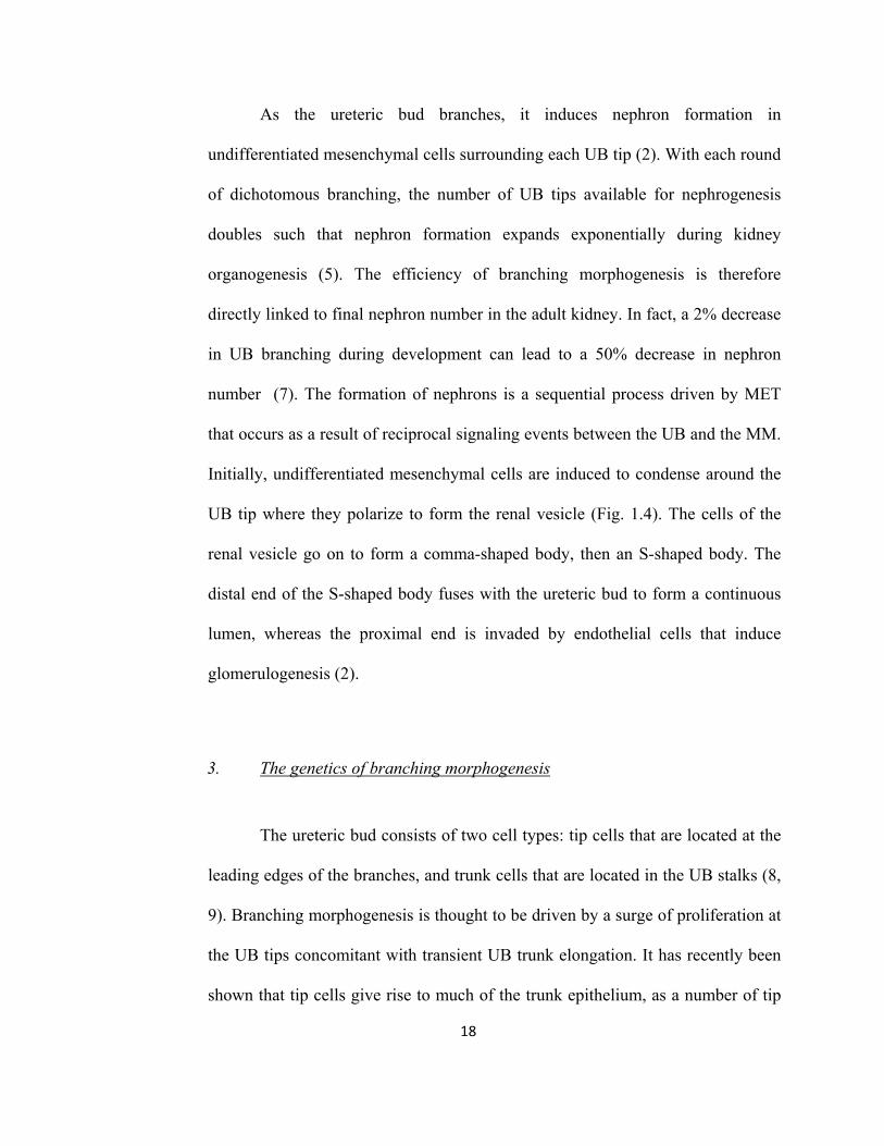
As the ureteric bud branches, it induces nephron formation in
undifferentiated mesenchymal cells surrounding each UB tip (2). With each round
of dichotomous branching, the number of UB tips available for nephrogenesis
doubles such that nephron formation expands exponentially during kidney
organogenesis (5). The efficiency of branching morphogenesis is therefore
directly linked to final nephron number in the adult kidney. In fact, a 2% decrease
in UB branching during development can lead to a 50% decrease in nephron
number (7). The formation of nephrons is a sequential process driven by MET
that occurs as a result of reciprocal signaling events between the UB and the MM.
Initially, undifferentiated mesenchymal cells are induced to condense around the
UB tip where they polarize to form the renal vesicle (Fig. 1.4). The cells of the
renal vesicle go on to form a comma-shaped body, then an S-shaped body. The
distal end of the S-shaped body fuses with the ureteric bud to form a continuous
lumen, whereas the proximal end is invaded by endothelial cells that induce
glomerulogenesis (2).
3. The genetics of branching morphogenesis
The ureteric bud consists of two cell types: tip cells that are located at the
leading edges of the branches, and trunk cells that are located in the UB stalks (8,
9). Branching morphogenesis is thought to be driven by a surge of proliferation at
the UB tips concomitant with transient UB trunk elongation. It has recently been
shown that tip cells give rise to much of the trunk epithelium, as a number of tip

UB
MMA B
C
D proximal
distal
collecting duct
Figure 1.4 - Nephron formation during metanephric kidney development is a sequential process. (A) At each ureteric bud (UB) tip, mesenchymal cells are induced to aggregate and condense. The condensed cells of the metanephric mesenchyme (MM) undergo mesenchymal-to-epithelial transition (MET) and become polarized to form arenal vesicle. (B) The newly polarized cells form a comma-shaped body with a luminalspace that fuses with that of the UB. (C) The epithelialized cells of the MM continue to proliferate and rearrange to form an S-shaped body. At this stage, endothelial cells (red) invade the proximal cleft of the S-shaped body to begin glomerulogenesis. (D) The fullydeveloped nephron contains a vascularized glomerulus that leads to a tubular segment that ultimately connects to the collecting duct at the distal end.

19
daughter cells are left behind in the trunks during the early stages of branching
morphogenesis (8).
3.1 The Gdnf/Ret/Gfr 1 pathway and Pax2
Various classes of genes, including transcription factors, soluble growth
factors, extracellular matrix and cell-adhesion molecules, have all been implicated
in renal branching morphogenesis (10). One pathway critical to initial UB
emergence and expansion involves signaling through the receptor tyrosine kinase,
Ret. The MM-secreted growth factor glial-derived neurotrophic factor (GDNF)
serves as the main ligand for Ret and its co-receptor GDNF family receptor (Gfr)
α1. Ret and Gfr 1 are exclusively expressed in UB tip cells, and following GDNF
binding, these cells are induced to proliferate in the direction of the GDNF-
secreting MM cells. GDNF/Gfrα1/Ret signaling in the UB is crucial for kidney
development, as the absence of any of these genes will lead to defective branching
morphogenesis (11-13). Besides its role in Ret signaling, GDNF is a direct
transcriptional target for other key proteins in kidney development such as Pax2, a
transcription factor expressed in both the UB and the MM (3). Mice lacking Pax2
fail to develop a metanephros and genital tract (14). Pax2 has further been shown
to promote the expression of other genes crucial for kidney development such as
the Wnt family of secreted proteins, as well as E-cadherin (Cdh1), an important
modulator of cell-adhesion (3, 15, 16).
20
3.2 Adhesion molecules
Given the epithelial nature of the ureteric bud, proteins involved in cell
adhesion and cell polarity are likely to be involved in patterning the UB. In order
for the UB to undergo branching morphogenesis, epithelial cell migration as a
result of both cell-cell and cell-matrix interactions, is important (17). For
example, αβ-integrins, cell surface receptors that mediate cell binding to the
extracellular matrix, have been shown to be important for UB branching. Mice
deficient in α3-integrin have fewer collecting ducts suggesting decreased
branching during development, while those lacking both α3- and α6-integrin do
not develop ureters (18, 19). A deficiency in α8-integrin also results in severe
renal malformations from defective branching morphogenesis (20). Polycystin-1
(PKD1) is a membrane-bound protein that has been shown to be important in
branching morphogenesis due to its role in cell-adhesion (21). Mutations in PKD1
are associated with autosomal dominant polycystic kidney disease, a common
renal disease characterized by the formation of macroscopic cysts that arise from
the renal tubular epithelium (22). During kidney development, PKD1 is expressed
in the UB in focal adhesions and adherens junctions (23, 24). Inhibition of PKD1
activity in embryonic kidney explants results in disrupted branching
morphogenesis such that there is a reduction in the number of branch tips, in
addition to a decrease in overall UB length, volume and area (21).
In a study investigating the mechanics of UB branching, it was suggested
that branching is mediated by the formation of outpouches at the tips of the

21
ureteric bud. These outpouches consist of wedge-shaped epithelial cells with
abundant levels of actin, myosin-2 and ezrin found on the apical domain of the
UB epithelium. It was proposed that this high concentration of actin and myosin
would promote cytoskeletal tightening along the apical domain, creating a “purse-
string” effect. Apical tightening would induce an increase in the surface area of
the basolateral membrane and bulging at the basolateral surface of the epithelium.
This process would in turn drive the protrusion of the epithelium into the
extracellular matrix and possibly promote the exocytotic release of morphogens
important for branching morphogenesis. The high proliferation rates at UB tips
would then drive the elongation of the new bud and by extension the growth of
the UB network as a whole (25). The authors further suggested that this
phenomenon could be due to the upregulation of tight junction proteins in the
apical domain of the UB epithelium. Indeed, in one microarray analysis
comparing the expression profile of UB tip cells with those of the trunk, as well as
the MM, it was found that the tight junction protein claudin-3 (Cldn3) was
upregulated in UB tip cells along with another claudin family member, claudin-7
(Cldn7) (26).
4. Tight junctions and claudins
4.1 Tight junctions are composed of various proteins
Tight junctions are the most apical of cell-cell junctions located between
epithelial cells and form a continuous paracellular barrier. As a result, they

22
promote cell-cell adhesion and regulate passive transepithelial ion transport
through the paracellular space (27, 28). Tight junctions consist of transmembrane
proteins that include the claudin family of proteins, occludin, tricellulin,
junctional adhesion molecules (JAM), scaffolding proteins such as ZO-1/-2/-3,
and various cytosolic transcription factors and kinases (29). Electron microscopy
is commonly used to identify tight junctions, and depending on how a particular
tissue is processed and then imaged, the morphology of the tight junction can
appear quite different. When transverse, ultra-thin sections of an epithelial tissue
are viewed under a transmission electron microscope, tight junctions appear as a
fusion of opposing cell membranes (30). In contrast, using freeze fracture electron
microscopy which permits the visualization of the surface of cells, tight junctions
appear as continuous, intramembranous strands (31).
4.2 Claudins are essential components of tight junctions
The barrier and cell adhesion functions of tight junctions are mediated by
transmembrane proteins such as claudins, occludin and JAMs. These proteins
contain extracellular domains that occupy the intercellular space and interact with
proteins on the opposing cell membranes as well as solutes that permeate through
the paracellular cleft (32). Of all these membrane-bound proteins, the claudin
family is the only one whose expression is sufficient for the formation of tight
junction strands (27, 33). Mice lacking occludin do not display altered tight
junction morphology, and its downregulation in cultured cell lines does not affect
paracellular ion permeability (34, 35). In contrast to claudins, when occludin or

23
JAM-A is overexpressed in fibroblasts, tight junctions are not established (36,
37).
4.3 Claudin structure and function
In mammals, the claudin family of proteins is composed of 24 members
that range in size from 20 to 27kDa (32). Claudins contain four transmembrane
domains, two extracellular loops, and an N- and C-terminus that are both located
intracellularly (30) (Fig. 1.2C). The extracellular loops mediate barrier and
adhesive functions through the formation of homo- or heterotypic interactions
with claudins in opposing cell membranes. The first extracellular loop consists of
approximately 50 amino acids that include two conserved cysteines which
promote barrier function, and several charged residues which determine the
electrostatic selectivity of the paracellular pore (30, 38, 39). The second
extracellular loop (16-31 amino acids) has not been well studied, but recent
evidence suggests it may be required for interaction with claudins on opposing
membranes (32, 40). The C-terminal tail displays the greatest amount of variation
among claudin species in terms of length, ranging from 22-55 amino acids (30).
The C-terminal tail contains a PDZ-binding motif (except for claudin-12) that
permits claudins to bind to tight junction associated scaffolding proteins such as
ZO-1/-2/-3 and MUPP1 (30). These interactions link membrane-bound claudins to
the actin cytoskeleton, possibly stabilizing them at the tight junction to maintain
the integrity of the epithelial barrier (41, 32). Claudin localization and function
has also been shown to be affected by phosphorylation of the C-terminal tail by

24
serine/threonine and tyrosine kinases (e.g. WNK4, PKC) (42, 43). For example,
mutating the phosphorylation site of claudin-1 (Cldn1) was shown to eliminate its
ability to mediate the normal, leftward direction of heart-looping during chick
embryogenesis (43). Furthermore, phosphorylation can either increase the barrier
function of tight junctions or decrease it making the epithelial barrier more
permeable (44, 45).
5. Claudins in the kidney
The expression pattern of over 12 claudin species has been established in
the mouse adult kidney (32) (Fig. 1.5). Most localize to tight junctions in the
epithelial cells of the renal tubules such that each nephron segment expresses a
distinct combination of claudins. The specific combination of claudins is thought
to shape the paracellular ion transport characteristics of each segment (32, 30).
For example claudin-8, which functions mainly as a cation barrier, is expressed
primarily in the distal portion of the nephron where high ion concentrations in the
filtrate must be maintained, and the flow of ions across the epithelial barrier is
limited (46, 47). In contrast, claudin-2 forms a highly permeable cation pore, and
is expressed in the proximal tubule where it contributes to the significant amount
of sodium reabsorption that occurs in this segment (48, 49). Claudins can also be
found in structures besides the tubular epithelium. Claudin-5 is expressed
exclusively in endothelial cells, including renal blood vessels while claudin-6 has
been shown to localize to tight junction-like structures in podocytes (50, 51).

glomerulus proximal tubule thin descendinglimb
thin ascendinglimb
thick ascending limb
distal tubule collecting duct
Cldn2
Cldn3
Cldn5
Cldn7 Cldn7
Cldn8 Cldn8
Cldn14
Cldn16
Cldn19
Figure 1.5 -The expression of claudins in the adult kidney varies in each segment of the nephron. The expression of selected claudins is summarized from several studies(32). Claudin-5 is expressed in the endothelial cells of the glomerular capillaries and is therefore not expressed in the epithelial cells of the nephron. Claudin-1, -4, -6, -9, -10 and-11 are also expressed along the nephron but are not shown. The expression pattern of over 12 claudins has been characterized in the kidney (32). The specific expression of claudins reflects the unique transport functions of each nephron segment. Cldn-12, -18, -20,-21,-22,-23 and -24 expression has not yet been charaterized in the kidney.

25
6. Claudins and human disease
The role of claudins in human disease has been extensively investigated
over the past decade. Familial hypercalciuric hypomagnesemia with
nephrocalcinosis (FHHNC) is an autosomal recessive disease characterized by
severe magnesium and calcium wasting in the urine leading to calcium deposition
in the renal tubules (52). In 1999, FHHNC was shown to be associated with loss-
of-function mutations in claudin-16 (Cldn16) (53). Cldn16 is expressed in the
thick ascending limb of Henle, the nephron segment responsible for the majority
of Mg2+ reabsorption in the kidney (1). To date, more than 30 loss-of-function
mutations in Cldn16 have been reported in families with FHHNC (32). In 2006,
mutations in the claudin-19 (Cldn19) locus were found in families with FHHNC
who also had severe ocular abnormalities (54). Cldn19 protein colocalizes with
CLDN16 in the thick ascending limb of Henle, and is also highly expressed in the
retina (54). It appears that CLDN16 and CLDN19 form heteromers in the cells of
the thick ascending limb of Henle, and together they promote magnesium
reabsorption (55). How loss-of-function mutations in Cldn19 lead to ocular
abnormalities has not yet been described.
Claudins have also been implicated in nonrenal diseases. Mutations in
Cldn1 were found in patients with neonatal ichthyosis and sclerosing cholangitis
(NISCH), a syndrome characterized by scaly skin, sparse hair and inflammation
of the bile ducts. Loss of CLDN1 from tight junctions may lead to increased
permeability of the skin and bile ducts, leading to dehydration and biliary leakage

26
(56). Human hereditary deafness has been attributed to loss-of-function mutations
in claudin-14 (Cldn14), which is normally expressed in the inner ear. Cldn14 is
expressed in the organ of Corti where it maintains the high cation gradient
between the endolymph and perilymph. Loss of this gradient prevents the
depolarization of sensory hair cells and inhibits signal transduction leading to
hearing loss (57, 58). In addition, claudin expression has been shown to be either
up- or downregulated in a variety of cancers, potentially modulating processes
such as epithelial-to-mesenchymal transition and metastasis (30, 59).
7. The function of Claudin-3 and Claudin-7
7.1 Claudin-3
In the adult mouse kidney, Cldn3 (23kDa) is expressed on the apical
surfaces of epithelial cells in the distal nephron and collecting ducts, where ion
flow is tightly regulated (50). Overexpressing Cldn3 in an epithelial ovarian
cancer cell line reduced tight junction-mediated ion permeability, suggesting that
one of its functions may be to “tighten” paracellular channels making them less
permeable to ions and other larger molecules (45). In addition to its role in
paracellular transport, Cldn3 also plays a role in cell motility and tumor
formation. Claudin-3 is one of the most highly expressed genes in ovarian tumors
and it is expressed in nearly 90% of ovarian cancers. This overexpression has
been associated with increased cellular motility and survival suggesting a role for
Cldn3 in promoting metastasis and malignancy (60). Indeed, downregulation of

27
Cldn3 in ovarian tumors resulted in a significant reduction in cell proliferation
concomitant with decreased tumor growth (61).
7.2 Claudin-7
Cldn7 (22kDa) is also expressed in the distal nephron and collecting ducts
as well as the descending limb of Henle, where ion exchange through the
paracellular pathway is more prominent (47). Overexpressing Cldn7 alone in the
porcine epithelial cell line LLC-PK1 resulted in decreased paracellular
permeability of chloride ions, and an increase in sodium transport (62). When
studied in MDCK type II cells however, Cldn7 was found to act as a chloride
channel (49). Indeed, when studying the ion permeability functions of claudins,
results have varied based on the cell line used (30).
An unusual feature of Cldn7 protein is that in epithelial tissues it localizes
primarily to the basolateral membrane, away from the conventional apical
location of tight junctions (47). The function of CLDN7 on the basolateral
membrane is not known, but recent evidence shows that it binds directly to the
adhesion molecule EpCAM (32, 63). The EpCAM-CLDN7 complex was found to
promote cell proliferation and tumorigenicity when both proteins were expressed
in the human kidney cell line HEK293 (64).
Both Cldn3 and Cldn7 appear to play important and separate roles in the
terminally differentiated epithelial cells of the adult kidney. However, their
function during UB branching morphogenesis has not been characterized. Given
the importance of cell-adhesion proteins in regulating branching morphogenesis,

28
and the presence of apically driven mechanical forces that could promote UB
branching at the tips, tight junction proteins may play a key role (18-20).
Microarray analysis has shown that Cldn3 and Cldn7 are upregulated in the UB
during embryonic kidney development (26). When studied in cancer cell models,
both claudins are associated with an increase in cell proliferation and migration,
which are processes that are also important in branching morphogenesis (60, 61,
64). If Cldn3 and Cldn7 are expressed in the epithelial ureteric bud, they may
therefore function as important modulators of UB branching during kidney
development.
8. Summary of objectives
The objectives of my research are to characterize the expression patterns
of Cldn3 and Cldn7 during metanephric kidney development and to describe their
function during renal branching morphogenesis. The expression of both claudin
species will be assayed at the transcript and protein levels at critical stages of
metanephric kidney development. The functional role of claudins in the branching
UB will be investigated through gain-of-function assays in a UB derived kidney
cell line that undergoes branching in vitro. We hypothesize that both claudin
species will be highly expressed in the ureteric bud epithelium, where they will
function to promote the process of branching morphogenesis.
This thesis contains two manuscript-based chapters. The first chapter
describes the expression patterns of Cldn3 and Cldn7 at critical stages of

29
metanephric kidney development, and examines the functional role of Cldn3 and
Cldn7 when overexpressed in a UB-derived epithelial cell line. The second
chapter describes the development of a model to study genetic perturbations in the
UB lineage in embryonic kidneys explants. Rodent embryonic kidneys can be
dissected and cultured as explants such that branching morphogenesis and
nephrogenesis can be observed ex vivo. We describe a new method in which DNA
constructs can be targeted to the UB lineage in cultured embryonic kidney
explants using microinjection and electroporation. The development of this model
is critical to our future goal of perturbing claudin expression in the UB lineage of
the developing mouse kidney.

30
CHAPTER II
The tight junction proteins Cldn3 and Cldn7 are expressed in the ureteric bud and
promote tubulogenesis in vitro
Nicholas Haddad, Bruno St. Jacques, Melissa Yu, Indra R. Gupta
Manuscript to be submitted

31
1. Abstract
The claudin family of proteins is required for the formation of tight junctions
(TJs) between epithelial cells. It has been shown that Cldn3 and Cldn7 are upregulated in
the ureteric bud (UB) versus the metanephric mesenchyme (MM) during kidney
development. We hypothesize that if Cldn3 and Cldn7 form TJs in the epithelial UB, they
will determine the pattern of UB branching. Using transmission electron microscopy, we
have established that TJs are situated between epithelial cells of the UB that undergo
branching. Whole-mount in situ hybridization has shown that Cldn3 and Cldn7
transcripts are expressed in the UB at E10.5, E13.5 and E16.5. Double
immunofluorescence experiments revealed that CLDN3 localizes to tight junctions at the
apical domain of UB cells, while CLDN7 is predominately expressed on the basolateral
membrane. To determine the functional role of these claudins, we used the mIMCD-3 cell
line which is derived from the embryonic UB. These cells undergo tubulogenesis when
placed in type-I collagen matrix in a manner morphologically similar to the UB. Double
immunofluorescence and Z-stacking showed that these cells express both Cldn3 and
Cldn7 at the tight junction. Stable cell lines expressing CLDN3 or CLDN7 fused at the
N-terminus to the red fluorescent protein (RFP) mCherry were isolated and seeded in
collagen matrix. Quantification of branching following 48 hours in culture revealed that
cell lines overexpressing either Cldn3 or Cldn7 underwent significantly increased
branching compared to mCherry controls. Cldn3 and Cldn7 may therefore promote
branching morphogenesis in the cells of the ureteric bud.

32
2. Introduction
The claudin family of proteins, of which there are 24 members in mammals, are
required for the formation of tight junctions between epithelial cells (30, 33). Tight
junctions form uninterrupted paracellular barriers on the apical surface linking adjacent
epithelial cells. They promote cell-cell adhesion and regulating passive transepithelial ion
transport through the paracellular pathway (28, 27). Tight junctions consist of
transmembrane proteins that include the claudin family of proteins, occludin, tricellulin,
junctional adhesion molecules (JAM), scaffolding proteins such as ZO-1/-2/-3, and
various cytosolic transcription factors and kinases (29). Of all these membrane bound
proteins, the claudin family is the only one whose expression is sufficient for the
formation of tight junction strands (33).
Claudins mediate both paracellular ion transport and cell adhesion (27, 30). They
have specific expression patterns that determine the different permeability properties of
epithelial tissues. In the adult kidney for example, over 12 different claudins are
expressed along the length of the nephron, and each one contributes to the specific
permeability properties of each nephron segment (32). With regards to cell adhesion, it
has been shown that expressing claudin-1, -2 or -3 in L-fibroblasts will lead to de novo
formation of tight junctions and epithelial strands of cells, suggesting claudin expression
is a key determinant in the development of an epithelial tissue (27). During
embryogenesis, claudins are crucial for the development of various structures by
maintaining tissue integrity and by separating fluids of different compositions. Claudin-4
and claudin-6 have been found to be critical for blastocyst formation during early

33
embryogenesis, while claudin-1 is important for epithelial stratification during skin
development (66, 67).
Mammalian kidney development involves the formation of three serial kidneys:
the pro-, meso- and metanephros. The metanephros forms the definitive adult kidney. All
three kidneys originate from two primordial tissues that are derived from the intermediate
mesoderm: the nephric duct and the nephric cord (2, 3). Metanephric kidney development
begins at embryonic day (E)10.5 after both the nephric duct and nephric cord have
elongated caudally to the level of the hindlimbs. At this point, an epithelial structure
known as the ureteric bud (UB) emerges out of the nephric duct and invades the
neighboring undifferentiated mesenchymal cells termed the metanephric mesenchyme
(MM) (2). As a result of reciprocal signaling and induction by the MM, the UB proceeds
to elongate and divide through repeated bifurcations in a process known as branching
morphogenesis, eventually forming the adult collecting duct system (2, 8). Branching
morphogenesis in the mouse involves 10-11 rounds of bifurcations which produce
approximately 1600 branches within each developing kidney (6). It is thought that
branching morphogenesis is largely driven by a surge of proliferation at the UB tips
followed by bifurcation, concomitant with transient UB trunk elongation (8). To date,
many transcription factors and growth factors have been reported to participate in
branching morphogenesis (3). Given the epithelial nature of the ureteric bud, proteins
involved in cell-cell adhesion and cell polarity are also likely to be involved in this
process. Indeed, in one microarray analysis comparing the expression profile of the cells
of UB tips with those of the MM, it was found that claudin-3 (Cldn3) was upregulated in
the UB tip cells along with another tight junction protein, claudin-7 (Cldn7) (26).

34
Cldn3 (23kDa) is expressed on the apical surfaces of cells in the distal nephron
and collecting ducts, where ion flow is highly regulated (50). Overexpressing Cldn3 in an
epithelial ovarian cancer cell line reduced tight junction-mediated ion permeability,
suggesting that one of its functions may be to “tighten” paracellular channels making
them less permeable to ions and other larger molecules (45). Cldn7 (22kDa) is also
expressed in the distal nephron and collecting ducts as well as the descending limb of
Henle where ion exchange is more essential (47). Overexpressing Cldn7 in the porcine
epithelial cell line LLC-PK1 resulted in decreased paracellular permeability of chloride
ions, and an increase in sodium transport (62). When studied in MDCK type II cells
however, Cldn7 was found to act as a chloride channel (49). Indeed, when studying the
ion permeability functions of claudins, results have varied based on the cell line used
(30). Both Cldn3 and Cldn7 appear to play important and separate physiological roles in
the terminally differentiated epithelial cells of the adult kidney. However, their function
during ureteric bud branching morphogenesis has not been characterized.
Here we show that Cldn3 and Cldn7 are expressed in the ureteric bud during
critical stages of branching morphogenesis. Using a cell line derived from renal collecting
ducts, we generated stable clones overexpressing either Cldn3 or Cldn7 and found that
both claudins stimulate branching morphogenesis in vitro.
35
3. Materials and methods
3.1 Animal care
Timed-pregnant CD1 mice (Charles River Laboratories, St. Constant, QC,
Canada) were sacrificed at embryonic day (E) 10.5, 12.5, 13.5, or 16.5 to retrieve
embryonic tissue as previously described (68). Whole embryos or metanephric kidneys
were dissected in phosphate buffered saline (PBS) using an M5A stereomicroscope (Wild
Leitz®, Willowdale, ON, Canada. The mouse studies were performed in accordance with
the rules and regulations of the Canadian Council of Animal Care.
3.2 Electron microscopy
Embryonic kidneys aged E12.5 were dissected and fixed in 2.5% glutaraldehyde
in 0.1M sodium cacodylate buffer for 24 hours, then washed in cacodylate buffer, and
then immersed in 1% osmium tetroxide for 2 hours. Samples were then dehydrated in
acetone and embedded in EPON®812. Sections were taken at 0.5μm thickness, stained
with 1% toluidine blue, and then imaged using light microscopy. For transmission
electron microscopy (TEM), ultrathin sections were collected on 200 mesh copper grids
(Electron Microscopy Sciences, Hatfield, PA, USA) and stained with uranyl acetate and
lead citrate. Images were obtained using a FEI Tecnai 12 transmission electron
microscope (FEI/Philips Electron Optics, Eindhoven, The Netherlands) that was attached
to a Gatan Bioscan CCD Camera Model 792.

36
3.3 Whole mount in situ hybridization
In situ hybridization was performed on partially eviscerated whole embryos at
E10.5 and on dissected kidneys at E13.5 and E16.5. All samples were fixed overnight in
4% PFA in PBS at 4 C. Coding sequences for mouse Cldn3, Cldn7, and Ret were
subcloned in the PCRII-Topo vector (Invitrogen, Carlsbad, CA, USA) and then linearized
using XhoI, XbaI, and BamHI restriction enzymes respectively. DIG-labeled UTP probes
were generated in vitro using the RNA polymerases SP6 for Cldn3 and Cldn7, and T3 for
Ret. Anti-DIG antibody conjugated to alkaline-phosphatase was used to detect target
RNA sequences. Treated samples were developed using NBT/BCIP substrate in NTMT.
Cryosections of treated embryonic kidneys aged E13.5 and E16.5 were obtained at a
thickness of 20μm and counterstained with eosin.
3.4 Double immunofluorescence
Whole embryos were dissected at E13.5 and E16.5 and fixed in a mixture of
ethanol, water, and 37% formaldehyde in a ratio of 6:3:1, prior to dehydration and
paraffin embedding. Transverse sections were obtained at the level of the kidneys at a
thickness of 7μm. Following rehydration, samples underwent antigen retrieval with
heated 10mM citrate buffer (pH 6.0), followed by blocking with 10% goat serum in PBS
and 0.3% TritonX. Double primary antibody incubations were performed with polyclonal
rabbit anti-CLDN3 (Spring Bioscience, Fremont, CA, USA) at a dilution of 1:100, or
rabbit anti-CLDN7 (Spring Bioscience, Fremont, CA, USA) at 1:200 together with

37
monoclonal mouse anti-ZO-1 (Invitrogen, Camarillo, CA, USA) antibody at 1:100 in
PBS with 5% goat serum and 0.3% TritonX at 4 C overnight. Secondary antibodies were
goat anti-rabbit IgG (Invitrogen, Camarillo, CA, USA) conjugated to Alexa Fluor 594
(TXR) and goat anti-mouse IgG (Invitrogen, Camarillo, CA, USA) conjugated to Alexa
Fluor 488 (FITC), both at a dilution of 1:500. The secondary antibodies were incubated
with the samples for 1 hour at room temperature. For double immunofluorescence
performed on mIMCD-3 cells, the same reagents and conditions were used with the
exception that mIMCD-3 cells were grown in monolayer to confluence on glass
coverslips and then fixed with 4% paraformaldehyde at 4 C for 10 minutes. All slides
and coverslips were mounted with Slowfade Gold anitfade reagent (Invitrogen, Eugene,
OR, USA).
3.5 Cell lines and constructs
mIMCD-3 cells were grown in DMEM/F12 medium containing 10% fetal bovine
serum (FBS), 1% penicillin/streptomycin (P/S) in a humidified atmosphere at 37°C with
5% CO2. The pcDNA3 expression vector containing the coding sequence of mCherry red
fluorescent protein RFP downstream of a CMV promoter was kindly provided by Aimee
K. Ryan (McGill University Health Centre, Montreal, QC, Canada). Complete coding
sequences of Cldn3 and Cldn7 were subcloned upstream of mCherry, in frame, to
produce a fusion protein in which the mCherry reporter tag was fused to the N-terminus
of either CLDN3 or CLDN7. Vectors expressing mCherry-CLDN3, mCherry-CLDN7 or
mCherry alone were stably transfected into mIMCD-3 cells using Lipofectamine 2000
Reagent (Invitrogen, Carlsbad, CA, USA). The selection drug G418 was added after 48

38
hours in culture and colonies expressing high levels of RFP were selected. Stable clones
were maintained in DMEM/F12 containing 500μg/ml of G418.
3.6 Immunoblotting
Confluent wildtype or stably transfected mIMCD-3 cells were dissolved in lysis
buffer (50 mM Tris-HCl pH 7.5, 150 mM NaCl, 1% NO-40, 1.5mM MgCl2, 10%
glycerol, 1mM EDTA, 25mM NaF, 1mM Na3VO4 and protease inhibitor cocktail; Roche,
Mannheim, Germany). Total protein extracts were separated on SDS-PAGE and
transferred to a nitrocellulose membrane. Blocking was performed in 5% milk in PBST,
followed by incubation with polyclonal rabbit anti-CLDN3 antibody (Invitrogen,
Camarillo, CA, USA) at a 1:500 dilution at 4 C overnight. This was followed by
incubation with peroxidase-labeled anti-rabbit IgG secondary antibody (Cell Signaling
Technology Inc, Denvers, MA, USA) at a 1:2500 dilution for 1hour at room temperature.
Antigen-antibody binding was detected using the ECL Western Blot Detection System
(Amersham, Buckinghamshire, UK). Molecular mass was determined relative to a
defined protein ladder (Fermentas, Burlington, ON, Canada).
3.7 Collagen gel assays
100,000 mIMCD-3 cells from either wild-type or stable transfectants expressing
mCherry-CLDN3, mCherry-CLDN7, or mCherry alone were seeded in a mixture of rat-
tail type I collagen (BD Biosciences, Mississauga, ON, Canada) containing HEPES,
NaHCO3 and DMEM-F12 (69). Cells were distributed in a 96-well plate such that
10,000 cells were seeded per well. Gels were left to solidify then 100μl of DMEM-F12

39
containing 10% FBS and 1% P/S was added to each well. Cultures were grown in a
humidified atmosphere at 37°C with 5% CO2 and observed over 48h. At 24h intervals,
gels were removed from culture and fixed in 4% paraformaldehyde, rinsed with PBS and
mounted on slides. The number of branches in a standardized area was determined for
each cell line by first randomizing the images collected such that quantification was
performed in a blinded manner. ImageJ Software (NIH, rsb.info.nih.gov/ij/) was used to
count the number of tubular structures. Statistical analysis was performed using the
Student’s t-test.
4. Results
4.1 Tight junction structures are present on the apical domain of UB cells
To establish the presence of tight junctions in the cells of the branching ureteric
bud (UB), transmission electron microscopy (TEM) was performed on ultra-thin sections
taken from embryonic kidneys at E12.5. At this time point the UB has already gone
several branching events and is the only epithelial tissue present in the developing
kidney. TEM confirmed that tight junctions are present at the most apical regions of cell-
cell contacts (Fig. 2.1). The presence of tight junctions in the ureteric bud suggests that
the claudin family of proteins may be important in the process of branching
morphogenesis.

A B
TJ
C
Figure 2.1 - Tight junction structures are present on the apical surface of ureteric bud cells. (A) An embryonic kidney at E12.5 was sectioned and the ureteric bud (UB) network was imaged. A UB-tip in cross-section is highlighted (black box) (scale bar = 50μm). (B) Ultra-thin sections were examined using transmission electron microscopy. Polarized epithelial cells of the UB lineage and their most apical lateral surfaces (black box) are shown (scale bar = 5μm). (C) Upon closer examination of apical cell-cell contacts, tight junctions (TJ) are detected at several points where the opposing cellmembranes appear to touch (black arrows) (scale bar = 200nm).

40
4.2 Cldn3 and Cldn7 mRNA are expressed in the UB throughout branching morphogenesis
To characterize the expression patterns of Cldn3 and Cldn7 mRNA in the
branching UB, whole mount in situ hybridization was performed at three stages of kidney
development: E10.5, E13.5 and E16.5. At E10.5, there is the initial outgrowth of the UB,
at E13.5 the UB has undergone several branching events, and at E16.5 the UB continues
branching against a backdrop of terminally differentiated mesenchymal tissue (i.e.
nephrons) and UB derivatives (i.e. collecting ducts). Both Cldn3 and Cldn7 transcripts
are expressed in the UB and its derivatives at all three time points (Fig. 2.2). The
expression of both Cldn3 and Cldn7 mRNA is detected in the nephric duct and the
emerging ureteric bud at E10.5. By E13.5, expression is found throughout the ureteric
bud lineage, but not in the differentiating mesenchymal cells of the MM. At E16.5, both
Cldn3 and Cldn7 are strongly expressed in the UB tips, but expression weakens in the
stalks. The UB tip marker Ret was used as a positive control to recognize the branching
ureteric bud. These hybridization assays established that the UB cell lineage expresses the
mRNA of both claudin species throughout kidney development.
4.3 Cldn3 protein localizes to the apical domain of the UB, while Cldn7 is expressed primarily on the basolateral domain
To determine the subcellular localization of Cldn3 and Cldn7 protein, double
immunoflourescence was performed at E13.5 and E16.5 with the tight junction marker
zona occludens-1 (ZO-1) and the adherens junction and basolateral domain marker E-
cadherin (Cdh1) (Fig. 2.3). Cldn3 is expressed in the apical domain of the UB at both

retclaudin -7
ndUB
nd
UB
E16.5
claudin-7 ter3-nidualc
E13.5
E10.5
UB
nd
G
Figure 2.2 - Cldn3 and Cldn7 transcripts are expressed in the ureteric bud (UB) during branching morphogenesis. Whole mount in situ hybridization was performed on wholeembryos (Embryonic day (E)10.5) or dissected embryonic kidneys (E13.5, E16.5) which were then cryosectioned. At the start of branching morphogenesis at E10.5, Cldn3 and Cldn7 mRNAwas expressed throughout the nephric duct (nd) and the emerging ureteric bud (UB, black box) ina pattern similar to the UB marker, Ret. At E13.5, Cldn3 and Cldn7 mRNA continues to be expressed in the UB lineage (upper row), and following cryosection both claudin species, are expressed throughout the UB and not in the differentiating mesenchyme (lower row). Ret expression is restricted to the tips of the UB. At E16.5, Cldn3 and Cldn7 expression persists exclusively in the UB, with the strongest levels found at the UB tips of the UB with weak to no signal in the stalks (upper, lower rows). Ret expression remains in the UB tips. White scale bar =0.5mm, black scale bar =50mm.

E13.5 CLDN3
CLDN7
ZO-1
ZO-1
merge
merge
E16.5
CLDN7 merge
CLDN3
CLDN7 mergeCDH1
ZO-1
ZO-1
merge
Figure 2.3 - The subcellular localization of CLDN3 and CLDN7 differs in the ureteric bud lineage. Double immunoflourescence was performed using antibodies toCLDN3 or CLDN7 (2° TXR) with either the tight junction marker zona occludens-1(ZO-1, 2° FITC) or basolateral membrane marker E-cadherin (CDH1). CLDN3 was found to colocalize with ZO-1 at both embryonic day (E)13.5 and E16.5, indicating thatCLDN3 is localized to tight junctions in the UB lineage (arrows). CLDN7 is foundprimarily at the basolateral surface (arrowheads) of UB cells and largely overlaps withCDH1. Insets represent a higher magnification of the UB at E16.5. Scale bar = 50μm.
41
E13.5 and E16.5, consistent with the known expression pattern of ZO-1. This suggests
that Cldn3 functions within tight junctions during branching morphogenesis. Cldn7
protein is expressed primarily on the basolateral domain of UB cells, with only limited
colocalization with ZO-1 at both E13.5 and E16.5. In contrast, CLDN7 colocalizes with
CDH1 at both embryonic stages. Cldn7 protein may therefore associate with different
membrane linked structures in the basolateral domain of the ureteric bud.
4.4 Overexpressing Cldn3 or Cldn7 in mIMCD-3 cells leads to increased tubulogenesis in vitro
The mIMCD-3 cell line is derived from adult mouse inner medullary collecting
ducts, an epithelial tissue that is a descendant of the embryonic ureteric bud (70). When
placed in type-I collagen matrix, these cells undergo tubulogenesis and branching in a
manner morphologically similar to the UB. Using double immunofluorescence, we have
established that Cldn3 and Cldn7 proteins colocalize with ZO-1 at the cell membrane of
mIMCD-3 cells (Fig. 2.4). Z-stacks confirm co-localization of CLDN3 and CLDN7 to
tight junctions at the apical membrane.
To determine the function of Cldn3 and Cldn7 during branching, stable mIMCD-3
clones expressing mCherry red fluorescent protein (RFP) fused to CLDN3 or CLDN7
(not shown), and unfused mCherry, were isolated. Western Blot analysis confirms
mCherry-CLDN3 expression in stably transfected clones (Fig. 2.5A). Endogenous
expression of Cldn3 is detected in wild-type mIMCD-3 cells and in empty vector
mCherry clones, and is also present in the mCherry-CLDN3 clones albeit at a lower level.

Cldn3 ZO-1 merge
Cldn7 ZO-1 merge
Figure 2.4 - Cldn3 and Cldn7 proteins colocalize with ZO-1 in mIMCD-3 cells. mIMCD-3 cells were grown in monolayer and double immunofluorescence wasperformed using antibodies to Cldn3 and Cldn7 (2° FITC), and ZO-1 (2° TXR). Cldn3, Cldn7 and ZO-1 are expressed at the cell membrane of mIMCD-3 cells. The mergedimages show the overlapping expression domains of CLDN-3 and -7 with ZO-1. Z-stacks indicate that both claudin species localize with ZO-1 to tight junctions in mIMCD-3 cells. Scale bar = 10μm

0
100
200
300
400
500
600
700C
B 0hr 24hr 48hr
mCherry
mCherry-CLDN3
AWT mCherry
mCherry-CLDN3
CLDN3
mCherry-CLDN3
actin
45kDa
23kDa
mCherry(n=16)
mCherry-CLDN3(n=16)
Num
ber o
f tub
ules
Figure 2.5 - Ectopic expression of Cldn3 or Cldn7 in mIMCD-3 cells leads to an increase in tubulogenesis. (A) Protein extracts were obtained from wild-type mIMCD-3cells and clones stably transfected with empty vector mCherry or mCherry- CLDN3 and Western Blot was performed. The fusion protein is approximately 45kDa in size and is expressed in the clones overexpressing Cldn3. Endogenous CLDN3 was detected in allthree cell lines, but was diminished in mCherry-CLDN3 cell line. (B) mIMCD-3 cell lines expressing mCherry-CLDN3 and mCherry-CLDN7 fusion constructs, as well asmCherry alone, were seeded in type I collagen matrix at a concentration of 10,000 cells per well. Cells were grown for 48 hours then fixed and imaged at 24 hour intervals underlight microscopy. A qualitative increase in tubule formation was first observed at 24hours in cells overexpressing Cldn3 or Cldn7 (data not shown) when compared to cellsexpressing the empty vector alone. (C) Quantification of branching after 48 hours revealed that cell lines overexpressing either Cldn3 or Cldn7 had significantly increased branching when compared to mCherry controls. (Student’s t-test, *P<0.001). Data is presented as mean ± s.e. from a representative experiment, out of three that were performed, such that n is the number of wells per cell line for that experiment.

42
To evaluate the effect of Cldn3 overexpression in mIMCD-3 cells, overexpressing
clones were seeded in type-1 collagen matrix and in vitro tubulogenesis was evaluated
over 48 hours in culture (Fig. 2.5B, C). Starting at 24 hours, a qualitative increase in
tubule formation is observed in cells lines ectopically expressing either mCherry-CLDN3
or mCherry-CLDN7 (data not shown). The tubules are longer and more branched as
compared to controls. Quantification following 48 hours in culture revealed that cell lines
overexpressing either claudin species have significantly increased tubule formation
compared to mCherry controls (P<0.001). Cldn3 and Cldn7 therefore promote branching
in an in vitro model of tubulogenesis, suggesting tight junction proteins may contribute to
the process of branching morphogenesis in the cells of the ureteric bud.
5. Discussion `
We have characterized the expression patterns of Cldn3 and Cldn7 throughout
embryonic kidney development and found that they are restricted to the epithelial UB
lineage. Although they are expressed in the same cell lineage during kidney
organogenesis, their subcellular localization is different. Cldn3 protein is expressed on
the apical surface of UB cells, while Cldn7 is primarily found on the basolateral surface.
We overexpressed Cldn3 and Cldn7 in a renal epithelial cell line that is a descendant of
the embryonic UB, and found that both promoted increased formation of tubular
structures. Our data suggests that tight junctions play a role in promoting branching
morphogenesis of the UB in vivo.
Studies on isolated UB cultures in vitro suggest that tight junction proteins may
generate the mechanical forces that guide UB branching. Meyer et al. showed that UB

43
branching proceeds through swelling at the tips of the ureteric bud caused by the
formation of “outpouches” (25). These outpouches share a continuous lumen with the UB
trunks and are composed of wedge-shaped epithelial cells with a concentration of actin
and myosin-2 on the apical domain of the cytoplasm. The authors proposed that an
apically driven “purse-string” mechanism could push the formation of outpouches, as the
surface area of the apical membrane decreases and that of the basolateral membrane
increases, changing the UB cells from columnar to wedge-shaped, and thereby
propagating the UB luminal network (25). In light of the essential role of claudins in
establishing tight junctions, the apical expression of Cldn3 suggests that this claudin
member may contribute to the “purse-string” mechanism that drives branching
morphogenesis.
Our results show that Cldn7 localizes to the basolateral domain of UB cells as
opposed to the traditional subcellular localization of tight junction proteins at the apical
domain. This unconventional localization of Cldn7 has been reported in the adult kidney,
as well as other epithelial tissues such as the distal colon, the epididymus and the
respiratory epithelium (47, 71-73). The function of Cldn7 in the basolateral membrane is
still not fully known, but it has been suggested that basolateral claudins may serve as a
storage pool that can be quickly recruited to tight junctions should there be a functional
need (47). Recently, Cldn7 protein has been shown to form a complex with the epithelial
cell adhesion marker (EpCAM) (63). The EpCAM-CLDN7 complex has been shown to
localize to areas of the cell membrane called tetraspanin-enriched microdomains where it
promotes cell proliferation, drug resistance and cell migration. The signaling pathways
triggered by localization of EpCAM-CLDN7 to these microdomains are unknown, but

44
their identification may shed new light on the signaling functions of claudins with regards
to proliferation and cell migration, which are essential processes in the progression of
branching morphogenesis (17, 64). EpCAM is also expressed in the ureteric bud,
therefore further characterization of the EpCAM-CLDN7 complex could provide new
insight into the mechanisms that induce UB branching (74).
The overexpression of Cldn3 and Cldn7 in mIMCD-3 cells promoted tubule
formation when these cells were seeded in a collagen matrix. Wild-type mIMCD-3 cells
exposed to hepatocyte growth factor (HGF) display increased cell proliferation and
migration and form more tubules when seeded in collagen (75). Interestingly, increased
proliferation and cell migration have also been observed in ovarian epithelial cells with
increased expression of Cldn3 (60, 61). In fact, Cldn3 is one of the most highly expressed
genes in ovarian tumors, and is also overexpressed in prostate, colorectal and breast
cancer (60, 76-79). Additionally, siRNA-mediated knockdown of Cldn3 in ovarian cancer
cells reduced migration (60). Moreover, when Cldn3 was downregulated in mice with
ovarian cancer xenografts there was a significant reduction in tumor cell proliferation and
growth with an increase in the number of apoptotic cells (61). Experiments are currently
underway to evaluate the proliferative and migratory response of mIMCD-3 cells
showing Cldn3 overexpression.
Although considerable evidence has been generated demonstrating the effects of
claudin-3 upregulation on cell migration and proliferation, it has also been associated
with other mechanisms that may promote branching morphogenesis such as degradation
of the extracellular matrix. Studies have shown that the overexpression of Cldn3 led to

45
increased matrix metalloproteinase-2 (MMP-2) activity (60, 80). Pro-MMP-2 activation
is mediated by a subtype of membrane-bound MMPs, so-called membrane-type (MT)-
MMPs. Claudins-1, -2, -3 and -5 stimulate the transformation of pro-MMP-2 into its
active form in cooperation with MT-MMPs. In fact, claudin-1 and MT1-MMP proteins
have been shown to interact directly in human embryonic kidney (HEK293) cells through
immunoprecipitation (80). Claudins may recruit MT1-MMP to the cell surface where the
activation of pro-MMP-2 occurs, thereby facilitating degradation of the extracellular
matrix and promoting UB branching. The cells of the UB express MMP-2, and the
inhibition of MMP activity has been shown to disrupt branching morphogenesis in kidney
organ culture, suggesting that CLDN3 and MMP-2 interaction may constitute a novel
mechanism for the regulation renal branching morphogenesis (81, 82).
Claudins form homo- and heteromeric interactions with other claudin species that
are either next to them or on the opposing cell membrane (30). Cldn3 protein specifically
interacts with Cldn-1, -2 and -5, but it is not known to interact with Cldn7 (83). The
specific combination of claudins expressed in an epithelial tissue has been shown to
affect the permeability properties of those cells (30). Heteromeric claudin interactions
may also influence other claudin-mediated molecular processes. Assessing the ability of
the mCherry-CLDN3 fusion protein to form heteromers will provide important
information as to whether it activates MMP-2 alone or in combination with other claudin
species. It will also provide insight into the signaling events that are mediating
tubulogenesis in mIMCD-3 cells.

46
We show that when Cldn3 or Cldn7 is overexpressed in mIMCD-3 cells, they
both promote an increase in tubulogenesis. In vivo, we also show that Cldn3 and Cldn7
proteins localize to different subcellular domains within the cells of the UB, suggesting
they may function differently, at least in the developing kidney. Studies on the role of
Cldn3 suggest it can influence cell proliferation and migration as well as MMP activity,
processes that are crucial for branching morphogenesis. The precise mechanism through
which Cldn3 influences these processes is currently under investigation through further
characterization of the stable cell lines and by assessing its function in cultured
embryonic kidneys.
47
CHAPTER III
Electroporation of embryonic kidney explants
Nicholas Haddad, Daniel Houle, Indra R. Gupta
Chapter 19: Electroporation of embryonic kidney explants. In: Nakamura H (ed) Electroporation and sonoporation in developmental biology. (2009) Springer, Tokyo
© Springer 2009
With kind permission of Springer Science and Business Media

48
1. Abstract
Explant cultures of embryonic kidneys provide an important ex vivo model to
study the molecular and cellular processes that govern kidney development. We have
developed a protocol in which we can successfully target the metanephric mesenchyme
or the ureteric bud lineages of embryonic kidneys by microinjecting plasmid DNA into
these tissue types followed by electroporation. Expression of a fluorochrome reporter
gene encoded in the vector persisted in the targeted cell population for up to 96 hours.
Microinjection and electroporation of embryonic mouse kidney explants is therefore an
effective technique to study the function of genes and their roles in ureteric bud
branching and nephrogenesis during kidney formation.

49
2. Introduction
Metanephric kidney development in the mouse begins at embryonic day (E) 10.5,
when the ureteric bud, an outgrowth of the epithelial nephric duct, invades the
neighboring metanephric mesenchyme. The ureteric bud then undergoes a series of
branching events to form the collecting duct network of the adult kidney (Fig. 3.1). As
each ureteric bud tip forms, the adjacent undifferentiated mesenchyme is induced to
epithelialize and form a nephron, the functional unit of the adult kidney that filters waste.
Rodent embryonic kidneys can be dissected and cultured as explants such that branching
morphogenesis and nephrogenesis can be observed ex vivo (69, 84-86).
The roles of signaling molecules and transcription factors during kidney
development have been studied in explant cultures by introducing small interfering RNA,
by treating with pharmaceutical agents, or by using viral transduction to overexpress
mutant proteins (87-89). More recently, microinjection followed by electroporation has
been used to successfully express plasmid DNA encoding proteins important for ureteric
bud induction, branching morphogenesis, and nephrogenesis in the developing kidney
(68, 90, 91). This approach allows for both gain-of-function and loss-of-function
experiments in a high-throughput, cost-effective manner. Tissue-specific effects can also
be analyzed: DNA constructs can be delivered to either the metanephric mesenchyme or
the ureteric bud lineages to evaluate the primary effects of genetic perturbation within a
target cell population. This method can be used to evaluate the function of any gene of
interest to determine whether it is then feasible to invest in the creation of more labour-
intensive conditional knock-out or knock-in mouse lines. From expression profiling

Figure 3.1 - Early stages of mouse metanephric kidney development used in microinjection and electroporation experiments. The Hoxb7/GFP+/- transgenic mouse can be used to monitor the morphological changes undergone by the ureteric bud during branching. The Hoxb7 promoter drives the expression of green fluorescent protein (GFP) within the nephric duct (nd), the ureteric bud (UB), and the branched derivatives. Themetanephric kidney arises at E10.5 (top panel, left) when the UB emerges from the nephric duct and invades the neighboring metanephric mesenchyme (MM), demarcatedby the dotted white line. As a result of reciprocal signaling and induction by the adjacent MM, the UB proceeds to elongate and divide repeatedly in a process termed branching morphogenesis (bottom panel, left and right). The undifferentiated mesenchymal cellssurrounding each UB tip undergo mesenchymal to epithelial transformation (MET),which is the first step in nephrogenesis. Microinjection and electroporation has beenused to study the process of UB budding at E10.5-11.5 (top row) as well as nephrogenesis and branching from E12.5-14 (bottom row).

50
technologies such as microarrays and SAGE, a large number of genes have been
implicated in kidney development, yet for most of these genes, their function in
organogenesis remains uncharacterized (25, 26). Microinjection with electroporation
therefore provides an economical means to rapidly evaluate the function of previously
described genes, and to characterize the role of genes more recently implicated in kidney
development.
3. Procedure
The method is based on microinjection of a DNA construct into specific regions
of the developing kidney. The kidney is then electroporated by exposing the tissue to an
electric field. This permeabilizes cell membranes by creating pores that permit DNA
constructs to enter targeted cells.
3.1 Mouse embryonic kidney cultures
Timed-pregnant CD1 mice (Charles River Laboratories, Wilmington, MA, USA)
were sacrificed at embryonic day (E) 12, 13, or 14 to retrieve the embryos.
Hoxb7/GFP+/- males (kindly provided by F.Costantini) were mated with CD1 females to
obtain heterozygous offspring that expressed green fluorescent protein (GFP) in the
ureteric bud and its branched derivatives. The mouse studies were performed in
accordance with the rules and regulations of the Canadian Council of Animal Care.
Using a M5A stereomicroscope (Wild Leitz ®, Willowdale, ON, Canada), metanephric
kidneys were dissected from the embryos, suspended in 20μl of ViaSpan® (Barr
Pharmaceuticals, Pomona, NY, USA), and cooled to 4ºC prior to and during

51
microinjection by placing them on a stage with a cooling system (20/20 technology inc.,
model BC-100, USA).
3.2 DNA constructs
The expression vectors used in these experiments were pEGFP-C1 (Clontech
Laboratories Inc., Palo Alto, CA, USA), containing enhanced green fluorescent protein
(EGFP) as a reporter tag, and pcDNA3 containing mCherry red fluorescent protein (RFP)
as a reporter tag. In both vectors, the human cytomegalovirus (CMV) promoter drives
gene expression.
3.3 Microinjection and electroporation
Microinjection was performed using an inverted microscope (Leitz Labovert FS,
Foster City, CA, USA) with Nomarksi differential interference optics and a Leitz®
micromanipulator. Microinjection needles were prepared from Pyrex® capillary tubing
containing Omega Dot fiber (FHC Inc., Bowdoinham, ME, USA) and were pulled using
the KOPF 720 needle puller (Narashige Co., Tokyo, Japan) and cut at the tips. Glass
needles were connected via plastic tubing to an IM 300 microinjector (Narishige) which
delivered the DNA expression vector at a concentration of either 2 or 5μg/μl diluted in
ddH2O. The DNA was mixed with 1% FastGreen FCF dye (Fisher Scientific Inc.,
Ottawa, ON, Canada) in PBS in a 50:1 ratio. Each kidney was injected at a single site,
receiving an average total volume of 0.025μl. To target the metanephric mesenchyme, the
needle was inserted into the cortex of the explanted kidney (Fig. 3.2). To target cells of
the ureteric bud, the needle was inserted into the lumen of the ureter such that after

Figure 3.2 - The method of microinjection and electroporation in embryonic kidney explants. To target cells of the metanephric mesenchyme (MM) (left panel, top row), the needle is inserted in the cortex of the kidney and DNA is injected into a single injection site. After electroporation, the transgene is expressed within the region of injection. For ureteric bud (UB) targeting (left panel, bottom row), the needle is inserted into the lumen of the severed ureter to ensure that the plasmid DNA is dispersed throughout the branching UB. Microinjection of DNA and FastGreen dye into the ureter of an embryonic day 12 kidney is shown (right panel). The dye is seen dispersed throughout the ureteric bud and its branches. scale bar: 0.5 mm

52
microinjection, the DNA solution was dispersed throughout the branching derivatives of
the ureteric bud (Fig. 3.2). Following microinjection, kidneys were placed on a platinum
plate Petri dish-base electrode (CUY701-P2E, Protech International Inc., San Antonio,
TX, USA) and covered with an L-shaped platinum plate covered electrode (CUY701-
P2L, Protech International). A BTX square wave electroporator (ECM830, Genetronics
Inc., San Diego, CA, USA) was used to deliver 5 pulses of 175V for 75μs with a 1s delay
between pulses. Following microinjection and electroporation, kidney explants were
grown on 0.4μm pore size PET track-etched membranes (Becton Dickinson Labware,
Franklin Lakes, NJ, USA) in DMEM/F-12 with 10% FBS and 0.1%
penicillin/streptomycin (Wisent Inc., St.Bruno, QC, Canada). Explants were cultured in
6-well plates at 37ºC for up to 96 hours.
4. Results
4.1 Establishing parameters for microinjection and electroporation
Most electroporation protocols in mammalian tissues have used conditions that
were originally described in the chick consisting of a low voltage of 25V applied in 1-5
pulses of 50ms each, which is considered a long pulse time. Microinjection and
electroporation of the anterior tibialis muscle of adult mice has demonstrated that the
parameters of high voltage, 900V, combined with a short pulse, 100μs, lead to higher and
more sustained transgene expression (92). Therefore, we screened a range of
electroporation parameters to identify the optimal conditions for widespread gene
expression and to minimize tissue necrosis in mouse embryonic kidney explants collected
from E12-14 (68). After microinjecting the expression vector pEGFP-C1, we showed

53
that kidney explants electroporated with a high voltage (250V) and a long pulse time
(100ms) had high levels of EGFP expression, yet grew poorly with excessive cell death.
Kidneys electroporated using a low voltage (25V) and a long pulse time (50ms) grew as
well in culture as non-electroporated kidneys, however EGFP expression was
significantly lower and more focal than in kidneys electroporated with a high voltage.
The number of pulses was also important for gene expression: kidneys that received at
least 5 pulses displayed the highest EGFP expression. The conditions for electroporation
were therefore established such that kidneys were electroporated with 5 short pulses,
75μs each, using a high voltage of 175V. Kidneys that were microinjected and
electroporated with these parameters grew well and underwent branching morphogenesis
and nephrogenesis. The kidneys expressed the reporter gene as early as 2 hours after
electroporation, and maintained high expression for 96 hours in culture.
4.2 Tissue-specific expression
Adjusting the site of microinjection allowed us to target either the metanephric
mesenchyme or the ureteric bud within the developing kidney. Peak levels of reporter
gene expression, mCherry RFP, were detected 24 hours after electroporation within the
metanephric mesenchyme (Fig. 3.3). The metanephric mesenchyme was successfully
targeted 95% of the time (n=19/20 explants), by manually inserting the needle within the
explant. The ureteric bud injections required more precision and were successfully
targeted 75% (n=30/40) of the time. Prior to microinjection, the ureter was severed at its
connection to the renal pelvis, generating a short ureteral stump. Plasmid DNA was
injected directly into the lumen of the cut ureter using a micromanipulator which

brightfield mCherry merge
4x
UB
MM
MM
+GFP
Figure 3.3 - Tissue-specific expression is achieved by targeted microinjection and electroporation of DNA constructs into different cell populations within mouse embryonic kidney explants. Embryonic day (E) 14 kidneys were dissected and thenmicroinjected in either the metanephric mesenchyme (MM, top and bottom rows) or ureteric bud (UB, middle row) lineages with a plasmid encoding the mCherry RFP (red fluorescent protein) fluorochrome, and subsequently electroporated. Peak tissue-specificexpression in the explants was detected after 24 hours in culture. Kidneys derived from the Hoxb7/GFP+/- transgenic mouse line can also be used for microinjection andelectroporation experiments (bottom row). An E12 GFP-expressing kidney was microinjected in the MM with mCherry RFP and then electroporated. The Hoxb7/GFP+/- explants can be used to monitor primary effects on UB branching by targeting the UB itself or the MM. scale bar: 0.5 mm

54
facilitated the distribution of DNA throughout the branching ureteric bud via the lumen of
the renal pelvis (Fig. 3.2). E14 kidneys were examined 24 hours later, and reporter gene
expression was detected within the ureteric bud branching network of the kidney explant
(Fig. 3.3). We were also able to successfully target the metanephric mesenchyme or the
ureteric bud when E12 or E13 kidneys were microinjected and electroporated.
Localized gene expression after microinjection and electroporation allows
researchers to compare a transgene’s effect in regions of expression versus regions of no
expression within the same kidney (Fig. 3.3). In this way, each kidney serves as its own
control. Alternatively, two different kidneys with similar amounts of fluorochrome
expression can be paired for comparative analyses (68). Kidneys can be analyzed as
whole mounts or they can be fixed and cryosectioned.
5. Comments
Microinjection and electroporation has been applied to a number of embryonic
stages beginning at E10.5 when the UB first emerges, and continuing up to E14 when
nephrogenesis is well established. Kidney explants taken at later stages of development
are more difficult to study because they fail to grow adequately in culture. Several
measures, that have been shown to be beneficial in other systems, were applied to this
model with the objective to reduce tissue necrosis and to improve transgene expression.
During dissection, kidney explants were suspended in ViaSpan®, a solution
originally designed to preserve organs that have been harvested for transplantation.
When cells are suspended in this solution and then electroporated with DNA, there is less

55
toxicity (93). ViaSpan is enriched in potassium and magnesium, and it is postulated this
may reduce the loss of these ions from the intracellular compartment during
electroporation, thereby reducing cytotoxicity.
When electroporation is performed at 4°C, cell viability is enhanced and the
duration of cell permeabilization is increased, permitting greater DNA entry and higher
expression (94). We therefore performed microinjection and electroporation at 4ºC to
minimize cytotoxicity and improve gene expression. Expression levels of reporter genes
have also been enhanced when plasmid DNA is linearized prior to electroporation (95).
We investigated this, but found no significant difference in expression when linearized as
opposed to circularized DNA was microinjected and electroporated in embryonic kidney
explants (data not shown).
Although our current protocol leads to high levels of gene expression with
minimal toxicity, additional measures may be beneficial. Transgene expression may be
significantly increased if microinjected kidneys are exposed sequentially to two different
electric fields: the first one containing a high voltage with a short pulse time followed by
the second one containing a low voltage and long pulse time (96). The initial high
voltage permits a greater cell surface area to be permeabilized, while the subsequent long
pulse time allows for increased DNA transport across the membrane (97). Gene transfer
and expression is also enhanced by increasing the proportion of the cell membrane that
interacts with DNA. DNA travels in the direction of the anode when an electric field is
applied. The area of the cell membrane that interacts with DNA can be increased when
electric pulses are applied in different orientations and polarities (98). It is therefore

56
possible to augment transgene expression in mouse kidney explants if the orientation of
the kidneys is changed during electroporation, or if the polarities of the paddle and plate
electrodes are reversed between pulses. Treatment with the enzyme hyaluronidase, which
is known to degrade components of the extracellular matrix (ECM), has been shown to
improve the expression levels of plasmid DNA injected into muscle fibers in vivo (99).
Degradation of the ECM enhanced cellular exposure to plasmid DNA and permitted the
use of lower voltages which decreased cytotoxicity (100). It is possible that pre-treating
kidney explants with this enzyme may also improve transgene expression and permit the
use of lower voltages.
In our experiments, reporter gene expression began to weaken as early as 48 hours
post-electroporation, and was significantly diminished after 5 days in culture (68). This
may be due to the high level of cell proliferation and differentiation taking place in the
developing kidney. Microinjection and electroporation of embryonic mouse brain in
utero has shown that this technique can generate long-term expression, up to 6 weeks
after treatment, when transfected DNA is not diluted by cell division (101). Longer
transgene expression may therefore be possible if terminally differentiated cells are
targeted within kidneys at later stages of development (102, 103).

57
CHAPTER IV
Future Experiments

58
The stably transfected mIMCD-3 cells expressing CLDN3 and CLDN7 fused to
mCherry RFP have been informative in our analysis of claudin function. With time
however, RFP expression in the stable cell lines has faded, hampering our efforts to
further evaluate the subcellular localization of the fusion protein and its effects on
tubulogenesis in vitro. Currently, we are isolating new colonies that express enhanced
green fluorescent protein (EGFP) at the N-terminus as EGFP-CLDN3 or EGFP-CLDN7.
The use of GFP for N-terminal fusions of other claudins has not prevented the protein’s
ability to localize to the cell membrane (62, 104). These new clones will be used to
confirm our data and to complete our analysis of the tubulogenesis phenotype.
Our results show that Cldn3 and Cldn7 are expressed throughout the UB during
branching morphogenesis, and that the overexpression of Cldn3 or Cldn7 promotes
tubulogenesis in vitro. The tubules observed were considerably longer and more branched
in our overexpressing clones when compared to mCherry controls. What remains to be
determined is the precise mechanism that drives the increase in tubule formation. Three
relevant cellular mechanisms will be examined: proliferation, migration and adhesion.
Cell proliferation may promote tubule formation due to an increase in the number of
available cells, which then favors the growth of longer tubules and perhaps increases the
number of branch points per tubule. A change in the proliferation rate of stable cell lines
overexpressing Cldn3 or Cldn7 will be assessed using a Bromodeoxyuridine (BrdU)
labeling assay, in which BrdU is incorporated in cells undergoing replication. These
results will be evaluated in conjunction with a more detailed analysis of the tubulogenesis
phenotype, such that the average tubule length and the number of branch points per
tubule will be quantified.

59
Increased tubule formation in collagen matrix has been observed when mIMCD-3
cells are treated with hepatocyte growth factor, HGF, and this has been attributed to an
increase in cell migration shown by scattering assays from cells grown in subconfluent
monolayers (75). Enhanced cell migration has been observed in cancer cell lines from
elevated Cldn3 and Cldn7 protein expression (60, 64). It is therefore possible that an
increased migratory response is contributing to increased tubule formation in our stable
transfectants. We will assess cell migration in our overexpressing clones using a transwell
migration assay to assess cellular movement across a porous membrane.
Tight junctions contribute to cell-cell adhesion, a cellular process that may affect
the behavior of mIMCD-3 cells once seeded in collagen. Increased levels of claudins may
promote cell adhesion leading to more tubule formation. To assess the effect of Cldn3 or
Cldn7 overexpression on cell adhesion, we will perform cell dissociation and re-
aggregation assays. Briefly, these assays are based on counting “clumps” of cells
following trypsinization and removal of Ca2+ from the culture medium (27). Calcium is
required for E-cadherin-mediated cell adhesion, and its removal allows us to evaluate
claudin-mediated cell adhesion which is Ca2+-independent. The more cells found in
clumps following trypsinization the more adherent the cells are. For re-aggregation
assays, the adherence of cells is measured by the time required for cells to form clumps
when cultured on a rotating platform.
Within tight junctions, claudins form both homo- and heteromeric interactions
with other claudin species that are either next to them or on the opposing cell membrane
(30). CLDN3 specifically interacts with CLDN1, 2, and 5 in addition to forming

60
homodimers, but it is unknown if it interacts with CLDN7 (83). The ability of Cldn3 and
Cldn7 proteins to interact with each other will be examined by transfecting cells that do
not endogenously express claudins (e.g. HEK293 cells) with mCherry-CLDN3 or GFP-
CLDN7 (or vice-versa). The cell lines can then be co-cultured to explore the potential
colocalization of fluorescent signals. Additionally, co-immunoprecipitation experiments
will be performed to confirm any CLDN3-CLDN7 interaction, as well as to “pull-down”
any other relevant proteins they may interact with to mediate proliferation and migration.
Assessing the ability of the CLDN3 and CLDN7 fusion proteins to form both homo- and
heteromers will provide important insight into the signaling events at the tight junction
that are mediating tubulogenesis.
The development of microinjection and electroporation to target DNA constructs
to the UB lineage of the embryonic mouse kidney is crucial for our goal of studying
claudin function in branching morphogenesis. We aim to microinject and electroporate
DNA constructs encoding either mCherry-CLDN3 or mCherry-CLDN7 into the UB
lineage, and to compare the effects with our results using the mIMCD-3 tubulogenesis
assay. To visualize UB branching in culture in real-time, we will perform these
experiments using dissected kidneys from the Hoxb7/GFP+/- transgenic mouse. In this
mouse, GFP expression is driven by the UB-specific Hoxb7 promoter, such that the
emerging UB and its derivatives express GFP (8). UB-targeted microinjections will be
performed on kidneys harvested at E11.5. At this early stage in kidney development,
genetic perturbation to the UB is likely to have the greatest effect. We expect to see
significant changes in branching morphology with regards to trunk length, number of

61
branching events and the number of UB tips in the cultured embryonic kidney explants as
a result of the increased levels of Cldn3 and Cldn7.

62
REFERENCES
1. Koeppen B,M. BAS. Renal Physiology, Third Edition edn. Mosby Inc.: St. Louis, 2001, 258pp.
2. Dressler GR. The cellular basis of kidney development. Annual review of cell and
developmental biology 2006; 22: 509-529. 3. Bouchard M. Transcriptional control of kidney development. Differentiation;
research in biological diversity 2004; 72: 295-306. 4. Saxen L. Organogenesis of the Kidney. Cambridge University Press: Cambridge,
1987, 173pp. 5. Pohl M, Stuart RO, Sakurai H, Nigam SK. Branching morphogenesis during
kidney development. Annual review of physiology 2000; 62: 595-620. 6. Cebrian C, Borodo K, Charles N, Herzlinger DA. Morphometric index of the
developing murine kidney. Dev Dyn 2004; 231: 601-608. 7. Sakurai H, Nigam SK. In vitro branching tubulogenesis: implications for
developmental and cystic disorders, nephron number, renal repair, and nephron engineering. Kidney international 1998; 54: 14-26.
8. Costantini F, Shakya R. GDNF/Ret signaling and the development of the kidney.
Bioessays 2006; 28: 117-127. 9. Shakya R, Jho EH, Kotka P, Wu Z, et al. The role of GDNF in patterning the
excretory system. Developmental biology 2005; 283: 70-84. 10. Burrow CR. Regulatory molecules in kidney development. Pediatr Nephrol 2000;
14: 240-253. 11. Durbec P, Marcos-Gutierrez CV, Kilkenny C, Grigoriou M, et al. GDNF
signalling through the Ret receptor tyrosine kinase. Nature 1996; 381: 789-793. 12. Enomoto H, Araki T, Jackman A, Heuckeroth RO, et al. GFR alpha1-deficient
mice have deficits in the enteric nervous system and kidneys. Neuron 1998; 21:317-324.
13. Schuchardt A, D'Agati V, Pachnis V, Costantini F. Renal agenesis and
hypodysplasia in ret-k- mutant mice result from defects in ureteric bud development. Development (Cambridge, England) 1996; 122: 1919-1929.

63
14. Bouchard M, Souabni A, Mandler M, Neubuser A, et al. Nephric lineage specification by Pax2 and Pax8. Genes & development 2002; 16: 2958-2970.
15. Torban E, Goodyer PR. Effects of PAX2 expression in a human fetal kidney
(HEK293) cell line. Biochim Biophys Acta 1998; 1401: 53-62. 16. Hartsock A, Nelson WJ. Adherens and tight junctions: structure, function and
connections to the actin cytoskeleton. Biochim Biophys Acta 2008; 1778: 660-669.
17. Kim D, Dressler GR. PTEN modulates GDNF/RET mediated chemotaxis and
branching morphogenesis in the developing kidney. Developmental biology 2007; 307: 290-299.
18. Kreidberg JA, Donovan MJ, Goldstein SL, Rennke H, et al. Alpha 3 beta 1
integrin has a crucial role in kidney and lung organogenesis. Development (Cambridge, England) 1996; 122: 3537-3547.
19. De Arcangelis A, Mark M, Kreidberg J, Sorokin L, et al. Synergistic activities of
alpha3 and alpha6 integrins are required during apical ectodermal ridge formation and organogenesis in the mouse. Development (Cambridge, England) 1999; 126:3957-3968.
20. Muller U, Wang D, Denda S, Meneses JJ, et al. Integrin alpha8beta1 is critically
important for epithelial-mesenchymal interactions during kidney morphogenesis. Cell 1997; 88: 603-613.
21. Polgar K, Burrow CR, Hyink DP, Fernandez H, et al. Disruption of polycystin-1
function interferes with branching morphogenesis of the ureteric bud in developing mouse kidneys. Developmental biology 2005; 286: 16-30.
22. Yu A, Kanzawa S, Usorov A, Lantinga-van Leeuwen I, et al. Tight junction
composition is altered in the epithelium of polycystic kidneys. The Journal of pathology 2008; 216: 120-128.
23. Wilson PD, Geng L, Li X, Burrow CR. The PKD1 gene product, "polycystin-1,"
is a tyrosine-phosphorylated protein that colocalizes with alpha2beta1-integrin in focal clusters in adherent renal epithelia. Laboratory investigation; a journal of technical methods and pathology 1999; 79: 1311-1323.
24. Li HP, Geng L, Burrow CR, Wilson PD. Identification of phosphorylation sites in
the PKD1-encoded protein C-terminal domain. Biochemical and biophysical research communications 1999; 259: 356-363.

64
25. Meyer TN, Schwesinger C, Bush KT, Stuart RO, et al. Spatiotemporal regulation of morphogenetic molecules during in vitro branching of the isolated ureteric bud: toward a model of branching through budding in the developing kidney. Developmental biology 2004; 275: 44-67.
26. Schmidt-Ott KM, Yang J, Chen X, Wang H, et al. Novel regulators of kidney
development from the tips of the ureteric bud. J Am Soc Nephrol 2005; 16: 1993-2002.
27. Kubota K, Furuse M, Sasaki H, Sonoda N, et al. Ca(2+)-independent cell-
adhesion activity of claudins, a family of integral membrane proteins localized at tight junctions. Curr Biol 1999; 9: 1035-1038.
28. Turksen K, Troy TC. Barriers built on claudins. Journal of cell science 2004; 117:
2435-2447. 29. Chiba H, Osanai M, Murata M, Kojima T, et al. Transmembrane proteins of tight
junctions. Biochim Biophys Acta 2008; 1778: 588-600. 30. Krause G, Winkler L, Mueller SL, Haseloff RF, et al. Structure and function of
claudins. Biochim Biophys Acta 2008; 1778: 631-645. 31. Umeda K, Ikenouchi J, Katahira-Tayama S, Furuse K, et al. ZO-1 and ZO-2
independently determine where claudins are polymerized in tight-junction strand formation. Cell 2006; 126: 741-754.
32. Angelow S, Ahlstrom R, Yu AS. Biology of Claudins. American journal of
physiology 2008. 33. Furuse M, Sasaki H, Fujimoto K, Tsukita S. A single gene product, claudin-1 or -
2, reconstitutes tight junction strands and recruits occludin in fibroblasts. The Journal of cell biology 1998; 143: 391-401.
34. Saitou M, Fujimoto K, Doi Y, Itoh M, et al. Occludin-deficient embryonic stem
cells can differentiate into polarized epithelial cells bearing tight junctions. The Journal of cell biology 1998; 141: 397-408.
35. Yu AS, McCarthy KM, Francis SA, McCormack JM, et al. Knockdown of
occludin expression leads to diverse phenotypic alterations in epithelial cells. Am J Physiol Cell Physiol 2005; 288: C1231-1241.
36. Furuse M, Fujimoto K, Sato N, Hirase T, et al. Overexpression of occludin, a
tight junction-associated integral membrane protein, induces the formation of intracellular multilamellar bodies bearing tight junction-like structures. Journal of cell science 1996; 109 ( Pt 2): 429-435.

65
37. Itoh M, Sasaki H, Furuse M, Ozaki H, et al. Junctional adhesion molecule (JAM)
binds to PAR-3: a possible mechanism for the recruitment of PAR-3 to tight junctions. The Journal of cell biology 2001; 154: 491-497.
38. Wen H, Watry DD, Marcondes MC, Fox HS. Selective decrease in paracellular
conductance of tight junctions: role of the first extracellular domain of claudin-5. Molecular and cellular biology 2004; 24: 8408-8417.
39. Van Itallie CM, Rogan S, Yu A, Vidal LS, et al. Two splice variants of claudin-10
in the kidney create paracellular pores with different ion selectivities. American journal of physiology 2006; 291: F1288-1299.
40. Piontek J, Winkler L, Wolburg H, Muller SL, et al. Formation of tight junction:
determinants of homophilic interaction between classic claudins. Faseb J 2008; 22: 146-158.
41. Muller D, Kausalya PJ, Claverie-Martin F, Meij IC, et al. A novel claudin 16
mutation associated with childhood hypercalciuria abolishes binding to ZO-1 and results in lysosomal mistargeting. American journal of human genetics 2003; 73:1293-1301.
42. Tatum R, Zhang Y, Lu Q, Kim K, et al. WNK4 phosphorylates ser(206) of
claudin-7 and promotes paracellular Cl(-) permeability. FEBS letters 2007; 581:3887-3891.
43. Simard A, Di Pietro E, Young CR, Plaza S, et al. Alterations in heart looping
induced by overexpression of the tight junction protein Claudin-1 are dependent on its C-terminal cytoplasmic tail. Mechanisms of development 2006; 123: 210-227.
44. Ikenouchi J, Matsuda M, Furuse M, Tsukita S. Regulation of tight junctions
during the epithelium-mesenchyme transition: direct repression of the gene expression of claudins/occludin by Snail. Journal of cell science 2003; 116: 1959-1967.
45. D'Souza T, Agarwal R, Morin PJ. Phosphorylation of claudin-3 at threonine 192
by cAMP-dependent protein kinase regulates tight junction barrier function in ovarian cancer cells. The Journal of biological chemistry 2005; 280: 26233-26240.
46. Yu AS, Enck AH, Lencer WI, Schneeberger EE. Claudin-8 expression in Madin-
Darby canine kidney cells augments the paracellular barrier to cation permeation. The Journal of biological chemistry 2003; 278: 17350-17359.
66
47. Li WY, Huey CL, Yu AS. Expression of claudin-7 and -8 along the mouse nephron. American journal of physiology 2004; 286: F1063-1071.
48. Enck AH, Berger UV, Yu AS. Claudin-2 is selectively expressed in proximal
nephron in mouse kidney. American journal of physiology 2001; 281: F966-974. 49. Hou J, Gomes AS, Paul DL, Goodenough DA. Study of claudin function by RNA
interference. The Journal of biological chemistry 2006; 281: 36117-36123. 50. Kiuchi-Saishin Y, Gotoh S, Furuse M, Takasuga A, et al. Differential expression
patterns of claudins, tight junction membrane proteins, in mouse nephron segments. J Am Soc Nephrol 2002; 13: 875-886.
51. Zhao L, Yaoita E, Nameta M, Zhang Y, et al. Claudin-6 localized in tight
junctions of rat podocytes. Am J Physiol Regul Integr Comp Physiol 2008; 294:R1856-1862.
52. Lee DB, Huang E, Ward HJ. Tight junction biology and kidney dysfunction.
American journal of physiology 2006; 290: F20-34. 53. Simon DB, Lu Y, Choate KA, Velazquez H, et al. Paracellin-1, a renal tight
junction protein required for paracellular Mg2+ resorption. Science (New York, NY 1999; 285: 103-106.
54. Konrad M, Schaller A, Seelow D, Pandey AV, et al. Mutations in the tight-
junction gene claudin 19 (CLDN19) are associated with renal magnesium wasting, renal failure, and severe ocular involvement. American journal of human genetics 2006; 79: 949-957.
55. Hou J, Renigunta A, Konrad M, Gomes AS, et al. Claudin-16 and claudin-19
interact and form a cation-selective tight junction complex. The Journal of clinical investigation 2008; 118: 619-628.
56. Hadj-Rabia S, Baala L, Vabres P, Hamel-Teillac D, et al. Claudin-1 gene
mutations in neonatal sclerosing cholangitis associated with ichthyosis: a tight junction disease. Gastroenterology 2004; 127: 1386-1390.
57. Wilcox ER, Burton QL, Naz S, Riazuddin S, et al. Mutations in the gene
encoding tight junction claudin-14 cause autosomal recessive deafness DFNB29. Cell 2001; 104: 165-172.
58. Ben-Yosef T, Belyantseva IA, Saunders TL, Hughes ED, et al. Claudin 14
knockout mice, a model for autosomal recessive deafness DFNB29, are deaf due to cochlear hair cell degeneration. Human molecular genetics 2003; 12: 2049-2061.

67
59. Soini Y. Expression of claudins 1, 2, 3, 4, 5 and 7 in various types of tumours.
Histopathology 2005; 46: 551-560. 60. Agarwal R, D'Souza T, Morin PJ. Claudin-3 and claudin-4 expression in ovarian
epithelial cells enhances invasion and is associated with increased matrix metalloproteinase-2 activity. Cancer research 2005; 65: 7378-7385.
61. Huang YH, Bao Y, Peng W, Goldberg M, et al. Claudin-3 gene silencing with
siRNA suppresses ovarian tumor growth and metastasis. Proceedings of the National Academy of Sciences of the United States of America 2009; 106: 3426-3430.
62. Alexandre MD, Lu Q, Chen YH. Overexpression of claudin-7 decreases the
paracellular Cl- conductance and increases the paracellular Na+ conductance in LLC-PK1 cells. Journal of cell science 2005; 118: 2683-2693.
63. Kuhn S, Koch M, Nubel T, Ladwein M, et al. A complex of EpCAM, claudin-7,
CD44 variant isoforms, and tetraspanins promotes colorectal cancer progression. Mol Cancer Res 2007; 5: 553-567.
64. Nubel T, Preobraschenski J, Tuncay H, Weiss T, et al. Claudin-7 regulates
EpCAM-mediated functions in tumor progression. Mol Cancer Res 2009; 7: 285-299.
65. Matter K, Balda MS. Signalling to and from tight junctions. Nat Rev Mol Cell
Biol 2003; 4: 225-236. 66. Moriwaki K, Tsukita S, Furuse M. Tight junctions containing claudin 4 and 6 are
essential for blastocyst formation in preimplantation mouse embryos. Developmental biology 2007; 312: 509-522.
67. Lopardo T, Lo Iacono N, Marinari B, Giustizieri ML, et al. Claudin-1 is a p63
target gene with a crucial role in epithelial development. PLoS ONE 2008; 3:e2715.
68. Alie TM, Vrljicak PJ, Myburgh DB, Gupta IR. Microinjection and electroporation
of embryonic kidney explants: an improved method. Kidney international 2007; 72: 121-125.
69. Piscione TD, Yager TD, Gupta IR, Grinfeld B, et al. BMP-2 and OP-1 exert
direct and opposite effects on renal branching morphogenesis. The American journal of physiology 1997; 273: F961-975.

68
70. Rauchman MI, Nigam SK, Delpire E, Gullans SR. An osmotically tolerant inner medullary collecting duct cell line from an SV40 transgenic mouse. The American journal of physiology 1993; 265: F416-424.
71. Holmes JL, Van Itallie CM, Rasmussen JE, Anderson JM. Claudin profiling in the
mouse during postnatal intestinal development and along the gastrointestinal tract reveals complex expression patterns. Gene Expr Patterns 2006; 6: 581-588.
72. Inai T, Sengoku A, Hirose E, Iida H, et al. Claudin-7 expressed on lateral
membrane of rat epididymal epithelium does not form aberrant tight junction strands. Anat Rec (Hoboken) 2007; 290: 1431-1438.
73. Coyne CB, Gambling TM, Boucher RC, Carson JL, et al. Role of claudin
interactions in airway tight junctional permeability. Am J Physiol Lung Cell Mol Physiol 2003; 285: L1166-1178.
74. Trzpis M, Popa ER, McLaughlin PM, van Goor H, et al. Spatial and temporal
expression patterns of the epithelial cell adhesion molecule (EpCAM/EGP-2) in developing and adult kidneys. Nephron Exp Nephrol 2007; 107: e119-131.
75. Cantley LG, Barros EJ, Gandhi M, Rauchman M, et al. Regulation of
mitogenesis, motogenesis, and tubulogenesis by hepatocyte growth factor in renal collecting duct cells. The American journal of physiology 1994; 267: F271-280.
76. Zhu Y, Brannstrom M, Janson PO, Sundfeldt K. Differences in expression
patterns of the tight junction proteins,claudin 1, 3, 4 and 5, in human ovarian surface epithelium as compared to epithelia in inclusion cysts and epithelial ovarian tumours. Int J Cancer 2006; 118: 1884-1891.
77. Sheehan GM, Kallakury BV, Sheehan CE, Fisher HA, et al. Loss of claudins-1
and -7 and expression of claudins-3 and -4 correlate with prognostic variables in prostatic adenocarcinomas. Hum Pathol 2007; 38: 564-569.
78. de Oliveira SS, de Oliveira IM, De Souza W, Morgado-Diaz JA. Claudins
upregulation in human colorectal cancer. FEBS letters 2005; 579: 6179-6185. 79. Kominsky SL, Vali M, Korz D, Gabig TG, et al. Clostridium perfringens
enterotoxin elicits rapid and specific cytolysis of breast carcinoma cells mediated through tight junction proteins claudin 3 and 4. Am J Pathol 2004; 164: 1627-1633.
80. Miyamori H, Takino T, Kobayashi Y, Tokai H, et al. Claudin promotes activation
of pro-matrix metalloproteinase-2 mediated by membrane-type matrix metalloproteinases. The Journal of biological chemistry 2001; 276: 28204-28211.

69
81. Barasch J, Yang J, Qiao J, Tempst P, et al. Tissue inhibitor of metalloproteinase-2 stimulates mesenchymal growth and regulates epithelial branching during morphogenesis of the rat metanephros. The Journal of clinical investigation 1999; 103: 1299-1307.
82. Pohl M, Sakurai H, Bush KT, Nigam SK. Matrix metalloproteinases and their
inhibitors regulate in vitro ureteric bud branching morphogenesis. American journal of physiology 2000; 279: F891-900.
83. Daugherty BL, Ward C, Smith T, Ritzenthaler JD, et al. Regulation of heterotypic
claudin compatibility. The Journal of biological chemistry 2007; 282: 30005-30013.
84. Gupta IR, Lapointe M, Yu OH. Morphogenesis during mouse embryonic kidney
explant culture. Kidney international 2003; 63: 365-376. 85. Rothenpieler UW, Dressler GR. Pax-2 is required for mesenchyme-to-epithelium
conversion during kidney development. Development (Cambridge, England) 1993; 119: 711-720.
86. Vega QC, Worby CA, Lechner MS, Dixon JE, et al. Glial cell line-derived
neurotrophic factor activates the receptor tyrosine kinase RET and promotes kidney morphogenesis. Proceedings of the National Academy of Sciences of the United States of America 1996; 93: 10657-10661.
87. Davies JA, Ladomery M, Hohenstein P, Michael L, et al. Development of an
siRNA-based method for repressing specific genes in renal organ culture and its use to show that the Wt1 tumour suppressor is required for nephron differentiation. Human molecular genetics 2004; 13: 235-246.
88. Tang MJ, Cai Y, Tsai SJ, Wang YK, et al. Ureteric bud outgrowth in response to
RET activation is mediated by phosphatidylinositol 3-kinase. Developmental biology 2002; 243: 128-136.
89. Favre D, Ferry N, Moullier P. Critical aspects of viral vectors for gene transfer
into the kidney. J Am Soc Nephrol 2000; 11 Suppl 16: S149-153. 90. Self M, Lagutin OV, Bowling B, Hendrix J, et al. Six2 is required for suppression
of nephrogenesis and progenitor renewal in the developing kidney. The EMBO journal 2006; 25: 5214-5228.
91. Gao X, Chen X, Taglienti M, Rumballe B, et al. Angioblast-mesenchyme
induction of early kidney development is mediated by Wt1 and Vegfa. Development (Cambridge, England) 2005; 132: 5437-5449.
70
92. Vicat JM, Boisseau S, Jourdes P, Laine M, et al. Muscle transfection by electroporation with high-voltage and short-pulse currents provides high-level and long-lasting gene expression. Human gene therapy 2000; 11: 909-916.
93. Baron S, Poast J, Rizzo D, McFarland E, et al. Electroporation of antibodies,
DNA, and other macromolecules into cells: a highly efficient method. Journal of immunological methods 2000; 242: 115-126.
94. Rols MP, Delteil C, Serin G, Teissie J. Temperature effects on electrotransfection
of mammalian cells. Nucleic acids research 1994; 22: 540. 95. Laursen CM, Krzyzek RA, Flick CE, Anderson PC, et al. Production of fertile
transgenic maize by electroporation of suspension culture cells. Plant molecular biology 1994; 24: 51-61.
96. Bureau MF, Gehl J, Deleuze V, Mir LM, et al. Importance of association between
permeabilization and electrophoretic forces for intramuscular DNA electrotransfer. Biochim Biophys Acta 2000; 1474: 353-359.
97. Satkauskas S, Bureau MF, Puc M, Mahfoudi A, et al. Mechanisms of in vivo
DNA electrotransfer: respective contributions of cell electropermeabilization and DNA electrophoresis. Mol Ther 2002; 5: 133-140.
98. Phez E, Faurie C, Golzio M, Teissie J, et al. New insights in the visualization of
membrane permeabilization and DNA/membrane interaction of cells submitted to electric pulses. Biochim Biophys Acta 2005; 1724: 248-254.
99. McMahon JM, Signori E, Wells KE, Fazio VM, et al. Optimisation of
electrotransfer of plasmid into skeletal muscle by pretreatment with hyaluronidase -- increased expression with reduced muscle damage. Gene therapy 2001; 8:1264-1270.
100. Mennuni C, Calvaruso F, Zampaglione I, Rizzuto G, et al. Hyaluronidase
increases electrogene transfer efficiency in skeletal muscle. Human gene therapy 2002; 13: 355-365.
101. Saito T, Nakatsuji N. Efficient gene transfer into the embryonic mouse brain
using in vivo electroporation. Developmental biology 2001; 240: 237-246. 102. Tsujie M, Isaka Y, Nakamura H, Imai E, et al. Electroporation-mediated gene
transfer that targets glomeruli. J Am Soc Nephrol 2001; 12: 949-954. 103. Takabatake Y, Isaka Y, Mizui M, Kawachi H, et al. Exploring RNA interference
as a therapeutic strategy for renal disease. Gene therapy 2005; 12: 965-973.

71
104. Michikawa H, Fujita-Yoshigaki J, Sugiya H. Enhancement of barrier function by overexpression of claudin-4 in tight junctions of submandibular gland cells. Cell and tissue research 2008; 334: 255-264.

APPENDIX A: Ethics approval and certificates
72

73
74

APPENDIX B: Permission to reprint
75

76