incorporation of plga nanoparticles into porous chitosan–gelatin scaffolds: influence on the...
TRANSCRIPT
J O U R N A L O F T H E M E C H A N I C A L B E H AV I O R O F B I O M E D I C A L M A T E R I A L S 4 ( 2 0 1 1 ) 1 3 1 8 – 1 3 2 7
available at www.sciencedirect.com
journal homepage: www.elsevier.com/locate/jmbbm
Research paper
Incorporation of PLGA nanoparticles into porouschitosan–gelatin scaffolds: Influence on the physicalproperties and cell behavior
Vijay Kumar Nandagiria,b,c,∗, Piergiorgio Gentilea, Valeria Chionoa, Chiara Tonda-Turoa,Amos Matsikoc, Zeibun Ramtoolab, Franco Maria Montevecchia, Gianluca Ciardellia
aDepartment of Mechanics, Politecnico di Torino, Corso Duca degli Abruzzi 24, 10129 Turin, Italyb School of Pharmacy, Royal College of Surgeons in Ireland, 123, St. Stephen Green, Dublin 2, IrelandcDepartment of Anatomy, Royal College of Surgeons in Ireland, 123, St. Stephen Green, Dublin 2, Ireland
A R T I C L E I N F O
Article history:
Received 8 February 2011
Received in revised form
23 April 2011
Accepted 25 April 2011
Published online 1 May 2011
Keywords:
Chitosan
Gelatin
Genipin
Poly(lactide-co-glycolide)
Porous scaffolds
Nanoparticles
A B S T R A C T
Bone regeneration can be accelerated by localized delivery of appropriate growth fac-
tors/biomolecules. Localized delivery can be achieved by a 2-level system: (i) incorporation
of biomolecules within biodegradable particulate carriers (nanoparticles), and (ii) inclusion
of such particulate carriers (nanoparticles) into suitable porous scaffolds. In this study,
freeze-dried porous chitosan–gelatin scaffolds (CH–G: 1:2 ratio by weight) were embedded
with various amounts of poly(lactide-co-glycolide) (PLGA) nanoparticles, precisely 16.6%,
33.3% and 66.6% (respect to CH–G weight). Scaffolds loaded with PLGA nanoparticles were
subjected to physico-mechanical and biological characterizations including morphologi-
cal analysis, swelling and dissolution tests, mechanical compression tests and cell viabil-
ity tests. Results showed that incorporation of PLGA nanoparticles into porous crosslinked
CH–G scaffolds: (i) changed the micro-architecture of the scaffolds in terms of mean pore
diameter and pore size distribution, (ii) reduced the dissolution degree of the scaffolds, and
(iii) increased the compressive modulus. On the other hand, the water uptake behavior of
CH–G scaffolds containing PLGA nanoparticles significantly decreased. The incorporation
of PLGA nanoparticles did not affect the biocompatibility of CH–G scaffolds.c⃝ 2011 Elsevier Ltd. All rights reserved.
d
1. Introduction
Bone regeneration is a complex cascade of biological eventscontrolled by numerous bioactive molecules that providesignals at local injury sites allowing progenitors and in-flammatory cells to migrate and trigger healing processes.
∗ Corresponding author at: Department of Mechanics, Politecnico di5646969; fax: +39 011 5646999.
E-mail address: [email protected] (V.K. Nandagiri).
1751-6161/$ - see front matter c⃝ 2011 Elsevier Ltd. All rights reservedoi:10.1016/j.jmbbm.2011.04.019
Torino, Corso Duca degli Abruzzi 24, 10129 Turin, Italy. Tel.: +39 011
Conventional tissue engineering strategies utilize combina-
tion of cells, biodegradable scaffolds and systemic adminis-
tration of bioactive molecules to promote natural processes of
tissue regeneration and development (Borenstein et al., 2007).
However, systemic administration of biomolecules such as
growth factors often produces poor results, probably due
.

J O U R N A L O F T H E M E C H A N I C A L B E H AV I O R O F B I O M E D I C A L M A T E R I A L S 4 ( 2 0 1 1 ) 1 3 1 8 – 1 3 2 7 1319
to their short biological half-life, lack of tissue specificity,long term instability, and potential dose dependent carcino-genicity (Kobsa and Saltzman, 2008; Lee and Shin, 2007).In addition to this, a well timed and localized delivery ofbiomolecules from the scaffold is necessary to achieve thedesired biomimetic effect (Vasita and Katti, 2006; Zisch et al.,2003).
A number of strategies for controlled biomolecule deliv-ery from scaffolds has been developed for bone tissue en-gineering. One of the most common methods to achievecontrolled and localized release of biomolecules is to incor-porate them within biomaterials during the phase of scaf-fold fabrication. According to this approach, the propertiesof the scaffolds, such as pore size and crosslinking density,control the biomolecule release rate by diffusion. In addi-tion, the rate of scaffold degradation affects the biomoleculerelease rate over a prolonged time period (Tachibana et al.,2006; Kim et al., 2003; Bonadio et al., 1999). Such approachesare often unsatisfactory, as the cells may be exposed initiallyto an excessive concentration of biological molecules whichcould result toxic and, subsequently, to ineffective concen-tration levels of the biomolecules as a consequence of theirshort half life and clearance. To overcome these problems,researchers have encapsulated biomolecule(s) into polymericmicro/nanoparticulate systems, to be subsequently incorpo-rated into scaffolds for localized and/or controlled delivery ofthe biomolecule(s). These micro/nanoparticulate carrier sys-tems allow controlled release of incorporated biomolecule(s)over time and, in addition, increase the biological half lifeof the biomolecule(s), as they protect them from degrada-tion/clearance. Furthermore, the release kinetics of the targetbiomolecules can be modulated by changing the compositionof the particulate carrier system, the amount of drug encap-sulated and the size of the micro/nanoparticles.
However, when micro/nanoparticles are incorporated intoprefabricated porous scaffolds, they often tend to aggregate,which may not serve the purpose of controlled/spatial de-livery of biomolecules (Langer, 1998; Jeong et al., 1997; Ma,2008). One approach to overcome these limitations is to sus-pend the biomolecules-loaded micro/nanoparticles into bio-material solutions during the crosslinking phase of scaffoldfabrication. Few studies in literature have reported the in-corporation of micro-scale particulate carrier systems withinporous scaffolds for localized delivery of biomolecules. Forexample, Perets et al. incorporated bFGF-loaded PLGA mi-croparticles into alginate porous scaffolds to enhance vas-cularization after implantation in rat peritoneum. Theyreported a fourfold increase in number of penetrating cap-illaries into the bFGF releasing scaffolds as compared totheir control counterparts (Perets et al., 2003). Furthermore,Khil et al. have designed a type of porous chitosan scaf-fold, containing chitosan microspheres loaded with TGF-β1,to enhance chondrogenesis (Khil et al., 2003). They demon-strated that the scaffolds containing the loaded chitosan mi-crospheres significantly increased the cell proliferation andproduction of ECM. A similar approach using chitosan-basedmaterials has been reported by Lee et al. (2004), where a three-dimensional collagen/chitosan/glycosaminoglycan scaffoldswere seeded with rabbit chondrocytes and combined withTGF-β1-loaded chitosan microspheres. This set-up allowed
for evaluating the effect of released TGF-β1 on the chondro-genic potential of rabbit chondrocytes in such combined sys-tems.
In all these studies, the authors have mainly emphasizedthe possibility to achieve a desired biological response bylocalized delivery of biomolecules. However, the influence ofparticle incorporation on the physico-mechanical propertiesof porous scaffolds has not been properly assessed. Recently,Banarjee et al. have reported the effect of poly(lactide-co-glycolide) (PLGA) microsphere incorporation on the physicalproperties as well as the cellular performance of the freeze-dried gelatin scaffolds (Banerjee et al., 2009). However, theseeffects may differ when blends of two or more polymersare used to fabricate porous scaffolds and these effectsare largely dependent on the size of incorporated particles.Nano/submicron particles offer numerous advantages overmicroparticles such as more homogeneous distributionof particles within the polymeric solution during thecrosslinking step of scaffold fabrication and availabilityof more particles for same equivalent weight of carriers.Moreover, the lengthy diffusion times of biomoleculesfrom microparticle(s) carrier matrix can be avoided whennano/submicron particles are used, which could facilitatethe pulsed release of incorporated biomolecules. A furtheradvantage with nano/submicron particles over microparticlesis the prevention of acidic microenvironment within particlematrix, which is a consequence of hydrolytic degradation ofPLGA into lactic and glycolic acids.
In this study, PLGA nanoparticles, containing a modelprotein, bovine serum albumin (BSA), were incorporated intofreeze-dried porous scaffolds based on a chitosan–gelatinblend (CH–G) crosslinked with genipin (GP). Such a systemprimarily acts as a local regulator to control doses andkinetics of released growth factor, thus increasing theirpotential retention time at therapeutic concentration levels(Kobsa and Saltzman, 2008; Silvia et al., 2007).
CH was selected as it is a biodegradable, biocompatibleand nontoxic naturally derived polysaccharide which exhibitshemostatic, antimicrobial and gel-forming properties (Madi-hally andMatthew, 1999). Scaffolds based on CH have been re-ported to display hydrophilic and cell adhesive/differentiatingcharacteristics (VandeVord et al., 2002; Suh and Matthew,2000). Furthermore, the inherent osteoconductive nature ofCH enhances its potential for bone tissue engineering appli-cations (Lahiji et al., 2000). G is a protein derived from colla-gen, and it has been frequently applied in artificial skin, bonegrafts, and scaffolds for tissue engineering (Esposito et al.,1996; Kawai et al., 2000; Zhao et al., 2002; Ito et al., 2003; Changet al., 2003). Its wide use in the biomedical field is motivatedby the presence of Arg-Gly-Asp (RGD)-like sequences that pro-mote cell adhesion and migration (Shen et al., 2000).
When CH and G are blended together, the spatialarrangement of G integrin ligands and CH polycationic groupsinteracting with the anionic cell surface is affected. Thus,blending influences cell adhesion, cellular bioactivity andtissue remodeling process and ultimately the quality ofthe regenerated tissue (Huang et al., 2005). The mechanicalproperties and water stability of CH–G blend can be increasedby crosslinking with suitable noncytotoxic crosslinkingagents. In this work, genipin, an aglycone derived fromgeniposide which is extracted from the fruit of Gardenia

1320 J O U R N A L O F T H E M E C H A N I C A L B E H AV I O R O F B I O M E D I C A L M A T E R I A L S 4 ( 2 0 1 1 ) 1 3 1 8 – 1 3 2 7
Jasminoides Ellis, was selected as a crosslinker for CH–G blend(Mi, 2005). PLGA nanoparticles were selected as they havea recognized biocompatibility and efficiency in the deliveryof growth factors, proteins or drugs, in a time dependentmanner, both in vitro and in vivo (Muthu, 2009).
Freeze-drying is one of the most applied methods forfabrication of scaffolds based on CH–G blends. Freeze-dryingmethod involves the formation of inter/intraconnected icecrystals inside the polymer solution(s) during the freezingstage, which then form pores during sublimation leading toa porous three-dimensional polymeric scaffold (Huang et al.,2005).
The introduction of hydrophobic particulate carriers toobtain a localized delivery of bioactive molecules may changethe pattern of ice crystal formation and distribution duringfreezing, which in turn may influence the scaffold micro-architecture.
In this work, the effect of PLGA nanoparticles incorpora-tion on the physical and biological properties of freeze-driedGP-crosslinked CH–G scaffold(s) were investigated by analyz-ing the scaffold micro-architecture, porosity, swelling degree,mechanical compressive strength, in vitro dissolution, cell at-tachment and cell viability using clonal human osteoblast cellline (hFOB). HFOB cell line is a clonal, conditionally immor-talized human fetal cell line capable of osteoblastic differen-tiation and bone formation, that provides a homogeneous,rapidly proliferating model system for studying human os-teoblast differentiation, physiology, and effects of cytokineson osteoblasts (Harris et al., 1995). HFOB is a widely used cellline to reflect human bone biology; hence, this cell line wasselected to analyze cell viability into CH–G scaffolds incorpo-rating PLGA nanoparticles.
2. Materials and methods
Chitosan (CH), gelatin (G), fraction V bovine serum albumin(BSA), poly vinyl alcohol (MW: 30–70 kDa, >87%–90% hy-drolyzed) (PVA) and trehalose were purchased from SigmaChemicals Co. Poly(DL-lactide-co-glycolide) (PLGA) 50:50 (RG504 H, MW 48,300 Da) was obtained from Boehringer Ingel-heim Pharma GmbH & Co. KG, (Ingelheim, Germany). Genipin(GP) was acquired from Challenge Bioproducts Co., Taiwan.For cell culture studies, hFOB (ATCC, MA) pre-osteoblasticcells cultured under standard conditions (5% CO2, 37
◦C) wereused. Other reagents for culture medium include Hams F12and Dulbecco’s modified Eagle’s medium (without phenol red)(Gibco, UK), 10% fetal bovine serum (Sigma-Aldrich), 1% peni-cillin/streptomycin 10 mg/ml (Sigma-Aldrich), trypsin–EDTA(Sigma-Aldrich). Alamar blue dye was obtained from Bio-science, Ireland. All reagents and solvents used were HPLCgrade or analytical grade.
2.1. Preparation of CH–G scaffolds
CH and G were dissolved in 0.5 M acetic acid (Sigma, Italy)at CH–G 1:2 weight ratio obtaining a solution with 3% (w/v)concentration by stirring for 12 h at 40 ◦C. GP crosslinkerwas added to the solution at defined weight percentage (2.5%wt/wt with respect to the CH–G amount). The mixture was
kept at 50 ◦C under stirring until a gel started to form(approximately 30 min). The gel was spread on Petri dishes,pre-freezed at −20 ◦C for 12 h and freeze-dried (Scanvac,CoolSafe) for 24 h to obtain porous CH–G matrices. Afterfreeze-drying, samples were washed in 70%, 90% and 100%w% ethanol, for 20 min to neutralize the acid content andthen repeatedly washed in de-mineralized water till pH ofwashing medium was 7. Washing was also performed toremove un-reacted GP residues (Chiono et al., 2008).
2.2. Preparation of BSA-loaded PLGA nanoparticles
PLGA nanoparticles were prepared using a modified doubleemulsion–solvent diffusion method (Cohen-Sela et al., 2009).The procedure in brief is as follows. One ml of BSA aqueousphase, containing 3% (w/v) trehalose in PBS, was added to4 ml of 25 mg/ml PLGA solution in ethyl acetate and subjectedto probe sonication (Branson sonifier 150, Branson UltrasonicsCorporation 41 Eagle Road, Danbury, CT) for 2 min at level3. The resulting emulsion was transferred into 4 ml of 2.5%(w/v) PVA (pH 4.5) solution and sonicated for 2 min at level3. After 2 min of sonication, the mixture was transferredinto 25 ml of 1% (w/v) PVA solution and homogenized for3 min at a speed of 13,500 rpm to form a double emulsion.The organic solvent was evaporated by stirring the doubleemulsion with 25 ml of normal saline at 30 ◦C for 3–4 h(until the solvent was evaporated). The nanoparticles werecollected by ultracentrifugation at 30,000 rpm for 30 min(Ultracentrifuge Sorvall RC 5C plus, Maryland, USA), washedthree times with purified water and freeze-dried (Freezone 6,Labconco, MO: −57 ◦C, 0.03 mbar, 24 h).
Blank nanoparticles were prepared similarly to the aboveprocedure except for the inclusion of BSA in internal aqueousphase.
2.3. Characterization of nanoparticles
Lyophilized nanoparticles were characterized for particle size,zeta potential, moisture content and surface morphology.For measuring particle size and zeta potential, freeze-dried PLGA nanoparticles were dispersed in deionized water(1 mg/ml). Approximately 0.1 ml of this suspension wasdiluted in filtered deionized water and transferred in a foldedcapillary cell avoiding the formation of any air bubbles. Themean particle diameter and polydispersity index of particleswas determined using non-invasive back scatter (NIBS)technology, which allows sample measurement in the rangeof 0.6–6000 nm by means of photon correlation spectroscopyusing a Zetasizer (Nano ZS/ZEN 3600, Malvern Instruments,UK). The measurement was carried out using a 4 mW He–Nelaser as a light source at a fixed angle of 173◦. The followingparameters were used for the measurements: 1.339 mediumrefractive index, 0.88 mPa.s medium viscosity, and 78.54dielectric constant, 25 ◦C temperature. Size measurementswere carried out by at least 5 runs and in triplicate foreach sample and results were expressed as the mean size± standard deviation (SD).
The morphology of PLGA nanoparticles was analyzedby scanning electron microscopy (SEM). Freeze-dried PLGAnanoparticles were fixed onto metallic studs with double-sided conductive tape (diameter 12 mm, Oxon, Oxfordinstruments, UK) and coated with gold for 4 min under
J O U R N A L O F T H E M E C H A N I C A L B E H AV I O R O F B I O M E D I C A L M A T E R I A L S 4 ( 2 0 1 1 ) 1 3 1 8 – 1 3 2 7 1321
nitrogen atmosphere in a Blazers of a sputter coatingunit (Agar Sputter coater, Agar Scientific Ltd., Essex, UK).A LEO 1450 VP (Leo Electron microscopy Ltd., Cambridge,UK) scanning electron microscope (SEM) was used with anacceleration voltage of 1.00 kV and a secondary detector(Holzer et al., 2009).
2.4. Preparation of PLGA nanoparticles-embedded CH–Gscaffolds
PLGA nanoparticles-embedded CH–G scaffolds were obtainedby dispersing an aqueous suspension of PLGA nanoparticlesinto CH–G blend solution (3% w/v) at different concentrations:5 mg, 10 mg and 20 mg per ml of CH–G blend solution,respectively (16.6; 33.3; 66.6 w/w PLGA loading with respectto CH–G weight). CH–G scaffolds without nanoparticleswere prepared for use as a control for experimentaltests. The ensuing preparative stages, such as crosslinking,lyophilization, and neutralization were performed using thesame protocols as described in paragraph 2.1.
2.5. Study of scaffold micro-architecture
Pore size analysis of the scaffolds was carried out using atechnique previously described by O’Brien et al. (2004).
A total of three scaffolds per group were used inthis analysis. Three fixed scaffold sections were analyzed:top, middle and bottom surface. In detail, samples werefirst embedded in JB-4-glycolmethacrylate (PolysciencesEurope, Eppelheim, Germany). The embedded samples weresectioned into 10 µm thick slices using a microtome (LeicaRM 2255, Leica, Germany) and the 20th slice (representative ofthe section located at 200 µm distance from the surface) wasused as themiddle section. Four serial sections were obtainedfrom each fixed location of each scaffold to obtain a total of12 serial sections. The sections were then mounted on glassslides and stained with toluidine blue, then observed undera microscope (Eclipse 90i, Nikon, Japan). Digital images werethen taken at 10X magnification (image quality 1280 × 1024– 16 bit, exposure time 3 ms) using a digital camera (DSRi1, Nikon, Japan). A total of 36 images of serial sectionsfrom each scaffold group were obtained. The digital imageswere evaluated using a specifically developed MatLab (TheMathWorks Inc., MA, USA) pore topology analyzer software.In order to yield correct results, the software was calibratedby setting the pixel to micron ratio using the scale bar on theimages. The software successively transformed the originalimages into binary images, removed unwanted blotches andgenerated the pore size. The pore size was defined as thediameter of a circle with a cross-sectional area equivalent tothat of the best fit ellipse generated by the software (at least 50pores/section were considered for each analysis). The meanpore size was calculated from the images of each scaffoldgroup statistical significance of data between the groups wasevaluated.
2.6. Scaffold morphology
Scaffold morphology was analyzed using SEM (LEO 1450VP; Leo Electron microscopy Ltd., Cambridge, UK) to study
the influence of PLGA nanoparticles into scaffold micro-architecture. Three samples were obtained by fracturing eachscaffold type and the fractured sections of these sampleswere analyzed by SEM. Prior to observation through SEM,scaffolds were sputter coated with gold and analyzed at anaccelerating voltage of 20 kV.
2.7. Study of the water uptake ability (swelling test)
The effect of nanoparticle incorporation on water absorptioncapacity of the scaffolds was determined after immersion ofcylindrical scaffolds with 8 mm diameter and approximately4 mm thickness in 3 ml of PBS (100 mM, pH 7.4) at 37 ◦C.Wet weight was determined after 24 h of incubation. Thepercentage of water absorption (Wsw) of the scaffolds wascalculated from the expression (Banerjee et al., 2009; Thein-Han et al., 2009):
Wsw = [(W24 h − W0)/W0] × 100 (1)
where, W24 h represents the wet weight of scaffolds after 24h of incubation and W0 is the initial weight of the scaffolds.The values were expressed as mean ± SD (n = 3).
2.8. Mechanical tests
Uniaxial compressive tests were carried out on cylindricalscaffolds with 8 mm diameter and 4 mm height. Sampleswere pre-hydrated in PBS (100 mM, pH 7.4) for 1 h. All testswere carried out in a bath of PBS (100 mM, pH 7.4) at roomtemperature, using a mechanical testing machine (Zwick-Roell, Germany) fitted with a 5 N load cell. The tests werecarried out on unconfined and unlubricated platens. Thecross-head speed was set at 0.007 mm s−1 and the load wasapplied until the specimen was compressed at approximately10% of its original length. The tests were conducted ata strain rate of 10% per minute. Each sample was testedin triplicate and the stress was calculated by dividing theapplied force with the initial scaffold surface area, whereasstrain was calculated from the displacement of the scaffoldsin relation to the original thickness. A Matlab program wasrun to obtain the stress–strain curves from the acquired data.The compressive modulus (E) was calculated as the slopeof a linear fit to the stress–strain curves over 2%–5% strain(Al-Munajjed and O’Brien, 2009). Data on the compressivemodulus were averaged on three samples for each scaffoldtype.
2.9. Dissolution study
To study the effect of nanoparticle loading on in vitrodissolution of scaffolds, cylindrical scaffold samples of 8 mmdiameter and approximately 4 mm thickness were incubatedin 3 ml of PBS (pH 7.4) for 10 days at 37 ◦C. The dissolutiondegree was calculated in terms of percentage weight loss(%WL) using the formula (Banerjee et al., 2009):
%WL = [(W10 − W0)/W0] × 100 (2)
where, W10 is the dry weight of scaffolds after 10 days ofincubation in PBS and W0 is the initial weight. The valueswere expressed as the mean ± SD (n = 3).

1322 J O U R N A L O F T H E M E C H A N I C A L B E H AV I O R O F B I O M E D I C A L M A T E R I A L S 4 ( 2 0 1 1 ) 1 3 1 8 – 1 3 2 7
Fig. 1 – SEM images of (a) unloaded PLGA and (b) BSA-loaded PLGA nanoparticles (bar: 1 µm).
2.10. Cell culturing and seeding on scaffolds
HFOB (ATCC, MA) pre-osteoblastic cells were cultured understandard conditions (5% CO2, 37 ◦C). Cells were routinelygrown to 80% confluency in T175 culture flasks (Sarstedt,Ireland) containing culture media; a 1:1 ratio of Hams F12 andDulbecco’s modified Eagle’s medium (without phenol red),10% fetal bovine serum, 1% penicillin/streptomycin (Sigma-Aldrich). Expanded hFOB cells of passage 5 were harvestedwith trypsin–EDTA treatment, centrifuged and resuspendedin the culture medium. Aliquots of cell suspensions werethen evenly seeded by instillation onto three samples of eachscaffold type for each time interval to be analyzed to formcell-seeded scaffold constructs with a final seeding density of4×106 cells (2×10⁶ cells/each side of scaffold). The constructswere then placed in sterile 6 well plates and 5ml of the growthmedium were added into each well after 4 h incubation ofcells to allow their attachment. During the culture period,the medium was exchanged every two days time interval.Scaffolds were incubated up to 11 days in the culturemedium.
2.11. Cell attachment and viability of hFOB cells on CH–Gscaffold
At fixed time intervals (1 day, 2 days, 5 days, and 11days), metabolic viability of hFOB cells on the scaffolds wasdetermined by replacing media surrounding the cell-seededconstructs with that containing 10% v/v Alamar blue dye(Bioscience). The samples (n = 3) were incubated in an orbitalshaker at 37 ◦C, at a shaking rate of 50 rpm for 4 h.After 4 h, 100 µl of media were transferred into a 96 wellmicroplate and their UV–visible absorbance at 570 and 610 nmwas measured using a spectrophotometer. Samples weremeasured in triplicate for each scaffold type.
After collecting samples for Alamar blue assay, allscaffolds were washed three times by immersing in sterilePBS and then incubated in fresh 5 ml growth medium.The percentage of reduced dye as a function of cellviability was calculated in accordance with manufacturer’srecommendations (Keogh et al., 2010).
2.12. Statistical analysis
Experiments were run in triplicate for each sample. Alldata were expressed as mean ± SD for n = 3. Statistical
analysis was determined by using Analyse-it v2.22 software.The statistical differences between groups were calculatedusing Kruskal–Wallis One Way Analysis of Variance on Ranks(ANOVA). Statistical significance was declared at p < 0.05.
3. Results
3.1. Characterization of PLGA nanoparticles
The PLGA nanoparticles prepared by double emulsion–solventevaporation method showed a mean diameter of 205.0 ± 3.9nm, with a polydispersity index of 0.23 ± 0.04 (n = 3)
(Fig. 1(a)–(b)). SEM images of the nanoparticles showed theirregular spherical shape, smooth surface and the absenceof aggregation. Moreover, no differences were observed inthe morphological properties of nanoparticles due to theincorporation of BSA protein (Fig. 1(b)).
3.2. Scaffold morphology
SEM micrographs of PLGA nanoparticles-embedded CH–Gscaffolds (Fig. 2(a)–(c)) showed that PLGA nanoparticles wereuniformly distributed on the pore walls independently onthe amount of PLGA nanoparticles incorporated. However,some aggregates of PLGA nanoparticles were observed as theamount of particles increased (Fig. 2(c)).
Incorporation of PLGA nanoparticles into freeze-driedCH–G scaffolds did not affect significantly the micro-architecture of scaffolds: all scaffold types showed a porousstructure with pore interconnection (Fig. 3(a)–(d)).
Fig. 4 shows the mean pore size calculated according tothe method described at paragraph 2.5. Incorporation of PLGAnanoparticles into CH–G scaffolds did not change significantlythe mean pore size for the scaffolds loaded with 16.6% and33.3% w/w nanoparticles (110 ± 40 µm at 16.6% (w/w) loadingand 146 ± 63 µm at 33.3% (w/w) loading) as compared to thecontrol scaffolds (mean pore size of 130 ± 37 µm). On theother hand, an increase in the mean pore size (194 ± 70 µm)was observed when 66.6% (w/w) PLGA nanoparticles wereincorporated.
Themean pore size distribution of the PLGA nanoparticles-embedded CH–G scaffolds is shown in Fig. 5. In the caseof control CH–G scaffolds, around 60% of pores were in the

J O U R N A L O F T H E M E C H A N I C A L B E H AV I O R O F B I O M E D I C A L M A T E R I A L S 4 ( 2 0 1 1 ) 1 3 1 8 – 1 3 2 7 1323
Fig. 2 – SEM images of fractured sections of CH–G scaffolds showing distribution of PLGA nanoparticles on pore walls ofCH–G scaffolds doped with: (a) 16.6% w/w, (b) 33.3% w/w and (c) 66.6% w/w of PLGA nanoparticles (bar: 2 µm).
Fig. 3 – SEM images of fractured sections of CH–G scaffolds embedding different amounts of PLGA nanoparticles: (a) 0%w/w (control), (b) 16.6% w/w, (c) 33.3% w/w, and (d) 66.6% w/w of PLGA nanoparticles (bar 200 µm).
Fig. 4 – Mean pore diameter of CH–G scaffolds as afunction of PLGA nanoparticle amount. For each scaffoldtype, 50 pores were analyzed to get the mean pore size.Values are mean ± SD (n = 3).
100–150 µm size range, around 5% of pores had a size lower
than 75 µm or higher than 200 µm and 15% of pores were in
the 75–100 µm or 150–200 µm size ranges.
Fig. 5 – Effect of the amount of nanoparticles on pore sizedistribution. At least, 50 pores were analyzed to get themean pore size distribution. Values are mean ± SD (n = 3).
The incorporation of 16.6% (w/w) PLGA nanoparticlesresulted in 15% of pores with a size lower than 75 µm, 5% ofpores with a size above 200 µm, 35% of pores in 75–100 µm sizerange, 35% in the 100–150 µm size range and the remaining10% in 150–200 µm size range.
1324 J O U R N A L O F T H E M E C H A N I C A L B E H AV I O R O F B I O M E D I C A L M A T E R I A L S 4 ( 2 0 1 1 ) 1 3 1 8 – 1 3 2 7
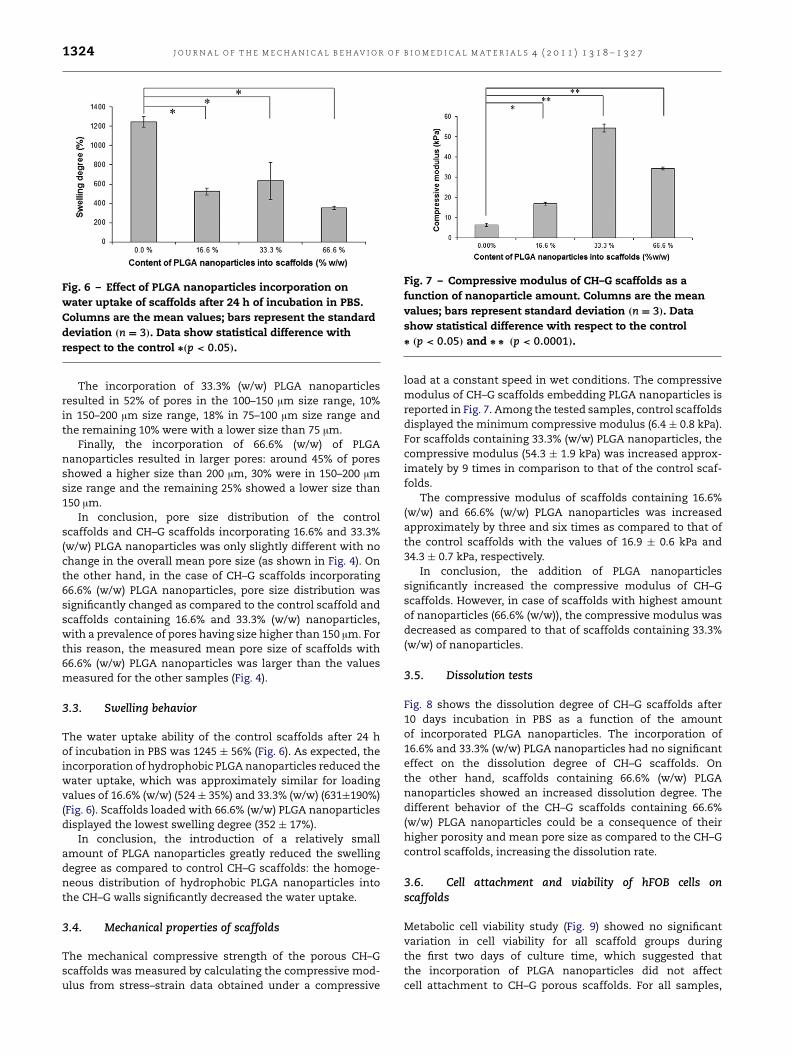
Fig. 6 – Effect of PLGA nanoparticles incorporation onwater uptake of scaffolds after 24 h of incubation in PBS.Columns are the mean values; bars represent the standarddeviation (n = 3). Data show statistical difference withrespect to the control ∗(p < 0.05).
The incorporation of 33.3% (w/w) PLGA nanoparticlesresulted in 52% of pores in the 100–150 µm size range, 10%in 150–200 µm size range, 18% in 75–100 µm size range andthe remaining 10% were with a lower size than 75 µm.
Finally, the incorporation of 66.6% (w/w) of PLGAnanoparticles resulted in larger pores: around 45% of poresshowed a higher size than 200 µm, 30% were in 150–200 µmsize range and the remaining 25% showed a lower size than150 µm.
In conclusion, pore size distribution of the controlscaffolds and CH–G scaffolds incorporating 16.6% and 33.3%(w/w) PLGA nanoparticles was only slightly different with nochange in the overall mean pore size (as shown in Fig. 4). Onthe other hand, in the case of CH–G scaffolds incorporating66.6% (w/w) PLGA nanoparticles, pore size distribution wassignificantly changed as compared to the control scaffold andscaffolds containing 16.6% and 33.3% (w/w) nanoparticles,with a prevalence of pores having size higher than 150 µm. Forthis reason, the measured mean pore size of scaffolds with66.6% (w/w) PLGA nanoparticles was larger than the valuesmeasured for the other samples (Fig. 4).
3.3. Swelling behavior
The water uptake ability of the control scaffolds after 24 hof incubation in PBS was 1245 ± 56% (Fig. 6). As expected, theincorporation of hydrophobic PLGA nanoparticles reduced thewater uptake, which was approximately similar for loadingvalues of 16.6% (w/w) (524 ± 35%) and 33.3% (w/w) (631±190%)(Fig. 6). Scaffolds loaded with 66.6% (w/w) PLGA nanoparticlesdisplayed the lowest swelling degree (352 ± 17%).
In conclusion, the introduction of a relatively smallamount of PLGA nanoparticles greatly reduced the swellingdegree as compared to control CH–G scaffolds: the homoge-neous distribution of hydrophobic PLGA nanoparticles intothe CH–G walls significantly decreased the water uptake.
3.4. Mechanical properties of scaffolds
The mechanical compressive strength of the porous CH–Gscaffolds was measured by calculating the compressive mod-ulus from stress–strain data obtained under a compressive
Fig. 7 – Compressive modulus of CH–G scaffolds as afunction of nanoparticle amount. Columns are the meanvalues; bars represent standard deviation (n = 3). Datashow statistical difference with respect to the control∗ (p < 0.05) and ∗ ∗ (p < 0.0001).
load at a constant speed in wet conditions. The compressivemodulus of CH–G scaffolds embedding PLGA nanoparticles isreported in Fig. 7. Among the tested samples, control scaffoldsdisplayed the minimum compressive modulus (6.4 ± 0.8 kPa).For scaffolds containing 33.3% (w/w) PLGA nanoparticles, thecompressive modulus (54.3 ± 1.9 kPa) was increased approx-imately by 9 times in comparison to that of the control scaf-folds.
The compressive modulus of scaffolds containing 16.6%(w/w) and 66.6% (w/w) PLGA nanoparticles was increasedapproximately by three and six times as compared to that ofthe control scaffolds with the values of 16.9 ± 0.6 kPa and34.3 ± 0.7 kPa, respectively.
In conclusion, the addition of PLGA nanoparticlessignificantly increased the compressive modulus of CH–Gscaffolds. However, in case of scaffolds with highest amountof nanoparticles (66.6% (w/w)), the compressive modulus wasdecreased as compared to that of scaffolds containing 33.3%(w/w) of nanoparticles.
3.5. Dissolution tests
Fig. 8 shows the dissolution degree of CH–G scaffolds after10 days incubation in PBS as a function of the amountof incorporated PLGA nanoparticles. The incorporation of16.6% and 33.3% (w/w) PLGA nanoparticles had no significanteffect on the dissolution degree of CH–G scaffolds. Onthe other hand, scaffolds containing 66.6% (w/w) PLGAnanoparticles showed an increased dissolution degree. Thedifferent behavior of the CH–G scaffolds containing 66.6%(w/w) PLGA nanoparticles could be a consequence of theirhigher porosity and mean pore size as compared to the CH–Gcontrol scaffolds, increasing the dissolution rate.
3.6. Cell attachment and viability of hFOB cells onscaffolds
Metabolic cell viability study (Fig. 9) showed no significantvariation in cell viability for all scaffold groups duringthe first two days of culture time, which suggested thatthe incorporation of PLGA nanoparticles did not affectcell attachment to CH–G porous scaffolds. For all samples,

J O U R N A L O F T H E M E C H A N I C A L B E H AV I O R O F B I O M E D I C A L M A T E R I A L S 4 ( 2 0 1 1 ) 1 3 1 8 – 1 3 2 7 1325
Fig. 8 – Effect of amount of PLGA nanoparticlesincorporation on dissolution properties of scaffolds after 10days of incubation in PBS. Columns are the mean values;bars represent standard deviation (n = 3). Data showstatistical difference with respect to the control ∗ (p < 0.05).
Fig. 9 – Effect of PLGA nanoparticle incorporation onmetabolic viability (Alamar Blue assay) of hFOB cellsseeded onto the scaffolds for 1, 2, 5 and 11 days. Columnsare the average data, bars are the standard deviation.
metabolic cell viability approximately doubled after 5 dayscell culture time and then further increased after 11 daysculture time. After 5 and 11 days culture time, viability of cellsadhered on scaffolds incorporating nanoparticles was onlyslightly decreased as compared to control samples. However,these differences in cell viability were not significant.
4. Discussion
The choice of the method for biomolecule encapsulationwithin nanoparticles is usually determined by the solubilitycharacteristics of the drug. In this study, the doubleemulsion–evaporation process was adopted since it is knownto be superior to other incorporation methods in terms ofstability of incorporated proteins (Tabata et al., 1993).
The encapsulation efficiency of BSA (used in this studyas a model protein) and the particle size were preliminarilyoptimized by varying the protein:polymer ratio and altering
external aqueous phase pH and osmolality. Based on thesestudies, the maximum encapsulation efficiency was reachedwhen the amount of polymer was about ten times higherthan that of the BSA protein (data not shown). The diffusionof BSA from nanoparticle core toward the aqueous externalphase was prevented by properly selecting the pH of externalaqueous phase (near to the i.e.p. of BSA) and by increasingits osmolality by adding sodium chloride (data not shown)(Muthu, 2009).
During freezing of CH–G solutions containing PLGAnanoparticles (0.00%–66.6% (w/w)), the interaction of watermolecules with the hydrophobic surface of PLGA nanopar-ticles affected the final pore size distribution of scaffolds.Water molecules in contact with the hydrophobic surfacesof PLGA nanoparticles could not form inter-molecular hy-drogen bonds with the hydrophobic surface. Instead, theyformed highly connected self-assembled structures by intra-molecular hydrogen bonding with other water molecules.However, an amount of PLGA nanoparticles of 16.6% (w/w)and 33.3% (w/w) only slightly influenced scaffold morphol-ogy. On the other hand, CH–G scaffolds loaded with 66.6%(w/w) PLGA nanoparticles showed an increased porosity de-gree and pore size (75% of pores were larger than 150 µm). Thisbehavior was a consequence of the distribution of nanoparti-cles within the scaffolds: the PLGA nanoparticles were homo-geneously distributed into the scaffold pore walls when theywere present at an amount of 16.6%–33.3% (w/w) (Fig. 2(a)–(b)).On the other hand, PLGA nanoparticles formed some aggre-gates when loaded at 66.6% (w/w) concentration (Fig. 2(c)). Asimilar result was found by Banerjee et al. for PLGA particlesembedded within porous gelatin scaffolds (Banerjee et al.,2009). In addition, the viscosity of the CH–G solution was ex-pected to increase due to PLGA nanoparticle addition in adose dependent manner (Gong et al., 2006), retarding the wa-ter molecule diffusion during freezing and leading to an irreg-ular porous structure as shown in Fig. 5.
Both the hydration degree and the degradation behaviorare the most important properties of materials aimed atbiomedical or environmental applications, as their lifetimeis mainly governed by these two intimately correlatedprocesses. For degradable polymers, degradation occurs as aresult of natural biological processes or other factors suchas hydrolysis. Additionally, the drug release rate is mostlyinfluenced by two factors: the diffusion of the drug out ofthe scaffold and the water uptake of the polymeric matrix.Therefore, the preparation of systems for controlled drugrelease applications requires the knowledge of water uptakeand degradation rate.
In the case of in vitro dissolution tests, scaffolds displayeda similar dissolution degree for PLGA nanoparticle loadingin the 0%–33.3% (w/w) range. A significant increase of thedissolution degree was found for the CH–G scaffold loadedwith 66.6% (w/w) PLGA nanoparticles: this behavior wasprobably a consequence of its superior porosity degree andpore size. Furthermore, the time dependent degradation ofPLGA particles themselves by means of hydrolysis could haveaugmented the weight loss percentage in scaffolds with thehighest amount of PLGA nanoparticles.
The swelling degree of CH–G scaffolds was stronglydecreased by the addition of a relatively low amount of

1326 J O U R N A L O F T H E M E C H A N I C A L B E H AV I O R O F B I O M E D I C A L M A T E R I A L S 4 ( 2 0 1 1 ) 1 3 1 8 – 1 3 2 7
PLGA nanoparticles (Fig. 6). Scaffolds with 16.6% (w/w) and33.3% (w/w) PLGA nanoparticles showed a similar swellingdegree; on the other hand, the loading of 66.6% (w/w) PLGAnanoparticles further decreased the swelling degree, probablyas a consequence of increased porosity degree andmean poresize.
The incorporation of PLGA nanoparticles within CH–Gscaffolds increased the compressive modulus of scaffolds(Fig. 7) in comparison to the control CH–G scaffolds.The compressive modulus increased with increasing PLGAnanoparticles amount from 0% w/w to 33.3% w/w. On theother hand, the compressive modulus of scaffolds containing66.6% (w/w) PLGA nanoparticles decreased as compared tothat of scaffolds containing 33.3% w/w PLGA nanoparticles,probably because of their increased porosity degree andmeanpore size. In general, the resistance area of a material sampledecreases with increasing pore size and porosity degree,reducing its mechanical resistance. Cell viability studieswere performed to examine the effect of the incorporationof hydrophobic nanoparticles within the hydrophilic CH–Gscaffolds on cell attachment and cell viability. Results after1 d and 2 d incubation time showed that all scaffolds induceda similar degree of cell attachment (Fig. 9) which indicatesthat incorporation of PLGA nanoparticles into CH–G scaffoldsdid not affect cell attachment behavior. However, for scaffoldsloaded with different amounts of PLGA nanoparticles, aslight, not significant decrease in cell viability was detectedafter 5 d and 11 d culture time. This behavior could beexplained by the degradation phenomena involving PLGAnanoparticles and making the local environment slightlyacidic.
5. Conclusion
Three-dimensional porous GP-crosslinked CH–G scaffolds in-corporated with PLGA nanoparticles were produced as suit-able systems for the localized delivery of bioactive agents inscaffolds for bone regeneration, such as growth factors, drugs,etc. This study disclosed the changes in physical propertiesof porous CH–G scaffolds as a consequence of incorporationof PLGA nanoparticles in three different percentages. Thestudy revealed that loading of hydrophobic PLGA nanopar-ticles in relatively hydrophilic GP-crosslinked CH–G scaffoldaltered the scaffold microenvironment and modulated wateruptake, compressive modulus, and dissolution properties. Onthe other hand, incorporation of PLGA nanoparticles withinCH–G scaffolds did not affect significantly cell attachmentand viability after 1–11 days cell culture time. This study wasaimed at the design of an optimized matrix for controlledrelease of biomolecules for bone tissue engineering applica-tions. Based on the results of this study, the incorporationof 33.3% w/w of PLGA nanoparticles within CH–G scaffoldsyielded scaffolds with enhanced mechanical properties, re-taining other desirable physical and cell attachment proper-ties. Further studies describing the encapsulation and releaseof therapeutic proteins, such as Bone Morphogenetic Protein(BMP2)/parathyroid hormone (PTH) from the optimized scaf-folds formulations are in progress.
Acknowledgments
The authors acknowledge support for this work provided byItalian Ministry for Research and the University (MIUR) forVijay Kumar Nandagiri’s grant. The authors also acknowledgethe assistance by Clara Mattu (Industrial Bio engineeringgroup, Department of Mechanics, Politecnico di Torino) forSEM analysis as well as by Dr. Jacqueline Daly, Dr. JohnGleeson andMs. Ciara Murphy (Department of Anatomy, Boneand Tissue engineering group, Royal College of surgeons inIreland, Dublin 2, Ireland) for their support in cell studies.
R E F E R E N C E S
Al-Munajjed, A.A., O’Brien, F.J., 2009. Influence of a novel calcium-phosphate coating on the mechanical properties of highlyporous collagen scaffolds for bone repair. J. Mech. Beh. Biomed.Mater. 2, 138–146.
Banerjee, I., Mishra, D., Maiti, T.K., 2009. PLGA microspheresincorporated gelatin scaffold: microspheres modulate scaffoldproperties. Int. J. Biomater. doi:10.1155/2009/143659. Article ID143659.
Bonadio, J., Smiley, E., Patil, P., Goldstein, S., 1999. Localized, directplasmid gene delivery in vivo: prolonged therapy results inreproducible tissue regeneration. Nature Med. 5, 753–759.
Borenstein, J.T., Weinberg, E.J., Orrick, B.K., Sundback, C.,Kaazempur-Mofrad, M.R., Vacanti, J.P., 2007. Microfabricationof three-dimensional engineered scaffolds. Tissue Eng. 13,1837–1844.
Chang, C.H., Liu, H.C., Lin, C.C., Chou, C.H., Lin, F.H., 2003.Gelatin–chondroitin–hyaluronan tri-copolymer scaffold forcartilage tissue engineering. Biomaterials 24, 4853–4858.
Chiono, V., Pulieri, E., Vozzi, G., Ciardelli, G., Ahluwalia, A.,Giusti, P., 2008. Genipin-crosslinked chitosan/gelatin blendsfor biomedical applications. J. Mater. Sci. Mater. Med. 19,889–898.
Cohen-Sela, E., Chorny, M., Koroukhov, N., Danenberg, H.D.,Golomb, G., 2009. A new double emulsion solvent diffusiontechnique for encapsulating hydrophilic molecules in PLGAnanoparticles. J. Contr. Rel. 33, 90–95.
Esposito, E., Cortesi, R., Nastruzzi, C., 1996. Gelatin microspheres:influence of preparation parameters and thermal treatment onchemico-physical and biopharmaceutical properties. Biomate-rials 17, 2009–2020.
Gong, Y.H., Ma, Z.W., Gao, C.Y., Wang,W., Shen, J.C., 2006. Speciallyelaborated thermally induced phase separation to fabricatepoly(l-lactic acid) scaffolds with ultra large pores and goodinterconnectivity. J. Appl. Polym. Sci. 101, 3336–3342.
Harris, S.A., Enger, R.J., Riggs, B.L., Spelsberg, T.C., 1995. Develop-ment and characterization of a conditionally immortalized fe-tal osteoblastic cell line. J. Bone. Miner. Res. 10, 178–186.
Holzer, M., Vogel, V., Mantele, W., Schwartz, D., Haase, W.,Langer, K., 2009. Physico-chemical characterization of PLGAnanoparticles after freeze-drying and storage. Eur. J. Pharm.Biopharm. 72, 428–437.
Huang, Y., Onyeri, S., Siewe, M., Moshfeghian, A., Madihally, S.V.,2005. In vitro characterization of chitosan–gelatin scaffolds fortissue engineering. Biomaterials 26, 7616–7627.
Ito, A., Mase, A., Takizawa, Y., Shinkai, M., Honda, H., Hata,K.I., Ueda, M., Kobayashi, T., 2003. Transglutaminase-mediatedgelatin matrices incorporating cell adhesion factors as abiomaterial for tissue engineering. J. Biosci. Bioeng. 95,196–199.
Jeong, B., Bae, Y.H., Lee, D.S., Kim, S.W., 1997. Biodegradable blockcopolymers as injectable drug-delivery systems. Nature 388,860–862.
J O U R N A L O F T H E M E C H A N I C A L B E H AV I O R O F B I O M E D I C A L M A T E R I A L S 4 ( 2 0 1 1 ) 1 3 1 8 – 1 3 2 7 1327
Kawai, K., Suzuki, S., Tabata, Y., Ikada, Y., Nishimura, Y., 2000.Accelerated tissue regeneration through incorporation of basicfibroblast growth factor-impregnated gelatin microspheresinto artificial dermis. Biomaterials 21, 489–499.
Keogh, M.B., O’Brien, F.J., Daly, J.S., 2010. A novel collagen scaffoldsupports human osteogenesis-applications for bone tissueengineering. Cell Tissue Res. 340, 169–177.
Khil, M.S., Cha, D.I., Kim, H.Y., Kim, I.S., Bhattarai, N., 2003.Electrospun nanofibrous polyurethane membrane as wounddressing. J. Biomed. Mater. Res. B Appl. Biomater. 67 (2),675–679.
Kim, H., Kim, W., Suh, H., 2003. Sustained release of ascorbate-2-phosphate and dexamethasone from porous PLGA scaffoldsfor bone tissue engineering. Biomaterials 24, 4671–4679.
Kobsa, S., Saltzman, M., 2008. Bioengineering approaches tocontrolled protein delivery. Pediatr. Res. 63, 513–519.
Lahiji, A., Sohrabi, A., Hungerford, D.S., Frondoza, C.G., 2000.Chitosan supports the expression of extra-cellular matrixproteins in human osteoblasts and chondrocytes. J. Biomed.Mater. Res. 51, 586–595.
Langer, R., 1998. Drug delivery and targeting. Nature 392,5–10.
Lee, J.E., Kim, K.E., Kwon, I.C., Ahn, H.J., Lee, S.H., Cho, H., Kim, H.J.,Seong, S.C., Lee, M.C., 2004. Effects of the controlled–releasedTGF-beta 1 from chitosan microspheres on chondrocytescultured in a collagen/chitosan/glycosaminoglycan scaffold.Biomaterials 25 (18), 4163–4173.
Lee, S.H., Shin, H., 2007. Matrices and scaffolds for delivery ofbioactive molecules in bone and cartilage tissue engineering.Adv. Drug. Deliv. Rev. 59, 339–359.
Ma, P.X., 2008. Biomimetic materials for tissue engineering. Adv.Drug. Deliv. Rev. 60, 184–198.
Madihally, S.V., Matthew, H.W.T., 1999. Porous chitosan scaffoldsfor tissue engineering. Biomaterials 20, 1133–1142.
Mi, F.L., 2005. Synthesis and characterization of a novelchitosan–gelatin bioconjugate with fluorescence emission.Biomacromolecules 6, 975–987.
Muthu, M.S., 2009. Nanoparticles based on PLGA and its co-polymer: an overview. Asian. J. Pharm. 3, 266–273.
O’Brien, F.J., Harley, B.A., Yannas, I.V., Gibson, L.J., 2004. Influenceof freezing rate on pore structure in freeze-dried collagen-GAGscaffolds. Biomaterials 25, 1077–1086.
Perets, A., Baruch, Y., Weisbuch, F., Shoshany, G., Neufeld, G.,Cohen, S., 2003. Enhancing the vascularization of three-dimensional porous alginate scaffolds by incorporatingcontrolled release basic fibroblast growth factor microspheres.J. Biomed. Mater. Res. A 65 (4), 489–497.
Shen, F., Cui, Y.L., Yang, L.F., Yao, K.D., Dong, X.H., Jia, W.Y., Shi,H.D., 2000. A study on the formation of porous chitosan/gelatinnetwork scaffold for tissue engineering. Polym. Int. 49,1596–1599.
Silvia, G.A., Coutinho, O.P., Duchevne, P., Reis, R.L., 2007. Materialsin particulate form for tissue engineering. 2. Application inbone. J. Tiss. Eng. Reg. Med. 1, 97–109.
Suh, J.K.F., Matthew, H.W.T., 2000. Application of chitosan-basedpolysaccharide biomaterials in cartilage tissue engineering: areview. Biomaterials 21, 2589–2598.
Tabata, Y., Takebayashi, Y., Ueda, T., Ikada, Y., 1993. A formulationmethod using D, L-lactic acid oligomer for protein releasedwith reduced initial burst. J. Control. Rel. 23, 55–64.
Tachibana, A., Nishikawa, Y., Nishino, M., Kaneko, S., Tanabe,T., Yamauchi, K., 2006. Modified keratin sponge: binding ofbone morphogenetic protein-2 and osteoblast differentiation.J. Biosci. Bioeng 102, 425–429.
Thein-Han, W.W., Saikhun, J., Pholpramoo, C., Misra, R.D.K.,Kitiyanant, Y., 2009. Chitosan–gelatin scaffolds for tissueengineering: physico-chemical properties and biological re-sponse of buffalo embryonic stem cells and transfectantof GFP–buffalo embryonic stem cells. Acta Biomater. 5,3453–3466.
VandeVord, P.J., Matthew, H.W., DeSilva, S.P., Mayton, L., Wu, B.,Wooley, P.H., 2002. Evaluation of the biocompatibility of achitosan scaffold in mice. J. Biomed. Mater. Res. 59, 585–590.
Vasita, R., Katti, D.S., 2006. Growth factor-delivery systems fortissue engineering: a materials perspective. Expert. Rev. Med.Dev. 3, 29–47.
Zhao, F., Yin, Y., Lu, W.W., Leong, J.C., Zhang, W., Zhang, J., Zhang,M., Yao, K., 2002. Preparation and histological evaluationof biomimetic three-dimensional hydroxyapatite/chitosan–gelatin network composite scaffolds. Biomaterials 23,3227–3234.
Zisch, A.H., Lutolf, M.P., Hubbell, J.A., 2003. Biopolymeric deliverymatrices for angiogenic growth factors. Cardiovasc. Pathol. 12,295–310.