incision care tutorial - massachusetts general hospital
TRANSCRIPT

Incision Care Tutorial
The following pictures show incisions not healed, infections, staples, brusing and blisters after orthopaedic oncology surgery. Some of the pictures can make people feel squeamish. These pictures are not meant to cause alarm or shock, but rather are meant to teach and help our patients identify when they should call their surgeon regarding changes to their incision. These photos predominately are in the immediate post-operative phase, not durig oncologic surveillance. However, there are basic guidelines about when to contact your surgeon’s office: when in doubt, call; don’t wait; report any new
suspicious findings (i.e. swelling, fever, redness, chills, flu-like symptoms, new lumps & bumps).
Glossary:Erythema: redness Hematoma: collection of blood Seroma: collection of fluidWound Dehiscence: opening of a surgical incisionEschar: a dry, dark scab on the incisionDelayed Healing: usually caused by radiation treatments and sometimes chemotherapy; this is when the surgical incision opens and can take several months to heal

Healing incision (left inner thigh) after a soft tissue sarcoma resection with normal bruising and a normal amount of erythema (redness).

Healing incision after a benign soft tissue mass was removed. This incision has dissolving sutures and steristrips.
There is a slight hematoma (blood collection) or seroma (fluid collection).

The is a common z-shaped incision, which shows a normal amount of swelling with normal small scabs from blisters and dry skin after excision of a bening soft tissue mass behind the knee.

This is an example of a wound dehiscence, which means an incision is primarily closed but a few days after surgery, the incision opens.
In this case, delayed surgical healing is due to prior radiation treatment.

This type of erythema (redness) and swelling should be reported to your surgeon.
The photo shows a post-operative seroma (fluid collection) under the horizontal incision.

This shows a post-operative infection. This type of redness and pustule-looking mass in the center of the incision are warning signs of infection. Call your surgeon.

This shows delayed wound healing after the removal of a soft tissue sarcoma on a knee that had radiation.
This wound closed after months of wound packing.

1. This is an incision that is healing well after radiation and surgery. Staples are intact in this photo.
2. Subsequent development of a large post-operative seroma (fluid collection). The incision remains healthy, but your surgeon should be notified of a new fluid collection.
1 2

Wound dehiscence that was not caused by radiation. This is a longitudinal incision on the spine.
1. The beginning of wound breakdown. There is yellow fibrinous tissue, but this incision does not looked infected. Any wound opening should be reported.
2. A week later, the would completely opened.
1 2

Beautifully healing thigh incision with staples intact two weeks after surgery.
The slight redness around the legs of the staple is normal.
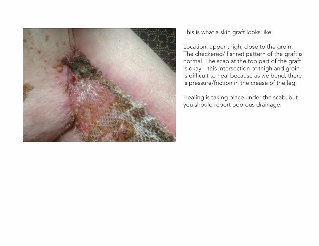
This is what a skin graft looks like.
Location: upper thigh, close to the groin. The checkered/ fishnet pattern of the graft is normal. The scab at the top part of the graft is okay – this intersection of thigh and groin is difficult to heal because as we bend, there is pressure/friction in the crease of the leg.
Healing is taking place under the scab, but you should report odorous drainage.

Lateral thigh superficial clean sinus tract, ¼” deep after total hip replacement infection.
Minimal redness, wrinkling of skin and no drainage are good signs of improvement.